Author Archives: rajani

Association between Body Composition and Disease Severity in Patients with Motor Neuron Disease / Amyotrophic Lateral Sclerosis

DOI: 10.31038/ASMHS.2019311

Abstract

The change in nutritional status is observed in patients with Motor Neuron Disease, in body composition. The electrical bio impedance method (BIA) determines lean mass, fat mass and phase angle, by estimating total body water. ALSFRS-R is used to monitor the functionality and progression of the disease.

Objective: To evaluate the composition with ALSFRS-R.

Methodology: Cross-sectional study in adult patients with conclusive diagnosis of MND. For the classification of nutritional status, BMI was used, and for body composition analysis, measurements of lean mass, fat mass, total body water and phase angle. Results: patients, with 54.8% of males with median age of 61.0 years. 77.4% presented the appendicular form of the disease. The median score of ALSFRS-R was 27 points, and the average BMI was 22.6 kg / m2. The phase angle had an average of 3.9 in women, and 4.1 in men. For statistical analysis, results with a probability of type I error lower than 5% (p <0.05 and p <0.001) were considered as statistically significant. The BMI and phase angle were correlated with the scale scores.

Conclusion: Nutritional status is directly related to disease progression, and depletion of body compartments influences the functionality of patients with MND.

Keywords

amyotrophic lateral sclerosis, body composition, nutritional state, functionality

Introduction

Motor neuron disease (MND) is a progressive, degenerative hyper catabolic disease with involvement of the motor neurons of the cortex, brain stem and spinal cord [1]. Due to the symptoms and the rapid progression of the disease, depletion of nutritional status is observed. Patients present a reduction in the lean mass inherent to disease progression, and the increase in body fat may be positively associated with disease progression [2].

Electrical bio impedance is a tool used to analyze body composition, and it is classified as a gold standard for evaluation in MND [3].The interpretation of the phase angle, allows the measurement of the body changes, and was proposed as an index of malnutrition, or prognostic factor of survival in other diseases. The association of the evaluation of the body composition with the functionality of these patients may help in the treatment of the evolution of the MND / ALS [4].

Hence, the objective of this work is to evaluate the body composition and to analyze the association of the nutritional state with the functionality of patients with MND.

Methods

A cross-sectional study carried out at the Neuromuscular Disease Research Section of the Federal University of São Paulo, approved by the Ethics and Research Committee of the University, under number 0606/2016, with authorization to participate, through a signed Free and Informed Consent Form.

The study was carried out from March to July 2016. Adult patients with a defined diagnosis were included [5]. Patients who did not tolerate being in the supine position for the examination were also excluded from the study.

For the analysis of the body composition, the Bio impedance device “BIODYNAMICS MODELO 450” was used, which provides clinical data of Body Composition and Hydration, such as body fat, total body water, lean mass and phase angle with precision of +/- 0.2 %.

The Amyotrophic Lateral Sclerosis Functional Rating Scale (ALSFRS-R) was applied, analyzed in two ways: by total score, and by domains (bulbar, appendicular and respiratory).

Regarding statistical analysis, the categorical variables were described in absolute value and relative frequency, whereas the continuous variables were described through measures of central tendency, dispersion and position.

Linear correlations between the anthropometric indicators (BMI, body water, lean mass, fat mass and phase angle) and the scores obtained in the ALSFRS-R functionality scale (total score and in the domains) were plotted according to Pearson’s method. After that, linear regression analyzes, the average to verify the extent (β) of the correlation between these indicators and the scale score were performed. The analyzes were performed with the aid of the statistical package IBM SPSS version 20.0.Results with a probability of Type I error of less than 5% were considered as statistically significant.

Results

The sample consisted of 31 patients, 12 adults and 19 elderly. There was prevalence of males and in a median age of 61 years, ranging from 22 to 80 years. Body composition was evaluated without distinction of age.

More than two thirds of the patients (77.4%) presented the appendicular form of the disease, and approximately 10% of the patients presented familial origin.

The mean time from onset of symptoms to the time of evaluation was 32.4 months (8.4 – 86.4). The median ALSFRS-R score was 27.0 points (8–39). The respiratory domain had the highest median score, followed by the appendicular and bulbar domains.

Regarding weight loss, 80.6% of the patients reported weight loss from the onset of symptoms, of which 64% had a loss greater than 10% of the previous weight. The median body mass index was 22.6 kg / m2 (11.8 – 34.6 kg/m2) and approximately 45% of the patients were in the eutrophic range, according to the BMI classification.

The bio impedance method identified 30.9% of fat mass, 67.6% of lean mass and 30.4% of total body water in the studied sample. The phase angle, measured using the bio impedance method, averaged 3.9 degrees in women and 4.1 degrees in men.

Table 2 shows the correlation matrix between the anthropometric indicators and the scale score in the three different domains, in addition to the total score observed. In general, BMI and Phase Angle were the best indicators correlated with the scale scores. The BMI showed significant correlations (p <0.001), directly proportional, with the score in the bulbar and respiratory domains, in addition to the total score.

Table 1. Demographics of the study population

Subject

N

%

 Gender

Male

17

54, 8

Female

14

45, 2

Age in years

Average (min – max)

61, 0 (22 – 80)

Elderly (≥ 60 years)

18

58, 1

Disease manifestation form

Appendicular

24

 77, 4

Bulbar

7

 22, 6

Time of onset of symptoms (months)

Average(min – max)

32, 4 (8, 4 – 86, 4)

ALSFRS-R, domains – average (min – max)

Bulbar

9, 0 (0 – 12)

Appendicular

9, 0 (0 – 20)

Respiratory

10, 0 (4 – 12)

ALSFRS-R, total score – average (min – max)

Total

27 (8 – 39)

Body mass index (kg/m²) – average (min – max)

 Male

 23, 0 (17, 6 – 32, 2)

 Female

 22, 9 (11, 8 – 32, 4)

Fat mass– average (min – max)

 Male

 29, 2 (13, 7 – 43, 3)

 Female

 34, 2 (19, 2 – 55, 2)

Lean mass –average (min – max)

 Male

 70, 7 (56, 7 – 86, 3)

 Female

 60, 6 (33, 7 – 74, 3)

Total body water (L) – average (min – max)

 Male

 34, 9 (16 – 51, 4)

 Female

 27, 6 (18, 4 -35, 4)

Phase angle (º) – average (min – max)

 Male

 4, 1 (2, 8 – 6)

 Female

 3, 9 (2, 4 – 6, 3)

Table 2. Matrix of correlations between anthropometric indicators and the scores obtained on the ALSFRS-R scale.

 ALSFRS-R

 Subject

Bulbar

Appendicular

Respiratory

Total

BMI (Kg/m2)

0, 555**

0, 169

0, 370**

0, 492**

Fat mass (%)

-0, 045

-0, 129

-0, 065

-0, 141

Lean mass (%)

-0, 024

0, 171

0, 014

0, 128

Body water (L)

0, 367**

0, 174

0, 335*

0, 402**

Phase angle (°)

0, 152

0, 659***

0, 516***

0, 744***

*P<0, 10
**P<0, 05
***P<0, 001

The phase angle showed significant correlations of at least moderate intensity (p <0.05), with the scores observed in the appendicular and respiratory domains of the scale, besides the total score obtained. The total body water measure also correlated positively with the bulbar domains and the total score. On the other hand, the indicators of body fat mass and lean mass did not show a significant correlation with the scale scores.

The results of the linear regression (Figure 1) point to an average increment of 5.63 (CI 95% 3.70 – 7.55) points in the scale at each degree measured in the phase angle. However, for each liter of total body water measured, there is an average increment of 0.42 (95% CI 0.05 – 0.75) points in the scale, and for each BMI unit, an average increase of approximately 0.95 (95% CI 0.31 – 1.58) points on the scale can be inferred.

ASMHS 2019-101- Rafella Brazil_F1

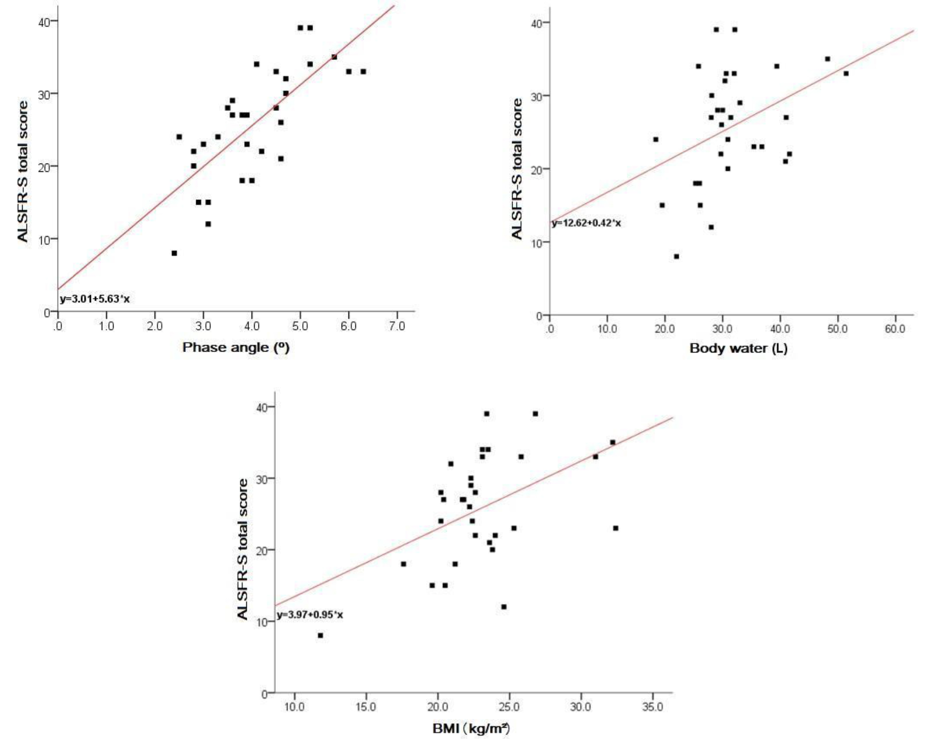
Figure 1. Body composition x functionality

Discussion

The epidemiological and clinical characteristics observed in patients with MND in this study are in agreement with the evidence described in the literature [6]. Being a relatively rare disease, the sample studied is sufficient to represent changes in the body composition of patients with DNM [7]. Patients older than 60 years were considered elderly, since, according to the World Health Organization, this is the cut age for developing countries, in which Brazil fits in [8]. Variables that were not nutritional factors were used to characterize the sample, but were excluded from the analysis.

Most of the patients in this study, representing 75% of the sample, presented the appendicular form as manifestation of the disease. It can also be observed that, there was predominance of the sporadic form on the familial one, which represented approximately 10% of the patients, as described in the literature [9, 10].

Studies have observed that patients with MND often present marked weight loss from the onset of symptoms [11], and JAWAID et al. described that weight loss is a negative prognostic factor for patients with MND [12]. In this study, the majority of patients (80.6%) reported weight loss from the onset of clinical symptoms, with 64% of them presenting a marked loss greater than 10% of the previous weight. This condition can be attributed to the characteristic picture of dysphagia, decreased food intake, hyper metabolism and increased energy expenditure [13]. It was observed that, in the present study, dysphagia was present in 100% of the bulbar patients, and among the patients with appendicitis, bulbar involvement was not recurrent. However, it is suggested that the hyper catabolic state may begin before the clinical manifestation of the disease [14].

BMI can be used as a predictor of progression and survival [14]. Some studies have described an association between the change in BMI and the clinical course of the disease, and evidence that survival is better in overweight patients compared to eutrophic or low-weight patients [11, 14, 15]. Other studies have shown that changes in BMI in the first two years after diagnosis correlate significantly with survival, and with the rate of progression of motor symptoms [12]. This faces the present study, where the BMI of the evaluated patients demonstrated significant, directly proportional correlations with the full score of the functionality scale. On the other hand, authors observed that there was an increase in mortality in patients with a higher BMI (above 35), and suggest that obesity may be related to a more rapid progression of the disease with reasons not yet elucidated [13, 16].

Previous studies have observed that there is a decrease in body weight, BMI, lean mass, phase angle, and an increase in fat mass during the course of the disease, and that depletion of lean mass has a negative impact on functionality and such thing is a prognostic factor for the survival decrease [17–19]. Fat mass is associated with a better outcome of the disease [20]. Authors suggest that increased LDL / HDL cholesterol may be related to neuronal protection and increased survival [15, 18]. Regarding body water, in this present study, the measure of this variable was positively correlated with the bulbar domains and the total score of the ALSFRS-R scale. Thus, for each liter of total body water measured, there was an average increase of 0.42 (95% CI 0.05 to 0.75) in the scale.

This fact is in agreement with the literature, which attributes to hydration as one of the most sensitive factors of malnutrition, and that the phase angle can also be interpreted as an indicator of water distribution between the extra and intra-cellular compartment, and that the better the phase angle, the better the cell membrane integrity [20].

In patients with MND, there is a probable relationship between phase angle and malnutrition, since the alteration of this indicator was higher in malnourished patients than in eutrophic ones [4]. In a study involving patients with MND and phase angle, the authors showed that there was significant worsening of the phase angle in these, and the same suggests that the phase angle could be used as a severity index [3]. The literature also describes that BMI and phase angle are independently associated with survival, and do not correlate with each other. This fact was also observed in this present study.

The reference value for healthy population of the phase angle is approximately 6.5º +/- 1º for females, and 7.5º +/- 1, 1º for males [3]. In this study, the phase angle averaged 3, 9º in women, and 4, 1º in men, as those found in the literature in patients with MND, where the average phase angle for women was 3, 8º , and for men of 4, 5º [20].

Authors demonstrated that there was a decline of the ALSFRS-R scale in the first 5 years of diagnosis of the evaluated patients [21]. The same study also noted that the higher the rate of decline in ALSFRS-R, the lower the survival. Considering that the literature assigns a gravity index to the phase angle, it was observed that in this present study there was a relation between the scale of functionality and the phase angle. The phase angle presented significant correlations of at least moderate intensity with the scores observed in the appendicular and respiratory domains of the scale, in addition to the total score obtained. There is agreement of the relationship between the phase angle and the appendicular domain, considering that the caloric restriction exacerbates the motor symptoms [18]. The results of the linear regression point to an average increase of 5.63 (95% CI 3.70 – 7.55) points in the scale, to every degree measured in the phase angle. However, as it is a cross-sectional study, it was not possible to evaluate the association between phase angle, ALSFRS-R and survival.

Bio impedance is a simple and easy to apply method, but the use in clinical practice to evaluate patients with MND may present limitations and bias for some measures, since the corporal distribution of these patients may behave differently, [20] once there is a difference in the fat distribution, as well as hydration. Although bio impedance monitors body composition more fractionally, body weight assessment should be a priority, once it is from its variation and monitoring that nutritional intervention is guided, and that BMI at the time of diagnosis can be considered a factor prognosis [17]. It is essential that the care invested be interdisciplinary to ensure the optimization of the quality of life of these patients. Studies confirm that patients who receive care from a multiprofessional team tend to have better survival compared to those who are deprived of these care [9]. All things considered, monitoring of nutritional status becomes indispensable for a better course of the disease.

Final considerations

The nutritional status is directly related to disease progression, and the depletion of the body compartments influences the functionality of patients with MND. BMI is a prognostic indicator and should be used as monitoring during the course of the disease. ALSFRS-R provides data that allows monitoring the evolution of the disease in a generalized way and by domains, allowing a more sensitive and focused observation of the bulbar, motor and / or respiratory alterations resulting from the disease, guiding the professional conduct and treatment.

The evaluation of body composition provides information that aggregates in the individualization of the nutritional therapy. The phase angle is an excellent predictor of severity, and concomitant with the application of ALSFRS-R, it is possible to notice that the alteration of the body composition has a direct and negative impact on the functionality of these patients.

References

  1. Bastos AF, Orsini M, Machado D, Mello MP, Nader S (2011) Amyotrophic lateral sclerosis: one or multiple causes? Neurol Int 3: e4. [crossref]
  2. Silva LBC, Mourão LF, Silva AA, Lima NMFV, Almeida SR, et al. (2010) Effect of nutritional supplementation with milk whey proteins in amyotrophic lateral sclerosis patients. Arq Neuro-Psiquiatr 68: 263–268. [crossref]
  3. Desport JC, Preux PM, Bouteloup-Demange C, Clavelou P, Beaufrère B, et al. (2003) Validation of bioelectrical impedance analysis in patients with amyotrophic lateral sclerosis. Am J Clin Nutr 77:1179–1185. [crossref]
  4. Desport JC, Marin B, Funalot BT, Preux PM, Couratier P (2008) Phase angle is a prognostic factor for survival in amyotrophic lateral sclerosis. Amyotroph Lateral Scler 9: 273–278. [crossref]
  5. Brooks BR, Miller RG, Swash M, Munsat TL (2000) El Escorial revisited: reviserd criterial for the diagnosis of amyotroph lateral sclerosis. Amyotroph Lateral Scler Other Motor Neuron Disord 1: 293–299. [crossref]
  6. Nieves JW, Gennings C, Factor-Litvak P, Hupf J, Singleton J, et al. (2016) Association Between Dietary Intake and Function in Amyotrophic Lateral Sclerosis. JAMA Neurol 73: 1425–1432. [crossref]
  7. Chiò A, Logroscino G, Traynor BJ, Collins J, Simeone JC, et al. (2013) Global epidemiology of amyotrophic lateral sclerosis: a systematic review of the published literature. Neuroepidemiology 41:118–130. [crossref]
  8. WHO (2002) Active Ageing-A Police Framework. A Contribution of the World Health Organization to the second United Nations World Assembly on Aging. Madrid, Spain.
  9. Phukan J, Hardiman O (2009) The management of amyotrophic lateral sclerosis. J Neurol 256: 176–186. [crossref]
  10. Leigh PN, Abrahams S, Al-Chalabi A, Ampong MA, Goldstein LH, et al. (2003) The management of motor neurone disease. J Neurol Neurosurg Psychiatry 74 Suppl 4: iv32–32iv47. [crossref]
  11. Shimizu T, Nagaoka U, Nakayama Y, Kawata A, Kugimoto C, et al. (2012) Reduction rate of body mass index predicts prognosis for survival in amyotrophic lateral sclerosis: A multicenter study in Japan. Amyotroph Lateral Scler 13: 363–366. [crossref]
  12. Jawaid L, Murthy SB, Wilson AM, Qureshi SU, Amro MJ, et al. (2010) A decrease in body mass index is associated with faster progression of motor symptoms and shorter survival in ALS. Amyotroph Lateral Scler 11: 542–548. [crossref]
  13. Paganoni S, Deng J, Jaffa M, Cudkwicz ME, Wills AN (2011) Body mass index, not dyslipidemia, is an independent predictor of survival in amyotrophic lateral sclerosis. Muscle Nerve 44: 20–24. [crossref]
  14. O’reilly JE, Wang H, Weisskopf MG, Fitzgerald KC, Falcone G, et al. (2012) Premorbid body mass index and risk of amyotrophic lateral sclerosis. Amyotroph Lateral Scler Frontotemporal Degener 14: 205–211. [crossref]
  15. Dorst J, Kühnlein P, Hendrich C, Kassubek J, Sperfeld AD, et al. (2011) Patients with elevated triglyceride and cholesterol serum levels have a prolonged survival in amyotrophic lateral sclerosis. J Neurol 258: 613–617. [crossref]
  16. Reich-Slotky R, Andrews J, Cheng B, Buchsbaum R, Leyv D, et al. (2013) Body mass index (BMI) as predictor of ALSFRS-R score decline in ALS patients. Amyotroph Lateral Scler Frontotemporal Degener 14: 212–216. [crossref]
  17. Marin B, Desport JC, Kajeu P, Jesus P, Nicolaud B, et al. (2010) Alteration of nutritional status at diagnosis is a prognostic factor for survival of amyotrophic lateral sclerosis patients. J Neurol Neurosurg Psychiatry 82: 628–634. [crossref]
  18. Dupuis L, Corcia P, Fergani A, Aguilar JLG, Bonnefont-Rousselor D, et al. (2008) Dyslipidemia is a protective factor in amyotrophic lateral sclerosis. Neurology 70: 1004–1009. [crossref]
  19. Dorst J, Cypionka J, Ludolph AC (2013) High-caloric food supplements in the treatment of amyotrophic lateral sclerosis: A prospective interventional study. Amyotroph Lateral Scler Frontotemporal Degener 14: 533–536. [crossref]
  20. Roubeau V, Blasco H, Maillot F, Corcia P, Praline J (2015) Nutritional Assessment of Amyotrophic lateral sclerosis in routine practice: value of weighing and bioelectrical impedance analysis. Muscle Nerve 51: 479–484. [crossref]
  21. Gordon PH, Cheng B, Salachas F, Pradat PF, Bruneteau G, et al. (2010) Progression in ALS is not linear but is curvilinear. J Neurol 257: 1713–1717. [crossref]

Neonatal Outcomes of Macrosomic New-Borns (4,000g +) of Diabetic and Non Diabetic Mothers: A Study of 1,391 Singleton New-Borns

DOI: 10.31038/IGOJ.2019212

Abstract

Objective: To explore the association between diabetic status of the mother and subsequent pregnancy outcomes in a cohort of macrosomic births (birth weight ≥ 4000 grams).

Design: Historical cohort study of macrosomic births comparing delivery method, newborn injury, and newborn morphology between diabetic and non-diabetic women.

Setting: Centre Hospitalier Universitaire Sud-Reunion’s maternity (island of La Reunion, French overseas department, Indian Ocean)

Population: All consecutive singleton live macrosomic births delivered from 2009–2017.

Methods: Macrosomic births were identified from the hospital and the medical records of the mother and newborn were abstracted. Pregnancy outcomes (method of delivery, newborn injury, and newborn morphology) were contrasted between diabetic and non-diabetic women. Among those delivered vaginally, we compared newborn injury between groups.

Results: Newborns from diabetic mothers (cases: 206) were slightly heavier while being younger in gestational ages than controls. There were more caesarean deliveries in the diabetic group (48.8% vs 22.5%, p< 0.001). Among diabetic newborns with vaginal deliveries (ap. half of all diabetic), there were more newborn injuries (brachial plexus, clavicle fractures) in the diabetic group (OR 2.5, p = 0.01) than in controls. A logistic regression model taking into account maternal pre-pregnancy BMI and fetal BW gave an adjusted Odds Ratio for newborn injuries of 2.29 (p = 0.03) in diabetic deliveries.

Conclusion: Among macrosomic deliveries (BW ≥ 4000g), newborns from diabetic mothers have more injuries than controls. This risk remains after controlling for pre-pregnant BMI and newborn birth weight. These data confirm that diabetic-macrosomic newborns may present a different truncular obesity than non-diabetics.

Keywords

Cesarean Delivery, Overweight, Shoulder dystocia, Pre-pregnancy Adiposity,

Introduction

This historical cohort study was conducted to test the hypothesis that pregnancy outcomes of macrosomic newborns (≥ 4000g) might be different according to the diabetic status of parturients. This information may have important implications for clinical management of these pregnancies. Some authors have reported a different morphology in heavy babies according to the diabetic status of the mother. In diabetic pregnancies, as compared to non-diabetic pregnancies, repartition of fetal adiposity may be predominant in the upper part of the body (fetal truncular obesity), inducing a greater risk of shoulder dystocia in these cases [1–6]. This may be particularity so in gestational diabetes and is therefore of paramount importance in clinical management with respect to mode of delivery. The data for this investigation were obtained from the computerized perinatal data base of more than 35,000 deliveries from nine years of practice at the Centre Hospitalier Universitaire Sud-Reunion’s maternity.

Material and Methods

From January 1st, 2009, to December 31st, 2017, the hospital records of all women delivered at the Centre Hospitalier Universitaire Sud-Reunion’s maternity were abstracted in standardized fashion. All data were entered into an epidemiological perinatal data base which contained information on obstetrical risk factors, description of deliveries and neonatal outcomes. As participants in the French national health care system, all pregnant women in Reunion Island have their prenatal visits, biological and echographical examinations, and anthropological characteristics recorded in their maternity booklet. Maternal pre-pregnant body mass index (BMI) was defined as the ratio of pre-pregnancy weight in kilograms divided by height in meters squared (kg/m²).

Screening for gestational diabetes was performed by the O’Sullivan test between 24 and 28 weeks gestation (ingestion of 50g glucose, followed one hour later by a glycaemia, the cut-off value being 1.4 g/l). The diagnostic test was then the oral glucose tolerance test (OGTT, ingestion of 100g glucose, followed by measurements of glycaemia 1, 2 and 3 hours after ingestion (cut-off values respectively being 0.95 g/l, 1.8 g/l, 1.55 g/l and 1.40 g/l). Diagnosis of gestational diabetes was performed when at least 2 glycemic measurements were above the cut-off values during the OGTT.

Epidemiological data have been recorded and analysed with the software EPI-INFO 7.1.5 (2008, CDC Atlanta, OMS), EPIDATA 3.0 and EPIDATA Analysis V2.2.2.183. and statistical analysis by Stata 7.

Results

During the nine year study period, there were 35,459 singleton live births of which 1,391 (3.9%) newborns weighing 4000g or more.

Table 1 compares macrosome newborns (BW ≥ 4000g) according the diabetes status of their mothers during pregnancy. In diabetic pregnancies, 88% (182/206) were gestational diabetes while 24 presented a preexisting diabetes mellitus. Newborns from diabetic mothers were slightly heavier than controls (44g in average, p = 0.002) but with a lower gestational age at birth (38.6 weeks vs 39.7 weeks,
p < 0,001).

Table 1. Diabetic and non diabetic macrosomes (≥ 4000g). Singleton live births

Macrosomes ≥ 4000g

Diabetic mothers

N=206

(%)

Macrosomes ≥ 4000g

Non-Diabetic mothers

N=1,185

(%)

Odds Ratio


[95% CI]

p

Mean Birthweight (g)

± SD

4247 ± 235

4203 ± 187

_

0.002

Mean Gestational Age (Weeks)

± SD

38.6 ± 1.2

39.7 ± 1.2

_

<0,001

Caesarian sections

(%)

101

(49.0)

266

(22.4)

3.3

[2.4–4.5]

<0.001

Induced deliveries

70

(33.9)

307

(25.9)

1.46

[1.05–2.0]

0.02

% of induced deliveries with a C-section issue

17/70

(24.3)

86/307

(28.0)

_

NS

Abnormal fetal monitoring*

18

(8.7)

157

(13.2)

_

NS

Fluid or thick meconium staining

36

(17.4)

279

(23.5)

0.68

[0.45–1.0]

0.05

APGAR ≤ 6

15

(7.3)

41

(3.4)

2.2

[1.1–4.2]

0.01

Transfers in neonatology

11

(5.3)

44

(3.7)

_

NS

Gestational diabetes

182

0

_

_

Preexisting Diabetes

24

0

_

_

Pre-pregnancy maternal BMI, Kg/m² ± SD

30.1 ± 6,8

n= 200

26.1 ± 5.6

N= 1140

< 0.001

* Abnormal fetal monitoring: Dip2, fetal bradycardia 10 minutes minimum, flat line.

There were significantly (incidence almost doubled) more Cesarean sections in diabetics (48.8% vs 22.5%, OR 3.3, p<0.001). There were more induced deliveries in diabetics than in controls (33.8% vs 25.9%, OR 1.4, p = 0.02), but in both groups, failures of induction (leading to a C-section) were similar. There was less meconium staining in diabetics (OR 0.68, p = 0.05) and a greater incidence of Apgar 3 mn scores less than 7, (7.2% vs 3.4% OR 2.2, p = 0.01) than in controls. There were no differences in transfers of newborns to a neonatal intensive care unit (NICU) or abnormal fetal monitoring during labour.

Table 2 analyzes vaginal deliveries (N = 1,024) in both groups of macrosomes. Instrumental extractions (vacuum, forceps) and transfers of newborns to the NICU were not statistically significant in both groups. There was significantly more fetal trauma (clavicle fractures and/or brachial plexus) in diabetics (OR 2.5, p = 0.02) and Apgar 3 mn scores less than 7 (OR 3.7, P< 0.001). In non-diabetic macrosomes, two infants presented with both clavicle fractures and brachial plexus.

Table 2. Obstetrical traumatisms in vaginal deliveries. Diabetic and non-diabetic macrosomes (≥ 4000g). Singleton live births

Macrosomes ≥ 4000g

Diabetic mothers

N= 106

(%)

Macrosomes ≥ 4000g

Nondiabetic mothers

N= 918

(%)

Odds Ratio


[95% IC]

p-value

Instrumental Extractions (vacuum, forceps)

9

(8.5)

102

(11.1)

_

NS

Obstetrical Traumatisms

(Clavicles and/or brachial plexus)

11

(10.4)

40

(4.4)

2.5

[1.2–5.3]

0.01

Clavicle fractures

8

(7.5)

26

(2.8)

2.8

[1.1–6.8]

0.02#

Brachial Plexus

3

(2.8)

16

(1.7)

_

NS

APGAR ≤ 6

11

(10.4)

28

(3.1)

3.7

[1.7–8.0]

<0.001

Transfers in neonatology

4

(5.7)

14

(3.2)

NS

Pre-pregnancy maternal BMI Kg/m² ± SD.

30.0 ± 6,6

n= 105

25.8 ± 5,6

n= 885

< 0.001

* Cephalic vaginal deliveries : breeches (N= 1) and deliveries « en route » (N= 3) excluded
# Fisher exact test

Table 3 depicts the logistic regression model for fetal trauma (brachial plexus and/or clavicle fracture) in cephalic vaginal deliveries, controlling for maternal pre-pregnancy body mass index and fetal birthweight in diabetic and nondiabetic mothers. Out of 1,024 vaginal deliveries, breech presentation (N = 1) and “en route” deliveries (N = 3) were excluded. Pre-pregnancy maternal body mass index were recorded in 990 mothers (96.5%). In this cohort, the crude odds ratio for fetal trauma was slightly different than the entire cohort of 1,024 women, see Table 2 (2.37 [1.14–4.91], p = 0.02 vs 2.5 [1.2–5.3], p = 0.01, respectively).

Table 3. Logistic regression model: Obstetrical trauma (brachial plexus and/or clavicle fracture) in vaginal birth of newborns ≥ 4000g BW.
Controlling for pre-pregnancy maternal Body Mass Index (BMI) and fetal birth weight.
There were 1,024 vaginal deliveries in our cohort, of which 990 (96.5%) record of pre-pregnancy maternal BMI.

Logistic model

Odds Ratio [95% CI]

P-value

BW 4250–4499g

0.67 [0.29–1.6]

0.36

BW 4500–4749g

2.5 [0.99–6.5]

0.053

BW ≥ 4750g

6.7 [1.9–23.0]

0.002

Maternal BMI

< 18.5 kg/m²

5.4 [1.6–18.2]

0.006

Maternal BMI

25.0–29.9 kg/m²

2.48 [1.17–5.2]

0.02

Maternal BMI

30.0–34.9 kg/m²

2.56 [1.08–6.08]

0.03

Maternal BMI

≥ 35.0 kg/m²

0.58 [0.12–2.9]

0.51

Diabetic mothers

2.29 [1.05–4.98]

0.03

BW= Birthweight. 4000–4249g as reference
Maternal BMI: Pre-pregnancy Body mass index (kg/m²). 18.5–24.9 kg/m² as reference

Controlling for fetal birthweight and maternal pre-pregnancy BMI, the adjusted odds ratio in diabetic mothers for fetal trauma was similar to the crude OR: 2.29 vs 2.37, p = 0.03.

The risk for fetal trauma was predominantly in newborns over 4750g as compared with the group 4000–4250g as well as a strong tendency for newborns 4500–4750g, see Table 3. Association with maternal BMI was less specific (notably in women over 35 kg/m²).

Discussion

The Centre Hospitalier Universitaire Sud-Reunion’s maternity (European standards of care) is the only public hospital in the southern part of Reunion Island (Indian Ocean, French overseas department). It serves the whole population of the area, and with 4,300 births per year, represents 80% of all births in the South. Results of the present study suggest that knowledge of macrosomia prior to delivery may affect obstetrical management between diabetic and non-diabetic mothers with respect to the risk of shoulder dystocia and possible consequences for the newborn. In our experience, macrosomes present more obstetrical trauma in vaginal delivery, even though the incidence of Cesareans is almost double that in non-diabetic mothers (OR 3.3, p< 0.001, Table 1), as already previously described in our population [7].

Several studies have reported higher neonatal morbidity and mortality risks in macrosomes delivered to diabetics as compared to non-diabetics [8–12]. Christoffersson et al describe a perinatal mortality of 1.2% in non-diabetic patients with shoulder dystocia versus 6.4% in diabetic mothers [12]. Nesbitt et al in a study of 175,886 births of newborns weighing more than 3,500g report a 3% incidence of shoulder dystocia (6,238 patients) [13]. Again, the incidence of shoulder dystocia was higher in diabetic mothers as compared with non-diabetics and directly correlated with the degree of macrosomia, diabetes (OR = 1.7), instumental extraction (OR 1.9) and induced delivery (OR = 1.3) being independently associated with shoulder dystocia. Saleh et al also describe a higher incidence of trauma (1.9% vs 0.2%) in macrosomes from diabetic mothers [14]. In studies comparing the incidence of fetal trauma, Casey et al [15] compared 61,209 non-diabetic patients with 874 diabetics and found that, among the diabetics, there was more shoulder dystocia and a significantly higher incidence of clavicle fractures while the incidence brachial was not significantly different. For Ecker et al , in 80 newborns having a plexus brachial injury at birth, 10 were from diabetic mothers (OR 2.84, p< 0.01) [16]. Conversely, in a study by Das et al, in the USA, reported a higher incidence of trauma in macrososmes of non-diabetic mothers. Vaginal deliveries occurred in 70% of cases in non-diabetic mothers with macrosomia while it was 34% in diabetic macrosomes [17]. In a recent study of 899 mothers whose babies weighed 3,500g or more, Mansor et al argue that macrosomia is the only reliable predictor of shoulder dystocia, while in their logistic model diabetes and instrumental deliveries were independently associated with that shoulder dystocia [18]. Recently, authors from Sweden however could not find an association between diabetes and shoulder dystocia (but their definition of macrosomia was ≥ 4500g) [19]

Our results are consistent with the hypothesis that foetuses weighing more than 4000g present a different anthropometry (adiposity) in diabetic and nondiabetic mothers. In our perinatal database the variable « shoulder dystocia » is not individualized as such. That is why in this study we used indirect measures of obstetrical complications (Apgar 3mn < 7, brachial plexus and/or clavicle fractures) in women having delivered vaginally. This finding could be interpreted as primarily associated with a higher rate of maternal obesity (see Table 2) which could influence negatively the obstetrical mechanics for maternal pre-pregnancy corpulence and babies’ birthweights. Results from the logistic regression (Table 3) on the 990 macrosomic vaginal births depicted similar odds ratios adjusting for BMI and birthweight: adjusted OR = 2.29 as compared to a unadjusted OR = 2.37, (p = 0.02 crude OR, p = 0.03 aOR), with a predominant risk for babies weighing more than 4,750g, and a strong tendency for those of 4,500–4,750g .

Other authors have described that diabetes by itself may be an independent risk factor responsible for a particular fetal morphology in macrosomes [1–3,5,20]. Macrosomes from diabetic mothers present an increase of the scapular diameter and a four centimeter average difference between the shoulder width and upper biparietal diameter as compared with macrosomes from non-diabetic mothers [5]. However, measurement of the shoulder width has low predictive value for shoulder dystocia, even if it can be evaluted by MRI [6]. For ultrasonographies, based one two-dimensional ultrasound formulae, accuracy is low, particularly at advanced gestation [21,22]. Three-dimensional ultrasound could be useful to monitor soft tissues [21]. Besides these problems, adiposity is well known to be more important in diabetic macrosomes. The fat mass evaluated by absorptiometry represents 30% of the body mass in newborns from diabetic mothers while it represents 15% in non-newborns of diabetic mothers [23–24]. Nasrat et al report a significant increase of sub-cutaneous fat thickness in 51 newborns from diabetic mothers, while height or biparietal diameter are similar in both groups, suggesting a disproportionate development of these fetuses [2]. McFarland et al report an increase of the shoulder width, a decrease of the head/shoulder ratio, an increase of the adipose tissue, and larger extremities in newborns of diabetic mothers [1]. Also Acker et al explain the higher risk of shoulder dystocia in newborns of diabetic mothers by a different composition of tissues and fat repartition than in controls [9].

Conclusion

Obstetricians or midwives face the dilemma of decisions on mode of delivery for women with preexisting or gestational diabetes mellitus. In these deliveries, the risk for shoulder dystocia is well-known. Our study suggests that diabetes by itself is an independent risk factor of fetal trauma in case of macrosomia. When a macrosomia is detected in the maternity ward, diabetes is a major contributor in the obstetrical decision for the mode of delivery.

References

  1. McFarland MB, Trylovich CG, Langer O (1998) Anthropometric differences in macrosomic infants of diabetic and nondiabetic mothers. J Matern Fetal Med 7: 292–295.
  2. Nasrat H, Abalkhail B, Fageeh W, Shabat A, el Zahrany F (1997) Anthropometric measurement of newborns of gestational diabetic mothers: does it indicate disproportionate fetal growth? J Matern Fetal Med 6: 291–295.
  3. Ballard JL, Rosenn B, Khoury JC, Miodovnik M (1993) Diabetic fetal macrosomia: significance of disproportionate growth. J Pediatr 122: 115–119.
  4. Collège National des Gynécologues et Obstétriciens Français (1999) Recommandations pour la pratique clinique. Diabète gestationnel. Encycl Méd Chir (Elservier, Paris), Gynécologie/Obstétrique 5-042-C-20.
  5. Modanlou HD, Komatsu G, Dorchester W, Freeman RK, Bosu SK (1982) Large-for-gestational-age neonates:anthropometric reasons for shoulder dystocia. Obstet Gynecol 60: 417–423.
  6. Verspyck E, Goffinet F, Hellot MF, Milliez J, Marpeau L (2000) Newborn shoulder width: physiological variations and predictive value for shoulder dystocia. J Gynecol Obstet Biol Reprod (Paris) 29: 192–196.
  7. Vivet-Lefébure A, Roman H, Robillard PY, Laffitte A, Hulsey TC, et al. (2007) Obstetrical and neonatal outcomes of gestatonal diabetes mellitus at Reunion island. Gynecol Obstet Fertil 35: 530–535.
  8. Esakoff TF, Cheng YW, Sparks TN, Caughey AB (2009) The association between birthweights 4000g or greater and perinatal outcomes in patients with and without gestational diabetes mellitus. Am J Obstet Gynecol 200: 672–674.
  9. Acker DB, Sachs BP, Friedman EA (1985) Risk factors for shoulder dystocia. Obstet Gynecol 66: 762–768.
  10. Dildy GA, Clark SL (2000) Shoulder dystocia: risk identification. Clin Obstet Gynecol 43: 265–282.
  11. Rouse DJ, Owen J, Goldenberg RL, Cliver SP (1996) the effectiveness and costs of elective cesarean delivery for fetal macrosomia diagnosed by ultrasound. JAMA 276:1480–1486.
  12. Christoffersson M, Rydhstroem H (2002) Shoulder dystocia and brachial plexus injury: a population-based study. Gynecol Obstet Invest 53: 42–47.
  13. Nesbitt TS, Gilbert WM, Herrchen B (1998) Shoulder dystocia and associated risk factors with macrosomic infants born in California. Am J Obstet Gynecol 179: 476–480.
  14. Saleh A, Al-Sultan SM, Moria AM, Rafak FI, Turkistani YM, et al. (2008) Fetal macrosomia greater or equal to 4000 grams. Comparing maternal and neonatal outcomes in diabetic and nondiabetic women. Saudi Med J 29: 1463–1469.
  15. Casey BM, Lucas MJ, Mcintire DD, Leveno KJ (1997) Pregnancy outcomes in women with gestational diabetes compared with the general obstetric population. Obstet Gynecol 90: 869–873.
  16. Ecker JL, Greenberg JA, Norwitz ER, Nadel AS, Repke JT (1997) Birth weight as a predictor of brachial plexus injury. Obstet Gynecol 89: 643–647.
  17. Das S, Irigoyen M, Patterson MB, Salvador A, Schutzman DL (2009) Neonatal outcomes of macrosomic births in diabetic and non-diabetic women. Arch Dis Child Fetal Neonatal 94: 419–422.
  18. Mansor A, Arumugam K, Omar SZ (2010) Macrosomia is the only reliable predictor of shoulder dystocia in babies weighing 3.5 kg or more. Eur J Obstet Gynecol Reprod Biol 149: 44–46.
  19. Turkmen S, Johansson S, Dahmoun M (2018) Foetal Macrosomia and Foetal-Maternal Outcomes at Birth. J Pregnancy 2018: 4790136.
  20. Kamana KC , Shakya S, Zhang H (2015) Gestational diabetes mellitus and macrosomia: a literature review. Ann Nutr Metab 66: 14–20.
  21. Araujo Júnior E, Peixoto AB, Zamarian AC, Elito Júnior J, Tonni G (2017) Macrosomia. Best Pract Res Clin Obstet Gynaecol 38: 83–96.
  22. Shmueli A, Salman L, Hadar E, et al. (2019) Sonographic prediction of macrosomia in pregnancies complicated by maternal diabetes: finding the best formula. Arch Gynecol Obstet 299: 97–103.
  23. Lapillonne A, Braillon P, Claris O, Chatelain PG, Delmas PD, et al. (1997) Body composition in appropriate and in small for gestational age infants. Acta Paediatr 86: 196–200.
  24. Stotland NE, Caughey AB, Breed EM, Escobar GJ (2004) Risk factors and obstetric complications associated with macrosomia. Int J Gynaecol Obstet 87: 220–226.

Formation of Distal Bone Elements in Amputated Neonatal Mouse Forelimbs

DOI: 10.31038/IJOT.2019212

Abstract

Mammalian limbs show little regeneration potency. Mouse and human limbs regenerate only finger tips. However, some Bone Morphogenetic Proteins (BMPs) stimulate the formation of a new radius and ulna on amputated distal elements of neonatal mouse forearms. In this article, we describe the formation of more distal bone elements, carpi and metacarpi-like structures, other than the radius and ulna, after forearm amputation and BMP-7 and hedgehog agonist (Hh-Ag 1.8) treatment. However, since skin and soft connective tissues at the hand level remained in the stumps in these experiments, the small bones may be formed from these hand tissues by BMP and Hh-Ag. Thus, amputation was carried out at the proximal level of the wrist. With BMP and Hh-Ag treatment, the distal ends of the radius and ulna showed bifurcated and segmented bone structures. These results suggest the formation of distal bone elements, carpi and metacarpi, from forearm tissues.

Keywords

Mouse limb, regeneration, BMP-7, hedgehog agonist

Introduction

It is well known that mammalian limbs regenerate only finger tips. In mice, a fingertip is regenerated after amputation and the regeneration is regulated by Bone Morphogenetic Proteins (BMPs) [1, 2]. We previously reported BMP-induced bone formation in amputated forearms [3]. Furthermore, BMP induced the pattern formation of forearm bones, radius and ulna, in the forelimb of a neonatal mouse from which distal halves of the radius and ulna had been removed previously [4]. A hedgehog agonist (Hh-Ag 1.8) promoted the function of BMP [4]. In an adult mouse, BMP induced bone pattern regeneration of the tibia and fibula in an amputated shank [5]. However, the regeneration stopped at the ankle and no regeneration was observed at the foot level.

In limb regeneration of urodele amphibians, an amputated forearm forms a distal forearm and a hand, new elements. Thus, it would be valuable to try to achieve formation of distal elements in mammalian limbs for regenerative medicine.

In this paper, we describe formation of the carpus and metacarpus-like branched long bone elements on the amputated radius/ulna of forelimbs of neonatal mice and also describe the formation of many skeletal protrusions on the distal ends of the radius/ulna after amputation at the wrist level.

Methods and Materials

Limb Amputation

Forelimbs were amputated with a knife at the palm, wrist or distal forearm level. When limbs were amputated at the palm level, proximal halves of metacarpi, all carpi, and distal halves of the radius and ulna were removed with small scissors.

Implantation of a Gelatin Rod Containing BMP-7 and Hh-Ag1.8

A gelatin rod containing 100 ng BMP-7 and 200 ng Hh-Ag1.8 was implanted according to the procedure described by Ide [4]. After the operation, the wound was treated with fibrin glue and surrounded with Tegaderm film.

Alcian Blue/Alizarin Red Staining

To observe the skeletal pattern, limbs were stained with Alcian blue for cartilage and Alizarin red for bone [6]. Prior to staining, most of the skin was removed and limbs were fixed in 100% ethanol.

Staining of Sectioned Limbs

Some limbs were fixed in paraformaldehyde and frozen samples were sectioned. The sections were stained with Alcian blue and/or Elastiica van Giesen [7].

Materials

Neonatals of DDY mice (SLC, Japan) at 1 to 2 days after birth were used. Procedures for care and use of mice for this study were in compliance with standard operating procedures approved by the Institutional Animal Care and Use Committee of Tohoku University and DARPA (Defence Advanced Research Project Agency).

BMP-7 (Recombinant, Human) was obtained from R&D Systems, Inc. (Minneapolis, MN). A hedgehog agonist (Hh-Ag1.8) was obtained from Curis, Inc. (Lexington MA). Tegaderm Film Roll was obtained from 3M Healthcare.

Results

Wrist and palm bone formation on truncated radius and ulna bones

As reported previously, BMP and Hh-Ag promoted formation of the radius-ulna in the forearm of neonatal mice from which distal halves of the radius and ulna had been removed previously [4]. Furthermore, isolated small bones were observed distal to the regenerated radius and ulna (Fig. 1). When only the distal halves of the radius and ulna were removed and the epidermis and soft connective tissues at the wrist and proximal palm levels remained, four to five small long bones were formed on the distal area of the newly formed radius and ulna (Fig. 2). An enlarged view revealed the formation of these small bones on a plane. Cross sections of these newly formed small bones revealed many branched structures on the distal region of forearm bones (Fig 3). This suggests that the formation of wrist and palm bones was induced by the application of BMP-7 and Hh-Ag. However, the possibility that these bones were formed from the soft connective tissues at the wrist and palm levels remains.

IJOT 19 - 107_F1

Figure 1. Skeletal pattern formation in a BMP-treated forearm amputated at the palm level.  A: Alcian blue and Alizarin red staining of the forearm showing the levels of amputation. The green line shows the amputation level. The distal half of the hand was removed. The red line shows the level of bone amputation. Metacarpi, carpi and distal halves of the radius and ulna were removed with small scissors from the hole of the amputated hand. B: Alcian blue and Alizarin red staining of newly formed bones 14 days after amputation. Two thick bones were formed at the distal side of the stump radius and ulna. Small bones were observed at the tips of these newly formed forearm bones.

IJOT 19 - 107_F2

Figure 2. Skeletal pattern formation in a BMP-treated forearm amputated at the palm level showing carpi, metacarpi and digit-like structures. Distal halves of the radius and ulna were also removed at the same time. Alcian blue and alizarin red staining. The red line in A shows the amputation level of the radius and ulna. Long bones were formed on the distal sides of the newly formed radius and ulna-like structures. Enlarged view of the distal region in A is shown in B.

IJOT 19 - 107_F3

Figure 3. Section of branched long bones stained with Elastica van Gieson and Alcian blue. The thin line in A shows the elbow. The area surrounded by a rectangle is enlarged in B. The red arrows show branched bones.

Formation of small distal bones after arm truncation at the wrist level

The styloid process of the ulna can be observed directly under a dissection microscope on the ulna of a neonatal mouse. Thus, we truncated the arm at the proximal portion of this process. Alizarin red and Alcian blue staining of the truncated arm showed no wrist tissues in the stump (Figure 4A). After 4 days, a gelatin rod containing BMP-7 and Hh-Ag 1.4 was inserted into the space between the amputated bones and wound epidermis. After 10 days, the arms were fixed and stained with Alizarin red and Alcian blue. Four or five protrusions were observed on the top of the truncated radius and ulna (Figure 4B). Some of the protrusions were separated from the radius-ulna bone. No such protrusions were observed on the radius-ulna bone without BMP-7. Only hypertrophy of the truncated portion of the radius-ulna was observed as in the case of amputated forearm bones (Figure 4C).

IJOT 19 - 107_F4

Figure 4A. Skeletal pattern of an arm amputated at the wrist level. Amputated hand and stump forearm were fixed and stained by Alcian blue and Alizarin red to confirm that hand tissues had been removed from the forearm. B: Skeletal pattern of a BMP-treated forearm showing many protrusions (red arrows) at the distal end of the forearm. C: Skeletal pattern of a non-treated forearm. Only hypertrophy (callus) of the amputation site is observed.

Discussion

Schematics of the experiments are shown in figure 5. In experiment A, distal halves of forearm bones, radius and ulna, were removed and BMP was added in the space. New distal halves of forearm bones were formed. In experiment B, hand bones, carpi and metacarpi, were formed on the distal region of the radius-ulna. It is possible that the new bones were formed from dedifferentiated tissues of the forearms. However, they may have been formed from soft tissues of the hand since only bone tissues of the hand were removed. BMP and Hh-Ag may induce differentiation of hand soft tissues that remained after hand bone removal to hand bones.

IJOT 19 - 107_F5

Figure 5. Summary of experiments on BMP-induced skeletal pattern formation. The upper diagram shows forearm and hand tissues. Red lines show the epidermis. The yellow area shows soft connective tissues and muscles. The middle diagrams show amputation levels. Violet and black dotted lines show forearm bones removed and hand bones removed, respectively. Gelatin gels containing BMP-7 and a hedgehog agonist were implanted. The lower diagrams show formation of bone patterns. Red rectangles show newly formed bones. A: Skeletal pattern of the forearm the was formed. B: Skeletal pattern of the hand that was formed. C: Many small bones were formed on the distal region of the forearm without hand tissues.

Thus, in experiment C, all hand tissues were removed and gelatin gel containing BMP and Hh-Ag was inserted in the epidermis, radius and ulna of the forearm stump. Some small bones other than the newly formed radius and ulna were formed from the forearm tissues, suggesting that these bones were formed from forearm tissues. However, no long thin bones shown in experiment B were observed. These results suggest that the soft connective tissues and/or muscle tissues of the mouse hand have the potency to form hand bones in the presence of BMP, but these tissues of the forearm have low potency to form hand bones even in the presence of BMP. That is, the acquisition of distal positional values, which is necessary for limb regeneration, may be difficult in the present mouse limb systems.

In mouse digit tips, bone formation is known to be regulated by the intercellular matrix [8] and macrophage systems [9]. These factors seem to promote bone formation also at the forearm level. By activating these factors in the BMP systems, further bone formation in the distal direction and regeneration in mouse limbs will be possible.

Acknowledgment

I thank Profs. K. Muneoka, T. Endo and A. Sato for their support and advice. We also thank Ms. N. Sagawa for assistance in staining of sections. I also thank the members of Prof. Tamura’s laboratory for providing the laboratory.

References

  1. Han M, Yang X, Farrington JE, Muneoka K (2003) Digit regeneration is regulated by Msx1 and BMP4 in fetal mice. Development 130: 5123–5132. [crossref]
  2. Yu L, Han M, Yan M, Lee EC, Lee J, et al. (2010) BMP signaling induces digit regeneration in neonatal mice. Development 137: 551–559. [crossref]
  3. Masaki H, Ide H (2007) Regeneration potency of mouse limbs. Dev Growth Differ 49: 89–98. [crossref]
  4. Ide H (2012) Bone pattern formation in mouse limbs after amputation at the forearm level. Dev Dyn 241: 435–441. [crossref]
  5. Ling Y, Han M, Yan M, Lee J, Muneoka K (2012) BMP2 induces segment-specific skeletal regeneration from digit and limb amputation by establishing a new endochondral ossification center. Dev Biol 372: 263–273. [crossref]
  6. Parr BA, McMahon AP (1995) Dorsalizing signal Wnt-7a required for normal polarity of D-V and A-P axes of mouse limb. Nature 374: 350–353. [crossref]
  7. Miura S, Hanaoka K, Togashi S (2008) Skeletogenesis in Xenopus tropicalis; Characteristic bone development in an anuran amphibian. Bone 43: 901–909. [crossref]
  8. Seifert AW, Muneoka K (2018) The blastema and epimorphic regeneration in mammals. Dev Biol 433: 190–199. [crossref]
  9. Simkin J, Sammarco MC, Marrero L, Dawson LA, Yan M, et al. (2017) Macrophages are required to coordinate mouse digit tip regeneration. Development 144: 3907–3916. [crossref]

Disgust and Sexual Dysfunctions: Treatment Implications

DOI: 10.31038/AWHC.2019215


Despite generally high prevalence of problems in sexual functioning in both men and women, the topic of sexual dysfunctioning remains an understudied phenomenon [1]. While traditionally, research focused on finding medicinal approaches to solve sexual dysfunctions (e.g. Viagra pills in erectile disorders), research increasingly started exploring psychological factors as well (e.g. sexual arousal [2]). To further attest to the relevance of psychological factors in etiological processes in sexual dysfunctions, most current psychological views consider sexual dysfunctions as the result of negative emotional responses following erotic stimulation. According to the Dual Control model [2], sexual dysfunctions may best be understood as a disturbance in the interaction between sexual excitatory and inhibitory processes. Negative emotional experiences could interfere with sexual excitatory processes and the generation of sufficient levels of sexual arousal, and render healthy sexual functioning problematic.

One negative emotion is disgust. Disgust is one of the basic human emotions and generally defined as: “revulsion at the prospect of (oral) incorporation of an offensive object.” [3]. Interestingly, disgust has a clear association with the sexual domain [4, 5]. Firstly, the function of disgust is to shield the individual from contamination with hazardous pathogens [6], so from an evolutionary perspective, disgust may also function to protect the individual against sexually transmitted diseases or to avoid sexual intercourse with partners with a strong genetic similarities (e.g., distant family) which could endanger the health of potential offspring. Secondly, sexual stimuli such as bodily fluids or contact with genitalia qualify as universally accepted disgust stimuli [3]. Further, research shows that sexual stimuli (such as pornographic imagery) can elicit disgust in healthy individuals [7, 8]. Indeed, recent research showed that disgust appraisals are significantly increased in some patients with sexual dysfunctions [9]. In sum, these findings suggest that there is a clear association between disgust and sexual (dys)functioning.

Recently, de Jong et al. [4] postulated the Give in or Get stuck Model on the interrelationship between disgust and arousal. The model suggests that experiences of disgust interfere with the generation of sexual excitatory processes, in particular, the generation of sexual arousal. While the generation of sexual arousal promotes approach behavior to a sex-relevant stimulus, disgust is to promote avoidance behavior towards the sex-relevant stimulus.

This likely disturbs the delicate balance between sexual excitatory and inhibitory processes. As disgust is a highly negative, aversive emotions, the sex-relevant stimulus could become associated with negative associations. This could create a dysfunctional feedback loop where (even the mere prospect of) future confrontations with that stimulus could already evoke negative emotions (e.g., disgust) and hinder effective sexual functioning.

As disgust is a highly negative, aversive emotion, the sex-relevant stimulus could become associated with negative associations, thus creating a dysfunctional feedback loop where (the prospect of) future confrontations with that stimulus could already evoke negative emotions and disturb the delicate balance between sexual excitatory and inhibitory processes.

Given that disgust and arousal seem important parts of healthy sexual functioning, treatment methods which focus on enhancing emotion regulation and/or arousal management, could be valuable in helping to restore the disturbed balance between excitatory and inhibitory processes. First, emotion regulation training could be a valuable treatment method. According to James Gross [10], emotion regulation entails “the processes by which we influence which emotions we have, when we have them, and how we experience and express them”. By using cognitive strategies (e.g., cognitive re-appraisal), participants could be trained to view their emotional experience from a different perspective. This could help them assign a different meaning to the negative emotional experiences which sex-relevant stimuli may hold for them, and help re-interpret negative disgust experiences into positive experiences. For example, a patient who learns to re-interpret a mild level of disgust as something normal to experience during the confrontation with sexual stimuli, is likely to avoid that disgust (inhibitory process) will become the predominant focus of attention during the sexual process.

A second method could be the use of biofeedback approaches. Disgust is associated with a strong reflective tendency [9]. This defensive reflex involves the contraction of pelvic musculature, which could consequently hamper sexual functioning and be responsible for creating various discomforts and negative experiences around sexual functioning (e.g., pain, stress, disgust). Biofeedback could help train patients in monitoring when pelvic floor muscles become tense or relaxed. Consequently, in some patient groups where the psychological factors could prove relatively important in the etiological processes of sexual dysfunctions [4, 9], biofeedback may even function as a tool which resembles exposure in vivo in this respect, whereby gradual exposure to the phobic stimuli and the emotions which are associated with them, allow the emotional response to be significantly reduced over time with repeated training.

A third method involves mindfulness. Mindfulness is derived from ancient Eastern traditions. However, when stripped from its philosophical and religious elements, mindfulness has established itself as a valid tool to help people manage psychological stress and improve their wellbeing [11]. Mindfulness involves teaching a series of emotion regulation and attention training techniques, which serve to help the individual to become more aware of their current state of emotions and cognitions. Such heightened awareness of emotional states could help prevent that these emotions unconsciously impact our behavior. In the context of sex-relevant stimuli, mindfulness could help train the patient to become aware of their attention towards negative emotions and thoughts surrounding the sex stimulus (inhibitory processes). Attention training could help people to shift away from these negative associations to the positive aspects of the sexual process (e.g. excitatory processes). Indeed, meta-analyses already revealed that mindfulness could be an efficacious treatment for female sexual dysfunctions [12].

In conclusion, disgust seems a highly interesting candidate to involve in the treatment of sexual dysfunctions. Based on the field of emotion research, several clinical tools can already be identified to explore and alleviate emotional disturbances in the process of sexual dysfunctioning, such as emotion regulation training, biofeedback or mindfulness. Future research should focus on the effectiveness of these interventions to examine whether they can be valuable additions to existing treatment programs.

References

  1. Heiman J (2010) Sexual dysfunction: overview of prevalence, etiological factors, and treatments. The Journal of Sex Research 39: 73–78. [crossref]
  2. Bancroft J, Janssen E (2000) The dual control model of male sexual response: a theoretical approach to centrally mediated erectile dysfunction. Neuroscience and Biobehavioral Reviews 24: 571–579. [crossref]
  3. Rozin P,  Fallon AE (1987) A perspective on disgust. Psychological Review 94: 23–41. [crossref]
  4. de Jong PJ, van Overveld M, Borg C (2013) Giving in to arousal or staying stuck in disgust? Disgust-based mechanisms in sex and sexual dysfunction. Journal of Sex Research 50: 247–262. [crossref]
  5. van Overveld M (2017) What to expect from sex? Contamination and harm relevant UCS-expectancy bias in individuals with high and low sexual complaints. Archives of Psychology 1: 1–13.
  6. Matchett G, Davey GC (1991) A test of a disease-avoidance model of animal phobias. Behavior Research and Therapy 29: 91–94. [crossref]
  7. Borg C, de Jong PJ (2012) Feelings of disgust and disgust-induces avoidance weaken following induces sexual arousal in women. PLOS ONE 7: e44111. [crossref]
  8. Koukounas E, McCabe M (1997) Sexual and emotional variables influencing sexual response to erotica. Behav Res Ther 35: 221–230. [crossref]
  9. van Overveld M, de Jong PJ, Peters ML, van Lankveld J, Melles R, et al. (2013) The Sexual Disgust Questionnaire; a psychometric study and a first exploration in patients with sexual dysfunctions. Journal of Sexual Medicine 10: 396–407. [crossref]
  10. Gross JJ (1998) The emerging field of emotion regulation: an integrative review. Review of General Psychology 2: 271–299.
  11. Goyal M, Singh S, Sibinga EM, Gould NF, Rowland-Seymour A, et al. (2014) Meditation programs for psychological stress and well-being: a systematic review and meta-analysis. JAMA Internal Medicine 174: 357–368. [crossref]
  12. Stephenson KR, Kerth J (2017) Effects of mindfulness-based therapies for female sexual dysfunction: a meta-analytic review. Journal of Sex Research 54: 832–849. [crossref]

Recent Cases of Women as Human Rights Defenders in Turkey and Near East

DOI: 10.31038/AWHC.2019214

Abstract

Human rights defenders and especially those active for women’s rights are under increasing pressure and face persecution due to the international tendency to disregard human rights that is supported by a shift to trust authoritarian governments. The article draws attention to recent cases of women active as key experts in human rights and for women’s human rights and summarises risks and strategies in the relevant cases. The article is based on data received from the respective countries. Professional ethics demands a firm stance against human rights abuses, but strategies appear to be so far insufficient to adequately support and protect those women at risk because of their unwavering compliance with these principles.

Keywords

Trauma, Persecution, Gender, Human Rights, Doctors at Risk, Torture

Introduction

The atrocities committed during the Second World War have led to a general agreement on basic human rights, such as the freedom from torture, to be recognised as “non-derogable”, which means they cannot be suspended under any circumstances, including national emergencies. This has been confirmed also in basic contractual systems, such as in the EU Charter of Fundamental Rights [1], as EU members states had been exposed to some of the most extreme atrocities, and in “the International Covenant on Civil and Political Rights”[2], followed by a number of treaties and humanitarian standards by the UN and other organisations.

It is a frightening development, that human rights and respect for these standards are now openly questioned by leading politicians not only in autocratic countries  or dictatorships but also in European democracies, such as Austria, where the right wing party Minister of interrior has reportedly been summoned to the countries’  president as he proposed that the European Human Rights Convention might have to be disregarded and legal standards should „obey politics“ and not rule of law, though he later tried to play down his statements [3]  (note: In Austria most fundamental human rights are protected by the constitution and by international treaties the country has signed). Other examples are the disregard for the prohibition of torture in US Guantanamo or the treatment of asylum seekers in Australia [4], but also in some EU countries.

The implementation of the above standards needs effective strategies requires international monitoring and control or sanctioning mechanisms and institutions such as the International Criminal Courts, (who can only become active in countries who have signed the “Rome” statute), the European Court of Human Rights, the UN Committee against Torture, the UN special rapporteurs, and the High Commissioner on Human Rights. The principle of  “Universal Jurisdiction” offers the option of starting a criminal prosecution against perpetrators of fundamental violations also in third countries, where a survivor might seek protection [5].

Human Rights Defenders as health care professionals [6] as the third important group besides journalists and lawyers – in the respective countries play a crucial role in calling attention to infrictions and in supporting victims, which are with increasing frequency women.

Medical doctors have together with other groups taken a strong position in many countries, speaking up against torture or gender specific severe human rights violations such as FGM [7–9]. This is also due to the fact that professional ethics as supported by the World Medical Association and outlined in the WMA “Code of Ethics” do not only prohibits any support or participation in human rights violations [10–14], but also encourages health care professionals to speak out against such acts that do not only endanger rule of law and civil society, but also lead to long term adverse mental health consequences. Professional umbrella organisations such as the WMA and World Psychiatric Association have underlined the need of doctors especially in „dual obligation situations“ [15] when they are employed by governments to speak up even when violations are ordered by governmental institutions or their superiors, and further publications have taken up this issue in regard to Turkey [16–20]. Comments also stress the need to support such human rights defenders, due to the substantial danger resulting from such actions to the professional, but also their own lives, safety and health but also to that of their families and relatives [21]. While guidelines are strict, no sufficient mechanisms exist so far to offer adequate protection to those professionals, who follow their ethical standards. The UN have consequently confirmed the right to protection in the General Assembly Resolution A/RES/53/144 adopting the Declaration on human rights [22] that states specifically

Articles 1, 5, 6, 7, 8, 9, 11, 12 and 13 of the Declaration provide specific protections to human rights defenders, including the rights:

  • To seek the protection and realization of human rights at the national and international levels;
  • To conduct human rights work individually and in association with others;
  • To form associations and non-governmental organizations;
  • To meet or assemble peacefully;
  • To seek, obtain, receive and hold information relating to human rights;
  • To develop and discuss new human rights ideas and principles and to advocate their acceptance;
  • To submit to governmental bodies and agencies and organizations concerned with public affairs criticism and proposals for improving their functioning and to draw attention to any aspect of their work that may impede the realization of human rights;
  • To make complaints about official policies and acts relating to human rights and to have such complaints reviewed;
  • To offer and provide professionally qualified legal assistance or other advice and assistance in defence of human rights;
  • To attend public hearings, proceedings and trials in order to assess their compliance with national law and international human rights obligations;
  • To unhindered access to and communication with non-governmental and intergovernmental organizations;
  • To benefit from an effective remedy;
  • To the lawful exercise of the occupation or profession of human rights defender;
  • To effective protection under national law in reacting against or opposing, through peaceful means, acts or omissions attributable to the State that result in violations of human rights;
  • To solicit, receive and utilize resources for the purpose of protecting human rights (including the receipt of funds from abroad).

Method

In the following article we explore recent cases of women active for human rights and especially women’s human rights that are health care professionals and the international support given (or not given) in each case.

Professor Sebnem Korur Fincanci, working originally as head of department of Forensic Medicine of Istanbul University, is one of the internationally most prominent experts in the forensic assessment of gross human rights violations, such as torture. She is co-author of the joint UN/WMA standard for the interdisciplinary documentation and investigation of torture and Inhuman and Degrading Treatment (“Istanbul Protocol”) [23]. She has participated in a number of important investigations, [24] including the UN lead investigation of mass graves in Srebrenica. She has been active in the peace movement and has been a trainer in the forensic documentation of human rights violations in many countries and projects.

The Turkish Medical Association and the Human Rights Foundation of Turkey (HRFT) that has close links to the Medical Association, and is led by former chairs of the Turkish Medical Association (TMA), have been active against the again increasing use of Torture [25,26] and gross human rights violations in the country [27–29] that have been observed and criticised by many international organisations including the EU, Amnesty International, Council of Europe CPT [30]  and the UN.

Women are frequently imprisoned together with their children and exposed to torture in Turkey, and suffer from special hardships such as exposure to sexual violence and to gender based problems during detention such as harassment and hygienic conditions [31] see also CPT report 2017 [30]. Both separation from children and joint detention under inadequate conditions or torture must be seen as especially traumatic for women.

Prof. Dr. Şebnem Korur Fincancı and the leadership of the TMA were imprisoned already as part of pre-trial detention in 2016, leading to international protests by many including the Austrian Medical Association, Amnesty International [32] , Amnesties network health professionals that supports human rights defenders [33]  and the World Medical Association, whose press release stated that “the WMA President Dr. Yoshitake Yokokura condemned the arrests and the threats of physical violence and the criminal complaint that has been made against the TMA.

‘The WMA fully supports our Turkish colleagues in their public statements that war is a public health problem. The WMA has clear policy that physicians and national medical associations should alert governments to the human consequence of warfare and armed conflicts. ‘The Turkish Medical Association has a duty to support human rights and peace and we are alarmed about the latest arrests and the criminal complaint. We strongly denounce these attacks on freedom of expression, which is enshrined in article 19 of the International Covenant on Civil and Political Rights that Turkey ratified in 2003.” [34]

At that time also World Psychiatric Association who strongly supports Human Rights and the Istanbul Protocol joined with a statement [35]  underlining that:

“We strongly denounce these attacks on the  freedom of expression of  our colleagues, freedoms  that are enshrined in article 19 of  the International Covenant on Civil and Political Rights ratified by Turkey in 2003.”

Shortly after these statements by WMA, WPA and local medical associations, the leaders of TMA were released, but the trial continued and the TMA leadership were sentenced by local courts with an appeal to the regional high court pending.

According to recent published reports by the HRFT HRFT President Prof. Dr. Şebnem Korur Fincancı was recently sentenced to 2 years 6 months imprisonment on a hearing held on December 19, 2018 in Istanbul, Turkey for signing the Peace Petition “We’ll not be a party to this crime!” in January 2016, Prof. Fincancı was being tried together with 542 other academics, with charges of “propagating for a terrorist organization”, in the scope of the„ Anti-Terror “Law.  She is the recipient of numerous Human Rights prices, including the German “Hessian Peace price”.

International statements were again published by PHR (https://phr.org/news/turkish-court-sentences-dr-sebnem-korur-fincanci-to-prison-on-false-charges) and the World Medical Association (WMA) [36] who stated that:

“WMA Chair Dr. Ardis Hoven said: ‘We are shocked at what is going on in Turkey. These physicians, along with many other doctors and health care workers, are being punished for supporting a petition calling on the Turkish government to stop the violence against civilians. These are just the latest examples of the Turkish authorities completely ignoring the most basic human rights by violating the right to free speech. The WMA has repeatedly called on the Turkish Government to call a halt to the appalling harassment of physicians and academics in Turkey following the failed coup in 2016’.

The WMA statement also noted that further women doctors active for human rights are endangered:

 “Former members of TMA’s boards, including Dr. Feride Aksu Tanik, a former Secretary General of TMA and official advisor to WMA, are facing trial or sentences on the same charges of supporting terrorism. Many of them have lost their jobs, had their passports cancelled and will never be able to work in public institutions.”

Further statements included one by the respected German Peace Research Institute “Hessische Stiftung Friedens- und Konfliktforschung (HSFK)”, at Hamburg University (IFSH) with the Bonn International Center for Conversion (BICC) https://idw-online.de/de/news708359, and a statement was passed by the Polish Bar Association, underlining interdisciplinary solidarity.

Also members of other professions are in danger, as for example the lawyer and human rights activist Eren Keskin, who is according to AI threatened by 40 different court cases [37], while women organisations are closed down [37].

At present it is difficult to seek international protection for Turkish human rights defenders, both due to the restrictions of granting passports and to increasingly restrictive asylum policies of most, including EU, countries.

Discussion

Limitations: Methodologically, it is challenging to get data on the specific situation of women as human rights defenders especially in authoritarian countries and totalitarian regimes due to the risks and inability to conduct research, any substantial research would potentially endanger both researchers and research subjects. The discussion must be based on well documented individual cases brought forward by independent entities, for example medical associations and by data available on public record. It appears justified to assume, that in other totalitarian regimes, larger numbers of less well published or supported human rights defenders have been persecuted, killed or imprisoned, but due to the persecution and risk to journalists and researchers that are equally at risk no reports or data have been published and case reports must be used to identify problems and discuss and later evaluate possible intervention models. Similar cases have been reported as in Egypt, where Prof. Aida El Dawla, a pioneer on women’s human rights and the fight against torture. She continues to support defenders of women’s rights such as bloggers in spite of the risks involved, and had been imprisoned for her outspoken position.

Conclusion

Women are under special danger if they defend women and patients against war or torture and similar gross human rights violations, and need special protection. Mechanisms are so far not sufficient in offering such protection, though international solidarity is strong. The „war on terror“ appears to be increasingly at risk to be abused against human rights defenders and women must be seen as being at a special risk to face imprisonment and torture.

Governments must be held accountable as also requested by the above United Nations declaration on human rights defenders, a point that must be seen as complimentary to the obligation of professionals as underlined in the WMA Ethics manual and the UN declarations article 11[38]. Universal Jurisdiction can be seen as a contribution if local Justice has been corrupted as part of totalitarian governments with disregard for human rights [5]. For first concrete steps, asylum (international protection) and a way to leave the country should be guaranteed to human rights defenders and international umbrella organisations and Universities should take all necessary steps to support them in respect for their willingness to risk their safety and live to protect women and other vulnerable groups against human rights violations.

Acknowledgement: We are grateful to the Turkish Medical Association members who have supplied data on the above case including summaries of court documents and Austrian Medical Association and to all those who take clear positions to support human rights defenders and women’s rights.

Abbreviations: TMA: Turkish Medical Association. WMA: World Medical Association, UN- United Nations, WPA: World Psychiatric Association

Funding Information: No funding was received by the authors from any side, government or party for this article. Open access was supported by the IERM institute of the University of Vienna.

References

  1. http://www.europarl.europa.eu/charter/pdf/text_en.pdf
  2. https://www.ohchr.org/en/professionalinterest/pages/ccpr.aspx
  3. https://derstandard.at/2000097149277/Wie-Oesterreich-ohne-Menschenrechte-aussaehe,
  4. den Otter J, Wenzel T, McGrath B, Osorio AL, Drozdek B (2018) Special Situations: Places of Immigrant Detention. An Uncertain Safety: Integrative Health Care for the 21st Century Refugees. T. Wenzel, Drozdek, B. New York, Springer: 283–299.
  5. Wenzel T, Völkl- and S. Kernstock, Wittek, T.U., Baron, D. (2018). Identifying needs, vulnerabilities, and resources in refugee persons and groups An Uncertain Safety: Integrative Health Care for the 21st Century Refugees. T. Wenzel, Droszdek, B. New York, Springer: 51–99.
  6. Kletecka-Pulker M, Fischer L, Wenzel T (2018) Handlungspflicht der Gesundheitsberufe nach dem Istanbul-Protokoll. J Med Gesundheitsrecht 3: 10.
  7. Koso-thomas O (1995) Sierra Leone. Secret societies leaders are engaged in the fight against FGM. Newsl Inter Afr Comm Tradit Pract Affect Health Women Child 17: 13.
  8. Mcwest C (1995) Empowering women: interventions. Nigeria: new law to end FGM. Afr Link [crossref]
  9. No authors listed (2013) A united fight against FGM. Midwives 16: 13. [crossref]
  10. No authors listed (1975) [General agreement on ethical declarations at the WMA general assembly in Tokyo]. Lakartidningen 72: 4808–4810. [Crossref]
  11. No authors Listed (1998) [The declaration of the WMA condemning female genital mutilation (Budapest, 1993). World Medical Association]. Chirurgia (Bucur) 93: 279–280. [Crossref]
  12. No authors Listed (1998) [The World Medical Association (WMA). The WMA declaration of Helsinki 1960 with recommendations on biomedical research on human subjects (modified in 1975, 1980 and 1989)]. Chirurgia (Bucur) 93: 138–140. [Crossref]
  13. Holm S (2006) The WMA on medical ethics–some critical comments. J Med Ethics 32: 161–162. [crossref]
  14. Yudkin JS, Ziv H, Menuchin I (2010) Medical complicity in torture. Time for WMA to take action. BMJ 341: 4105. [crossref]
  15. Sheather J, Beynon R, Davies T, Abbasi K (2015) Torture and doctors’ dual obligation. BMJ 350: h589. [crossref]
  16. Cilasun U (1991) Torture and the participation of doctors. J Med Ethics 17 Suppl: 21–22. [crossref]
  17. Malmstrom C (1992) [An international symposium on torture reflected ethical problems of the host country]. Lakartidningen 89: 4222–4227. [crossref]
  18. Iacopino V (1996) Turkish physicians coerced to conceal systematic torture. Lancet 348: 1500.
  19. Iacopino V, M Heisler, S Pishevar and R H Kirschner (1996) Physician complicity in misrepresentation and omission of evidence of torture in postdetention medical examinations in Turkey. JAMA 276: 396–402.
  20. Muñoz E (1996) Patient-physician confidentiality on trial in Turkey. JAMA 276: 1375–1376. [crossref]
  21. Parsell D (1996) Doctors in Turkey face attacks. Science 273: 517. [crossref]
  22. https://www.ohchr.org/en/issues/srhrdefenders/pages/declaration.aspx
  23. Haagensen J O (2007) The role of the Istanbul-Protocol in the uphill battle for torture survivors being granted asylum in Europe and ensuring the perpetrators pay. Torture 17: 236–239.
  24. Unuvar U, H Ulas and SK Fincanci (2014) Diagnosis of torture after 32 years: assessment of three alleged torture victims during the 1980 military coup in Turkey. Forensic Sci Int 244: 42–47.
  25. Egholm ML (1988) [Amnesty International. Electric torture in Turkey]. Sygeplejersken 88: 10. [crossref]
  26. Salinsky M (1998) Torture continues in Turkey: findings of new report. Lancet 352: 1854. [crossref]
  27. Yüksel S (1989) Torture in Turkey. Lancet 2: 866. [crossref]
  28. Yuzgun A (1993) Homosexuality and police terror in Turkey. J Homosex 24: 159–169. [crossref]
  29. Can B (2016) Human Rights, Humanitarianism, and State Violence: Medical Documentation of Torture in Turkey. Med Anthropol Q 30: 342–358.
  30. https://rm.coe.int/pdf/168075ec0a
  31. Lunde I, Ortmann J (1990) Prevalence and sequelae of sexual torture. Lancet 336: 289–291. [crossref]
  32. https://www.amnesty.de/2016/6/30/tuerkei-erol-oenderoglu-und-sebnem-korur-fincanci-sind-frei
  33. http://amnesty-heilberufe.de/tag/aerzte-als-opfer
  34. https://www.wma.net/news-post/arrest-of-turkish-medical-association-leaders-condemned-by-wma/
  35. https://www.wpanet.org/uploads/Latest_News/Other_News/WPA%20Statement%20on%20TMA%20Arrests_2018.pdf
  36. https://www.wma.net/news-post/wma-expresses-shock-at-turkish-physician-leaders-prison-sentences/
  37. AI report „Weathering the storm“, 2018, see www.amnesty.at/media
  38. Article 11 makes a special reference to the responsibilities of persons exercising professions that can affect the human rights of others, and is especially relevant for police officers, lawyers, judges, etc.“, see again https://www.ohchr.org/en/issues/srhrdefenders/pages/declaration.aspx, accessed 31.1.2019

Ensure Quality Assurance for Companies and Institutions

DOI: 10.31038/JPPR.2019215


Journal for outside or inside quality assurance personnel are trained and chartered to partner with companies and/or institutions instill quality assurance, maintain process and product requirement compliance thru in-house audits and evaluations and to provide oversight.

Vision: Quality is inclusive for creating a community working together and establishes an inspired future for business management, employees and customers.

Mission: Drive the growth of our people and our business through personal and professional development focused on disciplined execution and quality. Processes and Evaluation Audit Steps are:

  • Quality Planning
  • Perform Audits & Evaluations
  • Record and Report Audit & Evaluation results to Senior Management, organization and employees.

At the start of each review period, auditors prepare for audit and evaluation planning by identifying contracts and those processes that will be evaluated during that specific review period.  The identified contracts and processes evaluated during the review period require the right criteria derived from company and/or institution documentation (or associated plans and procedures). Using criteria derived from the documentation plus plans and procedures provides the performance of the audits and evaluations planned for each month.  The purpose of the audits and evaluations ensure that activities and/or tasks are completed as planned and are compliant with approved company and/or institution contracts, plans and procedures. Performing audits and evaluations includes:

  • Review of contracts, plans and procedures to determine and select appropriate evaluation criteria.
  • In performing the evaluations, auditors make an assessment as to whether the implemented processes are compliant or noncompliant.
  • The auditor identifies an issue or opportunity for improvement, as a result of the audit and evaluation.
  • Auditors are not limited to performing only the process audits and evaluations that have been planned for a given month, but can provide improvements outside the audit including discussions and suggestions for companies.
  • Auditors to perform company process audits is to verify, analyzed, communicate, and track technical, financial/costs, schedules, contractual, customer, suppliers and external and internal risks to ensure long-term success.
  • Interviews with employees and Senior Management to ensure quality assurance is implemented for compliance and promoting a professional environment.

Record and Report Process Audit and Evaluation Results:

Companies and/or institutions maintain historical records (electronic or paper) such that they accurately reflect the activities and status they represent. Manage configuration and control of audit and evaluation records as required by company requirements are retained records for compliance and use for future improvements. There are other and effective methods for audits and evaluations, but the number one method is to ensure “Quality Assurance is First” and the other methods come in second!

Effective Processes for Quality Assurance

DOI: 10.31038/JPPR.2019214


Quality Assurance will provide an understanding and importance for support in providing perspective and understanding from day to day activities and to provide effective and advocate a culture that supports commitment to customer integrity.

Index Terms

Delivery, Evaluations, Commitment, Consulting, Planning, Quality, Requirements, Audits, Evaluations, Verification and Validation.

Introduction

The primary purpose is to increase the implementation of Effective Processes for Quality Assurance for Companies and Institutions to increase communication, knowledge, and the visibility into the company operations. This journal will provide informative, interesting, and convey the methods for Quality Assurance to be more effective in current and future companies and institutions and could benefit as well by adopting these effective processes.

Driving innovation will help in controlling costs for companies, institutions, military programs, and successful businesses. Delivering complex products must have high quality to reduce customer problems and defects. Integration of Quality Assurance processes provides compliant work product management and gap analysis. The purpose of Quality Assurance is to provide a common operating framework in which best practices, improvements, and cost avoidance activities can be shared, and Quality Assurance responsibilities assigned results from converging on quality shared best practices are improved process execution and reduction of operational costs.

Quality Assurance personnel must support Business Companies by encouraging a cooperative, pro-active approach and ensure compliance through evaluations and management participation. All results are reported to management. Make sure that happens.

Quality Management

To have quality management in place it is simply, having documented paperwork, online instructions, execution with knowledgeable employees, monitoring or measuring and making continual improvements. The following improvements are to Plan and document to deliver results and Do implementation by a skilled work force. Always Check and Act to take actions and continually improve performance.

In order to have quality management implemented, the companies and institutions must be focused, process based, and improvement oriented. Say what you do, do what you say, prove it, and improve it. A quality management system can be used for internal application certifications and contractual purposes and the focus on the effectiveness of the quality management system in meeting customer requirements and expectations.

Do what you say: (Compliance): Follow all procedures and instructions that affect your work. You must say what you do (Documentation): Use current plans, procedures, and work instructions. Prove It: (Records): Demonstrate your work in accordance with compliant processes/procedures and provide objective evidence. Improve It: (Business Management/Continual Improvement and implement change based on information provided by Business Management.

Managers do not control change but need to manage change.”

– Dr. Boyd L. Summers

Policy

A policy is the key element in business process and there are organizational, planning and control documentation and/or procedures to support key elements. The significant activities are defined in this book. To conduct a successful business, we should understand the scope of the work to be accomplished. A policy provides a mission statement of direction and guidance for companies and institutions. Policies are the highest level of authority and are consistent with the visions that should be used to be successful.

A very effective policy to review over and over is a policy for Quality Assurance. The policy states that we are the difference such as:

  • I am personally responsible and accountable for the quality of my work.
  • I acquire/use the necessary tools and skills needed to meet quality requirements.
  • I know my objectives and needed process improvement goals.

Quality Engineering

Quality Engineering is associated with analysis, requirements understanding, and the importance of employer and/or consultant capabilities. Interfaces are defined externally and internal to ensure Quality Assurance is compatible supporting business activities and military programs. The Quality Engineering process methods are included in tasks or assignments to integrate all disciplines to meet all requirements and expectations. In years of working Defense and Space related to military and aerospace program technical Quality Assurance needs are very important.

Quality Engineering methods are used for application setting the ladder for rigorous business techniques to solve complex problems both technical and functional.

Driving innovation will help in controlling costs for companies, institutions, military programs, and successful businesses. Delivering complex products must have high quality to reduce customer problems and defects. Integration of Quality Assurance processes provides compliant work product management and gap analysis.

The purpose of Quality Assurance is to provide a common operating framework in which best practices, improvements, and cost avoidance activities can be shared, and Quality Assurance responsibilities assigned results from converging on quality shared best practices are improved process execution and reduction of operational costs.

Quality Assurance personnel must support Business Companies by encouraging a cooperative, pro-active approach and ensure compliance through evaluations and management participation. All results are reported to management. Make sure that happens.

Lean and Agile

Coming from a software and Quality Assurance technology background, I have supported many software companies, military and aerospace programs that are Lean and Agile and have a competitive advantage. By implementing these two principles, practices, development deliveries of products to the customers will show Quality Assurance has been applied and with fewer defects.

The definition of Lean is a new concept in the software world. Lean principals establish clear priorities by getting rid of bad multitasking, focus, and not finishing the task assigned to an individual within a business companies, military and aerospace programs. Lean principals will eliminate the release of software being late and require an early delivery. One must prepare, start, finish, and use checklists to prevent software defects and risk. Teams will face issues and resolve them on timely basis and drive daily software execution and quality products.

Applying the Agile management model per Figure 1 implements software development, supports many initiatives, and provides a Business Company and Institutions a strong management approach to emphasize short-term planning, risk mitigation, and adaptability to changes as well as close collaboration with the customers.

Proactive Approach to Quality Assurance

The elements of basic standards require identification to plans and procedures for production and service which can affect Quality Assurance processes. There are elements that should always be addressed. These elements are:

  • All plans and documents show how work is done
  • Effective tools for handling work used in a working environment
  • Compliance to monitor and control work products
  • Approval of Quality Assurance processes

What Quality Assurance auditors should always assess the operations and where all the work is done. The auditors need to talk to personnel and ensure they have the training and experience and the knowledge for process control for all data documentation. The auditors will interview personnel and ask about the workmanship activities, specifications and tie them to records. Education and training of personnel are the required standards and mush always be correct. The standards offer ways to address specific processes by continuous monitoring of all processes.

All Quality Assurance auditors need to demonstrate the capability to deliver effective and efficient data. There should always be an ongoing program for training to stay current and show improvement to satisfy customer needs.

Quality Assurance consultants must support by encouraging companies, institutions, military and aerospace programs, and successful businesses to be cooperative and a pro-active approach to quality and ensure process compliance through evaluations and management participation.

Compliance verification is performed using quality evaluations, assessments, reviews, or appraisals. Quality Assurance consultant’s witnesses/monitors activities in accordance with the project-level reviews and meetings.

Quality Management System

The Quality Management System (QMS) is a requirement to have processes documented and execute with knowledgeable people and teams. At times metrics are reviewed and monitored to ensure processes are showing improvement. I will have a chapter that defines and talks about metrics and a very good understanding of the importance of metrics and how they come into play with Quality Assurance.

All customer focus should be QMS and provide the framework that is followed to say; what you do, do what you say, prove it, and show improvement. The Standards for QMS is AS9100 AS9100C, AS9100D, SAE AS9110, and ISO 900 and is the model for:

  • Quality Assurance
  • Design and Development
  • Production and Delivery Results
  • Business Compliances
  • Customer Contracts

Biography

Boyd L. Summers has completed his Bachelor of Science (BS), Business Administration at Weber State University, USA. Areas of emphasis: Information Systems, Production and Operations Management, Quantitative Analysis and Methods, Human Resources, Economics, Business Management and Statistical Analysis and Computer Science.

He is a Software Technology and Quality Consultant for BL Summers Consulting LLC located in Florence, Arizona. With 30 years of experience in Software Engineering and a leader of multiple software development teams, Boyd continues to solve complex technical challenges to ensure that system and software engineering problems are addressed, resolved and include: System Design, Software Requirements, Software Design, Software Test and Evaluation, Configuration Management, Quality Assurance, Process and Product evaluations. Applies Processes in Agile, Lean and Six-Sigma including a Software Technology Speaker at conferences and member of the American Society Quality (ASQ).

Author of the three software and quality technology books titled; “Software Engineering Reviews and Audits.” and “Effective Methods for Software and Systems Integration. and Effective Process for Quality Assurance and Provides Software and Quality Articles to Journals and magazines.

Conversations and Action; Combining World Cafés with Experience-Based-Co-Design to Support Women to Breastfeed

DOI: 10.31038/AWHC.2019213

Abstract

The World Health Organisation recommends exclusive breastfeeding for the first six months of an infant’s life. Low breastfeeding initiation and duration rates remain of concern internationally including Ireland. One strategy to address these rates is to involve women and their families in designing healthcare services that are more responsive to their needs. Research approaches which emphasise consumer participation are therefore needed. We discuss combining two participatory approaches; World Cafés and Experience-Based-Co-Design. These approaches facilitate consumer and healthcare provider participation in designing and researching healthcare services. We conclude that World Cafés are useful when combined with Experience-Based-Co-Design to identify the important issues for women, families and healthcare providers to design responsive services to support women to breastfeed.

Keywords

Breastfeeding, Experience-Based-Co-Design, Health Service Improvement, Participatory Research Designs, Public-Patient Involvement, World Café

Introduction

Participatory research approaches are needed to engage women, families, and healthcare providers to design healthcare services to better support women to breastfeed in the weeks and months after delivery. The World Health Organisation [WHO] [1] continues to recommend exclusive breastfeeding for the first six months of the infant’s life. However, sub-optimal initiation and duration rates of breastfeeding remain of concern in industrialised nations. [2] Report that in 2010 in the United States (US), 76.5 % of U.S. mothers initiated breastfeeding, but by six months this had fallen to 49 %. The reasons why women may not begin to breastfeed or may discontinue breastfeeding before six months are complex and multi-factorial [3]. The woman’s decision may be influenced by peers, family, community, and the wider culture which may or may not support them to breastfeed. Healthcare services can enable women to breastfeed but may also function as a barrier. Government policies and corporate pressures from the dairy industry for example to promote bottle feeding may also influence the woman’s decision [4, 5].

All these factors are evident in Ireland. By international comparisons, Irish breastfeeding rates are reported as one of the lowest for breastfeeding initiation (56.9%) and duration (falling to 38% by month three). This is in comparison to initiation rates of 90% in Australia, 81% in the United Kingdom, and 79% in the US [6]. Furthermore, in the Mid-West Region of Ireland, breastfeeding rates are below the Irish national average. In 2016, the Mid-West figure for exclusive breastfeeding at the first visit by the community nurses was 49.6% where the national target was 56%. Promotion, support, and protection of breastfeeding are therefore identified as a priority area for children’s health in Ireland [6]. As nurses and midwives in this Region, we wanted to understand why our rates were so low. We also wanted to find ways to engage women, their families and healthcare providers to review and to change if necessary aspects of service provision in this Region. To achieve this, we combined a World Café approach [7] with Experience-Based-Co-Design (EBCD) [8–10]. World Cafés facilitate meaningful and co-operative dialogue around questions that count, leading to collective thinking, identification of innovative solutions, and collective action [11]. In EBCD, experiences of the service are collected from relevant stakeholders and used as a platform to co-design often small but meaningful changes in practice. We used the World Café to identify the issues that were important to women, their families, nurses, midwives and other stakeholders, which could then be taken forward as an EBCD project.

Experience-Based-Co-Design

The idea of consumers of healthcare services contributing to service design in healthcare has been around since the 1970s [12]. Experience-Based-Co-Design (EBCD) is a participatory approach to service improvement where consumers, stakeholders, and relevant others share experiences of the service. Drawing on these experiences, usable solutions to improve the service are identified. Together, the stakeholders design a new version of the service [8–10]. EBCD as an approach has been used in six countries with over 60 different projects instigated [13]. [12], in a rapid evidence synthesis identified 11 papers reporting studies from five countries in a variety of practice settings. These included out-patient facilities, emergency departments, mental health services and intensive care settings.

The EBCD process is divided into eight stages [14]. The first, is observing clinical areas to gain an understanding of what is happening on a daily basis. The second and third are to interview staff, patients, and families to explore the issues of concern to them. These interviews are then edited into a 25 to 30-minute film. The fourth, fifth, and sixth stages are feedback sessions to all stakeholders. The film can be used to trigger the discussion with staff and then patients. Areas of the service that could be improved are identified and agreed. Stage seven, is running small co-design groups to work on the identified improvement with stage eight being a celebration event.

Experience-Based-Co-Design is considered a useful approach for encouraging collaborative working between consumers of healthcare services, family, and staff in complex healthcare environments [8–10]. E.B.C.D. offers an inclusive way to design better services through an explicit focus on consumer experiences. Using EBCD, the project team aims for better engagement with those who typically may not be invited to contribute to quality improvement work [15–17]. E.B.C.D. represents a radical reconceptualization of the role of consumers of healthcare services with a structured process to involve them throughout all stages of research and quality improvement cycles [12]. There is some evidence that the processes used such as involving staff, patients, and generating ideas for service improvement are beneficial. There is however, little evidence of robust evaluations of cost effectiveness, sustainability, and possible impact on patient outcomes [12].

EBCD begins then with observing clinical practice in order to gain an understanding of what is happening on a daily basis. This works well in a single ward or clinical unit in which the areas that need to be improved might be clear. However, in a complex practice issue such as promoting breastfeeding there are many matters to consider. The inter-disciplinary project team, including the nurses and midwives working in community and hospital settings in the Region, were aware of how complex the decision to breastfeed is for women. Before the project began, we needed to identify the issues of most importance to all stakeholders including women to decide what the EBCD project should focus on. Guidance on how to achieve this in complex practice issues was not always evident in the EBCD literature. A rapid appraisal of the issues was needed. This was achieved through holding a World Café event which invited regional and national stakeholders including women. Rather than the project team deciding on what the focus of the project should be, the focus would be decided by the stakeholders. It was decided to use a World Café because the participatory ethos of the World Café approach complements the participatory ethos of EBCD.

The World Café Event

The project team identified participants who might be interested in attending this free, event. These included local women and their families (fathers and other family members), educationalists, healthcare professionals (midwives, public health nurses, general practitioners, obstetricians, and neonatologists), voluntary support groups, and policy makers. Invitations were sent by the project team through healthcare and university networks and local support groups for women. There was no expectation that participants must attend, rather that they would be very welcome if they wished to. 43 invitations were sent with 30 in total participating. The event was guided by the seven design principles of a World Café [11].

The first principle is to clarify the context. The context in this project was to explore the low rates of breastfeeding initiation and duration in the Mid-West Region of Ireland and what might be possible ways to support women to breastfeed if they wish. The second principle is to create a hospitable environment. A spacious and private restaurant area on a university campus with good parking and space for childcare was used. Attention was paid to providing comfortable surroundings with regular rest and refreshment periods. The area was set up in a Café style, with round tables, a ‘menu’ of the activities for the morning, and flip chart paper acting as a ‘tablecloth’ to record the conversations. Each table had four to six people hosted by a facilitator who had experience and training in hosting Café events. There is some debate as to whether a facilitator is necessary or desirable when using World Cafés [18]. From a participatory perspective, participants can self-facilitate without the need for external control or direction. However, the topic of infant feeding can be a sensitive one. As there was a mix of breastfeeding and non-breastfeeding women attending, we wanted to ensure that all participants felt safe to discuss their views. It was agreed therefore that experienced facilitators should be table hosts. Participants also came and went as they pleased throughout the event and babies and children were welcome.

The third principle is to explore questions that matter and these consisted of two:

  1. Why does the Mid-West region have the lowest recorded national breastfeeding initiation and duration rates?
  2. What can be done to increase breastfeeding rates in the Mid-West region?

A Café host oversaw the overall running of the event, introduced the topic guide and aimed with the table facilitators to encourage all contributions, the third principle. There were four rounds of conversations. Rounds one and two were to discuss the two questions posed above. Round three, was to identify the priority issues participants thought were most important to them. These issues were collated by the project team and the whole group then voted for their top three priority issues. In round four, participants were back into small groups to discuss actions that could be taken locally to address the issues.

The fifth and sixth principles are cross pollination and connecting diverse perspectives and listening together for patterns and deeper insights. To achieve these principles, each round lasted for approximately 30 minutes with notes and drawings made by participants on the paper tablecloths. The table host also kept a detailed written record of the conversations. Participants could change tables after each round, with the table host briefing them on the previous discussions.

The seventh and final principle is to harvest and share collective discoveries. Harvesting is collecting all the notes that are made during the Café. After round three, all the notes were summarised and collated by a university research team guided by the principles of thematic analysis [19]. This resulted in a list of priority issues. As a whole group, the participants anonymously voted for their top three priority issues. After collation of all votes, three final priorities were identified. To promote further sharing, all the material was summarised and compiled after the Café into a report and sent to all participants as a record of the event.

This harvesting process provided some answers to the two questions posed in round one. The reasons why breastfeeding rates were low in this Region reflected the complexities described by [3]. Broadly, these were a perceived lack of professional and family support for women, and that breastfeeding was not seen as the norm in this Region. To improve breastfeeding rates, the groups suggested, (a) that women and their families need to be better supported, (b) education about breastfeeding to the wider community including schoolchildren was also required and (c) some specific areas identified for improvement in the local hospital and community health service provision. From these three, the main priority identified by the participants was the need for intensive support of women in the first 48 hours after discharge from hospital. What intensive support might actually mean in practice in this region needed further clarification before it could be taken forward to the EBCD project. A further workshop with the same stakeholders was then convened to explore what intensive support entailed and how it might be offered.

Discussion

Engagement of consumers of healthcare services as partners in identifying health research and service improvement priorities is claimed to lead to optimisation in the design and delivery of a more patient-centred health service [18–20]. World Cafés can be used to generate the questions and issues important to all participants and lay the foundations for participatory action research strategies [21]. Using a World Café with EBCD was a useful strategy for consumers of healthcare, staff, and other stakeholders to identify their research and service priorities. It provided a forum to facilitate collaborative engagement with heterogeneous groups regarding health service and research prioritisation [18]. Exploring questions of importance together, also appeared to facilitate an examination of their own views surrounding breastfeeding and compare these to other stakeholders [22]. The World Café event allowed the participants to explore a complex issue such as breastfeeding and identify their priority areas for this region. The participants at the World Café clearly indicated that support for women and their families was their priority. Indeed, the ideas generated from the World Café to support women to breastfeed were comparable to a recent Cochrane systematic review findings in supporting women to breastfeed [3]. As the need for support was clearly identified, the project team could then make that their single focus in the EBCD phase.

Conclusion

Meaningful engagement and involvement of women, their families, healthcare providers and policy makers can be effective to develop services and identify research priorities. Using a World Café approach prior to an Experience-Based-Co-Design project, allowed a variety of stakeholders to meet, actively share their experiences and perspectives, and identified priority action points for practice and research. The World Café format has potential to be very useful when linked to Experience-Based-Co-Design to engage stakeholders in identifying their priority areas for health research and service improvement.

Acknowledgement

The authors acknowledge funding from the Nursing and Midwifery Planning and Development Unit HSE West/Mid-West, Ireland and would like to thank all who participated in the World Café event and members of the project group.

References

  1. World Health Organisation (2018) Exclusive breastfeeding for optimal growth, development and health of infants. E-Library of Evidence for Nutrition Actions eLENA. http://www.who.int/elena/titles/exclusive_breastfeeding/en/
  2. Dagher RK, McGovern PM, Schold JD, Randall XJ (2016) Determinants of breastfeeding initiation and cessation among employed mothers: a prospective cohort study. BMC Pregnancy and Childbirth 16: 194.
  3. McFadden A, Gavine A, Renfrew MJ, Wade A, Buchanan P, et al. (2017) Support for healthy breastfeeding mothers with healthy term babies. Cochrane Database of Systematic Reviews 2: CD001141. [crossref]
  4. Tarrant RC, Younger KM, Sheridan Pereira M, White MJ, Kearney JM (2009) The prevalence and determinants of breast-feeding initiation and duration in a sample of women in Ireland. Public Health Nutrition 13: 760–770. [crossref]
  5. McGorrian C, Shortt E, Doyle O, Kilroe J, Kelleher CC (2010) An assessment of the barriers to breastfeeding and the service needs of families and communities in Ireland with low breastfeeding rates. UCD: Dublin.
  6. Health Service Executive (HSE) (2016) Breastfeeding in a Healthy Ireland. Health Service Breastfeeding Action Plan 2016–2021. Dublin: HSE.
  7. Brown J, Isaacs D, the World Café Community (2005) The World Café: Shaping our Future through Conversations that Matter. San Francisco, CA: Berrett-Koehler.
  8. Bate P, Robert G (2006) Experience-based design: from redesigning the system around the patient to co-designing services with the patient. Quality & Safety in Health Care 15: 307–310. [crossref]
  9. Bate S, Robert G (2007a) Bringing user experience to healthcare improvement: the concepts, methods and practices of experience-based design. Oxford: Radcliffe Publishing.
  10. Bate, SP, Robert G (2007b) Towards more user-centric organisational development: lessons from a case study of experience-based design. Journal of Applied Behavioural Science 43: 41–66.
  11. Clarke D, Jones F, Harris R, Robert G3, Collaborative Rehabilitation Environments in Acute Stroke (CREATE) team (2017) What outcomes are associated with developing and implementing co-produced interventions in acute healthcare settings? A rapid evidence synthesis. BMJ Open 7: e014650. [crossref]
  12. Schieffer A, Isaacs D, Gyllenpalm B (2004) The World Café: Part One and Part two. Transformation 18: 1–7 & 18: 1–9.
  13. Robert G, Cornwell J, Locock L, Purushotham A, Sturmey G, et al. (2015) Patients and staff as codesigners of healthcare services. BMJ 350: g7714. [crossref]
  14. Point of Care Foundation (2014) Experience-Based-Co-Design Toolkit. https://www.pointofcarefoundation.org.uk/resource/experience-based-co-design-ebcd-toolkit/
  15. Blackwell RW, Lowton K, Robert G, Grudzen C, Grocott P (2017) Using Experience-based Co-design with older patients, their families and staff to improve palliative care experiences in the Emergency Department: A reflective critique on the process and outcomes. Int J Nurs Stud 68: 83–94. [crossref]
  16. Kenyon SL, Johns N, Dugal S, Hewston R, Gale N (2016) Improving the care pathway for women who request Caesarean section: an Experience-Based- Co-Design study. BMC Pregnancy and Childbirth 16: 348. [crossref]
  17. Donetto S, Tsianakas V, Robert G (2014) Using Experience-Based Co-design to improve the quality of healthcare: mapping where we are now and establishing future directions. London: King’s College London.
  18. McFarlane A, Galvin R, O’Sullivan M, McInerney C, Meagher E, Burke D, et al. (2017) Participatory methods for research prioritization in primary care: an analysis of the World Café approach in Ireland and the USA. Family Practice 34: 278–284. [crossref]
  19. Braun V, Clarke V (2013) Successful Qualitative Research a Practical Guide for Beginners. London: Sage.
  20. Morrow E, Boaz A, Brearley S, Ross F (2012) Handbook of Service User Involvement in Nursing & Healthcare Research. Oxford: Wiley-Blackwell.
  21. Shippee ND, Domecq Garces JP, Prutsky Lopez GJ, Wang Z, Elraiyah TA, Nabhan M, et al. (2015) Patient and service user engagement in research: a systematic review and synthesized framework. Health Expectations 18: 1151–1166. [crossref]
  22. Terry J, Raithby M, Cutter J, Murphy F (2015) A menu for learning: a World Café approach for user involvement and inter-professional learning on mental health. Social Work Education 34: 437–458.

Observations on and Challenges to Research for the Future Treatments for Envenomation

DOI: 10.31038/JPPR.2019213


In this limited space I intend to make a few observations and opinions, and raise some questions, in order to stimulate thinking about future treatments for envenomation, mainly but not exclusively for the benefit of newer investigators and investigators new to this field. There is no way to be definitive completely in this short opinion piece. It seems fair to disclaim that while not all questions have simple answers, the process of consideration, including debate, stimulated in part by controversial statements and open questions, can lead to improved understanding, and hopefully better clinical outcomes.

Envenomation is a significant health challenge worldwide. Aside from medically serious hypersensitivities to venom peptides/proteins from envenomation that would not otherwise be medically serious, e.g. by insects, the main concerns are snake and spider envenomation. The effects range from significant tissue loss to death. Traditional treatments have been the use of antisera in severe cases, and supportive care. The importance of treating envenomation and conducting research toward better treatment has periodically been specifically addressed by the World Health Organization.

Some time ago it was held in this country that you might acquire public research funding for a variety of projects, but not for a better understanding of the pathology of envenomation. The entrenched approach included reliance on antisera, and led to the Wyeth product for snake envenomation, helpful but with limitations for which it was difficult to impress many scientists that these existed and a better product was needed. Now it is no longer deemed safe and efficacious for human use, supplanted by a newer and more satisfactory product from abroad. Concerns limiting commercial development for many years, arguably with some validity, considered profitability of the development of such products. The recent involvement of at least one company in Mexico to develop products is another encouraging step, as has recent investment by the NIH, after a checkered history in this area. For neurotoxic snake venoms, one opinion expressed was that if the patient received timely supportive care in hospital, including respiratory support, they generally recover with minimal effects, without the use of anti-venom. Those are a lot of qualifications. One might ask what does real data say, both overall and for locales, and how uniformly is supportive care available.

We may also ask “Given the toll on populations in Africa and Asia, is the expenditure on research toward therapies for their envenomation at an appropriate level?”, and if this is an economically disadvantaged area, “Is there a responsibility by the more economically advantaged countries to engage in meaningful contributions?” The less common the envenomation, and less profitable the market, the lower the likelihood of product development. “What is the value to humankind of the development of an effective, accessible treatment, that if found will be useful in the future in perpetuity?”

As has been often mentioned including in print, traditional treatments have had limitations. In the context of snake envenomation, hospitalization is not universal, antisera if locally available are expensive and perishable, and effectiveness varies. It is not always clear which species of animal was responsible, and the composition of venoms can vary substantially within a species, making the targeting of therapies a challenge.

Is there a single path of knowledge development, a single research approach to develop the new therapy, acceptable to the general body of researchers? I have had a reviewer indicate as much. But historically no; and for the future there seem developing options and pitfalls. A substantial volume of work has been to deconstruct venoms, with detailed in vitro enzymology, pharmacology and lately proteomics, studies of each toxin present, with much less of the venom’s pathology in vivo. How comparable are studies done in vitro with reactions in simple buffers, occasionally with simple cell systems, compared to the clinically relevant in vivo complicated microenvironments of plasma and parenchymal tissue? For tissue destructive venoms, if a therapy to arrest tissue damage is found, can additional insights from wound management be applied to control excessive acute inflammation and hasten recovery, perhaps with tissue regeneration?

Lessons from and methods in drug development, from high throughput screening to lead optimization seem directly applicable. But to what extent are researchers trained in the study of purified enzymes in model reactions ready to consider the inhibition of the mixture of toxins, often enzymes in venoms, as is the actual clinical challenge, broadening their experimental systems, approaches and expectations, both in their own work and as grant or manuscript reviewers? Will established investigators adapt to new methodologies?

Is there a potential for enzyme inhibitors as a major component of anti-venom therapy, an idea that appears in print from the nineteen eighties? This idea has had a surge of interest recently, I hope in at least some small part due my own modest contributions, but recently by many others as well. Under what circumstances would the enzyme inhibitor approach have the best chances of success? Is the best target pathology due to venom with a single enzyme as the main virulence factor? Is the composition of venom across related species, within a species at different locales and developmental age of animal, sufficiently consistent a target for us to construct a simple yet effective cocktail of inhibitors, which if used in a single or brief dosing, will be effective but minimally toxic to the patient? Will it be possible to devise better, rapid, accurate diagnostic tests for the clinical lab to identify which venom is harming the patient, and point to specific therapies? The challenges are substantial, but equally so are the opportunities.

JAK Inhibitors: New Treatments for RA and beyond

DOI: 10.31038/JPPR.2019212


Recent years have brought great progress in our understanding of the pathogenesis of inflammatory and immunologic diseases, thereby uncovering novel therapeutic targets. One of these newly identified targets is the Janus Kinase (JAK) / Signal Transducer and Activator of Transcription (STAT) pathway.

Janus kinases are a family of intra-cellular tyrosine kinases that are activated after stimulation of several cell surface receptors by their specific growth factors, growth hormones, chemokines and cytokines. After activation, they phosphorylate STAT transcription factors, resulting in the transportation of these STATs to the nucleus and affecting expression of specific genes. These transduced cytokine-mediated signals via the JAK-STAT pathway are pivotal for the downstream signaling of inflammatory responses and their desired, as well as pathologic affects. As such, JAK kinases are a critical conduit for translating information from a cell’s extracellular environment to its nucleus, resulting in gene expression profiles corresponding to these extracellular cues.

There are four known types of JAKs: JAK1, JAK2, JAK3, and TYK2, which are predominantly, but not exclusively, expressed in hematopoietic cells. As such, JAKs can contribute substantially to the immunologic processes involved in inflammatory diseases, and with autoimmune pathologies in particular. There are currently three FDA approved oral JAK inhibitors in clinical use: Tofacitinib (Xeljanz), Ruxolitinib (Jakafi) and, most recently, Baricitinib (Olumiant). There are also a significant number of additional JAK inhibitors, with varying JAK selectivity profiles, currently undergoing clinical trials for a number of indications.

Tofacitinib (a JAK1/3 inhibitor) and Baricitinib (a JAK1/2 inhibitor) are approved to treat moderate-to-severe rheumatoid arthritis, with Tofacitinib also approved for ulcerative colitis, and active psoriatic arthritis. Ruxolitinib (a potent JAK 1/2 inhibitor) is approved for the treatment of myelofibrosis and polycythemia vera in cases where specific mutations lead to constitutive activation of JAK 2, contributing to dysregulated JAK signaling in the JAK/STAT pathway and growth factor hypersensitivity/independence.

Rheumatoid Arthritis (RA) is a well characterized autoimmune disease that affects a large patient population. Additional treatment options to methotrexate and TNF blocking injectable biologics are desirable for patients that don’t respond well to these therapies, so it made sense for first-in-class JAK inhibitors that can attenuate a dysregulated immune response to initially target RA. Other prominent autoimmune indications, in addition to ulcerative colitis and inflammatory bowel diseases such as Crohn’s, are currently being studies in clinical trials. A significant number of these targeted indications are dermatologic in nature, such as psoriasis (especially plaque psoriasis), atopic dermatitis, and vitiligo [1].

However, beyond these above stated indications for JAK inhibitors, there are other potential therapeutic utilities that have emerged. For example, Alopecia Areata (AA) (spot baldness), was identified as a possible indication for treatment with JAK inhibitors when a patient being treated with Tofacitinib for plaque psoriasis also saw improvements in his alopecia, which did not occur when on corticosteroids [2]. Thus, both oral and topical treatments for AA are currently being studied in clinical trials [3].

Furthermore, it has recently been demonstrated that chronic itch is dependent on neuronal JAK1 signaling in a conditional JAK 1 KO mouse model of itch, as well as efficacy in a mouse model of itch with a small molecule JAK inhibitor [4]. Approximately 15% of the general population suffers from chronic itch, which has been shown to be equivalent in terms of impact on quality of life as chronic pain. In contrast to pain, there are currently no FDA-approved treatments for chronic itch. Patients with recalcitrant itch that failed other immunosuppressive therapies showed marked improvement when treated off-label with the JAK inhibitor Tofacitinib [4]. In this case, signaling mechanisms attributed mainly to the immune system may represent novel therapeutic targets within the nervous system as well.

Graft-versus-Host Disease (GvHD) is a major and sometimes life-threatening complication of bone marrow transplantation in the treatment of blood cancers and in whole organ transplants. There are over 20,000 allogenic Hematopoietic Stem Cell Transplantations (allo-HSCT) performed annually, and approximately 30–60% get GvHD, which can result in death or significant decrease in quality of life, carrying a 50% mortality rate. Additionally, more than 30,000 solid organ transplants are performed in the US alone, of which 25–40% experience episodes of organ rejection. Current therapies to treat GvHD include intravenously administered glucocorticoids, which are often not effective and can have serious side effects such as chronic and life threatening infections. These complications limit wider application of allo-HSCT as a therapeutic approach to patients with high risk hematologic malignancies. Thus, new, safer and more effective therapies to treat GvHD are needed. Recent advances have shown that JAK inhibitors can, in animal models and small clinical trials, reduce graft-versus-host disease while maintaining their anti-cancerous effects against leukemia [5,6,7]. Optimization of such inhibitors as a therapeutic option for GvHD and whole organ transplant would provide clinicians with a much needed alternative to current standard of care.

Other potential uses for JAK inhibitors include Multiple Myeloma (MM) (in combination with other chemotherapeutic regimens), and Peutz-Jeghers syndrome.

In the multiple myeloma case, it is the tumor bone marrow stromal cell microenvironment that stimulates a JAK-STAT proliferative program in myeloma cells [8]. In another case, it was shown that a JAK-STAT pathway stimulated in an IL-6 environment down regulated CD38 expression on multiple myeloma cells, thus making patients on the anti-CD38 antibody daratumumab become resistant to this therapy. In vitro experiments with MM cells from these relapsed patients demonstrated that significant recovery of CD38 expression on these cells could be achieved following treatment with the JAK inhibitor Ruxolitinib, co-cultured with supernatant from bone marrow stromal cells [9].

Peutz-Jeghers Syndrome (PJS) is an autosomal dominant genetic disorder characterized by the development of benign hamartomatous polyps in the gastrointestinal tract. Germline mutations in the gene encoding tumor suppressor kinase LKB1 lead to gastrointestinal tumorigenesis in PJS patients and in mouse models. Loss of Lkb1 in stromal cells was associated with induction of an inflammatory program and activation of the JAK/STAT3 pathway in tumor epithelia concomitant with proliferation. PJS patients display hallmarks of chronic inflammation, marked by inflammatory immune-cell infiltration, the stated STAT3 activation, and increased expression of inflammatory factors associated with cancer progression. Targeting either T cells, IL-6, or STAT3 signaling reduced polyp growth in Stk11+/− animals [10]. Importantly, treatment of LKB1-defcient mice with the JAK1/2 inhibitor Ruxolitinib dramatically decreased polyposis [11]. These data indicate that the cytokine mediated induction of JAK/STAT3 is critical in gastrointestinal tumorigenesis following Lkb1 mutations and suggest that targeting this pathway has therapeutic potential in Peutz-Jeghers syndrome.

Lastly, it was recently reported that a JAK 1 inhibitor delivered locally to the lungs via inhalation suppressed ovalbumin-induced lung inflammation in both murine and guinea pig asthma models and improved allergen-induced airway hyper responsiveness in mice. In a mouse model driven by human allergens, this inhibitor had a more potent suppressive effect on neutrophil-driven inflammation compared to systemic corticosteroid administration. The inhibitor reduced lung pathology, without affecting systemic Jak1 activity in these rodents [12]. Thus local inhibition of Jak1 in the lung has the potential to suppress lung inflammation without significant exposure to Jak inhibition systemically, a strategy that might be effective for the treatment of asthma if this pre-clinical data translates to humans.

These examples highlight how seemingly disparate diseases with different patho-mechanisms may be affected positively by a single agent, in this case a JAK inhibitor. This illuminates the interplay between advances in basic science and clinical therapeutics and provides a compelling narrative of the ways in which an increasingly complex understanding of medicine and ingenuity in new treatment designs can benefit patients.

Chronic inflammation has been suspected to play a contributing role to disease progression in cancer, cardiovascular disease, neurodegeneration, and organ fibrosis, to name a few. The obvious beneficial effects of the immune system in neutralizing invading pathogens, wound healing, etc. are essential to overall well-being. But when optimum homeostatic control mechanisms go awry, and dysfunctional and pathogenic inflammatory signaling mechanisms stay locked in a perpetual “on” position, such chronic, unregulated signaling leads to non-homeostatic and disease enabling gene expression profiles. A number of key cytokines that are drivers of inflammation signal through the JAK-STAT pathways. If JAK inhibitors could be dosed and utilized in such a manner as to attenuate this dysregulated signaling and reset conditions back to a more reasonable homeostatic state, then perhaps JAK inhibitors can become a more versatile therapeutic tool in the treatment of multiple diseases driven in part by chronic inflammation.

Much of the positioning of such drugs will eventually also depends on the safety profile of JAK inhibitors. Increased susceptibility to opportunistic infections, sometimes fatal, is an obvious drawback to suppressing the immune system, and this has been observed with current JAK inhibitors. Other side effects will present themselves with increased usage over time, some being related to off-target effects specific to an inhibitor’s particular chemical structure. Clearly, the safety of long-term use will need to be assessed in follow-up clinical studies and safety registries. It is possible that strategic dosing regimens, where drug holidays are employed, can reduce pathologic inflammatory conditions to a satisfactory degree without significantly impairing immune surveillance abilities. Topical or other localized delivery options would further reduce systemic exposure and limit unwanted side effects. Also, JAK inhibitors with different chemical structures may have similar JAK inhibition profiles, but may interact variably in a heterogeneous patient population in regards to efficacy and, as implied above, side effect profiles. Thus, with the promise that JAK inhibitors can play a therapeutic role in the treatment of a wide range of diseases with an inflammatory and autoimmune pathology, development of multiple and chemically diverse JAK inhibitors would be desirable.

References

  1. Shreberk-Hassidim R, Ramot Y, Zlotogorski A (2017) Janus kinase inhibitors in dermatology: A systematic review. J Am Acad Dermatol 76: 745–753. [Crossref]
  2. Craiglow BG, King BA (2014) Killing two birds with one stone: oral tofacitinib reverses alopecia universalis in a patient with plaque psoriasis. Journal of Investigative Dermatology 134: 2988–2990.
  3. Laita Bokhari, Rodney Sinclair (2018) Treatment of alopecia universalis with topical Janus kinase inhibitors – a double blind, placebo, and active controlled pilot study. International Journal of Dermatology 57: 1464–1470.
  4. Oetjen LK, Mack MR, Feng J, Whelan TM, Niu H, et al (2017) Sensory Neurons Co-opt Classical Immune Signaling Pathways to Mediate Chronic Itch. Cell 171: 217–228. [Crossref]
  5. Jaebok Choi , Matthew L. Cooper, Bader Alahmari, Julie Ritchey, Lynne Collins, et al (2014) Pharmacologic Blockade of JAK1/JAK2 Reduces GvHD and Preserves the Graft-Versus-Leukemia Effect. PLOS ONE 9: 109799.
  6. Choi J, Cooper ML, Staser K,  Ashami K, Vij KR (2018) Baricitinib-induced blockade of interferon gamma receptor and interleukin-6 receptor for the prevention and treatment of graft-versus-host disease. Leukemia 32: 2483–2494. [Crossref]
  7. Madan Jagasia, Robert Zeiser, Michael Arbushites, Patricia Delaite, Brian Gadbaw, et al (2018)Ruxolitinib for the treatment of patients with steroid-refractory GVHD: an introduction to the REACH trials. Immunotherapy. 10: 391–402.
  8. Christine Lam, Ian D. Ferguson, Margarette C. Mariano, Yu-Hsiu T. Lin, Megan Murnane, et al (2018) Repurposing Tofacitinib As An Anti-Myeloma Therapeutic To Reverse Growth-Promoting Effects Of The Bone Marrow Microenvironment. Haematologica 103: 1218–1228.
  9. Daisuke Ogiya, Jiye Liu, Hiroto Ohguchi, Yu-Tzu Tai, Teru Hideshima,et al (2018) 4440 JAK-STAT3 Pathway Regulates CD38 Expression on Multiple Myeloma Cells. ASH Annual Meeting, San Diego Convention Center, San Diego, California, USA.
  10. M. C. Poffenberger, A. Metcalfe-Roach, E. Aguilar, J. Chen, B. E. Hsu (2018) LKB1 deficiency in T cells promotes the development of gastrointestinal polyposis. Science 361: 406–411.
  11. Saara Ollila,  Eva Domènech-Moreno,  Kaisa Laajanen,  Iris P.L. Wong, Sushil Tripathi, et al (2018) Stromal Lkb1 deficiency leads to gastrointestinal tumorigenesis involving the IL-11–JAK/STAT3 pathway. J Clin Invest 128: 402–414.
  12. Dengler HS, Wu X, Peng I, Rinderknecht CH, Kwon Y, et al (2018) Lung-restricted inhibition of Janus kinase 1 is effective in rodent models of asthma. “Science translational medicine.” Sci Transl Med  10: 468. [Crossref]