Author Archives: rajani

Per oral vitamin B12 replacement therapy after gastrectomy and its optimal dose; retrospective study

DOI: 10.31038/JCRM.2018141

Abstract

Background: Postgastrectomy vitamin B12 deficiency is common metabolic sequel and worsens the quality of life of gastric cancer survivors. We usually selected intramuscular injection of vitamin B12 for vitamin B12 deficiency after gastrectomy. Recently, oral vitamin B12 replacement is reported. Therefore, we investigated retrospectively the efficacy of oral vitamin B12 replacement for gastric cancer patients with vitamin B12 deficiencyafter total gastrectomy.

Methods: We reviewed 73 patients with gastric cancer who underwent total gastrectomy and were treated vitamin B12 replacement. Patients were consisted of 56 males and 17 females and median age was 70 y/o. We investigated time to vitamin B12 deficiency after total gastrectomy, initial treatment of vitamin B12 replacement, and improvement of vitamin B12 deficiency.

Results: The median time to vitamin B12 deficiency was about 9 months. Initial treatment of vitamin B12 replacements were intramuscular injection for 42 patients, per oral replacement for 28 patients and intravenous injection for 3 patients. Finally, all patients were treated with per oral replacement and the serum vitamin B12 levels became within normal range. Final vitamin B12 doses of replacement therapy were 500 µg of 20 out of 73 pts, respectively.

Conclusions: After total gastrectomy, vitamin B12 deficiency is occurred in 100% patients and within 1 year after TG frequently. Vitamin B12 replacement therapy should be necessary and continued. According to our results, one vitamin B12 tablet a day is enough. The vitamin B12 deficiency symptoms could be prevented. 500 micrograms vitamin B12 replacement orally is maybe effective and necessary.

Keywords

vitamin B12, replacement therapy, total gastrectomy, gastric cancer, oral vitamin B12 replacement

Background

A variety of factors affects some patients after gastrectomy for gastric cancer. Some nutrition deficiencies result from disturbance of the normal anatomic and physiologic mechanism that control gastric function. Gastrectomised patients commonly lose weight. The degree of weight loss tends to parallel the magnitude of the operation. The cause of weight loss after gastric surgery generally are altered dietary intake or malabsorption. Vitamin B12 deficiency as malnutrition after gastrectomy is common complication [1, 2].

Normally, there is a vast excess of intrinsic factor. But after gastrectomy, there is little intrinsic factor. Especially, after total gastrectomy (TG), there is no intrinsic factor. Because of this, vitamin B12 deficiency is an inevitable complication after TG. Cumulative vitamin B12 deficiency rates were 100% for TG and 15.7% for distal gastrectomy (DG) 4 years after surgery. The median time to vitamin B12 deficiency was 15 months after TG, whereas the median time was not reached after DG [3].

In spite of vitamin B12 administration orally is not a reliable route after TG, Kim et al previously reported that serum vitamin B12 increased after oral and intramuscular administration of vitamin B12 in TG patients. In this report, for the oral vitamin B12 replacement, mecobalamin was administrated. The dosage comprised three 500 µg tablets of mecobalamin for a total of 1500 µg daily. For the intramuscular vitamin B12 replacement, cyanocobalamin was administered. The dosage was 1000 µg weekly for 5 weeks and monthly thereafter [4].

Even if an oral vitamin B12 replacement is effective, a question whether is necessary for three tablets a day, 1500 µg daily, or not is left. Are one or two tablets, 500 or 1000 µg daily, not an effective dose? Thus, we evaluated retrospectively the efficacy and safety of oral vitamin B12 replacement for vitamin B12 deficiency after TG in gastric cancer patients.

Materials and Methods

The study was a single-center retrospective assessment and was conducted at Yokohama City University Hospital, Department of Surgery, Yokohama, Japan.

Patients

Serum vitamin B12 was measured after the surgery that is total gastrectomy for gastric cancer. All the patients whose serum vitamin B12 levels were below 180 pg/ml were treated with the intramuscular or intravenous or oral vitamin B12 replacement. Inclusion criteria were a history of TG for gastric cancer irrespective of receiving neoadjuvant chemotherapy or postoperative adjuvant chemotherapy and level of vitamin B12 below 180 pg/ml regardless of vitamin B12 deficiency related symptoms. The exclusion criteria were as a history of receiving any vitamin B12 supplementation such as multivitamins or nutritive supplement food. We reviewed 73 patients (pts) who underwent TG for gastric cancer between July 2004 and April 2016. These pts were diagnosed vitamin B12 deficiency and treated vitamin B12 replacement. Pts were consisted of 56 males and 17 females and median age was 70 y/o (range: 39–85) (Fig. 1). We investigated time to vitamin B12 deficiency after TG, initial treatment of vitamin B12 replacement, and improvement of vitamin B12 deficiency.
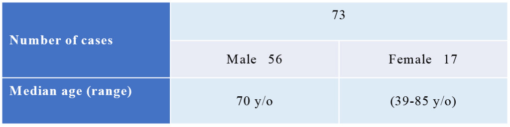
JCRM 2018-120 - Yasushi Rino Japan_F1

Figure 1. Patients

Pts were consisted of 56 males and 17 females and median age was 70 y/o (range: 39–85).

pts; patients

Replacement therapy

For the intra-muscular vitamin B12 replacement (IM), cyanocobalamin was administered. The dosage was 500 µg every 1–3 month.

For the oral vitamin B12 replacement (PO), mecobalamin was administrated. The dosage comprised 500 µg tablet of mecobalamin for a total of 500–1500 µg daily.

For the intra-venous vitamin B12 replacement (IV), cyanocobalamin was administered. The dosage was 500 µg every 1–3 month.

Measurement of Vitamin B12

Before the start of treatment and every 1 to 6 months after vitamin B12 replacement, serum specimens were obtained. Serum vitamin B12 were measured with the chemiluminescent enzyme immunoassay in a UniCel DxH 800 analyzer (Beckman Coulter, USA).

Results

The median time to vitamin B12 deficiency was 273.5 days (about 9 months, range: 66–953 days) after TG (Fig. 2). Initial treatment of vitamin B12 replacements were IM (dosage: 500 µg cyanocobalamin) for 42 pts, PO (dosage: 1,000–1,500 µg mecobalamin a day) for 28 pts and IV (dosage: 500 µg cyanocobalamin) for 3 pts. Serum vitamin B12 levels were not normalized in 10 (23.8%) out of 42 pts of IM, 4 (14.3%) out of 28 pts of PO, 1 (33.3%) out of 3 pts of IV, respectively. There is no significant difference between IM, IV and PO. Finally, all pts were treated with PO. But serum vitamin B12 levels decreased in 4 (5.5%) out of 73 pts of PO. These 4 pts were not continuously administered vitamin B12. After explaining it not to discontinue vitamin B12 replacement therapy to these 4 pts, the serum vitamin B12 levels became within normal range (Fig. 3). Final vitamin B12 doses of replacement therapy were 500 µg of 20, 1,000 µg of 17, and 1,500 µg of 36 out of 73 pts, respectively (Fig. 4).

JCRM 2018-120 - Yasushi Rino Japan_F2

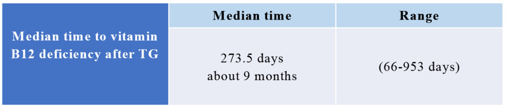
Figure 2. Median time of vitamin B12 deficiency

The median time to vitamin B12 deficiency was 273.5 days (about 9 months, range: 66–953 days) after TG.

TG; total gastrectomy

JCRM 2018-120 - Yasushi Rino Japan_F3

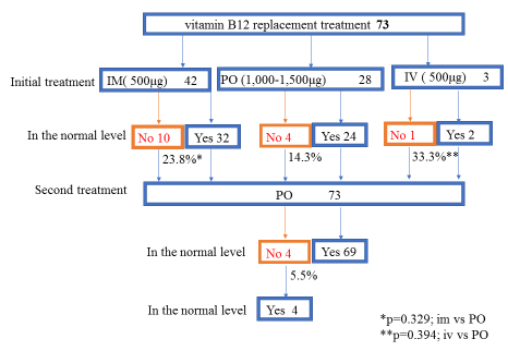
Figure 3. Vitamin B12 replacement therapy

Initial treatment of vitamin B12 replacements were IM (dosage: 500 µg cyanocobalamin) for 42 pts, PO (dosage: 1,000–1,500 µg mecobalamin a day) for 28 pts and IV (dosage: 500 µg cyanocobalamin) for 3 pts. “No” means that serum vitamin B12 levels were not normalized. “Yes” means that serum vitamin B12 levels were normalized. “No” was 10 (23.8%) out of 42 pts of IM, 4 (14.3%) out of 28 pts of PO, 1 (33.3%) out of 3 pts of IV, respectively. There is no significant difference between IM, IV and PO. Finally, all pts were treated with PO. But serum vitamin B12 levels decreased in 4 (5.5%) out of 73 pts of PO. These 4 pts were not continuously administered vitamin B12. After explaining it not to discontinue vitamin B12 replacement therapy to these 4 pts, the serum vitamin B12 levels became within normal range.

IM; intra-muscular vitamin B12 replacement, PO; oral vitamin B12 replacement, IV; intra-venous vitamin B12 replacement, pts; patients

JCRM 2018-120 - Yasushi Rino Japan_F4

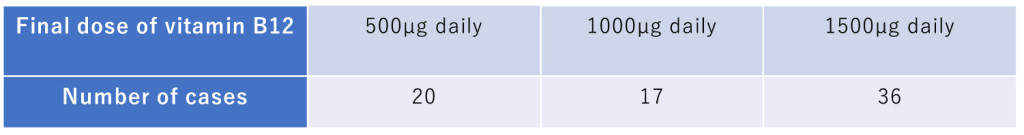
Figure 4. Final dose of vitamin B12 PO

Final vitamin B12 doses of replacement therapy were 500 µg of 20, 1,000 µg of 17, and 1,500 µg of 36 out of 73 pts, respectively.

Discussion

Normally, there is a vast excess of intrinsic factor. But after gastrectomy, there is little intrinsic factor. Especially, after TG, there is no intrinsic factor. Because of this, vitamin B12 deficiency is an inevitable complication after TG. Cumulative vitamin B12 deficiency rates were 100% for TG and 15.7% for DG 4 years after surgery. The median time to vitamin B12 deficiency was 15 months after TG, whereas the median time was not reached after DG [3]. In this report, the median time to vitamin B12 deficiency was about 9 months after TG. Our result was shorter than previous report from Korea [3]. The difference between Japan and Korean dietary habits may be the cause of this result. It is described in the textbook as follows [5], body stores are of the order of 2–3mg, sufficient for 3–4 years if supplies are completely cut off. May TG progress a daily choler excretion or a consumption of the vitamin B12? Gastric atrophy is background of gastric cancer. Most gastric cancer patients are complicated with atrophic gastritis. A severe lack of intrinsic factor due to gastric atrophy [5]. Vitamin B12 storage before TG would be decreased in gastric cancer patients with gastric atrophy. This small storage of vitamin B12 may be the cause of the shorter median time to vitamin B12 deficiency after TG.

Vitamin B12 absorption has two mechanisms. One is passive, occurring equally through buccal, duodenal, and ileal mucosa; it is rapid but extremely inefficient, with < 1% of an oral dose being absorbed by this process. The normal physiologic mechanism is active; it occurs through the ileum and is efficient for small oral dose of vitamin B12, and it is mediated be gastric intrinsic factor. Dietary vitamin B12 is released from protein complex by enzymes in the stomach, duodenum, and jejunum; it combines rapidly with a salivary glycoprotein that belongs to the family of cobalamin-binding proteins known as haptocorrins. In the intestine, the haptocorrin is digested by pancreatic tripsin and the cobalamin is transferred to intrinsic factor. Intrinsic factor is produced in the gastric parietal cells of the fundus and body of the stomach. The intrinsic factor-cobalamin complex passed to the ileum, where intrinsic factor attaches to specific receptor (cubilin) on the microvillus membrane of the enterocytes. The intrinsic factor-cobalamin complex enters the ileal cells, where intrinsic factor is destroyed. After a delay of about 6 hours, the cobalamin appears in portal blood attached to transcobalamin II. Between 0.5–5 µg of cobalamin enter the bile each day. This binds to intrinsic factor, and a major portion of biliary cobalamin normally is reabsorbed together with cobalamin derived from sloughed intestinal cells. Because of the appreciable amount of cobalamin undergoing enterohaptic circulation, cobalamin deficiency develops more rapidly in individuals who malabsorb cobalamin that it does in vegans, in whom reabsorption of biliary cobalamin is intact [5, 6].

There is an absorption disorder by lack of intrinsic factor and daily choler excretion of vitamin B12. And altered dietary intake and malabsorption caused body weight loss. Because of these variety of factors after TG, vitamin B12 deficiency would be occurred within one year after total gastrectomy, frequently.

Recently, body weight loss after gastrectomy was independent risk factor for continuation of adjuvant chemotherapy and survival [7, 8].

Pts who was undergone TG have only passive absorption of vitamin B12. In this reason, intramuscular injection of vitamin B12 is recommended as a parenteral chronic treatment for pts with vitamin B12 deficiency after TG [1, 2]. Previous reports indicated that oral vitamin B12 successfully treats patients with pernicious anemia who have defects in the main vitamin B12 absorption mechanisms as a result of the presence of autoantibodies specific for intrinsic factor or a history of surgical resection of the ileum [9–11].

In spite of vitamin B12 administration orally is not a reliable route after TG, Kim et al previously reported that serum vitamin B12 increased after oral and intramuscular administration of vitamin B12 in TG patients. In this report, for the oral vitamin B12 replacement, mecobalamin was administrated. The dosage comprised three 500 µg tablets of mecobalamin for a total of 1500 µg daily. For the intramuscular vitamin B12 replacement, cyanocobalamin was administered. The dosage was 1000 µg weekly for 5 weeks and monthly thereafter [4]. After TG, vitamin B12 absorption has only one mechanism that is passive absorption with <1 % of an oral dose being absorbed by this process. When three 500 µg tablets of mecobalamin for a total of 1500 µg daily are administered, total amount of vitamin B12 absorption is about <15µg. On the other hand, between 0.5–5 µg of cobalamin excreted choler each day [5, 6]. Because of passive absorption and choler excretion, three 500 µg tablets of mecobalamin administration is reasonable replacement therapy for vitamin B12 deficiency after TG. But mecobalamin tablet has high concentration of vitamin B12. We presume that passive absorption rate would be higher than 1% when mecobalamin tablet is administered orally. Because our results showed that 500 µg tablets of mecobalamin oral administration maintained normal serum vitamin B12 levels in 20 out of 73 pts. We guessed it in this way. When only 500 µg of mecobalamin were administered orally, total amount of vitamin B12 absorption should be over 5 µg. Otherwise there is more quantity of passive absorption than the quantity of choler excretion, and it is impossible that serum vitamin B12 maintains within normal levels.

Vitamin B12 deficiency worsen quality of life after TG. Because pernicious anemia, bilateral peripheral neuropathy or degeneration(demyelination) of the posterior and pyramidal tracts of the spinal cord, and optic atrophy, cerebral symptoms or dementia would be occurred [12,13]. Hwang et al. [14] reported a patient who developed ataxic gait 9 years after TG for gastric cancer. Spinal cord degeneration due to vitamin B12 deficiency should be suspected in patients with neurological disorders, including gait disturbance, after gastrectomy, even as a long-term complication. Like this case, vitamin V12 deficiency symptoms are long-term complications. Vitamin B12 replacement therapy should be necessary and continued.

In conclusion, after TG, vitamin B12 deficiency is occurred in 100% pts and within 1 year after TG frequently. Vitamin B12 replacement therapy should be necessary and continued routinely. IM is a reliable therapy. But IM needs to go to clinic a month and is painful. According to our results, one tablet PO a day is enough. The vitamin B12 deficiency symptoms could be prevented and number of the going to clinic on pts could be reduced using this replacement therapy.

Conflict of interest statement:  The authors declare no conflicts of interest in association with the present study.

Acknowledgments: None

Abbreviations

TG: total gastrectomy

DG: distal gastrectomy

pts: patients

IM: intra-muscular vitamin B12 replacement

PO: oral vitamin B12 replacement

IV: intra-venous vitamin B12 replacement

References

  1. Brunicardi CF, Andersen DK, Billiar TR, Dunn DL, Hunter JG, Pollock RE. (2005) Schwartz’s Principles of Surgery, In: Dempsy DT, editor. Stomach. 8th ed. New York, McGraw-Hill: pp. 933–95.
  2. Townsent CM, Beauchamp RD, Evers BM, Mattox KL. (2012) Sabiston Textbook of Surgery, In: Mahvi DM, Krantz SK, editor. Stomach. 19th ed. Philadelphia: Elsevier Saunders; pp.1182–1226.
  3. Hu Y, Kim HI, Hyung WJ, Song KJ, Lee JH, Kim YM, et al. (2013) Vitamin B(12) Deficiency after gastrectomy for gastric cancer: an analysis of clinical patterns and risk factors. Ann Surg. 258(6): 970–5. doi: 10.1097/SLA.0000000000000214. [Crossref]
  4. Kim HI, Hyung WJ, Song KJ, Choi SH, Kim CB, Noh SH. (2011) Oral vitamin B12 replacement: an effective treatment for vitamin B12 deficiency after total gastrectomy in gastric cancer patients. Ann Surg Oncol. 18(13): 3711–7. doi: 10.1245/s10434-011-1764-6. [Crossref]
  5. Kasper DL, Fauci AS, Hauser SL, Longo DL, Jameson JL, Loscalzo J. (2015) Harrison’s Principles of Internal Medicine vol. 2, In: Hoffbrand AV, editor. Megaloblastic anemias. 19th ed. New York; McGraw-Hill; pp.640–9.
  6. Brunicardi CF, Andersen DK, Billiar TR, Dunn DL, Hunter JG, Pollock RE. (2005) Schwartz’s Principles of Surgery, In: Whang EE, Ashley SW, Zinner MJ, editor. Small intestine. 8th ed. New York: McGraw-Hill; pp.1017–5.
  7. Aoyama T, Sato T, Maezawa Y, Kano K, Hayashi T, Yamada T, et al. (2017) Postoperative weight loss leads to poor survival through poor S-1 efficacy in patients with stage II/III gastric cancer. Int J Clin Oncol. Feb 7. doi: 10.1007/s10147-017-1089-y. [Crossref]
  8. Aoyama T, Yoshikawa T, Shirai J, Hayashi T, Yamada T, Tsuchida K, et al. (2013) Body weight loss after surgery is an independent risk factor for continuation of S-1 adjuvant chemotherapy for gastric cancer. Ann Surg Oncol. 20(6): 2000–6. doi: 10.1245/s10434-012-2776-6. [Crossref]
  9. Berlin H, Berlin R, Brante G. (1968) Oral treatment of pernicious anemia with high doses of vitamin B12 without intrinsic factor. Acta Med cand. 184: 247–58 [Crossref]
  10. Kuzminski AM, Del Giacco EJ, Allen RH, Stabler SP, Lindenbaum J.(1998) Effective treatment of cobalamin deficiency with oral cobalamin. Blood. 92: 1191–8. [Crossref]
  11. Berlin R, Berlin H, Brante G, Pilbrant A. (1978) Vitamin B12 body stores during oral and parenteral treatment of pernicious anemia. Acta Med Scand. 204: 81–4. ) [Crossref]
  12. Kasper DL, Fauci AS, Hauser SL, Longo DL, Jameson JL, Loscalzo J. (2015) Harrison’s Principles of Internal Medicine vol. 2, In: Seeley WW,Miller BL, editor. Alzheimer’s disease and other dementias. 19th ed. New York; McGraw-Hill; pp. 2598–608.
  13. Kasper DL, Fauci AS, Hauser SL, Longo DL, Jameson JL, Loscalzo J. (2015) Harrison’s Principles of Internal Medicine vol. 2, In: Amato AA, Barohn RJ, editor. Peripheral neuropathy. 19th ed. New York; McGraw-Hill; pp. 2674–94.
  14. Hwang CH, Park DJ, Kim GY. (2016) Ataxic gait following total gastrectomy for gastric cancer. World J Gastroenterol. Oct 7; 22(37): 8435–8438. DOI: 10.3748/wjg.v22.i37.8435. [Crossref]

Burroughs Wellcome: The Seminal Link between Academia and the Pharmaceutical Industry

DOI: 10.31038/IMROJ.2018341

Abstract

This article reviews the research carried out by outstanding scientists to underscore the significant role played by Burroughs Wellcome Research Laboratories in erasing the differences in the objectives of scientists in academia and those in industry. These enlightened policies not only markedly advanced our fund of scientific knowledge in the biomedical sciences but led to the production of drugs that were of major benefit to mankind.

Introduction

Henry S. Wellcome (1853–1936) was an American-British entrepreneur who established the Burroughs Wellcome pharmaceutical conglomerate in London with his partner Silas Burroughs in the late 1880’s. Four years later, Wellcome formed a research component, which he named The Wellcome Physiological Research Laboratories. The creation of laboratories to conduct research was quite unusual in the late 1800’s, especially in association with a pharmaceutical enterprise [1–3]. When Henry Wellcome passed away in 1936, he left two legacies, his pharmaceutical company, The Wellcome Foundation and The Wellcome Trust, which distributed the financial resources for biomedical research [4].

This article will convey the company’s long time commitment to research by the fact that the staff scientists highlighted herein won a share of five Nobel Prizes (see below). At the same time, as a result of its long term involvement in basic research, Burroughs Wellcome became a major factor in bridging the gap that existed between academia and the pharmaceutical industry.

Sir Henry Hallett Dale (1875–1968)

Henry Dale (Figure 1), the renowned pioneer and leader in the discipline of Physiology/ Pharmacology, was the first major recruit to join Henry Wellcome’s new research initiative when he reluctantly accepted a research position at The Wellcome Research Laboratories in 1904 [5]. In those days it was unusual for a researcher at a university to give up his academic freedom to work in industry, and several colleagues advised him to decline the offer. However, Wellcome convinced Dale that he would be able to conduct basic research without concern for the business side of the organization.

Although Dale was free to select his own topics of research to investigate, Wellcome requested that Dale undertake the problem of ergot, which was marketed by the company as an abortifacient. Wellcome’s interest in ergot was in part commercially driven by the fact that Parke Davis was also marketing an ergot preparation for use in obstetrics. This competition prompted Henry Wellcome to also recruit a chemist, George Barger, whom he also encouraged to investigate ergot. Dale did not plan on ergot studies occupying a major portion of his time; however, his initial investigations into ergot properties proved to be unexpected and exciting and led him on a path that would ultimately provide the foundation for understanding the pharmacology of autonomic drugs and culminate in the awarding of the Nobel Prize.

IMROJ 2018-108 - Ronald Rubin USA_F1

Figure 1. Sir Henry Hallett Dale (1875–1968)

In 1906, Dale provided the first example of an adrenergic blocking agent by demonstrating that a substance obtained from ergot called ergotoxine reversed the hypertensive effect of sympathetic nerve stimulation and epinephrine (adrenaline) [6]. The sympatholytic action of ergotoxine prompted Dale to interpret his own studies in the light of recent work by Thomas Elliott, who in 1905 observed that the action of exogenous epinephrine mimicked the effects of sympathetic nerve stimulation [7]. Thus, ergotoxine became important in medical history because Dale’s observation that it inhibited sympathetic activity eventually led to the discovery of chemical synaptic transmission. In 1910, Dale also published a detailed account of the sympathomimetic actions of a number of biogenic amines synthesized by George Barger [8]. Unfortunately, Dale chose to exclude the epinephrine (adrenaline) series of sympathomimetics and overlooked the most physiologically relevant derivative – norepinephrine (noradrenaline) – and thus delayed for several more decades the discovery of norepinephrine as a physiological neurotransmitter.

Ergot yielded additional constituents, including histamine in 1907 and acetylcholine in 1913, although neither provided any results that could be marketed for sale. A few years later, an accidental observation made with a particular extract of ergot prompted Dale’s interest in the possible existence of chemical transmission across neuronal synapses. A conventional dose of this extract caused a profound inhibition of heart rate, and was later identified as the labile substance, acetylcholine. In a paper published in 1914, Dale identified a nicotinic and muscarine-like substance in ergot as acetylcholine [9]. In this article, Dale summarizes his work by noting that “acetylcholine occurs occasionally in ergot, but its instability renders it improbable that its occurrence has any therapeutic significance [10].” Nevertheless, such findings set the stage for the classical experiments of Otto Loewi in 1921 and beyond, which provided direct evidence in favor of the theory of chemical synaptic transmission.

Thus, because of Dale’s commitment to deciphering the puzzling effects of ergot, much of our knowledge of the action of autonomic drugs on the physiological components of the autonomic nervous system stems directly from the work of Henry Dale carried out at Burroughs Wellcome Research Laboratories. The quality of Dale’s work was recognized by his academic peers and had much to do with reducing the prevailing negative opinion of the scientific mission of pharmaceutical companies. Dale was subsequently elected to the Royal Society and later served as President of the Royal Society of Medicine. He was knighted in 1932, and shared the Nobel Prize with Otto Loewi for a discovery of fundamental physiological significance that had its origins in a drug company interested in the pharmacological properties of ergot.

Dale spent 10 years at the Burroughs Wellcome Research Laboratories at Brockwell Park, where a great deal of his most productive work was carried out. Although Dale was appointed the first Director of the Medical Research Council at the National Institute for Medical Research in 1928, his link to Burroughs Wellcome was not at an end. In 1936, he became associated with the Trust which had been created by the will of Henry Wellcome. He first served as a Trustee, then as Chairman from 1938 to 1960. He spent the last eight years of his life as its scientific advisor [11]. In addition, a special Henry Dale Fellowship sponsored by the Wellcome Trust provides funds for biomedical research. The basic research fostered by Henry Wellcome and implemented by Henry Dale was not only profoundly significant in its day, but it led Burroughs Wellcome to become a dominant force in biomedical research. And, it was Sir Henry Dale who set the landscape for those who were to follow.

Sir John Robert Vane (1927–2004)

John Vane (Figure 2) was considered one of Britain’s most eminent pharmacologists [12]. He began working with Joshua Harold Burn at Oxford in 1946, where he learned to utilize bioassays. At the time, chemical methods were generally unavailable and bioassays, which detected and measured sensitivity of tissue strips to biologically active substances, required laborious procedures. As a graduate student, I myself toiled at a bath containing aortic strips to measure catecholamines by bioassay, and my task was made much easier when I learned the fluorometric method of assaying adrenomedullary catecholamines at Burroughs Wellcome.

IMROJ 2018-108 - Ronald Rubin USA_F2

Figure 2. Sir John Vane

After graduating in Pharmacology and obtaining additional experience in the United States at Yale University, Vane returned to the United Kingdom where he was offered a position in The Department of Pharmacology at the University of London, which was headed by Sir William Paton. During those years, Vane, striving to move beyond outdated methodologies and antiquated concepts, further developed the blood-bathed organ bioassay system. By slowly perfusing mammalian blood over a series of isolated tissues in a cascade, Vane was able to measure the release of biologically active substances in a manner that simulated release in vivo. One of the first major biochemical processes to be discovered using the blood-bathed organ system was the conversion of angiotensin I to angiotensin II in the pulmonary vasculature. This finding led to the development of Angiotensin Converting Enzyme Inhibitors, which at the time revolutionized the treatment of hypertension. But, it was at the College of Surgeons that John Vane made an indelible mark on the scientific world by elucidating the mechanism of action of aspirin [13].

Vane left the Chair at the Royal College of Physicians in 1973, and followed the example of Henry Dale by joining The Wellcome Research Laboratories in the UK [14]. Vane, like Henry Dale, found that friends and colleagues were dubious about his accepting the offer to enter the industrial realm. Nevertheless, Vane was impressed by the fact that some seventy years before, Henry Dale had accepted a position at Burroughs Wellcome after experiencing academic life. Understanding that good science was not limited to academia, Vane undertook his new role as Director of Research and Development for a major pharmaceutical company.

The fact that he was able to take a number of his research team with him was a major factor in his final decision, and Vane never expressed any regrets about this move. The colleagues he recruited from the Royal College of Surgeons, included Salvador Moncada, Richard Gryglewski, and Rod Flower [15].This research group composed of very talented individuals of diverse ethnic origins, backgrounds, and traditions worked together in a highly competitive research environment. Vane’s laboratory became known as the Prostaglandin Research Group and served as a venue where basic pharmacological research could be carried out without being limited by outdated and narrow approaches to biomedical research. An example of the rewards that could be achieved by this philosophy was the other watershed in Vane’s storied history, the discovery of prostacyclin.

The years spent at Burroughs Wellcome was a challenging period for John Vane since he assumed a new set of managerial responsibilities, as well as research goals. Imbuing colleagues with the concept that it was possible to carry out quality science in an industrial setting, Vane advised them to follow their instincts with regard to drug discovery. This concept soon reaped rewards when in 1976 the Prostaglandin Research Group under the leadership of Salvador Moncada discovered prostacyclin and elucidated its pharmacological properties by utilizing the bioassay of extracts from platelets and vascular tissues [16]. Capitalizing on the versatility of the bioassay cascade, prostacyclin was found to be the main product of arachidonic acid metabolism in arteries and veins and its major effect was to inhibit platelet aggregation by stimulating adenylate cyclase.

John Vane presided over an environment in which there was a strong interaction with academia and the pharmaceutical industry. He, like Henry Dale, clearly demonstrated how it was possible to conduct quality scientific research in an industrial setting. During those years, Vane was awarded several honors, including Fellowship in the Royal Society, The Lasker Prize, and in 1982 the Nobel Prize for Medicine [17]. Salvador Moncada, who was also involved in the discovery of nitric oxide, was considered by some as deserving of a share of the Nobel Prize [18].

The work carried out by John Vane and his associates at the Wellcome Foundation spawned important research around the world that provided additional insights into the key factors that regulate blood circulation. In 1993, after much more information was accumulated about prostacyclin, Vane eventually reached the conclusion that the endothelium occupied a key role in regulating blood circulation and that prostacyclin, as well as nitric oxide, was responsible for defending against atherosclerotic angiopathies [19].

One of Vane’s other major contributions was to promote the link between scientists at academic institutions with those in the pharmaceutical industry, and he did a great deal to blur the boundaries that had separated these two groups of research scientists. In 1985, Vane returned to academia by establishing the William Harvey Research Institute at the Medical College of St. Bartholomew’s Hospital, where his research group focused its attention on cyclooxygenase-2 inhibitors and the interplay between nitric oxide and endothelin in the regulation of vascular function [20].

Sir James Whyte Black (1924- 2010)

The Nobelist, James Black (Figure 3), was one of the first scientists who utilized “rational design” for discovering new drugs [21,22]. Much of Black’s early work was carried out at the now defunct Imperial Chemical Industries (ICI Pharmaceuticals) in the United Kingdom from 1958–1964. Becoming aware of the importance of a balance between experimental research and drug development, Black and coworkers developed propranolol, the first clinically effective beta-adrenergic antagonist. The development of this drug not only represented a marked advance in the pharmacotherapy of hypertension, angina pectoris, and arrhythmias, but it also initiated further studies on the physiological role of beta adrenergic receptors by subsequently dividing them into beta-1 and beta-2 subtypes. At Black’s next position at Smith Kline and French (now GlaxoSmithKline), he introduced a new concept in the treatment of gastric ulcers by producing a drug that blocks histamine (H2) receptors.

IMROJ 2018-108 - Ronald Rubin USA_F3

Figure 3. Sir James Black (left), Gertrude Elion (middle), and George Hitchings (right)

Black wanted to escape from commercial constraints in order to have the freedom to pursue his research interests, so he returned to academia by accepting a Chair in Pharmacology at University College London. But, it was not long before John Vane invited Black to join him at Burroughs Wellcome in the United Kingdom in 1977. Black accepted the offer to serve as Director of Therapeutic Research in order to implement ideas he held about the reasons for the success and failure of industrial projects.

During the next six years at Burroughs Wellcome, Black failed to make much progress in his managerial role, but his research, now involving analytical pharmacology, produced a major advance in the description of the functional effects of drugs and their therapeutic potential. A collaboration with Paul Leff, which compared pharmacological data to quantitative models, developed a new framework for categorizing and analyzing drug actions. The most significant tool employed was the operational model, in which the quantitation of agonist activity in one test system enabled the prediction of activity in another system [23]. The principles of this analytical approach have since been employed in drug classification and the mechanisms of drug action [24].

However, despite the fact that Burroughs Wellcome enjoyed an impeccable reputation with regard to its research activity, Black spent seven years dealing with what he felt were traditional and conservative attitudes. For Black, the interplay between corporate commercial needs and personal scientific aspirations provided an ongoing dilemma. The perceived counterproductive policies were resolved when a small independent research unit in King’s College, London was established for him in 1984 and financially supported by Burroughs Wellcome. It had modern facilities, and together with talented researchers and doctoral students, Black was able to carry out non-profit research with complete independence. Black received his Nobel Prize there in 1988, together with George Hitchings and Gertrude Elion (Figure 3), and remained at Kings College as Professor of Analytical Pharmacology until 1993 when he became Professor Emeritus. In 1988, Black also established the James Black Foundation in the United Kingdom to promote his own vision of pharmacological research [25].

As a fulltime employee of pharmaceutical companies, including Burroughs Wellcome, Black was provided with the independence and resources to be successful. In this way, he was able to offer benefit to both his company and for the good of mankind. Although he derived little personal gain from his discoveries, his strong sense of independence, combined with his dislike for large institutions, caused him to frequently abandon positions whenever he felt the short-sightedness of corporations was obstructing progress in his research. Black’s outstanding quality as a researcher can best be described as being able to discover drugs by meticulous structural design based upon known agonists, rather than by random screening.

George Herbert Hitchings (1905–1998) and Gertrude Belle Elion (1918–1999)

George Hitchings and Gertrude Elion (Figure 3) were the only Nobel Laureates who spent their entire careers at Burroughs Wellcome, even when the company moved from Tuckahoe, New York to North Carolina during a period of sustained research activity. Their investigations covered a span of nearly 40 years and were previously chronicled in some detail [26].

Hitchings received his doctoral degree in Biochemistry from Harvard in 1933, where he studied analytical methods used in physiological studies of purines at a time when little was known about nucleic acid metabolism. After working at several colleges for ten years, Hitchings was hired in 1942 as the only scientist in the Biochemistry Department at Burroughs Wellcome at the Tuckahoe New York facility. Two years later, he recruited Gertrude Elion, a chemist by training, to join his small research group. Elion was then able to leave a rather tedious job of food analyst to join Hitchings when World War II made research positions available for women.

Although up to that time women had difficulty finding jobs in scientific research, Hitchings had no trouble working with women or men from different ethnic backgrounds or religious beliefs, and he encouraged Elion to learn as rapidly and as much as she could. Because she never felt constrained to restrict herself to the subject of chemistry, Elion, who possessed only a Bachelor’s and a Master’s degree, greatly expanded her scope of knowledge in biochemistry, pharmacology, immunology and virology. As a result, Elion began to take on more and more responsibility by concentrating almost exclusively on purines. Because of residency requirements at Brooklyn Polytechnic University, which would take her away from Burroughs Wellcome, Elion never obtained a formal doctorate. However, she was later awarded an honorary PhD degree from Polytechnic University in 1989 and an honorary SD degree from Harvard in 1998.

As previously noted, drug development had historically been a consequence of random trial and error, as in the case of sulfa drugs for example [27]. Because of the legacy provided by the vision of Henry Wellcome, Hitchings and Elion, like James Black, were free to diverge from this approach by using what then was called “rational drug design [28].” It was based upon the supposition that the understanding of basic biochemical and physiological processes formed the basis for the design and development of drugs. Because their research was based upon the premise that drugs could be designed which were based upon differences in nucleic acid metabolism in normal and abnormal cells, Elion and Hitchings employed specifically designed chemicals to form atypical DNA in abnormal cells which did not affect normal cells. By blocking nucleic acid synthesis, the growth of the abnormal cells would be inhibited. Thus, for example, Hitchings postulated that folic acid deficiency would lead to alterations in the synthesis of purines and pyrimidines and thus DNA.

By 1950, this line of research reaped major dividends when Hitchings and Elion synthesized two antimetabolites, diaminopurine and thioguanine. These substances proved to be effective in the treatment of leukemia. In 1957, further alterations in chemical structure led to the production of azathioprine (AZT). This immunosupressant is now used to prevent the rejection of transplanted organs and to treat rheumatoid arthritis and other autoimmune disorders. However, in the 1980’s, because AZT was the primary treatment for AIDS, the United States government allowed Burroughs Wellcome to apply for full patent rights to the drug. As a result, Burroughs Wellcome was able to charge an exorbitant price for AZT to patients with AIDS, despite the fact that the majority of the company was owned by a charitable Foundation, the Wellcome Trust [29,30]. Thus, there was an aspect of the policies of Burroughs Wellcome that dimmed the luster of its legacy.

In 1967 Hitchings became Vice President in charge of research at Burroughs Wellcome, which virtually terminated his involvement in research and redirected his attention to philanthropy. Elion took over his position as Head of the Department of Experimental Therapy. In 1970, the group headed by Hitchings and Elion moved to Research Triangle Park, North Carolina, where they developed the first antiviral drug acyclovir, as well as allopurinol, which is used in the treatment of gout.

Although Henry Wellcome had always been resolute in his commitment to unencumbered biomedical research, Hitchings and Elion did not always find that their efforts were totally supported by management. Hitchings and Elion were subjected to interference by the Head of the Tuckahoe laboratories, William Creasy, who tried to persuade the chemists to work on projects that he favored. Eventually, Creasy relented, realizing that the successes achieved by Hitchings and Elion made it unwise to interfere with their work [31]. In marked contrast, Hitchings and his elite group had key collaboration from the Sloan-Kettering Institute to examine whether purines/pyrimidines possessed anti-neoplastic activity. Moreover, the financial support afforded by Sloan- Kettering enabled Burroughs Wellcome to expand and eventually become self-sustaining [32]. Thus, the ability of Hitchings and Elion to test their theories without interference by commercial considerations led to discoveries of important principles for drug treatment resulting in the development of new approaches to pharmacotherapy.

Hitchings and Elion were initially overlooked by the Nobel Committee. One reason perhaps had to do with the fact that the Nobel Prize Committee rarely honors the work of scientists who develop new drugs. However, in 1988 they were awarded the Nobel Prize, some 30 years after most of their major discoveries. Gertrude Elion underscored the profound significance of her work in a review published in Science in 1989, “…chemotherapeutic agents are not only ends in themselves but also serve as tools for unlocking doors and probing Nature’s mysteries [33].” When Hitchings retired in 1975, and Elion followed eight years later, another memorable chapter in the history of Burroughs Wellcome came to an end.

John J. Burns (1920–2007) and Allan H. Conney (1930–2013)

During the same period that Hitchings and Elion were making their invaluable drug discoveries in the Biochemistry Department, John Burns (Figure 4) joined Burroughs Wellcome as Vice-President and Director of Research in 1960. Prior to his arrival at the Tuckahoe New York facility, Burns had worked at the NIH and had provided valuable information about the biosynthesis and metabolism of Vitamin C (ascorbic acid) and the etiology of scurvy [34]. At Burroughs Wellcome, his seminal investigations demonstrated the clinical importance of microsomal enzyme induction. In particular, Burns demonstrated that phenylbutazone is converted in man to two major metabolites, one with anti-rheumatic activity, the other possessing uricosuric actions [35]. The importance of this basic research was underscored by the fact that during the 1960’s, the NIH provided financial support for the research being conducted at the Tuckahoe facility.

IMROJ 2018-108 - Ronald Rubin USA_F4

Figure 4. John J. Burns. Courtesy of ASPET)

Coincident with the advent of John Burns, a talented research group was formed in the Department of Biochemistry that included Allan Conney, Ronald Kuntzman, and Richard Welch. Providing fundamental knowledge concerning drug metabolism and its clinical implications, this group was the first to demonstrate the clinical significance of microsomal enzyme induction by showing that chronic administration of several drugs to animals stimulated their metabolism and decreased their toxicity [36]. Also by employing selective inhibitors, they were able to determine whether a drug possessed intrinsic pharmacological activity or owed its activity to a metabolite. This work was of considerable significance in the field of drug metabolism and led to early studies on individual differences in the metabolism of drugs in humans.

John Burns wore many hats as a scientist. While at Burroughs Wellcome, he was also an advisor to a number of biotech companies, a member and officer in a large number of national and international scientific committees, and served as a Visiting Professor of Pharmacology at Albert Einstein College of Medicine. In his capacity as an adjunct faculty member, Burns became thesis advisor to a graduate student, Louis Lemberger. Alfred Gilman, the Chairman of the Pharmacology Department was not enamored of the fact that Lemberger had graduated from a Pharmacy School. Nevertheless, Gilman allowed Lemberger to carry out his doctoral thesis with John Burns. At the time, I was a graduate student at Albert Einstein, and because of the prevailing views I was surprised that one of my fellow students had been allowed to carry out his research at an industrial setting.

Despite the vestiges of prejudice that still existed in academia about drug companies at the time, the legacy generated by Henry Wellcome endured. Subsequently, John Burns encouraged Lemberger to obtain his MD degree and gain further clinical training; and so, Lemberger went on to an outstanding career as Director of Clinical Pharmacology at Eli Lilly in Indianapolis Indiana and as Professor of Pharmacology Medicine and Psychiatry at the Indiana School of Medicine [37]. He was involved in the development of several centrally acting drugs, including Prozac, a commonly prescribed anti-depressant.

John Burns subsequently left Burroughs Wellcome in 1968 to serve as Vice President of Research & Development at Hoffmann LaRoche, where he helped to develop the famed Roche Institute of Molecular Biology. Adhering to the view that basic research would lead to practical results, Burns supported basic research as much as any pharmaceutical executive. The extensive research conducted by Burns and his colleagues on the metabolic fate and the mechanism of action of drugs provided a fundamental basis for discovering new drugs and improving their therapeutic use. After Dr. Burns retired from Hoffman LaRoche, he served as Adjunct Professor of Pharmacology at Weill Medical College and was scientific advisor to many biotech companies and a member of the National Academy of Sciences. However, his work at Burroughs Wellcome proved to be seminal.

The Biochemistry group led by Allan Conney (Figure 5) was also involved in investigating other areas of drug metabolism, including cytochrome P-450, a family of enzymes responsible for the biotransformation of many medications, toxic substances, and environmental chemicals [38,39]. Conney’s work provided the molecular basis for understanding how drugs induce tolerance and environmental chemicals produce mutagenesis and carcinogenesis.

Much of Conney’s career was spent in the pharmaceutical industry, first at Burroughs Wellcome and then at Hoffman-LaRoche, where he rejoined John Burns. Further recognition of Conney’s work came from a prestigious faculty appointment at Rutgers University in 1987, where he established the Department of Chemical Biology and founded the Laboratory for Cancer Research. At Rutgers University, Conney continued to carry out research mainly on cancer prevention [40]. His contributions were recognized by his election to the National Academy of Sciences in 1982, and as President of the American Society for Pharmacology and Experimental Therapeutics (ASPET) (1983–1984). During the years 1965–1978, Dr. Conney was among the 40 most cited scientists in the field of pharmacology.

IMROJ 2018-108 - Ronald Rubin USA_F5

Figure 5. Allan Conney

It was fitting that we end this article by recounting the work of Allan Conney, because it defines a gifted scientist who readily bridged the gap between industry and academia. The now entrenched alliances between academia and industry provided another important advance in mankind’s search for more effective medications. Once again, it took some time, but the overall lesson learned by scientists is that forward thinking and cooperation will always trump unfounded biases.

Epilogue

The research laboratories that Henry Wellcome set up first in the United Kingdom in 1880 and then throughout the world employed elite researchers who performed rational and outstanding biomedical research. As a result, the company set the stage for the advent of Pharmacology as an established biomedical discipline. Although the Burroughs Wellcome Research Institute is no longer a functional entity, having been assimilated by Smith/Kline/Glaxo in the 1980’s, the research arm of the company provided the path for academicians to join forces with industrial companies to produce medications that have extended human life and reduced human suffering.

References

  1. Church R, Tansey EM (2007) Burroughs Wellcome and Company: Knowledge, Trust, Profit and the Transformation of the British Pharmaceutical Industry  Crucible Books, Lancaster UK, Pg No :1880–1940
  2. Bailey P (2008) The birth and growth of Burroughs-Wellcome & Co. The Wellcome Trust, 2008.
  3. James RR. Henry Wellcome (1994) Hodder & Stoughton, London, United Kingdom.
  4. Bailey P (2008) Henry Wellcome’s Physiological and Chemical Research Laboratories. Wellcome Trust. 2008. http//www.wellcome.ac.uk./About-us/History/WTX052313.htm.
  5. Dale HH (1938) An autobiographical sketch. Perspec. Biol. Med. 1938; 1: 128–130. See also Dale HH. Introduction; In: Adventures in Physiology. London. Pergamon Press. pp x-xvi.
  6. Dale HH (1906) On some physiological actions of ergot. J Physiol 34: 163–206. [crossref]
  7. Elliott TR (1905) The action of adrenalin. J Physiol 32: 401–467. [crossref]
  8. Barger G, Dale HH (1910) Chemical structure and sympathomimetic action of amines. J Physiol 41: 19–59. [crossref]
  9. Dale HH (1914) The action of certain esters and ethers of choline, and their relation to muscarine. J. Pharmacol. Exp. Ther. 6: 147–190.
  10. Dale HH. (op cit. Ref 7). pg. 189.
  11. Sánchez García P (2010) [The neurotransmision from the other side]. An R Acad Nac Med (Madr) 127: 257–268. [crossref]
  12. Moncada S (2006) Sir John Robert Vane: 29 March 1927 – 19 November 2004. Biogr Mem Fellows R Soc 52: 401–411. [crossref]
  13. Vane JR (1971) Inhibition of prostaglandin synthesis as a mechanism of action for aspirin-like drugs. Nat New Biol 231: 232–235. [crossref]
  14. John R. Vane- Biographical. Nobelprize.org. Nobel Media AB 2014. Web. 7 Apr 2016.
  15. Flower R (2006) John Vane. A Biographical Sketch. In Memory of Sir John Vane. (Nistico, G. McGiff J & Born G. Editors); 2006; Roma; Exorma. pp. 53–55.
  16. Moncada S (1982) Eighth Gaddum Memorial Lecture. Biological importance of prostacyclin. Br. J. Pharmacol. 76: 3–31.
  17. Obituary (2004) Sir John Vane. The Telegraph.
  18. Howlett R (1998) Nobel award stirs up debate on nitric oxide breakthrough. Nature 395: 625–626. [crossref]
  19. Vane JR (1994) The Croonian Lecture, 1993. The endothelium: maestro of the blood circulation. Philos Trans R Soc Lond B Biol Sci 343: 225–246. [crossref]
  20. Moncada S (2006) Sir John Robert Vane: 29 March 1927 – 19 November 2004. Biogr Mem Fellows R Soc 52: 401–411. [crossref]
  21. Braunwald E (2010) Sir James W. Black MD, FRS. In Memoriam. Circ. Res. 107: 3–5.
  22. Black’s autobiographical sketch can be found in: Les Prix Nobel. The Nobel Prizes 1988; Editor Tore Frangsmyr. (Nobel Foundation) 1989. Stockholm.
  23. Black JW, Leff P (1983) Operational models of pharmacological agonism. Proc R Soc Lond B Biol Sci 220: 141–162. [crossref]
  24. Kenakin T, Christopoulos A (2011) Analytical pharmacology: the impact of numbers on pharmacology. Trends Pharmacol Sci 32: 189–196. [crossref]
  25. Wright P (2010) Sir James Black obituary: Nobel prizewinning pharmacologist who invented new drugs such as beta-blockers. The Guardian.
  26. Rubin RP. A brief history of great discoveries in pharmacology. Pharmacol. Rev. 2007; 59: pp. 311–314.
  27. Rubin RP (op. cit. Ref. 23). pp. 315–318.
  28. Turney J (2009) Rational drug design: Gertrude Elion and George Hitchings. Wellcome Trust. London, England.
  29. http://www.nytimes.com/1989/08/28/opinion/azt-s-inhuman-cost.html
  30. Emmons WM (1991) Burroughs Wellcome and AZT. (A): Faculty & Research. Harvard Business School Case 792–004.
  31. Turney J. (op. cit. Ref. 25).
  32. George H. Hitchings- Biographical. The Nobel Prize in Physiology or Medicine 1988. From Les Prix Nobel; The Nobel Prize 1988. Editor Tore Frangsmyr [Nobel Foundation]. Stockholm.1988.
  33. Elion G The purine path to chemotherapy. Science. 1989; 244: pg. 46.
  34. Kuntzman R and Conney A (2008) Dr. John J. Burns. 1920–2007. Obituary. Neuropsychopharmacology. 33: 458–459.
  35. Kuntzman R and Conney A. (op. cit. Ref. 34). pg. 458.
  36. Burns JJ (1964) Editorial. Implications of enzyme induction for drug therapy. Amer. J. Med. 37: 327–331.
  37. Albert Einstein College of Medicine (1989) Alumni. Louis Lemberger M.D, PH.D. Distinguished Alumnus.
  38. Conney AH (1967) Pharmacological implications of microsomal enzyme induction. Pharmacol. Rev. 19: 317–366.
  39. Conney AH (2003) Induction of drug-metabolizing enzymes: a path to the discovery of multiple cytochromes P450. Annu Rev Pharmacol Toxicol 43: 1–30. [crossref]
  40. Yang CS Suh N Stoner G, Dong, Z and Surh Y-J. Allan H (2013) Conney: In Memoriam (1930–2013). Cancer Prev. Res. 6: 1376–1377.

Effect of a Traditional Chinese Medicine combined therapy on Spleen-Qi deficiency HIV/AIDS patients: a randomized controlled trial

DOI: 10.31038/JCRM.2018135

Abstract

Objective: In order to observe the clinical therapeutic effects of the immune reconstitution (IR) granule on treating HIV/AIDS patients with Spleen-Qi deficiency.

Methods: One hundred Spleen-Qi deficiency HIV/AIDS patients were randomly divided into a TCM treatment group (TCMG) and a placebo-controlled group (PCG). TCMG patients were given oral administration of IR granule, in combination with Highly Active Anti-Retroviral Therapy (HAART). PCG patients were given oral administration of placebo granule in combination with HARRT therapy. Spleen-Qi deficiency symptoms of HIV/AIDS patients was determined after 6 months treatment , and patient’s CD4+ and CD8+ T lymphocyte absolute count was measured after treatment. The D values ratio, average polymorphism and discreteness among TCR- receptor gene in the surface γδ-T cells was measured after 6 months of treatment.

Results: a 6-month cohort study of 80 outpatients with Spleen-Qi deficiency HIV/AIDS was performed. In the treated group, the results showed that the Spleen-Qi deficiency symptoms, such as weakness and fatigue, diarrhea and abnormal defecation, had been significantly improved compared to control group, the CD4+ T lymphocyte absolute count of Spleen-Qi deficiency HIV/AIDS patients present improvement, Although it showed no significant difference (P > 0.05). It was found that in HIV/AIDS patients of Spleen-Qi deficiency, the average D value was significantly ameliorated after TCM treatment, and it suggested that the PCR products of γδ-T cell receptors (TCR-γ chain) had less variation and they were closer to normal distribution through treatment.

Conclusion: In this study, we proposed the pathogenesis of Spleen-Qi deficiency in HIV/AIDS patients, revealed some of the cellular immune pathogenesis, such as attenuation of the polymorphism of γδ-T cell receptor genes and enhancement mucosal immune activation profile in HIV / AIDS patients of Spleen-Qi deficiency. Through the exploration, we provided a new way to evaluate the effects of TCM dialectical Therapy on the HIV / AIDS patients of Spleen-Qi deficiency, which can guide clinical treatment of Traditional Chinese Medicine to improve treatment efficacy in HIV/AIDS.

Keywords

HIV/AIDS; Spleen-Qi deficiency; T lymphocyte; γδ-T cell receptor genes.

Introduction

AIDS is a chronic wasting disease, accompanied by the deficiency of the body’s immunity and resistance to disease [1]. In Chinese HIV/AIDS patients, the Spleen (Pi)-Qi deficiency symptom is the most proportion of Symptoms, up to 80% [2]. Usually, in theory of Traditional Chinese medicine (TCM) Spleen Qi is closely related to the immunity and body’s resistance to disease, especially linked to the integrity of mucosal immunity [3]. Recent studies showed that the mucosal immune system played important roles in HIV infection and pathological progress of AIDS. Most HIV infections are transmitted through mucosa, and it was observed that HIV / AIDS carriers have defects on the mucosal immune system during the pathogenesis of HIV infection [4]. Many researches indicated that the mucosal immune system of HIV carriers were activated by destroyed gastrointestinal mucosa, inflammatory cytokines and long-term consumption of highly active antiretroviral therapy (HAART) medications [5].

Therefore in this study, With randomized double-blinded placebo-controlled method adopted, clinical observation on 80 HIV/AIDS patients of Spleen-Qi deficiency symptom treated with the immune reconstitution (IR) granule combined  HARRT had been conducted. We compared the polymorphism of γδ-T cell receptors (TCR-γ chain) in HIV/AIDS patients of Spleen-Qi deficiency with them after treatment, which revealed their cellular pathogenesis of mucosal immunity in Spleen-Qi deficiency HIV/AIDS patients, and provide theoretical evidence for HIV /AIDS prevention and dialectical therapy by Traditional Chinese Medicine.

Materials and Methods

Research subjects

  • Case Collection and Grouping:  From September 2013 to January 2016, this study included HIV/AIDS patients from the Capital Medical University affiliated Beijing Ditan Hospital, Guangzhou Eighth People’s Hospital, Yunnan traditional Chinese Medicine Research Institute. Diagnostic criteria of HIV/AIDS patients was made according to the Ministry of health “AIDS and HIV infection diagnosis standard” (WS293-2008) and “AIDS diagnosis and treatment guide (2011 Edition)” Spleen-qi Deficiency Syndrome Diagnostic Criteria in TCM diagnostic criteria: According to “clinical technology solutions to treatment of AIDS using Chinese Traditional Medicine.” described previously [6]. TCM diagnostic criteria: According to “clinical technology solutions to treatment of AIDS using Chinese traditional medicine.” issued by the State Administration of traditional Chinese medicine of the P.R.C. in 2005. The TCM diagnostic criteria for deficiency of Spleen Qi syndrome include the primary symptoms. The primary symptoms were weakness and fatigue, food less anorexia, diarrhea and abnormal Defecation. Patients with 3 items of the primary symptoms could be determined by the Rome III diagnostic criteria.
  • Inclusion Criteria: The HIV/AIDS patients were recognized as an HIV antibody positive by western blot test. The absolute count of CD4+ T cell in HIV/AIDS patients was more than 50 cells/ul, and HIV RNA below the detection value. The HIV/AIDS patients who received HAART from more than 12 months to less than 24 months was less than 200 cells/ul, and who received HAART for more than 24 months was less than 350 cells/ul. All HIV/AIDS patients voluntarily participated in this study and signed the informed consent form. Standards of syndrome differentiation were based on “Clinical Technology Solutions of National Free TCM HIV/AIDS Treatment.
  • Exclusion criteria: Before entering the group, patients with serious opportunistic infection which not been controlled; Patients who are participating in clinical trials of other drugs; Patients with autoimmune diseases; Pregnant or lactating women, or ready to pregnant women; Any patient with renal insufficiency; patients with mental or language barriers, cannot fully understand the content of the test or to give good cooperation.

Randomization, double-blind method

  • This trial was adopted randomized double blinded placebo parallel controlled method, and was registered in the Chinese clinical trial registry, the registration number: ChiCTR-TRC-13003716. One hundred HIV/AIDS cases enrolled in this study were randomized into two groups. The whole observation course was 6 months. Statistical professionals using SAS software to generate random number table, a random process was achieved by clinical evaluation center of China Academy of Traditional Chinese Medicine. After determining the inclusion and exclusion criteria of the subjects, the researcher obtained the “subjects number” and “drug number” through the central randomization system.

Therapy

  • One hundred HIV/AIDS cases enrolled in the study were randomized into two groups, The treatment group with 50 cases received IR granule +HAART (d4T +ddI +NVP), while the placebo group with 50 cases received placebo +HAART (d4T+ddI +NVP);the treatment last for 6 months in either groups.
  • The IR granule were provided by Jiangyin Tian Jiang Pharmaceutical Co., Ltd. Specification: 5.5g/bag; Lot number: 1311327; The recipe consisted of Huangqi (Astragalus membranaceus), Dangshen (Codonopsis Pilosula), Lingzhi (Ganoderma Lucidum), Gouqizi (Fruit of Chinese wolfberry), Bajitian (Morinda Officinalis). The placebo is designed and manufactured by the above manufacturers. It was taken orally twice a day.
  • In the course of the study, 80 subjects completed the trial, in which include the treatment group 40 cases and the placebo group 40 cases. The treatment group: (43.64 + 11.2) years old, Height (165.87 + 7.52) cm, weight (58.56 + 8.41) kg; the placebo group: (43.92 ± 10.54) years old, Height (166.77 ± 7.12) cm, and weight (59.89 ± 9.35) kg. Immune indicators (The absolute count of CD4+ and CD8+ T cell), body temperature, heart rate, respiration, blood pressure, the baseline of symptoms and signs had no significant difference between the two groups.

Research methods

  • 2ml peripheral venous blood was collected in the morning, Mix the blood samples well in the blood collection tubes. Take 300ul anti-coagulated whole blood. DNA samples from whole blood cell were extracted with Promega Maxwell 16 Blood DNA Purification Kit. Protein concentrations in all samples were confirmed to meet the requirementsof fluorescentPCR (OD 260/280 values between 1.8and 2.0). CD4+an CD8+ T lymphocyte absolute count in patients and other relevant information were described previously [7].
  • Fluorescent PCR The primers were synthesized by Shanghai Shenggong bioengineer company. PCR amplifications were carried out in two tubes. Primersets for TCR-γchain and PCR program were described previously [8]. After PCR amplification, fluorescent PCR products were analyzed with ABI -3100 gene fragment analyzer and GeneMarker V1.95 scan analysis software. The area of Mean values, standard deviations, and Distances to mean (D value) of TCR-γchain distribution in samples and controls were recorded.

Medical ethics

In accordance with the Helsinki declaration, all patients signed informed consent, And a comprehensive understanding of the purpose, procedures, possible risks and benefits of this study before participating in the trial approved by China Academy of Traditional Chinese Medicine ethics committee.

Statistical analysis

All analyses were performed using SPSS software version 17.0. Data are shown with mean ± standard deviation. An independent sample t-test was used for quantitative data fitting a normal distribution. A paired sample t-test was used to compare the data from the two groups before and after treatment. P < 0.05 was considered as a statistically significant difference.

Results and Discussion

After 6 months of treatment, in the treated group, the results showed that the Spleen-Qi deficiency symptoms, such as weakness and fatigue, food less anorexia, diarrhea and abnormal Defecation, had been significantly improved, which compared to that of the control (Table 1).

Table 1. The score of Spleen-Qi deficiency symptoms before and after treatment

Treatment group

Control group

Before

After

Before

After

weakness and fatigue

2.08 ± 0.27

0.86 ± 0.58ab

2.06 ± 0.38

1.25 ± 0.35a

food less anorexia

2.13 ± 0.45

1.31 ± 0.38a

2.11 ± 0.41

1.35 ± 0.35a

diarrhea

2.15 ± 0.46

0.89 ± 0.39ab

1.96 ± 0.45

1.33 ± 0.50a

Data were shown as mean ± SD (n = 40). Statistical comparisons were made by the paired sample t-test.a indicates P < 0.05, compared to that of before; b indicates P < 0.05, compared to that of control.

In the treated group, CD4+ count increased on average, while in the control group it decreased on Average. Although it showed no significant difference as compared with before treatment, while CD8+ count in the IR granule group and control group presented an elevating rather than a decreasing trend (Table 2).

Table 2. Comparison of CD4+ and CD8+ Count between the two groups after 6 months of treatment

Treatment group

Control group

Before

After

Before

After

CD4+

209.12 ± 72.09

225.45 ± 76.23

212.78 ± 79.01

211.11 ± 80.45

CD8+

658.09 ± 251.12

632.65 ± 263.17

787.64 ± 441.98

736.52 ± 350.64

Data were shown as mean ± SD (n = 40). Statistical comparisons were made by the paired sample t-test

Compared to control group, the average D value of γδ-T cell receptor genes (TCR-γ f2 chain) in Spleen-Qi deficiency HIV/AIDS patients was significantly ameliorated after treatment. The discreteness of TCR-γ f1 chain did not show significant difference, yet indicating greater polymorphism after treatment (figure 1). And it suggested that the PCR products of γδ-T cell receptors (TCR-γ chain) in Spleen-Qi deficiency HIV/AIDS patients had less variation and they were closer to normal distribution through TCM dialectical therapy.

Defensing, one major function of spleen as stated in Miraculous Pivot (Lingshu), is closely associated with its immune function in Western Medicine. A famous saying, quoted from Master Zhang Zhongjing, that a healthy spleen can keep one from evils of the four seasons, explicitly points out the significance of spleen in human defensing function. Qi and blood transformation and production insufficiency due to the spleen deficiency could lead to weakening of human defensing strength. The rules of distribution and development of Traditional Chinese Medicine syndromes of HIV/AIDS cases was reported by Our research Team, the disease location of HIV infector is spleen, main syndrome is deficiency of Spleen Qi [9]. Therefore, strengthening Spleen Qi is a fundamental TCM treatment therapy. For example, our research Team reported that one randomized, doubleblinded, placebo-controlled clinical trial tested Aining granule plus HAART on 100 HIV/AIDS cases [10]. Aining granule consists of Astragali mongolici, Fructus lycii, Panax notoginseng, Poria cocos, and Radixet Rhizoma glycyrrhizae, which can tonify qi and invigorate the spleen. Although there was no significant difference between the two groups in viral load after treatment, CD4 + T counts declined less in the Aining group than in the control group. The treatment group showed significant improvement of symptoms such as fatigue, anorexia, and diarrhea. Meanwhile, Yi Ai Kang capsule, as a Chinese hospital herbal preparation, has been used to treat HIV/AIDS in Henan Province, and more than 4,500 infectors take this preparation every day [11]. Its main ingredients include Astragalus membranaceus, ginseng, Ligusticum chuanxiong, which can strengthens the Spleen Qi, invigorates vital energy. The treatment was effective in improving Spleen-Qi deficiency symptoms, such as reducing fatigue and spontaneous perspiration.

The purpose of TCM is to maintain the Spleen Qi and body resistance so as to arrest the progressing of the disease. If someone has enough strong Spleen Qi and body immunity through TCM, even if infected with HIV, he can live with it, being a long term HIV carrier, so as to postpone onset and death [12].

Current idea of Western Medicine to cure HIV/AIDS patients is firstly against for HIV virus (such as HAART therapy can suppress HIV replication), though HAART therapy can significantly reduce plasma viral load and reduce the morbidity and mortality of AIDS, but it is difficult to remove virus lurking in the cell, and long-term HAART therapy of HIV infection in asymptomatic people will directly impair the infector’s gastrointestinal mucosa, led to digestive discomfort (including diarrhea, abdominal distension) [13]. While Traditional Chinese Medicine (TCM) attaches great importance to regulate Spleen Qi and enhance the body’s resistance to disease. It was found that a lot of Chinese medicine could improve the gastrointestinal mucosa, strong Spleen-Qi and body immunity [14]. For example, it was reported Wendan granule can reduce the incidence of adverse symptoms caused by HARRT, especially gastrointestinal adverse symptoms [15]. In this study, the IR immune granule consists of Huangqi (Astragalus membranaceus), Dangshen (Codonopsis Pilosula), Lingzhi (Ganoderma Lucidum), Gouqizi (Fruit of Chinese wolfberry), Bajitian (Morinda Officinalis). It tonifies Spleen Qi, nourishes yin-yang, clears detoxifies. It was demonstrated that the IR granule plus HAART can improve the Spleen-Qi deficiency symptoms, such as weakness, diarrhea and abnormal Defecation, compared to the control group.

Now the research of immune reconstitution (IR) of HIV/AIDS patients is gradually paid more attention, but the indicators of clinical evaluation of Traditional Chinese Medicine (TCM) treatment’s efficacy focus more on HIV/AIDS patients’ clinical complaints, such as abnormal symptoms and their immune system function are still mainly based on CD4+ T lymphocyte count and viral load, which become the key problems to be concerned as soon as possible [16]. Although CD4 + T cell counts and HIV loads are gold standard evaluation criteria for AIDS therapy as recommended by the World Health Organization (WHO), CD4+ T lymphocyte count cannot fully reflect the functions of the immune system, such as the integrity of mucosal immunity; on the other hand, viral load measurement is not suitable for evaluation of TCM treatment’s efficacy on HIV/AIDS patients [17]. Studies have shown that the pathogenesis of AIDS that HIV virus directly killing CD4+ T cells in the body caused the decline in CD4+ T cells and thus triggered AIDS remains to be questionable, the degree of immune activation in HIV/AIDS patients can predict the progression of the disease better than viral load in plasma [18]. Recent studies showed that the mucosal immune system played important roles in HIV infection and pathological progress of AIDS [19]. Most HIV infections are transmitted through mucosa, and it was observed that HIV / AIDS carriers have defects on the mucosal immune system during the pathogenesis of HIV infection. Many researches indicated that the mucosal immune system of HIV carriers were activated by destroyed gastrointestinal mucosa, inflammatory cytokines and long-term consumption of highly active antiretroviral therapy (HAART) medications [20].

JCRM 2018-119 - Liuying China_F1

Figure 1. Comparison of gene polymorphism among γδ-T cell receptors (TCR-γ chain) between the two groups after 6 months of treatment.

D values of TCR-γ f1 chain in Treatment group (A1) and Control group (A2). D values of TCR-γ f2 chain in Treatment group (B1) and Control group (B2). *indicates P < 0.05, compared to that of before.

Recently, more and more reports have uncovered the associations between mucosal immune function and Spleen-Qi deficiency [21, 22]. It was summarized that immune activation mediators involved in the animal models of Spleen-Qi deficiency and found out that cytokines, inflammation in gastrointestinal mucosa were all closely related to the syndrome of Spleen-Qi deficiency, which directly certified the tight connection between mucosal immune function and Spleen-Qi deficiency [23]. In this research, a 6-month cohort study of 80 outpatients with Spleen-Qi deficiency HIV/AIDS tested the IR granule’s effects on regulation of immune activation molecules γδ-T cell receptors related to mucosal immune activation. The results suggested that one potential mechanism of action for the IR granule may lie in its ability to up-regulate polymorphism levels on γδ-T cell receptors, thereby modulating immune activation of γδ-T cell. Although γδ-T cells do not have CD4 receptors and HIV virus does not infect and damage them directly, they are critical in regulating mucosal immune system. Since they mainly exist on mucosal membranes, they are considered as the first line of defense when attacked by pathogens [24]. For example, γδ-T cell impairment in early stage of HIV-1 infection gets more and more attention, in the early stage of infection, the count of γδ-T cells decreased dramatically [25]. It has been reported that γδ-T cells could be marker cells during HIV infection and AIDS progress [26].

Theoretically, without specific stimulation of foreign antigens, TCR-γchain gene fragments of γδ-T receptors recombinate randomly. Correspondingly, the TCR-γchain gene fragment scans presented a typical bell-shaped Gaussian distribution in healthy individuale. Undamaged polymorphism of healthy γδ-T cell receptors ensures their potentials of recognizing various foreign antigens. In cases of activated immune system, such as in virus infected and lymphocytoma patients, TCR genes of γδ-T cell receptors may exhibit specific single combination and expression. Once the polymorphism of the surface receptor genes is damaged, the probability of being infected by other pathogens increases correspondingly. Recently, the changes of γδ-T cells in HIV-infected peripheral blood have been reported. Our post study revealed that TCR-γchain genes of γδ-T cell receptors in HIV/AIDS patients exhibited single recombination, impaired polymorphism, lowered complexity and increased system entropy [8]. The numbers and functions of γδ-T cells remain normal in HIV-1 infected patients without long-term non-progressor HAART therapy (natural viral suppressors, NVSs), while those of other HIV infected individuals couldn’t restore to the normal level even after HARRT treatment [27, 28].

From this point of view, it is worth knowing whether certain Chinese herbs, which directly regulate the functions of digestive mucosa and improve the Spleen-Qi deficiency symptoms, could help to recover γδ-T cell functions in HIV/AIDS patients of Spleen-Qi defiency. In this study, to some extent our results verified the immune activation state of γδ-T cell was improved in the Spleen-Qi deficiency HIV/AIDS patients through the treatment of the IR granule.

Conclusion

Currently, the research on the mechanism on TCM dialectical therapy among HIV/AIDS patients of Spleen-Qi deficiency is absent. In this study, with randomized, double-blind, controlled method and multiple-primer fluorescence PCR fragment analysis, gene recombination of γδ-T cell receptors (TCR-γ chain) following treatment of the IR granule was detected. Compared with control group, the study showed that the polymorphism of γδ-T cell receptor genes appeared recovered, and the mucosal immune activation modulated by γδ-T cell receptor genes in HIV / AIDS patients of of Spleen-Qi deficiency presented obviously improvement. This study provides a new way to evaluate the effects of TCM dialectical Therapy on the HIV / AIDS patients of Spleen-Qi deficiency.

Acknowledgments

This work was supported by the National Major Science and Technology Specific Project of China (No.2013zx10005001) and National Natural Science Foundation of China (No. 81573816) and National Natural Science Foundation of China (No. 81403271)

References

  1. Killian MS, Levy JA (2011) HIV/AIDS: 30 years of progress and future challenges. Eur J Immunol 12(41): 3401–11. [Crossref]
  2. Liu Y,  Zou W,. Xian QF, et al. (2013) Four hundred and eighty five cases of HIV/AIDS patients based on expert interpretation of characteristics of syndromes. Zhongguo Zhong Yao Za Zhi 38(15): 2468–71. [Crossref]
  3. Wang CF, Li Z, Xu LR. (2012) Discuss the spleen deficiency of AIDS pathogenesis from the point of view intestinal mucosal immune system damage. CJTCMP, 27(1): 72–74.
  4. Cao W, Mehraj V, Vyboh K, Li T, Routy JP. (2015) Antiretroviral therapy in primary HIV-1 Infection: Influences on Immune Activation and Gut Mucosal Barrier Dysfunction. AIDS Rev. 17(3): 135–46. [Crossref]
  5. George MD, Asmuth DM. (2014) Mucosal immunity in HIV infection: what can be done to restore gastrointestinal-associated lymphoid tissue function? Curr Opin Infect Dis.  27(3): 275–81. [Crossref]
  6. Jiang FL, Fu C, Ma JP, et al. (2011)Traditional Chinese medicine syndrome factors of patients with HIV infection or AIDS in China. Zhong Xi Yi Jie He Xue Bao 9(9): 955–64.  [Crossref]
  7. Xu JQ, Wang JJ, Han LF, et al. (2006) Epidemiology, clinical and laboratory characteristics of currently alive HIV-1 infected former blood donors naive to antiretroviral therapy in Anhui Province, China.Chinese medical journal 119(23): 1941–8. [Crossref]
  8. Sun M, Liu Y, Wang KL, et al. (2014) Characteristics study on the diversity of γδ-T lymphocyte receptor gene in the HIV/AIDS patients. Chinese Journal of Basic Medicine in Traditional Chinese Medicine. 20(12): 1661–1664.
  9. Liu Y, Wang Y, Zou W, Wang J. (2010) Chinese Medical clinical research Analysis of HIV /AIDS, Chinese Journal of Basic Medicine in Traditional Chinese Medicine. 161(1): 80–81.
  10. Wang J, Liu Y, Zou W, He YL, Yan SY, Yuan YH. (2008) Clinical observations on 100 HIV/AIDS cases treated with Chinese herb Aining Granule plus HAART. Chin J AIDS STD (Chin). 14(2): 101–7.
  11. Xu LR, Guo HJ, Liu ZB, Li Q, Yang JP, He Y. (2015) Unified-Planning, gradedadministration, and centralized controlling: a management modality for treating acquired immune deficiency syndrome with Chinese Medicine in Henan Province of China. Chin J Integr Med. 21: 243–8. [Crossref]
  12. Yang FZ, Yan JH, Wang J. (2005) The research on the pathogenesis of AIDS in Spleen-Qi deficiency in TCM. Chinese Journal of Basic Medicine in Traditional Chinese Medicine. 11(2): 147–148.
  13. Cai J, Xiao J, Zhang Q et al. (2014) Side effects and tolerability of post-exposure prophylaxis with zidovudine, lamivudine, and lopinavir/ritonavir: a comparative study with HIV/AIDS patients. Chinese medical journal (Engl) 127(14): 2632–6. [Crossref]
  14. Liu ZB, Yang JP and Xu LR. (2015) Effectiveness and safety of traditional Chinese medicine in treating acquired immune deficiency syndrome: 2004–2014. Infectious Diseases of Poverty. 4: 59. [Crossref]
  15. Zhang XW, Guo HJ, Jiang F, Chen XM. (2012) Study on Wendan granule in treatment of gastrointestinal adverse caused by antiretroviral therapy. Chin J Exp Tradit Med Form (Chin). 18(7): 252–4.
  16. Wang J,  Zou W,  Liu Y. (2013) Analysis on efficacy action points of HIV/AIDS treatment with traditional Chinese medicine and antiretroviral drugs. Zhongguo Zhong Yao Za Zhi 38(15): 2504–6. [Crossref]
  17. Wang K L. (2006) The sense of reduction of AIDS mortality in China by early intervention. Journal of Public Health and Preventive Medicine 17(5): 118.
  18. Brenchley JM, Price DA, Schacker TW, et al. (2006) Microbial transloction is a cause of systemic immune activation in chronic HIV infection. Nat Med 12: 1365–1371. [Crossref]
  19. Mudd JC, Brenchley JM. (2016) Gut Mucosal Barrier Dysfunction, Microbial Dysbiosis, and Their Role in HIV-1 Disease Progression. J Infect Dis. 214 Suppl 2: S58–66. [Crossref]
  20. Torres RA, Lewis W. (2014) Aging and HIV/AIDS: pathogenetic role of therapeutic side effects. Lab Invest 94(2): 120–8. [Crossref]
  21. Wang FY, Su M, Zheng YQ, et al. (2015) Herbal prescription Chang’an II repairs intestinal mucosal barrier in rats with post-inflammation irritable bowel syndrome. Acta Pharmacol Sin. 36(6): 708–15. [Crossref]
  22. Liu F, Liu Y, Tian C. (2015) Effect of Rhizoma Atractylodis extract in protecting gastric mucosa and modulating gastrointestinalimmune function in a rat model of spleen deficiency. Nan Fang Yi Ke Da Xue Xue Bao. 35(3): 343–7, 354 [Crossref]
  23. Yu B, Zhou C, Zhang J, Ling Y, Hu Q, Wang Y, Bai K. (2014) Latest Study on the Relationship between Pathological Process of Inflammatory Injury and the Syndrome of Spleen Deficiency and Fluid Retention in Alzheimer’s Disease. Evid Based Complement Alternat Med.   2014: 743541. [Crossref]
  24. Cimini E, Agrati C, D’Offizi G, et al. (2015) Primary and Chronic HIV Infection Differently Modulates Mucosal Vδ1 and Vδ2 T-Cells Differentiation Profile and Effector Functions. PLoS One. 10(6): e0129771.
  25. Espíndola MS, Soares LS, Galvão-Lima LJ, et al. (2016) HIV infection: focus on the innate immune cells. Immunol Res. 64(5–6): 1118–1132 [Crossref]
  26. Pauza CD, Poonia B, Li H, Cairo C, Chaudhry S. (2015) γδ T Cells in HIV Disease: Past, Present, and Future. Front Immunol. 30 (5): 687. [Crossref]
  27. David J. Riedel DJ, Sajadi MM, et al. (2009) Natural viral suppressors of HIV-1 have a unique capacity to maintain γδ T cells. AIDS, 23(15): 1955–1964. [Crossref]
  28. Andrew M. Hebbeler AM, Propp N, Cairo C, et al. (2008) Failure to Restore the Vγ2-Jγ 1.2 Repertoire in HIV-infected Men Receiving Highly Active Antiretroviral Therapy ( HAART). Clin Immunol, 128(3): 349–357. [Crossref]

Impact of Chronic Disease State Management by Clinical Pharmacists on Diabetes Outcomes: Interim Results of a Prospective Pilot Study

DOI: 10.31038/EDMJ.2018245

Abstract

Objective: To provide program methodology and outcomes data identifying the impact of clinical pharmacy services provided to patients with diabetes mellitus.

Design: Prospective pilot study.

Patients: Adult patients with diabetes mellitus identified by a member of the primary care team were referred to the pharmacist-led disease state management program, a patient-centered medication therapy management (MTM) program developed through university collaboration with a local Federally Qualified Health Center.

Interventions: Pharmacist-delivered disease state management and medication therapy management across three or more face-to-face encounters over the course of six months.

Main outcome measures: Clinical outcomes were followed for 6 months from the time of referral and enrollment into the program. The primary diabetes endpoint, glycosylated hemoglobin, and patient-reported experience with care were collected at baseline and the end of the study. Clinical pharmacists documented the content of clinical visits, including the number of visits per patient, duration of encounters, number and proportion of identified medication therapy problems, and the number and proportion of associated interventions to optimize pharmacotherapy.

Results: Glycosylated hemoglobin was significantly reduced versus baseline at the 6-month assessment in both the intent-to-treat (−2.7%; P < 0.0001) and the per-protocol groups (−3.0%; P < 0.0001). Patient-reported satisfaction with care was higher for the pharmacists as compared to the primary care providers with significantly more patients rating the care received from the pharmacist as excellent (P = 0.001). The pharmacists completed 158 visits, identifying and resolving an average of 7.7 medication therapy problems for each subject included in the analysis.

Conclusion: In this model of MTM, the clinical pharmacists were able to identify and resolve interventions which subsequently resulted in statistically significant reductions observed in the primary diabetes endpoint and high levels of satisfaction with care.

Background

The pharmacist’s role in direct patient care is ever evolving. Traditionally, the pharmacist’s ability to provide care to patients has been associated with the provision of a medication product. However, the past several decades have witnessed a significant expansion in how pharmacists may provide care. Unlike other healthcare providers, pharmacists are unique in their extensive training in performing medication therapy management (MTM) as part of their clinical service, whereby MTM is defined by the American Pharmacists Association (APhA) [1] as “a distinct service or group of services that optimize therapeutic outcomes for individual patients.” The goal of MTM is to promote safe and effective medication use via collaboration among pharmacists and other healthcare professionals in order to achieve optimal patient outcomes. As described by the joint framework developed by the APhA and the National Association of Chain Drug Stores Foundation, [2] MTM services incorporate five core elements:

  1. A medication therapy review
  2. A personal medical record
  3. A medication-related action plan
  4. Intervention and/or referral, and
  5. Documentation and follow up.

There are a large number of publications in the literature that support the role of a pharmacist in disease state management via MTM services. However, there is a relative scarcity of structured studies evaluating the potential impact of MTM in the primary care medical home. In a review of randomized-controlled trials evaluating MTM services in this setting, it was determined that the most appropriate delivery methods were those that resulted in efficient implementation of recommended interventions [3]. In those studies that demonstrated positive impact, the practice settings were associated with an educational institution and promoted pharmacist autonomy, such as through the use of collaborative practice agreements authorizing pharmacists to directly adjust medications without need to consult the primary care provider (PCP) first. Study authors concluded that in order for MTM to be successful in the primary care medical home and to benefit patient care, it should be delivered discriminately to patients with a specific therapeutic problem as determined by the PCP. Also, MTM services should involve timely communication with PCPs to discuss therapeutic problems and routine patient follow up should be incorporated into the model to support adherence to changes in therapy.

In order to add to the evidence supporting the role of clinical pharmacists in delivering MTM in the primary care collaborative practice setting and to expand our current knowledge of the role of the pharmacist in the indigent population, a descriptive report of MTM was conducted at the FOCUS Clinic in Newark, New Jersey [4]. Over the course of 8 months, there were 313 documented patient-centered pharmacist interventions (PCPIs) affecting 69 unique patients across a range of chronic illnesses, such as hypertension, diabetes mellitus, dyslipidemia, and asthma.

While this report provides preliminary evidence supporting the role of a pharmacist in the management of a variety of disease states, it does not consider clinical outcome measures. Therefore, in 2017 we began a prospective pilot study to assess the impact of adding a pharmacist to the healthcare team in a Federally Qualified Health Center (FQHC). Through this study, pharmacists provide MTM services as suggested by Kucukarslan et al [3]. in the context of a structured study protocol that quantifies MTPs and interventions while also documenting impact on clinical outcome measures. This report presents interim data demonstrating the impact of chronic disease state management by clinical pharmacists on patients with diabetes mellitus at Henry J. Austin Health Center (HJAHC) in Trenton, New Jersey.

Methods

Between March 21, 2017 and March 21, 2018, 50 patients were referred to and enrolled into the study for management of diabetes mellitus by a clinical pharmacist. To be included in the study, patients may be referred to the pharmacist by any member of the healthcare team. In instances when the patient is referred by someone other than the PCP, the PCP is contacted to ensure that the PCP agrees with and supports the referral. If the PCP does not agree with the referral, the patient is not enrolled. Patients may be enrolled if they are 18 years old or older with at least one chronic medical condition in which pharmacotherapy is indicated and the total duration of pharmacotherapy is expected to continue for at least 6 months after referral. The disease state may be newly diagnosed or pre-existing. Patients are excluded from participation if they are less than 18 years old, pregnant, or referred for a disease state that is managed by an outside provider, is not chronic, or is one in which pharmacotherapy is not indicated and/or the total duration of pharmacotherapy is not expected to continue for at least 6 months after referral. Patients are administratively withdrawn from the study under the following circumstances:

  • The patient is no longer being managed by a HJAHC PCP for the referred medical condition;
  • The patient misses three appointments during the study period;
  • The pharmacist is unable to schedule an appointment within 30 days of a missed appointment;
  • The patient does not complete a minimum of three visits within the 6 month study period or the third appointment does not occur at least 30 days prior to the ideal study close out date; or
  • The patient becomes pregnant at any point during the course of the study.

Patients who are seen by an outside provider for management of the referred disease state are excluded as the pharmacists cannot work directly with that provider and will not be able to make interventions within a team-based setting. As the purpose of this study is to assess the impact a pharmacist can make on chronic disease states over a 6 month period of time through MTM, patients who do not have at least one chronic medical condition that is expected to last for at least six months and requires pharmacotherapy are excluded. Any patient who fails to adhere to the study protocol is withdrawn to allow the pharmacists to enroll additional patients who may benefit from the intervention. Pediatric patients and/or pregnant patients are excluded due to variable disease course throughout the six month period of time and potential to create a non-homogenous sample. Upon enrollment, the pharmacists collect demographic information, clinical information, including the primary outcome measure, the patient’s self-rated health, which is reported on a 5-point Likert scale, healthcare utilization over the 6 months prior to enrollment, including visits to the PCP, emergency departments visits, and hospital admissions, and the patient’s satisfaction with care as it relates to his or her PCP measured by a seven question patient-reported survey.

Patients meet with clinical pharmacists for a minimum of three study visits over the course of 6 months. Visits include a comprehensive medication review and MTM, with a specific focus on improving clinical outcomes for the referred disease state. Clinical pharmacists document all clinical measures, vital signs, and contents of the encounters in HJAHC’s Electronic Medical Record (EMR) system to make this information available to all HJAHC team members. Recommendations are also communicated to PCPs verbally or via electronic notifications within the EMR. Patients complete routine blood work as it relates to their medical conditions. Meetings with the clinical pharmacists are conducted independently or in conjunction with the PCP or other healthcare professional visits. The following data is collected at each visit:

  1. Date of study visit,
  2. Duration of study visit,
  3. Number and type of MTPs identified by the pharmacist, and
  4. Number and type of PCPIs.

Upon completion of the study, patients undergo a final study evaluation. In addition to collection of the patient’s self-rated health and healthcare utilization, the patient completes a satisfaction survey regarding care received from the pharmacist.

Outcomes

The primary outcome of this interim report is to evaluate the change in glycosylated hemoglobin (A1C) from baseline to end of study for those patients referred to the pharmacist for chronic disease state management of diabetes mellitus.

Secondarily, this report evaluates the content of the interaction between clinical pharmacists and ambulatory care patients by reporting on the frequency and duration of clinical pharmacy appointments, total number and type of MTPs identified and associated disease states, and total number and type of pharmacist-initiated interventions. Additionally, patient satisfaction with care is compared between care received from the PCP and care received from the pharmacist.

Statistical Analysis

The intention-to-treat (ITT) analysis for the primary outcome includes all the participants who had at least one additional A1C measurement after the baseline assessment and within six months of enrollment. The per-protocol (PP) population includes all participants who adhered adequately to the study protocol, which is defined as follows. The patients completed at least three visits with the clinical pharmacist with the third visit conducted no later than 30 days prior to the ideal end of study date. The patients continued to receive treatment for diabetes from a HJAHC PCP throughout the duration of the study. Patients did not miss three or more scheduled appointments with the pharmacists, and in the event that the patient did miss an appointment, that appointment was rescheduled within 30 days of the missed appointment. Patients did not become pregnant at any time during the trial.

Change in A1C is compared from baseline to end of study using two-sample T-test assuming unequal variances for the ITT analysis and a paired T-test for the PP analysis. Differences in patient satisfaction with care between care received from the PCP and the pharmacist are compared using a chi-square test. Only patients who completed both baseline and final provider satisfaction surveys are included in the analysis of patients’ satisfaction with care. Statistical analysis is performed in Microsoft Excel. Descriptive statistics are reported for the MTM analysis.

The point of care (POC) A1C machine used to measure the A1C is unable to detect the exact value for any A1C that is greater than 15%. Therefore, the A1C value is reported as 15% for any patient who experienced an undetectable A1C greater than 15% at baseline.

Results

Participant Population

Fifty patients were referred and enrolled into the study between March 21, 2017 and March 21, 2018 for management of diabetes mellitus. Of those patients, 47 were referred for type 2 diabetes mellitus (T2DM), one was referred for type 1 diabetes mellitus, one was referred for gestational diabetes, and one was referred for pre-diabetes. Thirty-one patients were referred by a nurse practitioner, 18 by a physician, and one by a registered dietician. Of the 50 patients referred and enrolled into the study, 27 patients were included in the ITT population and 15 were included in the PP analysis. All patients included in either analysis were referred for T2DM. Demographic information for the patients included in the ITT analysis is summarized in (Table 1).

Table 1. Demographic Data

Age (mean ± SD years)

53 ± 11.9

Sex, n (%)

Female

16 (59.3)

Male

11 (40.7)

Race, n (%)

Back or African American

18 (66.7)

White

9 (33.3)

Highest Level of Education, n (%)

None

1 (3.7)

Elementary/Middle School

5 (18.5)

Some High School

3 (11.1)

High School Degree

8 (29.6)

Some College

2 (7.4)

Associate Degree

5 (18.5)

Declined to Answer

3 (11.1)

Employment Status, n (%)

None

17 (63.0)

Part Time

3 (11.1)

Full Time

5 (18.5)

Declined to Answer

2 (7.4)

Annual Income, n (%)

$0 to $4999

9 (33.3)

$5000 to $9999

2 (7.4)

$10000 to $14999

1 (3.7)

$20000 to $25000

4 (14.8)

Greater than $25000

2 (7.4)

Declined to Answer

9 (33.3)

Housing Status, n (%)

Renting

12 (44.4)

Living with Others

5 (18.5)

Living in Own Home

4 (14.8)

Living in Public Housing

2 (7.4)

Declined to Answer

4 (14.8)

Number of Comorbidities (mean ± SD)

3.2 ± 1.8

Years with Diabetes (mean ± SD)

12.1 ± 9.9 (n = 26)

Of the 27 patients with at least one additional A1C result prior to the end of the study period, twelve patients were not included in the PP analysis. Three patients were lost to follow up, three patients did not reschedule a missed appointment within 30 days, two patients did not complete three visits with the pharmacists within the study period, two patients transferred care to an outside facility, one patient missed three appointments within the study period, and one patient was inappropriately enrolled into the study for diabetes management as the patient was referred for medication reconciliation. Four of the 27 patients included in the ITT analysis were newly diagnosed with T2DM at the time of referral.

Impact on A1C

Impact on A1C for the ITT and PP groups are summarized in Figures 1a and 1b. For the 27 subjects included in the ITT analysis, the mean baseline and final A1C results were 11.1% and 8.4%, respectively, resulting in a mean reduction in A1C of 2.7% (P=0.00003). For the 15 subjects included in the PP analysis, the mean baseline A1C was 10.9% and the mean final A1C was 7.9% to provide a mean reduction of 3% (P = 0.00007). (Figure 1a, 1b).

EDMJ 2018-114 - C. McCarthy USA_F1

Figure 1a. Impact of Pharmacist Management of Diabetes on A1C for Intent-To-Treat Population

EDMJ 2018-114 - C. McCarthy USA_F2

Figure 1b. Impact of Pharmacist Management of Diabetes on A1C for Per Protocol Population

Medication Therapy Management

Overall, the pharmacists conducted 158 visits with those patients included in the ITT analysis. During these visits, the pharmacists identified a total of 209 MTPs and associated PCPIs (Table 2).

Table 2. Summary of Medication Therapy Management Visits

Number of Patients Included in Analysis (n)

27

Number of Visits with the Pharmacist (n)

158

Average Duration of Visits with the Pharmacist (minutes)

26.2

Total Number of Medication Therapy Problems Identified (n)

209

Average Number of Interventions Per Visit (n)

1.3

Average Number of Interventions Per Patient (n)

7.7

The most common disease states requiring pharmacist intervention were T2DM (61.2%), hypertension (11.5%), dyslipidemia (5.3%), and vitamin D deficiency (3.3%). The most common MTPs identified by the pharmacists included an indication for additional medication therapy (29.7%), medication nonadherence (26.8%), sub-therapeutic medication dosage (20.5%), and presence of an adverse drug reaction (10.0%). Common associated PCPIs included counseling the patient or caregiver (35.4%), initiating therapy (30.1%), increasing dosage (20.6%), and discontinuing therapy (7.2%).

(Table 3) provides a summary of the number of patients for whom diabetic medication therapy required adjustment and the types of interventions made.

Table 3. Patients Requiring Adjustment of Diabetes Medication Therapy

Type of Intervention

Number of Patients [n (%)]

Any Intervention to Adjust Therapy

23

85.2

Dose Adjustment

17

63.0

Initiation of Therapy

16

59.3

Discontinuation of Therapy

5

18.5

Change in Formulation

3

11.1

Change in Frequency of Administration

1

3.7

(Table 4) highlights the number of patients for whom other interventions were recommended by the pharmacy team with relative frequency.

Table 4. Patients Requiring Other Interventions

Type of Intervention

Number of Patients [n(%)]

Identification and Management of Nonadherence to Any Medication

18

66.7

Identification and Management of Adverse Drug Reaction to Any Medication

17

63.0

Initiation or Adjustment of Antihypertensive for Uncontrolled Blood Pressure

10

37.0

Initiation of Vitamin D Therapy

6

22.2

Initiation of Vaccination

6

22.2

Initiation or Adjustment of Statin Therapy Based on ASCVD Risk

5

18.5

Initiation of Antiplatelet Therapy Based on ASCVD Risk

4

14.8

ASCVD = Atherosclerotic Cardiovascular Disease

Patient Satisfaction with Care

Eleven patients finished the study and successfully completed both pre-study and post-study provider satisfaction surveys. Two of the 11 patients (18.2%) reported an excellent experience with the PCP whereas 10 of the 11 patients (90.9%) reported an excellent experience with the pharmacist (P = 0.001). A comprehensive listing of all evaluated satisfaction criteria is listed in (Table 5).

Table 5. Patient Experience with Medical Provider Survey

Satisfaction Criterion

Provider

n (%)

Pharmacist

n (%)

P-value

The provider always explains things in a way that is easy to understand

4 (36.3)

11 (100)

0.001*

The provider always listens carefully to the patient

11 (100)

11 (100)

The provider always asks the patient to describe how the patient follows the provider’s instructions

7 (63.6)

11 (100)

0.027*

The provider has talked with the patient about, or helped the patient to make, specific goals for the patient’s health

9 (81.8)

11 (100)

0.138

The provider has asked the patient if there was a period of time when the patient felt sad, empty, or depressed

6 (54.5)

11 (100)

0.011*

The provider has talked with the patient about, or offered to talk to the patient about, a personal or family problem, alcohol or drug use, or mental and emotional illness

4 (36.3)

10 (90.9)

0.008*

The patient’s experience with the provider was rated as excellent

2 (18.2)

10 (90.9)

0.001*

*Statistically significant P-values

Discussion

It is estimated that approximately 30.3 million people of all ages have diabetes in the United States, with higher prevalence noted among Asians, non-Hispanic blacks, and Hispanics as compared to their non-Hispanic white counterparts. Prevalence of diabetes also varies significantly by education level, a marker for socioeconomic status, with higher rates among those of lower academic achievement. Uncontrolled diabetes is associated with a number of significant complications, including cardiovascular disease, extremity amputation, and diabetic ketoacidosis. As such, diabetes is a leading cause of death in the United States, ranking seventh in 2015, and results in significant healthcare costs. Total healthcare costs associated with diabetes in 2012 was 245 billion US dollars, and after adjusting for age group and sex, average medical expenditures among patients with diabetes were about 2.3 times higher than expenditures for those without diabetes [5].

Given the significant morbidity and mortality associated with diabetes, it is of paramount importance to explore novel methods of delivering healthcare services to this vulnerable population. One of the challenges facing the nation’s ability to adequately manage diabetes is the projected physician shortage over the next decade. It is estimated that due to population growth, an increase in the number of aging Americans, and retirement of practicing physicians, the United States could experience a shortage of up to 120,000 physicians by the year 2030. More specifically, the United States may experience a deficit of between 14,800 and 49,800 primary care physicians by the same year [6].

When considering this looming barrier, it is necessary to reflect upon how other members of the healthcare team may complement the physician and increase access to care. One of the methods of addressing this need is via disease state management by clinical pharmacists. This study adds to the growing body of literature to support the role of pharmacist-provided disease state management in the primary care setting, particularly as it relates to the management and control of T2DM.

Because patients included in the analysis were referred from PCPs, baseline A1C results are representative of level of disease state control when managed by the PCP alone, with the exception of four patients who were newly diagnosed with diabetes at the time of referral. Final study results are representative of the care provided by the clinical pharmacist via direct collaboration with the PCP. Mean reductions in A1C of 2.7% and 3% in the ITT and PP analyses, respectively, show that clinical outcomes can be improved when patients receive additional care from the clinical pharmacist in an FQHC. It is possible that the impact on A1C was even larger than reported as the baseline A1C values for two of the patients was not precise. For these two patients, the POC result was > 15% and the baseline result with labeled as 15% per protocol. By listing this number as 15%, the baseline A1C value is underestimated. Additionally, provider satisfaction survey results show that patients report receiving a high level of care from clinical pharmacists. With the exception of goal-setting and listening carefully to the patient, results of which were high for both PCPs and pharmacists, results for the pharmacists were statistically significantly higher than for the PCP regarding all other criteria. Though the focus of this interim analysis is management of diabetes mellitus, results from the MTM analysis indicate that pharmacists are able to manage a number of interrelated disease states. This demonstrates the comprehensive nature of disease state management.

Limitations

While results are compelling, one key limitation of this study is the lack of an active control arm. Without such, it may be argued that the improvement in outcomes is not solely due to the study intervention. This is particularly true for those patients who were newly diagnosed. While the baseline A1C may serve as a historic control for those patients with preexisting diabetes, this is not the case for patients who are new to treatment. To confirm that the results are truly due to pharmacist-delivered disease state management, a more robust study including an active control arm is advised.

Fortunately, similar studies have been conducted in patients with T2DM in which a control arm was included. For example, Brummel AR, et al [6]. published a study in which patients were invited to receive MTM services from pharmacists across a large healthcare organization. In this evaluation, patients who had diabetes and received MTM services were compared to a random sample of patients with diabetes who did not receive the service but were eligible to do so. While the magnitude of A1C reduction was not as large as what was seen in our study, when considering difference-in-differences, the results remained statistically significant. Results from this study indicate that even when a control arm is included, disease state management impacts clinical outcome measures.

Another limitation of our study was the lack of an economical evaluation. A key challenge when implementing pharmacy within the primary care medical home is the questionable cost-effectiveness of such an intervention. Considering that pharmacists are not recognized as medical providers in all states, reimbursement from third party payers for clinical pharmacy services is limited. However, a number of studies have been conducted that indicate pharmacist-led pharmacotherapy management of T2DM can improve clinical outcomes and result in a reduction in overall costs associated with the disease [7–10].

Finally, the number of patients included in our study was relatively small. As nearly 50% of patients were lost to follow up, this could have self-selected for the more adherent patients to be included in the analysis. Nonetheless, our results indicate statistically significant improvement in A1C. A larger sample size would be advised in order to determine if there is impact on healthcare utilization, which may offer additional insight into potential cost-savings. As this is an interim analysis, it is anticipated that the sample size will continue to grow, allowing for a more robust evaluation regarding cost-effectiveness. In addition, as the study progresses, a comparison arm may be included to control for potential confounders.

Conclusion

In summary, our analyses demonstrate that clinical pharmacists can improve clinical outcomes in patients with diabetes in an FQHC and that patients report high levels of satisfaction in the care that they receive. Therefore, primary care settings should consider methods of incorporating clinical pharmacist services into the healthcare model to increase patient access to MTM.

Acknowledgments

The authors would like to thank the following individuals for providing valuable assistance with data collection:

Dana Chippi, PharmD Candidate, 2019

Jinsu Im, PharmD Candidate, 2019

Young Kim, PharmD Candidate, 2019

Affan Aamir, PharmD Candidate, 2021

Nandini Patel, PharmD Candidate, 2020

Author Disclosure Statement

Drs. McCarthy and Bateman are employees of the Ernest Mario School of Pharmacy at Rutgers, the State University of New Jersey and practice clinical pharmacy at HJAHC. Drs. McCarthy and Bateman disclose no conflicts of interest in the research, authorship, and/or publication of this article.

References

  1. Medication Therapy Management (MTM) (2018) American Pharmacists Association Foundation website. https://www.aphafoundation.org/medication-therapy-management. Accessed September 30, 2018.
  2. American Pharmacists Association; National Association of Chain Drug Stores Foundation (2008) Medication therapy management in pharmacy practice: core elements of an MTM service model (version 2.0). J Am Pharm Assoc (2003) 48: 341–353.
  3. Kucukarslan SN, Hagan AM, Shimp LA, Gaither CA, Lewis NJ (2011) Integrating medication therapy management in the primary care medical home: A review of randomized controlled trials. Am J Health Syst Pharm 68: 335–345.
  4. McCarthy C, Gunkel K, Willard S, Findley PA, Wagner M (2014) Impact of neuropsychiatric pharmacists on an urban interdisciplinary primary care collaborative practice [abstract]. Journal of Pharmacy Practice 27: 318.
  5. Centers for Disease Control and Prevention (2017) National Diabetes Statistics Report, 2017. Atlanta, GA: Centers for Disease Control and Prevention, U.S. Dept of Health and Human Services; 2017.
  6. Association of American Medical Colleges (2018) New Research Shows Increasing Physician Shortages in Both Primary and Specialty Care. Published April 11, 2018. Accessed September 30, 2018.
  7. Monte SV, Slazak EM, Albanese NP, Adelman M, Rao G, et al. (2009) Clinical and economic impact of a diabetes clinical pharmacy service program in a university and primary care-based collaboration model. J Am Pharm Assoc (2003) 49: 200–208.
  8. Fera T, Bluml BM, Ellis WM (2009) Diabetes Ten City Challenge: final economic and clinical results. J Am Pharm Assoc (2003) 49: 383–391. [crossref]
  9. Brummel A, Lustig A, Westrich K, Evans MA, Plank GS, et al. (2014) Best practices: improving patient outcomes and costs in an ACO through comprehensive medication therapy management. J Manag Care Spec Pharm. 20: 1152–1158.
  10. Jameson JP, Baty PJ (2010) Pharmacist collaborative management of poorly controlled diabetes mellitus: a randomized controlled trial. Am J Manag Care 16: 250–255.

Imperative for Action and Advocacy: Relevance of Sexually Transmitted Infection (STI) Education for adolescents

DOI: 10.31038/AWHC.2018131


In the United States, prevalence rates of many sexually acquired infections are highest among adolescents and young adults [1]. While sexually transmitted diseases (STIs) affect individuals of all ages, STIs have a high incidence among young people. The Centers for Disease Control (CDC) estimates that youth ages 15–24 make up just over one quarter of the sexually active population, but account for half of the 20 million new sexually transmitted infections that occur in the United States each year[2]. In the United States, prevalence rates of many sexually acquired infections are highest among adolescents and young adults [2].

The reported rates of chlamydia and gonorrhea are highest among females during their adolescent and young adult years, and many persons acquire HPV infection at this time. Persons who initiate sex early in adolescence are at higher risk for STIs, along with adolescents residing in detention facilities, those who use injection drugs, adolescents attending STD clinics, and young men who have sex with men [1]. Factors contributing to this increased risk during adolescence include having multiple sexual partners concurrently, having sequential sexual partnerships of limited duration, failing to use barrier protection consistently and correctly, having increased biologic susceptibility to infection, and facing multiple obstacles to accessing health care [3]. The CDC recommends initiatives to effectively prevent and control the spread of STIs that include health services sexual risk assessment, chlamydia screening for sexually active women aged ≤ 25 years, and risk-based testing for other STIs [3].

STI prevention education should be integrated into the traditional educational systems to help combat this growing problem before it becomes an epidemic. Primary prevention and anticipatory guidance to recognize symptoms and behaviors associated with STIs are strategies that can be incorporated into any or all types of health-care visits for adolescents and young adults. Advocacy for education that is age relevant and realistic has to be integrated in equational strategies aimed at adolescents about risky behaviors that can predispose adolescents to an STI.

The identification and examination of influences on the rising number of STIs among youth and adolescents is needed. Health care providers should work with youth advocates, teachers and community agencies to seek to identify what factors are contributing to the increasing rate of STIs among adolescents; what types of education in the areas of STI prevention are utilized that have demonstrated efficacy.

Initiatives aimed at community outreach, preventative education and peer educator trainings have proven to be effective modalities are proven to be effective practices to help decrease rates of STIs in any given population [4]. Engagement will provide an effective strategy to help minimize the incidence of STIs. Advocacy for the mobilization of community resources and community engagement is advocated as a strategy to communicate effectively with adolescents about risky behaviors and STI risk factors and approaches to decrease the incidence of STIs among adolescents. Health care providers must work diligently to identify and implement strategies that include education, screenings for STIs, per counseling to mobilize resources to address the problem of STIs in adolescents. STI prevention initiatives should focus on behaviors that are relevant and focus on adolescents that consider social, psychological, and structural barriers that may impede health seeking for screening and treatment of STIs. Healthcare providers must consider influences on the incidence of STIs and provide information in the form of education, screening, and treatment modalities that are effective in providing health care and education to the adolescent population.

References

  1. Centers for Disease Control (2014) Sexually transmitted disease surveillance 2013. Atlanta: US Department of Health and Human Services.
  2. Leichliter JS, Copen C, Dittus PJ (2017) Confidentiality Issues and Use of Sexually Transmitted Disease Services Among Sexually Experienced Persons Aged 15–25 Years United States, 2013–2015. MMWR Morb Mortal Wkly Rep 66: 237–241.
  3. Centers for Disease Control (2015) Sexually Transmitted Diseases Treatment Guidelines, 2015. Recommendations and Reports. June 5, 2015 / 64(RR3/
  4. LaChausse RG (2006) Evaluation of the Positive Prevention HIV/STD Curriculum. American Journal of Health Education 37: 203–209.

Schistosoma haematobium Infection and the Gynecological and Obstetric Effects on Girls and Women: A Short Note

DOI: 10.31038/IGOJ.2018131

Short Commentary

Schistosoma haematobium (blood fluke) has a complex life cycle, involving freshwater snails as intermediate hosts and humans as definitive host.

Humans are passive victims of this parasite, not as occurs with other parasites that have insects as vectors that are actively introduced when the insects have a blood meal, when humans enter into fresh water they are infected from snails parasitized with S. haematobium in the form of numerous infective cercariae. These cercariae swim in the water and penetrate the human skin.. After penetration, the cercariae are known as schistosomulae. These migrate and develop into mature adult schistosome worms in and around the vesical plexus, and occasionally in the rectal region, the mesenteric portal system and ectopic sites [1]. Then, the parasite can have several negative effects on female health, and here our objective is to alert medical practitioners of the gynecological and obstetric effects on girls and women. Considering the mode of transmission of S. haematobium, we must recognize the fact that the high risk of infection occurs in regions where the human population is exposed to snails where there is a lack of a water canalization system, and basic sanitation. In general, these are in conditions of severe poverty. In Africa where these situations are frequent schistosomiasis caused by S. haematobium is endemic, and girls and women are more exposed to infection because of their domestic work, bathing and fishing and agricultural, activities that in several African countries are especially attributed to girls and women.. Accordingly [2] women suffer considerably from female genital schistosomiasis that causes infertility, preterm labor, anemia, menstrual disorders, and dyspareunia. Also infected pregnant women have low birth weight infants, and an increased infant and maternal mortality rates. In Africa approximately 10 million women have schistosomiasis in pregnancy [3–5]. Schistosomiasis has been detected in the placenta and in newborns thus confirming congenital infection. Data suggest that infected women have a higher rate of spontaneous abortions and a higher risk for ectopic pregnancies [6,7].

Concerning schistosomiasis treatment [8], Praziquantet (PZQ) was made available in 1979 and has been the mainstay of schistosomiasis control programs for decades. PZQ was never studied in pregnant and lactating women and was, therefore, designated as a “pregnancy class B” drug. In this class, the drugs are presumed to be safe based on animal studies but lack safety data from studies in pregnant women. In practice, this has led to the withholding of treatment for pregnant and lactating women in most schistosomiasis endemic countries.

In [8] we have a good and recent article on PZQ for the treatment of schistosomiasis during human pregnancy. The authors cite in their conclusion: “based on recent evidence of the safety of praziquantel in human pregnancy, we recommend that, in areas with endemic schistosomiasis, women of reproductive age, including pregnant and breastfeeding women, be treated with PZQ either individually, in local antenatal clinics, or in mass drug administration”.

Considering our personal knowledge acquired during our scientific activities on S. haematobium / schistosomiasis carried out in Guinea Bissau [9], we found that many girls and women had a history of menstrual disorders, infertility and spontaneous abortions.

Final conclusion: in view of the negative effects of S. haematobium on physical health here related and, certainly, on psychological health of girls and women, treatment with PZD may be ministered to pregnant and lactating girls and women.

Keywords

Schistosoma haematobium; Girls; Women; Gynecology; Obstetric; Praziquantel

References

  1. Eddington GM, Nwabuelo I, Junaid TA (1975) The pathology of schistosomiasis in Ibadan Nigeria, with special reference to the appendix, brain, pancreas and genital organs. Trans R Soc trop Med Hyg 69: 153–156.
  2. Nour NM (2010) Schistosomiasis: health effects on women. Rev Obstet Gynecol winter; 3(1): 28–32.
  3. Friedman JF, Mital P, Kanzaria HK, Olds GR, Kurtis JD (2007) Schistosomiasis and pregnancy. Trends in Parasitology 23(4): 159–164.
  4. Slegrist D, Slegrist-Obimpeh P (1992) Schistosoma haematobium infection in pregnancy. Acta Trop 50: 317/322.
  5. Helling-Giese G, Kjetland EF, Gundersen SG et al (1996). Schistosomiasis in woman: manifestations in the upper reproductive tract. Acta Trop 62: 225–238.
  6. Bahrain S, Alatassi H, Slone SP, O’Connor DM (2006) Tubal gestation and schistosomiasis: a case report. J Reprod Med 51: 595–598.
  7. Laxma VV, Adamson B, Mahmood T (2008). Recurrent ectopic pregnancy due to Schistosoma haematobium. J Obstet Gynaecol 461–462.
  8. Friedman JF, Olveda RM, Mirochnich MH, Bustinduy AL, Elliott (2018). Praziquantel for the treatment has of schistosomiasis during human pregnancy. Bull World Health Organ. Jan 1; 96(1): 59–61.
  9. Grácio MA, Nhaque AT, Rollinson D (1987-1991). Schistosomiasis in Guinea Bissau, contract TS2 -0205- Science and Technology for Development, Second program, European Commission, vol 2 – Parasitology: 239–247.

Off-Pump Myocardial Revascularization from the Beginning till Now

DOI: 10.31038/JCCP.2018114

Editorial

Surgical myocardial revascularization was introduced in the cardiovascular surgical practice late, just after two decades of experience in congenital heart disease and valvular dysfunctions. The landmark of direct myocardial revascularization was the possibility to access the coronary arteries through coronary angiography developed by Mason Jones in Cleveland Clinic in early sixties [1].

The possibility to identify the presence and severity of coronary obstructions was the birth of modern coronary surgery and more than this, to evaluate and select really effective surgical techniques.

Of several indirect surgical procedures made in the past only the Vineberg technique showed some efficiency [2].

The modern era of direct myocardial revascularization started with the saphenous vein bypass grafts, with massive information coming mainly from Cleveland Clinic regarding diagnosis, selection of patients and technical challenges [3].  Nevertheless, Garret utilized years before a bypass graft instead of endarterectomy in 1962 and published a successful follow-up in 1973. It is important to remind that at that time he didn´t have information of cinecoronarygraphy and did this option during a valve operation [4].

Although coronary arteries are located on the surface of the heart and it is not necessary to open cardiac chambers, surgeons started to use cardiopulmonary bypass to make the graft anastomosis due to development of myocardial protection methods, familiarity with extracorporeal circuit and mainly due to perform a difficult anastomosis in a beating field.

Nevertheless, the idea to make a coronary artery anastomosis in a beating heart was applied before the concept of doing a saphenous vein graft bypass under extracorporeal circulation and cardiac arrest.

Goetz et al 1961 [5] did a direct myocardial revascularization utilizing the mammary artery and mechanical suture with tantalum ring.

Vasilii Kolesov and Potashov [6,7] published in Russia (1965) and later in United States (1967) their experience with mammary artery to left anterior descending coronary artery in a beating heart through a left thoracotomy and in few cases with mechanical suture. It is interesting to observe that he didn´t have informations about the coronary arteries and made the operation only in clinical basis.

Years later Trapp & Bisarya [8] in Canada and in the same year Ankeney [9] in Western Reserve – Cleveland independently reported the first series of patients with acceptable results. This alternative of myocardial revascularization did not have acceptance due to technical difficulties and the concept that it was not possible to occlude a coronary artery even for a few minutes without causing a myocardial infarction. Due to this fact complicated distal perfusion devices were necessary introducing more difficulties to perform coronary anastomosis.

Only years later Buffolo et al [10] and independently Benetti [11] published a consecutive series of patients with saphenous or mammary artery grafts to left anterior descending, diagonal and right coronary arteries, calling attention to feasibly and safety of this alternative of myocardial revascularization. Technical maneuvers were described to facilitate anastomosis as position of the table, anesthesia expertise and utilization of pharmacological “stabilizers” like verapamil to reduce heart rate, oxygen consumption and arterial pressure.

During the next decade some papers tried to demonstrate the advantages of myocardial revascularization without pump, but did not have wide acceptance by local and international community [12–18].

Despite the evidence of the feasibility with good results, the confort to perform a conventional coronary artery bypass in an arrested heart and the concerns about the quality of anastomosis led to only isolated experiences for many years, and sometimes with strong criticisms in editorials like: “Look ma no hands!” or “myocardial revascularization without pump, a benefit or a risk for the patient”.

The development and persistence in performing off-pump coronary operation by pioneer groups culminated with the concept proposed by Benetti [19] to make a mammary-artery-LAD anastomosis in a beating heart through a mini-left thoracotomy. This idea was presented in a meeting in Rome in 1994 and received the name of MIDCAB (Minimally Invasive Direct Coronary Artery Bypass). The clinical experience was disseminated all around the world by Benetti and popularized by Calafiore et al [20], among others, with the name of “Last operation” (left anterior small thoracotomy).

This idea was strongly attractive and was in our opinion the key to the interest in beating heart surgery. Many groups started to learn how to operate without pump and discovered that was really possible to achieve good quality anastomosis even more, utilizing sternotomy.

Curiously in 1982, we collected only 5 direct publications in off-pump coronary surgery, in 1992,18 and in 1998 an explosion of 18.423 papers in the subject.

With the widespread application of beating heart coronary surgery many important contributions were made like the concept of hybrid approach and the use of stabilizers [21–23].

In the hybrid approach, we use a mammary-artery-left anterior descending anastomosis (LAD) with minimal left thoracotomy (MIDCAB) and before or after, percutaneous angioplasty to other coronary arteries combining the five star treatment of the LAD with a minimally invasive treatment of multivessel coronary artery disease [24–26].

The stabilizers by compression or suction like OCTOPUS® facilitate the anastomosis making a regional “cardiac arrest”.   It can be used with devices like “STARFISH”® that put the “apex cordis” up to permit visibility of marginal branches of circumflex artery. The use of the stabilizers was very important to get better quality anastomosis and now they are essential in off pump coronary surgery.

In the following decade, we can observe a lot of contributions  and randomized controlled trials comparing off-pump and on-pump surgery regarding mortality, morbidity, inflammatory response, patency rates, stroke, blood transfusion, costs, results in high risk patients among others [27–32].

Off-pump coronary surgery was studied under various aspects: advantages and criticisms and opinion makers like Thomas Salerno, Michael Mack, David Taggart, R. Ascione, Gianni Angelini, John Puskas, Antonio Calafiore [33–39] among others established the main concepts, advantages and disadvantages of this alternative of myocardial revascularization.

The different results regarding patency rates and benefits of the off-pump technique in our opinion is mainly due to improper training, selection of cases and surgical skills.

Although some groups are now performing 90% or even 100% of the operations off-pump, we believe that is difficult to achieve a 100% application of the technique. In our experience, patients with hypertrophic left ventricles, diffuse athermanous coronary arteries that sometimes need endarterectomy intramyocardial coronary arteries among others need the conventional approach, and the key to avoid unacceptable conversion rates is the proper preoperative selection of patients.

In retrospective analysis of our experience, we think that off-pump myocardial revascularization have strong evidences of advantages regarding reduction in mortality rates, reduction in stroke, lesser major postoperative complications, shorter hospital stay and lower costs.  The controversies that we observe in the literature regarding mortality and morbidity are mainly due to excess of enthusiasm trying to apply the technique to all patients, untrained surgeons and selection of patients.

It is very difficult to demonstrate advantages in low risk patients [40]. The differences appears in patients with co-morbidities; worse the patient, better the outcome.

Recent large international randomized trials did show advantages of off-pump coronary surgery compared with conventional revascularization for high risk patients with important co-morbidities like previous CVA, renal insufficiency, pulmonary insufficiency or porcelain aorta. Off-pump surgery in the hands of well trained teams offers a reduced risk regarding mortality and mobility [41–43]

In recent years it was demonstrated that it is possible to perform myocardial revascularization using a robotic system with closed chest in a selected group of patients (TECAB) Totally Endoscopic Coronary Artery Bypass.

This technique using port access and robotics represents the maximum application of a minimally invasive concept using the technology of the new millennium and the applicability and results will be analysed in the next years.

Comparative studies between off-pump coronary surgery and angioplasties will be necessary to stablish new strategies in the management of coronary artery disease because the parameters we have now compares conventional surgery and percutaneous intervention in the majority of randomized controlled trials.

In summary, in our long and large experience with CABG without CPB, the indications for operation with this method has been identified; the method can be used in approximately 25% of patients undergoing coronary revascularization. Arterial conduits can be used, and the patency rate is similar to that of conventional techniques. The mortality rate is acceptably low, and complication rates were lower compared with conventional techniques. In selected cases, the procedure is cost-effective due to lower use of hospital resources in the operating room, intensive care unit, and ward. The continuing use of this technique of coronary artery surgery is therefore justified.

References

  1. Sones FM Jr, Shirey EK (1962) Cine coronary arteriography. Mod Concepts Cardiovasc Dis 31: 735–738. [crossref]
  2. Vineberg AM (1946) Development of an anastomosis between the coronary vessels and a transplanted internal mammary artery. Can Med Assoc J  55: 117–119.
  3. Favaloro RG (1968) Saphenous vein autograft replacement of severe segmental coronary artery occlusion: operative technique. Ann Thorac Surg Apr;5: 334–339.
  4. Garrett HE, Dennis EW, DeBakey ME (1996) Aortocoronary bypass with saphenous vein graft. Seven-year follow-up. 1973. JAMA 276: 1519–1520. [crossref
  5. Goetz RH, Rohman M, Haller JD, et al. (1961) Internal mammary-coronary artery anastomosis. A nonsuture method employing tantalum rings. J Thorac Cardiovasc Surgm41: 378–386
  6. Kolesov VI, Potashov LV (1965) [Surgery of coronary arteries]. Eksp Khir Anesteziol 10: 3–8. [crossref]
  7. Kolesov VI (1967) Mammary artery-coronary artery anastomosis as method of treatment for angina pectoris.  J Thorac Cardiovasc Surg 54: 535–544
  8. Trapp WG, Bisarya R (1975) Placement of coronary artery bypass graft without pump oxygenator. Ann Thorac Surg 19: 1–9. [crossref]
  9. Ankeney JL (1975) Editorial: To use or not to use the pump oxygenator in coronary bypass operations. Ann Thorac Surg 19: 108–109. [crossref]
  10. Buffolo E, Andrade JCS, Succi JF, et al (1982) Revascularização direta do miocárdio sem circulação extracorpórea. descrição da técnica e resultados iniciais. Arq Bras Cardiol  38: 365–373
  11. Benetti FJ (1985) Direct coronary surgery with saphenous vein bypass without either cardiopulmonary bypass or cardiac arrest J Cardiovasc Surg (Torino) 26: 217–22.
  12. Buffolo E, Andrade JC, Succi J, et al (1985) Direct myocardial revascularization without cardiopulmonary bypass. Thorac Cardiovasc Surg 33: 26–29
  13. Buffolo E, Andrade JC, Branco JN, et al (1990) “Myocardial revascularization without extracorporeal circulation. seven-year experience in 593 cases.”.Eur J Cardiothorac Surg 4: 504–507
  14. Buffolo E, De Andrade CS, Branco JN, et al (1996) Coronary artery bypass grafting without cardiopulmonary bypass. Ann Thorac Surg 61: 63–66
  15. Archer R, Ott DA, Parravicini R, Cooley DA, Reul GJ, et al. (1984) Coronary artery revascularization without cardiopulmonary bypass. Tex Heart Inst J 11: 52–57. [crossref]
  16. Laborde F, Abdelmequid I, Piwnica A (1989) Aortocoronary bypass without extracorporeal circulation: why and when? Eur J Cardiothorac Surg 3: 152–154
  17. Rivetti LA, Gandra SM (1997) Initial experience using an intraluminal shunt during revascularization of the beating heart. Ann Thorac Surg 63: 1742–1747
  18. Lima RC, Soares M A, Lobo FJG, et al (2003) Resultados cirúrgicos na revascularização do miocárdio sem circulação extracorpórea. analise de 3.410 pacientes.  Rev. Bras. Cir. Cardiovascular18: 261–267
  19. Benetti FJ, Ballester C, Sani G, Doonstra P, Grandjean J (1995) Video assisted coronary bypass surgery. J Card Surg 10: 620–625. [crossref]
  20. Calafiore AM, Giammarco GD, Teodori G, et al (1996) Left anterior descending coronary artery grafting via left anterior small thoracotomy without cardiopulmonary bypass. Ann Thorac Surg 61: 1658–1663
  21. Borst C, Jansen EW, Tulleken CA, et al  (1996) Coronary artery bypass grafting without cardiopulmonary bypass and without interruption of native coronary flow using a novel anastomosis site restraining device (“octopus”) J Am Coll Cardiol. 27: 1356–1364
  22. Izzat MB, Yim AP (1997) Cardiac stabilizer for minimally invasive direct coronary artery bypass. Ann Thorac Surg 64: 570–571. [crossref]
  23. Shennib H, Lee AG, Akin J (1997) Safe and effective method of stabilization for coronary artery bypass grafting on the beating heart. Ann Thorac Surg 63: 988–992. [crossref]
  24. Angelini GD, Wilde P. Salerno T. A. et al (1966) Integrated left small thoracitomy and angioplasty for multivessel coronary artery revascularization Lancet 347: 757–758
  25. Wittwer T, Cremer J, Boonstra P, Grandjean J, Mariani M, et al. (2000) Myocardial “hybrid” revascularisation with minimally invasive direct coronary artery bypass grafting combined with coronary angioplasty: preliminary results of a multicentre study. Heart 83: 58–63. [crossref]
  26. Riess FC, Bader R, Kremer P, Kühn C, Kormann J, et al. (2002) Coronary hybrid revascularization from January 1997 to January 2001: a clinical follow-up. Ann Thorac Surg 73: 1849–1855. [crossref]
  27. Cleveland JC Jr, Shroyer AL, Chen AY, et al (2001)  Off-pump coronary artery bypass grafting decreases risk-adjusted mortality and morbidity. Ann Thorac Surg 72: 1282–1288.
  28. Al-Ruzzeh S, Ambler G, Asimakopoulos G, et al (2003) Off-pump coronary artery bypass (OPCAB) surgery reduces risk-stratified morbidity and mortality: a United Kingdom multi-center comparative analysis of early clinical outcome.  Circulation 108: 115–120
  29. Sergeant P, Wouters P, Meyns B, et al (2004) Opcab versus early mortality and morbidity: an issue between clinical relevance and statistical significance. Eur J Cardiothorac Surg 25: 779–785
  30. Brasil LA, Gomes WJ, Salomão R, Buffolo E (1998) Inflammatory response after myocardial revascularization with or without cardiopulmonary bypass. Ann Thorac Surg 66: 56–59
  31. Ascione R, Lloyd CT, Underwood MJ, et al (2000) Inflammatory response after coronary revascularization with or without cardiopulmonary bypass. Ann Thorac Surg 69: 1198–1204.
  32. Edmunds LH Jr (1998) Inflammatory response to cardiopulmonary bypass. Ann Thorac Surg 66: 12–16. [crossref]
  33. Mack MJ (2000) Beating heart surgery for coronary revascularization: is it the most important development since the introduction of heart lung machine? Ann Thorac Surg 70: 1779–1781
  34. Salerno T, Ricci M, Karamanoukian HL et al (2001) Beating heart coronary artery surgery” Futura Publishing Company Inc 2001
  35. Mack MJ, Pfister A, Bachand D, et al (2004) Comparison of coronary bypass surgery with and without cardiopulmonary bypass in patients with multivessel disease. J Thorac Cardiovasc Surg 127(1): 167–173
  36. Calafiore AM, Di Mauro M, Canosa C, et al (2003) Myocardial revascularization with and without cardiopulmonary bypass: advantages, disadvantages and similarities.” Eur J Cardiothorac Surg 24: 953–960
  37. Angelini GD, Taylor FC, Reeves BC, Ascione R (2002) Early and midterm outcome after off-pump and on-pump surgery in Beating Heart Against Cardioplegic Arrest Studies (BHACAS 1 and 2): a pooled analysis of two randomised controlled trials. Lancet 359: 1194–1199. [crossref]
  38. Puskas JD, Williams WH, Duke PG, et al (2003) Off-pump coronary artery bypass grafting provides complete revascularization with reduced myocardial injury, transfusion requirements, and length of stay: a prospective randomized comparison of two hundred unselected patients undergoing off-pump versus conventional coronary artery bypass grafting. J Thorac Cardiovasc Surg 125: 797–808
  39. Taggart DP1, Westaby S (2001) Neurological and cognitive disorders after coronary artery bypass grafting. Curr Opin Cardiol 16: 271–276. [crossref]
  40. Gerola LR, Buffolo E, Jasbik W, Botelho B, Bosco J, et al. (2004) Off-pump versus on-pump myocardial revascularization in low-risk patients with one or two vessel disease: perioperative results in a multicenter randomized controlled trial. Ann Thorac Surg 77: 569–573. [crossref]
  41. Diegeler A, Börgermann J, Kappert U, Breuer M, Böning A, et al. (2013) Off-pump versus on-pump coronary-artery bypass grafting in elderly patients. N Engl J Med 368: 1189–1198. [crossref]
  42. Lamy A, Devereaux PJ, Prabhakaran D, Taggart DP, Hu S, et al. (2013) Effects of off-pump and on-pump coronary-artery bypass grafting at 1 year. N Engl J Med 368: 1179–1188. [crossref]
  43. Sá MP, Ferraz PE, Escobar RR, Martins WN, Lustosa PC, et al. (2012) Off-pump versus on-pump coronary artery bypass surgery: meta-analysis and meta-regression of 13,524 patients from randomized trials. Rev Bras Cir Cardiovasc 27: 631–641. [crossref]

First Fabulous Fifty – An Initial Experience of Dulaglutide from a Tertiary Care Centre in Eastern India

DOI: 10.31038/EDMJ.2018244

Abstract

Objective: This retrospective single centred real world observational study was undertaken with the aim to introspect the glycaemic control, weight loss, changes in lipid parameters, adverse events and treatment adherence with Dulaglutide therapy.

Methodology: Single centered, retrospective, real world, observational study conducted on subjects taking liraglutide for a mean duration of 41 weeks in the endocrine out-patient department.

Results: Data of 45 subjects were available. Mean age was 46.67 ± 5.53years. Glycosylated haemoglobin (HbA1c) significantly decreased from 8.68 ± 0.43% at baseline to 7.58 ± 0.19% at end of therapy. Body weight significantly reduced from 74.2 ± 8.07 kg at baseline to 69.27 ± 4.74kg at end of therapy and BMI significantly declined from 33.06 ± 4.5 to 30.09 ± 0.93 at end of therapy respectively. Nausea, vomiting and diarrhoea (15.55%) were the major adverse events noted in the study. Only one patient developed acute pancreatitis (2.22%).

Conclusion: Treatment with Dulaglutide resulted in clinically meaningful HbA1c, FPG and weight reductions. The overall safety profile is consistent with the GLP-1 receptor agonist class. However, Dulaglutide did not show statistically greater reduction of glycaemic parameters in the subset of Indian patients compared to RCT data of Western population.

Keywords

Dulaglutide, obesity, Indian, type 2 diabetes

Introduction

Glucagon-like peptide-1 (GLP-1) agonists act at GLP-1 receptors in pancreatic beta cells to increase glucose-dependent insulin secretion, in pancreatic alpha cells to decrease glucagon release and slow gastric emptying. Over the years, glucagon-like peptide-1 receptor agonists (GLP-1 RAs) have become integral in diabetes management as demonstrated by various publications from India [1–7]. Short-acting GLP-1 RAs requires either a once-daily (e.g. liraglutide) or twice-daily dosing (e.g., exenatide and lixisenatide). Studies published as back as 2005 from India by Vijan et. Al [8]. Showed that the injection burden was definitely an issue to be considered. When adherence to injectable treatment was looked into, the increased number of injection burden was found to be responsible for missed doses and non-adherence to treatment (GAAP study) [9]. Dulaglutide is longer acting GLP-1RA for the treatment of type 2 diabetes mellitus (T2D) and requires once-weekly dosing [10]. Hence the launch of Dulaglutide since march 2016 in India, the novel once weekly GLP1 RA was an welcome step and expected to increase the adherence to GLP1 RA treatment. However, adverse effects if any with one shot of the weekly once Dulaglutide would carry on for the entire week relentlessly. This retrospective single centred real world observational study was undertaken with the aim to introspect the glycaemic control, weight loss, changes in lipid parameters, adverse events and treatment adherence with Dulaglutide therapy.

Materials and Methods

This retrospective real world observational study was conducted in the Endocrinology Department of KPC Medical College and Hospital. It is a 700 bedded tertiary care hospital, situated in the southern fringes of the city of Kolkata, in the eastern part of India. The Endocrine out-patient database was frisked to tease out the initial 50 patients who were prescribed Dulaglutide over and above standard of care (with the exception that DPP 4 inhibitors if any on board) and weren’t lost to follow-up thereafter irrespective of the fact whether they were able to initiate or carry on Dulaglutide therapy continuously or not. No patients with e GFR <30, family history of medullary carcinoma of the thyroid and history of pancreatitis were offered the Dulaglutide as a standard of care of the Department.

The inclusion and exclusion criteria used while selecting the cohort of patients were as follows:

Inclusion Criteria:

  1. Adult type 2 diabetes between 18–75 year age
  2. HbA1C >= 7% and < 11% on a combination of OAD ± insulin
  3. First 50 patients to be prescribed Dulaglutide therapy and who came for a second follow up irrespective of whether he/she had started Dulaglutide.

Exclusion Criteria:

  1. Patients who were initially prescribed Dulaglutide but were lost to follow up after 1st visit
  2. Pregnancy
  3. Hospitalisation during follow-up

Statistical Analysis

Descriptive statistical analysis has been carried out in the present study. Results on continuous measurements are presented on Mean ± SEM and results on categorical measurements are presented in Number (%). Significance is assessed at a level of 5 %.

The following assumptions on data are made.

Assumptions:

  1. Cases of the samples should be independent.
  2. The populations from which the samples are drawn have the same variance (or standard deviation).
  3. The samples drawn from different populations are random.

Normality of data tested by Anderson Darling test, Shapiro-Wilk, Kolmogorov-Smirnoff test and visually by QQ plot. Paired t-test has been used to find the significance of study parameters within groups of patients measured on two occasions.

Statistical software: The Statistical software namely SAS (Statistical Analysis System) version 9.2 for windows, SAS Institute Inc. Cary, NC, USA and Statistical Package for Social Sciences (SPSS Complex Samples) Version 21.0 for windows, SPSS, Inc., Chicago, IL, USA were used for the analysis of the data and Microsoft word and Excel have been used to generate graphs and tables.

Results and Analysis

In the present analyses, a total of 50 patients with T2D were included out of which 45 did actually initiate the drug as was revealed in the first follow-up visit. Patient numbers for gender, age, height, BMI, duration of diabetes, baseline blood pressure, FPG, PPPG, HbA1c, Cholesterol, HDL, LDL, TG and duration of follow up are listed in Table 1. Out of 45, 24 were female and 21 were male having a mean age of 46.67 ± 5.53 years .The patients had a mean height, mean body weight of 74.2 ± 8.07 kg and mean BMI of 33.06 ± 1.47 kg /m2. The mean FPG was 169.18 ± 11.38 mg/dl, PPPG was 222.46 ± 23.77 mg/dl and mean Hba1c was 8.68+- 0.43% before the initiation of Dulaglutide. The baseline demographic and clinical characteristics of the study subjects are enumerated in (Table 1). Post analysis it was revealed that the mean follow up period for the 45 patients who ultimately initiated dulaglutide therapy was 41.2 ± 11.71 weeks.

Table 1. Baseline Characteristics of the Patients (N = 45)

Demographic & Clinical Profile

Values

Male, n (%)

21 (46.67)

Female, n (%)

24 (53.33)

Age(years), Mean ± SEM

46.67 ± 5.53

Height (centimeters), Mean ± SEM

161.63 ± 11.42

Body weight (Kg), Mean ± SEM

74.2 ± 8.07

SBP (mmHg), Mean ± SEM

133.68 ± 12.11

DBP (mmHg), Mean ± SEM

84.03 ± 10.51

BMI (kg/m2), Mean ± SEM

33.06 ± 1.47

BMI – 23 -29.9

22 (48.89%)

BMI – 30–34.9

11 (24.44%)

BMI – 35–39.9

11 (24.44%)

BMI – ≥ 40

1 (2.22%)

FPG (mg/dL), Mean ± SEM

169.18 ± 11.38

PPPG (mg/dL), Mean ± SEM

222.46 ± 23.77

HbA1c(%), Mean ± SEM

8.68 ± 0.43

Total Cholesterol (mg/dL), Mean ± SEM

165.68 ± 6.23

LDL- Cholesterol (mg/dL), Mean ± SEM

115.06 ± 27.2

HDL- Cholesterol (mg/dL), Mean ± SEM

42.35 ± 1.70

Triglycerides (mg/dL), Mean ± SEM

189.87 ± 12.35

Duration of follow-up (weeks), Mean ± SEM

41.2 ± 11.71

HbA1c, FBG reductions and weight changes

All the glycaemic parameters viz. FPG, PPPG and HbA1C had statistically significant reductions, with the respective p values achieved being 0.044, 0.018 and 0.032 during the study period. FPG was reduced by 31.14 ± 0.17 mg/dl, PPPG was reduced by 53.02 ± 10.52 mg/dl and HBA1C was also reduced by 1.10 ± 0.24%. (Table 2) In this small subgroup of patients 22.2% achieved a target HBA1C of less than 7% and 55.56% achieved a target of less than 7.5%, which is a significant proportion considering the fact that the average HBA1C of Indian Diabetic patients undergoing treatment is far higher than this (18). 26.67% of patients were able to achieve a reduction of greater than 1% HBA1C , 17.78% achieved a reduction between 0.5%-1.0%, however interesting is the fact that 37.78 % showed no change in HBA1C and 13.33 % were showing an increased HBA1C than that at baseline (Table 3, 4).

Table 2. Change in study parameters during the follow-up period, (N = 45)**

Parameter

Baseline
Mean ± SEM

Follow-up**
Mean ± SEM

P value

Body weight (kg)

74.2 ± 8.07

69.27 ± 4.74

<0.001

BMI (kg/m2)

33.06 ± 1.47

30.09 ± 0.93

0.041

SBP (mmHg)

133.68 ± 12.11

130.92 ± 3.63

0.731

DBP (mmHg

84.03 ± 10.51

81.65 ± 8.03

0.930

FPG(mg/dl)

169.18 ± 11.38

138.04 ± 11.21

0.044

PPPG(mg/dl)

222.46 ± 23.77

169.44 ± 13.25

0.018

HbA1c (%)

8.68 ± 0.43

7.58 ± 0.19

0.032

Total Cholesterol (mg/dL)

165.68 ± 6.23

142.11 ± 6.17

0.020

LDL- Cholesterol (mg/dL)

115.06 ± 27.2

71.95 ± 5.57

0.43

HDL- Cholesterol (mg/dL)

42.35 ± 1.70

42.26 ± 3.15

0.178

Triglycerides (mg/dL)

189.87 ± 12.35

137.21 ± 10.05

0.068

p < 0.05 considered as statistically significant, p computed by paired-t-test,
** Calculated as per the data available at last follow-up visit

Table 3. Proportion of patients achieving HbA1c less than 7%, 7%-7.5%, 7.5%-8.5% and beyond, (N = 45)

Follow-up HbA1c (in %)

Number of subjects

% of subjects

<7%

8

17.78

7%-7.5%

10

22.22

7.5–8.5%

8

17.78

>8.5%

2

4.44

Drop-out

17

37.78

Table 4. Change in HbA1c from baseline to follow-up, (N = 45)

Change in HbA1c (in %)

Number of subjects

% of subjects

Drop of 1% and more

12

26.67

Drop of 0.5% to 1%

8

17.78

Drop of less than 0.5%

2

4.44

Increase from baseline

6

13.33

Drop out at 3 months

17

37.78

The statistical analysis of the cohort of 45 patients revealed a weight loss of 4.93 ± 3.33 kg which had a p value of <0.001 and thereby also achieved a statistically significant reduction in BMI from an initial value of 33.06 ± 1.47 kg/m2 to 30.09 ± 0.93 kg/m2 (p value 0.041). (Table 2) Weight benefits were more robust with 40% showing a weight loss of 5% or less from the baseline and another 20%showing a weight loss between 5.1–10 % from the baseline. 3 patients who had Insulin and Pioglitazone on board showed an increase in weight from the baseline and as many as 28.87% of patient showed no appreciable change in bodyweight despite addition of Dulaglutide reiterating the presence of non-responders to GLP 1 RA therapy with respect to reduction of weight (Table 5).

Table 5. Percentage Change in Weight during the 3 months follow-up period, (N = 45)


Number of subjects

% of subjects

Weight gain

3

6.67

Weight loss (Less than 5%)

14

31.11

Weight loss (5.1% to 10%)

9

20

Weight loss
(Greater than 10%)

2

4.44

Drop out

17

28.89

Blood pressure and lipid changes

When the blood pressure and lipid data of the 45 patients were analysed, systolic and diastolic pressure did not show any statistically significant reduction and amidst the lipid parameters only the total cholesterol values showed a significant reduction with a p value of 0.20 (Table 2).

Hypoglycaemia Gastrointestinal adverse events

Nausea, vomiting and diarrhoea (15.55%) were the major adverse events noted in the study. Only one patient developed acute pancreatitis (2.22%). Ten patients (22.22%) had to discontinue Dulaglutide due to financial constraints. (Table 6, 7)

Table 6. Reason for Drop-out

Reason for Drop-out

Number of subjects

% of subjects

Financial constraint

10

22.22

Nausea/Vomiting

6

13.33

Acute Pancreatitis

1

2.22

Diarrhea

1

2.22

Table 7. Adverse Effect Profile

Reason for Drop-out

Number of subjects

% of subjects

Nausea/Vomiting

6

13.33

Acute Pancreatitis

1

2.22

Diarrhea

1

2.22

Discussion

In this analysis of the 45 patients who (out of the 50 patients prescribed) we observed significant reduction of HbA1c with the initiation of Dulaglutide which was similar in either sex and as expected with all anti diabetic agents the fall was greater in the group with a higher baseline HbA1c (8.5% and above) and the drop of HbA1c achieved was 1.1 ± 0.24%. Fasting plasma glucose was reduced by 31.14 ± 0.14 mg/dl and the post prandial values dropped by 53.02 ± 10.52 mg/dl at the end of the analysis period. The change in the glycaemic indices namely HbA1c, FPG and PPPG all achieved statistical significance with p values of 0.032, 0.044 and 0.018 respectively.

Amidst the other parameters measured and the lipid parameters did not achieve statistical significance – except for the total cholesterol value which showed a drop of 23.57 ± 0.06 mg/dl and had a p value of 0.020 which was statistically significant. Weight however showed an overwhelming drop of 4.93 ± 3.33 kg and BMI also showed a drop of 2.97 ± 0.54 kg/m2 -both thus achieving statistical significance with p values of < 0.001 and 0.041. When we compare this data with the data of the various AWARD trials some stark differences do stand out all of which can perhaps be explained and some of which can be expected as a part of standard differences which occur in between RCTs and RWE (real world evidence) generated data. Dulaglutide being an once weekly GLP-1RAs is structurally a large molecule and is expected to have a more profound action over fasting plasma glucose rather than on the post prandial plasma glucose levels [11], however in this real world generated data the same was not reflected due to the heterogeneity of concomitant anti diabetic medication which perhaps played a differential role in the control of fasting and post prandial blood glucose levels. AWARD 3 assessed dulaglutide monotherapy at 1.5 gm dose over a 52 week period and achieved an HbA1C reduction of 0.78 ± 0.06 % and this data from the series of AWARD studies was less than the HbA1c reduction achieved in the subset of patients which we included in our study cohort [12]. AWARD 2 studied the effect of Dulaglutide on top of existing glimepiride and metformin therapy and over a period of 72 weeks and the HbA1c reduction of 1.08 ± 0.06 achieved, was a wee bit less than that achieved in our subjects; who, however had a mean duration of follow up of just over 41 weeks [13]. AWARD 1 studied Dulaglutide 1.5 mg in addition to Pioglitazone (30- 45 mg) and Metformin (2000- 3000mg) over a period of 24 weeks and showed a robust reduction of HbA1c of -1.51 ± 0.06 which was substantially greater than that achieved in our real world study of just over 41 weeks [14]. This discrepancy between the two reductions achieved may be attributed to the fact that both Pioglitazone and Metformin were used in lower doses of 7.5–15 mg and 1500–2000 mg respectively. AWARD-4 [15] studied prandial doses of insulin Lispro in addition to Dulaglutide over a period of 26 weeks and the combination achieved the highest HbA1c reduction of -1.64%( 95% CI -1.50 to – 1.78) and AWARD-10 studied effect of Dulaglutide 1.5 mg and SGLT2 inhibitor combination over a similar time period and achieved a HbA1c reduction of 1.34% [16]. The HbA1c reductions in these two RCTs were however significantly more than that achieved in our real world data of just over 41 weeks of Dulaglutide therapy.

In general, Incretin based therapies are more efficacious in the south-east Asian population suffering from Type 2 Diabetes than in their counterparts coming from the Western world [17]. With the previously available once daily GLP1RA – Liraglutide; the Indian experience (1–7) when taken together also showed superior glycaemic control and weight reduction than all the LEAD trials [18] which were RCTs performed with the same drug in Western population used at a dose of 1.8 mg /day – a dose which was not always used in the Indian real world studies. Doses as low as 0.6 mg/day were used and 1.2 mg/day rather than 1.8mg/day was the most frequently used dosage) [19].

The weight loss achieved by the subjects in this real world study is quite robust – a loss of 4.93 ± 3.33 kg. Considering the impact of weight loss on remission of diabetes as shown in the DIRECT trial [20] published in The Lancet, this weight loss, if it can be sustained over longer periods may have substantial role to play in redirecting the future management of diabetes in these subjects. If we closely assess the data 15 out of 50 subjects were not able to carry on Dulaglutide and dropped out on economic grounds. Of these, five patients came back to state that although prescribed reconsidering their finances they were unable to start the drug. Of the rest, ten more patients dropped out within the observation period, cumulating to a drop-out rate of 30% within the first year. GLP-1 RAs usually are thought to exert their cardiovascular benefit via modification of the atherosclerotic pathway [21] due to the delayed bifurcation of the outcomes graph in contrast to that of SGLT2 inhibitors [22]. Thus choosing the right patient who can carry on the GLP-1 therapy for longer periods to harness the CV outcome benefits also should be a clinical consideration before initiating the therapy.

GI Side effect and Drop-out

The incidence of gastrointestinal adverse events on dulaglutide treatment was observed in 41.47 % of patient’s. Out of 45 subjects, 18 had stopped treatment. Limitations in these analyses restrict the application of these data to the larger population of patients with T2D. No placebo or active comparator data were included in the analyses. The number of patients was small and may not necessarily be representative of the entire T2D patient population in clinical practice. The mean duration of diabetes of years and the mean age of 46 years were typical for the real world study, but may differ from the wider T2D population. Moreover, the durations of the study in the present analysis were limited to 32.2 weeks, which may not reflect the effect of longer‐term use of dulaglutide.

Conclusion

Treatment with dulaglutide resulted in clinically meaningful HbA1c, FBG and weight reductions. The overall safety profile is consistent with the GLP‐1 receptor agonist class. However, Dulaglutide did not show statistically greater reduction of glycaemic parameters in the subset of Indian patients compared to RCT data of Western population.

References

  1. Kaur P, Mahendru S, Mithal A (2016) Long-term efficacy of liraglutide in Indian patients with Type 2 diabetes in a real-world setting. Indian J Endocrinol Metab 20: 595–599. [crossref]
  2. Kaur P, Mishra SK, Mittal A, Saxena M, Makkar A, et al.  (2014) Clinical experience with Liraglutide in 196 patients with type 2 diabetes from a tertiary care center in India. Indian J Endocrinol Metab 18: 77–82.
  3. Kesavadev J, Shankar A, Gopalakrishnan G, Jothydev S (2015) Efficacy and safety of liraglutide therapy in 195 Indian patients with type 2 diabetes in real world setting. Diabetes MetabSyndr 9: 30–33.
  4. Roy Chaudhuri S, Sanyal D, Majumder A, Bhattacharjee K (2016) LIRA 365 Plus-A Real World Experience of 82 week Use of Liraglutide in the Obese Indian Type 2 Diabetic Subjects. AdvObes Weight Manag Control 5: 00136.
  5. Roy Chaudhuri S, Sanyal D, Majumder A,  Bhattacharjee K (2016) Short Term Outcomes of Low Dose Liraglutide in Obese Non Diabetic Indian Subjects-A Real World Experience. Diabetes ObesInt J 1: 000140.
  6. Sanyal D, Majumdar A (2013) Low dose liraglutide in Indian patients with type 2 diabetes in the real world setting. Indian J Endocrinol Metab 17: 301–303. [crossref]
  7. Majumder Anirban, Bhattacharjee K (2017) Beginning With Very Low Dose (0.2mg) Liraglutide in Indian Type 2 Diabetic Patients Appears Better Tolerated: Experience from Real Life Practice. J Diabetes MetabDisord Control 4: 00127.
  8. Vijan S, Hayward RA, Ronis DL (2005) The Burden of Diabetes Therapy: Implications for the Design of Effective Patient-centered Treatment Regimens. J Gen Int Med 20: 479–482.
  9. Peyrot M, Barnett AH, Meneghini LF, Schumm-Draeger P-M (2012) Insulin adherence behaviours and barriers in the multinational Global Attitudes of Patients and Physicians in Insulin Therapy study. Diabetic Medicine 29: 682–689.
  10. Kalra S, Baruah MP, Sahay RK, Unnikrishnan AG, Uppal S, et al.  (2016) Glucagon-like peptide-1 receptor agonists in the treatment of type 2 diabetes: Past, present, and future. Indian Journal of Endocrinology and Metabolism 20: 254–267.
  11. Miñambres I, Pérez A (2017) Is there a justification for classifying GLP-1 receptor agonists as basal and prandial? Diabetology & Metabolic Syndrome 9: 1–6.
  12. Umpierrez G, ToféPovedano S, Pérez Manghi F, Shurzinske L, Pechtner V (2014) Efficacy and safety of dulaglutide monotherapy versus metformin in type 2 diabetes in a randomized controlled trial (AWARD-3). Diabetes Care 37: 2168–2176.
  13. Giorgino F, Benroubi M, Sun JH, Zimmermann AG, Pechtner V (2015) Efficacy and Safety of Once-Weekly Dulaglutide Versus Insulin Glargine in Patients With Type 2 Diabetes on Metformin and Glimepiride (AWARD-2). Diabetes Care 38: 2241–2249
  14. Wysham C, Blevins T, Arakaki R, Colon G, Garcia P, et al. (2014) Efficacy and Safety of Dulaglutide Added Onto Pioglitazone and Metformin Versus Exenatide in Type 2 Diabetes in a Randomized Controlled Trial (AWARD-1). Diabetes Care 37: 2159–2167.
  15. Blonde L, Jendle J, Gross J, Woo V, Jiang H, et al. (2015) Once-weekly dulaglutide versus bedtime insulin glargine, both in combination with prandial insulin lispro, in patients with type 2 diabetes (AWARD-4): a randomised, open-label, phase 3, non-inferiority study. Lancet 385: 2057–2066.
  16. Ludvik B, Frías JP, Tinahones FJ, Wainstein J, Jiang H, et al. (2018) Dulaglutide as add-on therapy to SGLT2 inhibitors in patients with inadequately controlled type 2 diabetes (AWARD-10): a 24-week, randomised, double-blind, placebo-controlled trial. Lancet Diabetes Endocrinol 6: 370–381.
  17. Wong MCS, Wang HHX, Kwan MWM (2014) Comparative Effectiveness of Dipeptidyl Peptidase-4 (DPP-4) Inhibitors and Human Glucagon-Like Peptide-1 (GLP-1) Analogue as Add-On Therapies to Sulphonylurea among Diabetes Patients in the Asia-Pacific Region: A Systematic Review. Blachier F, ed. PLoS ONE 9: 90963.
  18. Rigato M, Fadini GP (2014) Comparative effectiveness of liraglutide in the treatment of type 2 diabetes. Diabetes, Metabolic Syndrome and Obesity. Targets and Therapy 7: 107–120.
  19. Sanyal D, Majumdar A (2013) Low dose liraglutide in Indian patients with type 2 diabetes in the real world setting. Indian J Endocrinol Metab 17: S301–303. [crossref]
  20. Lean ME, Leslie WS, Barnes AC, Brosnahan N, Thom G, et al. (2018) Primary care-led weight management for remission of type 2 diabetes (DiRECT): an open-label, cluster-randomised trial. Lancet 391: 541–551. [crossref]
  21. Álvarez-Villalobos NA, Treviño-Alvarez AM, González-González JG (2016) Liraglutide and Cardiovascular Outcomes in Type 2 Diabetes. N Engl J Med 375: 1797–1798. [crossref]
  22. Rosenstein R, Hough A (2016) Empagliflozin, Cardiovascular Outcomes, and Mortality in Type 2 Diabetes. N Engl J Med 374: 1093–1094. [crossref]

Virtual Reality Based Training Environments for Orthopedic Surgery

DOI: 10.31038/SRR.2018113

Abstract

In recent years, Virtual Reality based platforms and technologies have come to play a key role in supporting training of medical residents and doctors in medicine including surgery. The focus of this paper is to discuss several platforms including haptic, Virtual Reality, Mixed Reality and distributed collaborative web based cyber training technologies. The design and role of these simulators in the context of two surgical procedures (LISS plating and Condylar plating surgery) is discussed for training application contexts. These surgical procedures are performed to treat the fractures of the femur bone. A comparison of these simulation technologies including emerging low cost platforms are discussed along with an overview of the impact of using such cyber based approaches on medical training.

Keywords

Virtual Reality (VR), Simulator, Haptic, Immersive, Cloud bases, Orthopedic surgery

Introduction

VR based surgical training environments have been developed for various surgical domains such as heart surgery [1, 2], laparoscopic surgery [3, 4] among others. There are a number of simulators available in the orthopedic surgery field; however, most of them have been built for arthroscopic surgical training. Only few researchers have focused on the design of VR based simulators for the complex fracture related surgical training [5, 6].  Orthopedic medical training is currently limited by use of traditional methods. Such methods involve training by observing an expert surgeon performing surgery, practicing on cadavers or animals, etc.

Developing VR based simulators for surgical training will help improve and supplement the traditional methods used to train the residents and medical students. There are several platforms and technologies available which can be used for creating VR based simulators for orthopedic surgical training. Each of the platforms have unique attributes which make them suitable for training residents in surgical training. In this paper, the focus is on the emerging as well as traditional platforms and technologies used in the creation of such simulators. The discussion is presented in the context of orthopedic surgical procedures including Condylar plating [7] and LISS (Less Invasive Stabilization System) plating surgery [8, 9]. Both these surgical procedures are performed by orthopedic surgeons to treat fractures of the femur bone. Four platforms and technologies discussed in this context are

  • Haptic Platform
  • Immersive Virtual Reality (VR) Platform
  • Mixed Reality (MR) based Platform
  • Distributed Web based Platform

Haptic based simulator platforms

Many researchers have created VR based simulators using haptic interfaces and technologies. For example, the design and validation of a bone drilling simulator was presented in [10]. In [11], a VR based simulator to improve the bone-sawing skills of residents using haptic designed and presented. Other researchers have utilized haptic technologies as well for arthroplasty, osteotomy and open reduction training in [12] and cardiac catheter navigation in [13].

Haptic based simulators built for training medical residents in LISS plating surgery and Condylar plating surgery has been demonstrated to improve understanding of these surgical procedures [7, 8, 14, 41, 42]. These simulators have been developed using Geomagic Touch™ haptic device which provides a haptic interface allowing users to touch, grasp and interact with various surgical tools during the training activities. An intuitive ‘feel’ for various tasks during the training activities is provided through the haptic interface. Such tasks include picking up various plates or tools, placing them accurately in a certain location, drilling the bone, screwing the bolts etc. The haptic device consists of a stylus which is used to interact with the virtual objects. The stylus has with two buttons. A resident can pick up an instrument or other virtual object by pressing and holding the dark colored button (figure 1). The pressing of the button allows user to make a virtual contact with the target object. The haptic simulator was built using the Unity 3DTM simulation engine using C# and JavaScript. A haptic plugin was used to create support for the haptic based interface. The various virtual objects in the simulation environment such as bones, surgical implants such as Condylar plates and supports, were designed using SolidworksTM.  A view of the haptic based LISS plating surgical environment has been shown in figure 2.

SRR 2018-101 - J. Cecil USA_F1

Figure 1. A haptic device used in surgical training

SRR 2018-101 - J. Cecil USA_F2

Figure 2. A view of one of LISS plating surgical training environments

Immersive VR (IVR) Simulator Platforms

Recently, immersive platforms such as ViveTM and Oculus RiftTM have been explored to design VR simulators for medical surgical training contexts [16–18]. These emerging platforms support immersive capabilities at a lower cost compared to traditional technologies such as the VR CAVETM and PowerWallTM. In [18], a comparison between non-immersive and immersive Vive laparoscopic simulator has been presented. In [19], residents were tested on scenarios such as appropriate completion of primary survey, responding to vital cues from the monitor and recognizing fatal situations in a fully immersive VR blunt thoracic trauma simulator. Test and survey were conducted to assess the simulator and the results suggested that it can be used as a viable platform for training.

An innovative Immersive VR (IVR) simulator has been developed using the HTC Vive system for both LISS plating and Condylar plating surgical procedures [7]. This Vive based simulator system consists of a fully immersive headset, pair of wireless handheld controllers and two tracking cameras (figure 3). Cameras are used to track the position of the user in the virtual environment. The Vive headset has a field of view of 110˚ which increases the level of immersion allowing users to interact with the environment more naturally. The two wireless handheld controllers allow users to freely explore and interact with the virtual environment. In the context of LISS plating surgery (for example), users (or residents) can perform various surgical tasks such as assembly, screw insertion and tightening, drilling among others using the controllers. A view of a user interacting with the virtual environment wearing the Vive headset and using the controllers can be seen in figure 3. This IVR simulator was also developed using the Unity 3D game simulation engine C# and JavaScript were used to program the simulation environments. Steam VR toolkit and a third party VR toolkit were used to build this simulator. The major modules of the IVR simulator include a VR manager, Simulation Training manager and User Interface Manager. The Simulation Training manager coordinates all the interactions between the simulator and the users. The User Interface manager coordinates the input from the users and transmits it to the required training environment. The VR manager module serves as a bridge between Vive plugins and the Unity based simulation (graphical) environment. In figure 4, a view of the Condylar plate assembly environment for the IVR simulator is shown.

SRR 2018-101 - J. Cecil USA_F3

Figure 3. A user interacting with the IVR simulator

SRR 2018-101 - J. Cecil USA_F4

Figure 4. A view of Plate Insertion Training Environment (one of the six training environments developed for IVR condylar plating simulator)

Mixed Reality based Platforms

The Mixed Reality (MR) platform typically allows users to interact with both the real and virtual worlds at the same time; the primary advantage of such an MR platform is that a user can be guided by a simulation scenario to practice specific surgical steps using components in a physical environment; this allows a more integrated training scenario where residents and budding surgeons can practice various steps in a procedure with appropriate guidance from the virtual environment (running on the MR headset). A MR based simulator has been built using the Microsoft HoloLensTM platform and the Unity 3D engine [7, 31] to support training activities in both LISS plating and Condylar plating procedures and developed using the Unity 3D engine. The HoloLens is an untethered and portable MR based device in which user can interact with the virtual environment immersively without losing the sense of the real world environment. In the context of surgical training, the user can interact with virtual surgical objects using the finger based gesture supported by HoloLens and practice the same steps with corresponding physical surgical instruments, plates and other components. A programming toolkit known as the Windows 10 SDK was used during the building of the simulator. A user can be seen interacting with the HoloLens using the finger gesture in the figure 5. In figure 6, a view of the user assembling the LISS plating components using finger gestures can be seen.

SRR 2018-101 - J. Cecil USA_F5

Figure 5. A medical resident interacting with the HoloLens using finger gestures

SRR 2018-101 - J. Cecil USA_F6

Figure 6. A view of one of the HoloLens’ based training scenarios (to assemble the LISS plate)

Distributed/Web based Platforms

Information centric approaches have gained prominence recently due to the emergence of smart technologies that support web based interactions. However, the need to create such information centric approaches has been underscored by earlier research in manufacturing and engineering; one of the earliest initiatives involved creating an information centric function model to understand the complexities of designing fixtures for use in computer aided manufacturing contexts [33]; other more recent efforts have focused on creating information centric process models to understand the complexities of surgical activities prior to designing and building simulation based training environments in orthopedic surgery [6, 9, 34–38].

Another emerging area revolves around the term ‘Internet of Things (IoT)’, which can play an important role in the development of distributed/web based collaborative framework in engineering, medical and other application contexts. IoT, in general, can be described as a network of physical objects or ‘things’ enhanced with electronics, software and sensors [20]; the ‘things’ refer to sensors and other data exchange or processing devices which are part of a connected network linking cyber and physical resources in various contexts.  In the healthcare and medical context, the adoption of IoT based approaches can be useful in reducing costs and improving the health care quality while facilitating use of distributed (and remote) software and physical resources. The term ‘Internet of Medical Things’ (IoMT) has been proposed by the researchers to refer to IoT for medical applications [21, 22]. However, the term IoMT can lead to misinterpretation as this term is also being used by manufacturing researchers to refer to ‘Internet of Manufacturing Things’ [23, 24]. We propose to use the acronym ‘IoMedT’ to refer to the Internet of Medical Things, which can be defined as an IoT network which links medical devices, software, sensors and other cyber/physical resources through a network to support various activities including tele-medicine, web based surgical training, remote patient monitoring, among others. In the surgical training context, IoMedT based approaches can provide collaborative training to the medial residents and students along with proving crucial patient data online to improve their surgical capabilities. The role of collaborative learning has been underscored by only few researchers in medical education; for such cyber training activities, users and students can interact from different locations through the Internet [25–29]. One such environment is discussed in [30] which outlines a collaborative VR based environment for temporal bone surgery. They used a private gigabit Intranet to send only the modifications in drill positions/forces through the network whereas the model was made available in both locations.  Another approach involving collaborative haptic surgical system is described in [25] where users can simulate surgical processes using virtual tools independently in two different locations. The literature shows an evident lack of cloud based training simulators through which medical residents can practice surgical steps remotely and collaboratively.

The use of cloud based approaches along with Virtual Reality (VR) based technologies can transform the way medical training is performed. Software Defined Networking (SDN) principles can be used to support such cloud based training approaches which not only reduce the complexity seen in today’s networks, but also help Cloud service providers host millions of virtual networks without the need for common separation/isolation methods [9, 15, 32]. In [15], a cloud based training framework to support haptic interface based training activities in LISS plating surgery has been discussed. Using such framework (figure 7), residents from different locations can interact with an expert surgeon who can guide them or supervise their virtual training activities in a collaborative manner. Initial results on the performance of the network as well as assessment of learning/training [8, 14, 39, 40, 43] have underscored the potential of such distributed approaches based on IoMedT principles.

SRR 2018-101 - J. Cecil USA_F7

Figure 7. A Cloud based IoMedT Framework for Surgical Training

Discussion

An important aspect to emphasize is that during the creation of these four VR based simulation platforms, the role of the expert surgeons was important. They were involved with the design, planning and development of the content of the training modules from the beginning; they provided reading materials and surgery videos along with discussing the surgical procedures in detail to ensure the software engineers gained a better understanding of the details involved. They also provided suggestions on improving the user interface and making it user friendly for the students and residents.

The impact of the four platforms supporting VR based training have been assessed through interactions with surgeons and medical residents at Texas Tech University Health Sciences Center, El Paso, Texas [7–9, 15, 39] and the Burrell College of Osteopathic Medicine (BCOM), Las Cruces, Mexico.

Survey based comparisons [6, 7, 14, 40] was conducted involving haptic based, IVR and MR based simulators; the criteria considered included:

Head movement/Comfort: did the users feel comfortable interacting with the training environments using the VR headsets? (Vive and HoloLens)

Field of view: how much of the training scenarios within the simulation environments could be seen when wearing when wearing the HoloLens or Vive device?

Navigation: how easy and intuitive were these platforms to allow users to explore a target training scenario including walking around, changing direction, zooming in on a specific area, etc. using a haptic pen (shown in figure 1), finger gestures in a HoloLens platform (figure 5) or a controller in a Vive platform (figure 3).

Ease of interaction: how easy was it for a user to interact with the training environment using the available user interface options?

Mobility during training: how much mobility did users have while interacting with the training simulator? (could they move freely or was their mobility restricted to a specific position during training)

The IVR simulator platform received a higher score on the first criterion. In terms of the Field of View, both IVR and MR simulators received comparable scores as both allowed a user to turn their heads and interact with the environment. In terms of Navigation, the three platforms (haptic, IVR and MR simulator) received similar scores. For criterion 4 (ease of user interaction), the controllers provided with the IVR simulator and the haptic simulators received a higher score than the MR based simulator (some users had difficulty using the finger gestures to interact with the user interfaces). For criterion 5 (Mobility during user interaction), the MR based simulator received the higher score as it allowed users unhindered movement during training. The IVR simulator had wires connected to the Headset which could potentially hinder or trip users when they were moving and navigating during a training session (the wires can be seen figure 3). The cost of these platforms devices were comparable. In general, the emergence of both the HoloLens and Vive based platforms provide a low cost alternative to the more traditional expensive VR environments such as the VR CAVE or Powerwall.

Conclusion

In this paper, a discussion of four simulator platforms for training medical residents in orthopedic surgery has been discussed. The four platforms discussed were haptic based, immersive, mixed reality based and distributed/web based platforms. These simulators were built to provide residents training in two orthopedic surgical procedures (LISS plating and condylar plating) to address fractures of the femur bone. A comparison of their interactive capabilities along with results from studies assessing their impact on education and training was also provided. The results from the learning interactions were predominantly positive and the comparison studies revealed that the low cost emerging platforms have the potential to be incorporated into medical training activities.

Acknowledgment

We would like to express our thanks to the surgeons, residents and students at the Paul L. Foster School of in El Paso (Texas) and Burrell College of Osteopathic Medicine (BCOM), Las Cruces, Mexico for participating in some of these project activities.

Competing interests

The author(s) declare(s) that they have no competing interests.

Funding Information

This material is based upon work supported by the National Science Foundation [under grant number CNS 1257803].

References

  1. Sørensen, T. S., Therkildsen, S. V., Makowski, P., Knudsen, J. L., & Pedersen, E. M. (2001). A new virtual reality approach for planning of cardiac interventions. Artificial Intelligence in Medicine, 22(3), 193–214.
  2. M. Peters, C. A. Linte, J. More, D. Bainbridge, D. L. Jones, G. M. Guiraudon (2008). Towards a medical virtual reality environment for minimally invasive cardiac surgery. In Miar 2008, Lncs 5128, pp. 1–11
  3. Azzie, JT. Gerstle, A. Nasr, D. Lasko, J. Green, O. Henao, M. Farcas, A. Okrainec (2011) Development and validation of a pediatric laparoscopic surgery simulator. In Journal of Pediatric Surgery, vol. 46, pp. 897–903 [Crossref]
  4. Makiyama, M. Nagasaka, T. Inuiya, K. Takanami, M. Ogata, Y. Kubota (2012) Development of a patient-specific simulator for laparoscopic renal surgery. In International Journal of Urology 19, pp. 829–835 [Crossref]
  5. Pettersson, J., Palmerius, K. L., Knutsson, H., Wahlstrom, O., Tillander, B., & Borga, M. (2008). Simulation of patient specific cervical hip fracture surgery with a volume haptic interface. IEEE Transactions on Biomedical Engineering, 55(4), 1255–1265.
  6. Cecil, J., Gupta, A., & Pirela-Cruz, M. (2018). An advanced simulator for orthopedic surgical training. International journal of computer assisted radiology and surgery, 13(2), 305–319. [Crossref]
  7. Gupta, A., Cecil, J., & Pirela-Cruz, M. (2018, August). A Virtual Reality enhanced Cyber Physical Framework to support Simulation based Training of Orthopedic Surgical Procedures. In 2018 14th IEEE Conference on Automation Science and Engineering (CASE). IEEE.
  8. Cecil, J., Gupta, A., Pirela-Cruz, M., & Ramanathan, P. (2017). A cyber training framework for orthopedic surgery. Cogent Medicine, 4(1), 1419792.
  9. Cecil, J., Kumar, M. B. R., Gupta, A., Pirela-Cruz, M., Chan-Tin, E., & Yu, J. (2016, October). Development of a virtual reality based simulation environment for orthopedic surgical training. In OTM Confederated International Conferences” On the Move to Meaningful Internet Systems” (pp. 206–214). Springer, Cham.
  10. Ming-Dar Tsai, Ming-Shium Hsieh, Chiung-Hsin Tsai (2007) Bone drilling haptic interaction for orthopedic surgical simulator. In Computers in Biology and Medicine, vol. 37, pp. 1709 – 1718 [Crossref]
  11. Lin, Y., Wang, X., Wu, F., Chen, X., Wang, C., & Shen, G. (2014). Development and validation of a surgical training simulator with haptic feedback for learning bone-sawing skill. Journal of biomedical informatics, 48, 122–129. [Crossref]
  12. Ming-Dar Tsai, Ming-Shium Hsieh, Shyan-Bin Jou (2001) Virtual reality orthopedic surgery simulator. In Computers in Biology and Medicine, vol. 31, pp. 333–351 [Crossref]
  13. J.W. Park, J. Choi, Y. Park, K. Sun (2011) Haptic Virtual Fixture for Robotic Cardiac Catheter Navigation. In Artificial Organs, ISSN 0160-564X, Vol. 35, Issue 11, pp. 1127–1131 [Crossref]
  14. Cecil, J., Gupta, A., Pirela-Cruz, M., & Ramanathan, P. (2018). A Network-Based Virtual Reality Simulation Training Approach for Orthopedic Surgery. ACM Transactions on Multimedia Computing, Communications, and Applications (TOMM), 14(3), 77.
  15. Cecil, J., Gupta, A., Ramanathan, P., & Pirela-Cruz, M. (2017, April). A distributed collaborative simulation environment for orthopedic surgical training. In 2017 Annual IEEE International Systems Conference (SysCon) (pp. 1–8). IEEE.
  16. Suresh, P. and Schulze, J.P., 2017. Oculus Rift with Stereo Camera for Augmented Reality Medical Intubation Training. Electronic Imaging, 2017(3), pp.5–10.
  17. Egger, J., Gall, M., Wallner, J., Boechat, P., Hann, A., Li, X., Chen, X. and Schmalstieg, D., 2017, March. Integration of the HTC Vive into the medical platform MeVisLab. In SPIE Medical Imaging (pp.
  18. Huber, T., Paschold, M., Hansen, C., Wunderling, T., Lang, H. and Kneist, W., 2017. New dimensions in surgical training: immersive virtual reality laparoscopic simulation exhilarates surgical staff. Surgical Endoscopy, pp.1–6. [Crossref]
  19. Harrington, C. M., Kavanagh, D. O., Quinlan, J. F., Ryan, D., Dicker, P., O’Keeffe, D., & Tierney, S. (2018). Development and evaluation of a trauma decision-making simulator in Oculus virtual reality. The American Journal of Surgery, 215(1), 42–47.  [Crossref]
  20. http://www.techopedia.com/definition/28247/internet-of-things-iot
  21. https://www.healthcaretechnologies.com/the-internet-of-medical-things-internet-of-things-health
  22. Jha, N. K. (2017, May). Internet-of-medical-things. In Proceedings of the on Great Lakes Symposium on VLSI 2017 (pp. 7–7). ACM.
  23. Zhang, Y., Zhang, G., Wang, J., Sun, S., Si, S., & Yang, T. (2015). Real-time information capturing and integration framework of the internet of manufacturing things. International Journal of Computer Integrated Manufacturing, 28(8), 811–822.
  24. Cutler, T. R. (2014). The internet of manufacturing things. Ind. Eng, 46(8), 37–41.
  25. Chebbi, B., Lazaroff, D., Bogsany, F., Liu, P. X., Ni, L., & Rossi, M. (2005, July). Design and implementation of a collaborative virtual haptic surgical training system. In IEEE International Conference Mechatronics and Automation, 2005 (Vol. 1, pp. 315–320). IEEE.
  26. Paiva, P. V., Machado, L. D. S., Valença, A. M. G., De Moraes, R. M., & Batista, T. V. (2016, June). Enhancing Collaboration on a Cloud-Based CVE for Supporting Surgical Education. In 2016 XVIII Symposium on Virtual and Augmented Reality (SVR) (pp. 29–36). IEEE.
  27. Tang, S. W., Chong, K. L., Qin, J., Chui, Y. P., Ho, S. S. M., & Heng, P. A. (2007, March). ECiSS: A middleware based development framework for enhancing collaboration in surgical simulation. In Integration Technology, 2007. ICIT’07. IEEE International Conference on (pp. 15–20). IEEE.
  28. Jay, C., Glencross, M., & Hubbold, R. (2007). Modeling the effects of delayed haptic and visual feedback in a collaborative virtual environment. ACM Transactions on Computer-Human Interaction (TOCHI), 14(2), 8.
  29. Acosta, E., & Liu, A. (2007, March). Real-time interactions and synchronization of voxel-based collaborative virtual environments. In 2007 IEEE Symposium on 3D User Interfaces. IEEE.
  30. Morris, D., Sewell, C., Blevins, N., Barbagli, F., & Salisbury, K. (2004, September). A collaborative virtual environment for the simulation of temporal bone surgery. In International Conference on Medical Image Computing and Computer-Assisted Intervention (pp. 319–327). Springer Berlin Heidelberg.
  31. Cecil, J., Gupta, A., & Pirela-Cruz, M. (2018, April). Design of VR based orthopedic simulation environments using emerging technologies. In 2018 Annual IEEE International Systems Conference (SysCon) (pp. 1–7). IEEE.
  32. Cecil, J., Cecil-Xavier, A., & Gupta, A. (2017, August). Foundational elements of next generation cyber physical and IoT frameworks for distributed collaboration. In 2017 13th IEEE Conference on Automation Science and Engineering (CASE) (pp. 789–794). IEEE.
  33. Cecil, J., (2002) A Functional Model of Fixture Design to aid in the design and development of automated Fixture Design systems’, Journal of Manufacturing Systems, Vol. 21, No.1, pp.58–72
  34. Cecil, J. and Cruz, M., (2015) An Information Centric framework for creating virtual environments to support Micro Surgery, International Journal of Virtual Reality, Vol. 15, No. 2, pp. 3–18.
  35. Cecil, J., and Cruz, M., (2011) An Information Model based framework for Virtual Surgery, International Journal of Virtual Reality, International Journal of Virtual Reality, Vol 10 (2), pp. 17–31
  36. Cecil, J., Pirela-Cruz, M., (2013) An information model for designing virtual environments for orthopedic surgery, Proceedings of the 2013 EI2N workshop, OTM Workshops, Graz, Austria. Vol 8186, Lecture Notes in Computer Science, page 218–227. Springer,
  37. Cecil, J., Pirela-Cruz, M., Information Model for a Virtual Surgical Environment, presentation at the 2010 Industrial Engineering Research Conference, Cancun, Mexico, June 5 -9, 2010
  38. Cecil, J., Pirela-Cruz, M., Development of an Information Model for Virtual Surgical Environments, Proceedings of the 2010 Tools and Methods in Competitive Engineering, Ancona, Italy, April 12–15, 2010.
  39. Cecil, J., Gupta, A., Pirela-Cruz, M., & Ramanathan, P. (2018). An IoMT-based cyber training framework for orthopedic surgery using next generation internet technologies. Informatics in Medicine Unlocked.
  40. Cecil, J., Gupta, A., Pirela-Cruz, M., The Design of IoT based smart simulation environments for orthopedic surgical training, accepted for presentation and publication in the proceedings of the Twelfth International Symposium on Tools and Methods of Competitive Engineering (TMCE 2018),Las Palmas de Gran Canaria, Gran Canaria, Spain, May 7 – 11 2018.
  41. Cecil, J., Gupta, A., Pirela-Cruz, M., Design of a Virtual Reality Based Simulation Environment and its Impact on Surgical Education, 2016 International Conference of Education, Research and Innovation (ICERI), Seville, Spain, Nov 14–16, 2016.
  42. Cecil, J., P. Ramanathan. M. Cruz, and Raj Kumar, M.B., A Virtual Reality environment for orthopedic surgery, Proceedings of the 2014 EI2N workshop, Amantea, Calabria, Italy, Oct 29–30.
  43. Cecil, J., Ramanathan, P., Prakash, A., Pirela-Cruz, M., et al., Collaborative Virtual Environments for Orthopedic Surgery, Proceedings of the 9th annual IEEE International Conference on Automation Science and Engineering (IEEE CASE 2013), August 17 to 21, 2013, Madison, WI.

Don’t Break My Heart: A Case Series Report of Acute Traumatic injury in Left Ventricular Assist Device (LVAD) Patients

DOI: 10.31038/SRR.2018112

Abstract

Purpose

The left ventricular assist device (LVAD) population is rapidly expanding. Unique characteristics, including lack of knowledge concerning LVADs and necessary anticoagulation, complicate acute traumatic injury.

Methods

A descriptive retrospective case series was created by cross-referencing our LVAD and trauma databases at our academic, Level 1 trauma center from 2004 – Present. A chart review was performed to obtain demographics, admission data, traumatic work-up and management, LVAD data, and outcomes.

Results

Three patients sustained traumatic injuries with a LVAD in place. None were admitted by the Trauma service; 2 had routine consultations, delaying imaging and injury diagnoses. Arrival vitals were not predictive of life threatening injuries; no palpable pulse with no recorded blood pressure. No mechanical pump complications resulted; 1 patient experienced a pump flow pulsatility/suction event.

Conclusion

Trauma providers must understand LVAD function to effectively evaluate these patients. Initial work-up should involve the trauma service to prevent delays in diagnosis and cardiothoracic surgery to evaluate the LVAD.

Keywords

Left ventricular assist device (LVAD); Trauma; Complications; Acute injury; Presentation

Introduction

Heart disease continues to plague Americans. It remains the leading cause of death for both men and women, and 1 out of every 4 deaths in America is due to heart disease [1]. For those with advanced or end stage heart failure aside from medical management, left ventricular assist devices (LVADs) are the most common therapy. Between 2,500 and 3,000 LVADs are implanted annually in the United States at nearly 200 facilities [2]. LVADs can serve as a bridge to heart transplantation (BTT) or as destination therapy (DT) for patients not meeting transplant eligibility which has exponentially increased the number of LVAD implantations in recent years [3]. Interestingly, one and two year survival rates of patients with LVADs is equivalent to those undergoing heart transplantation [4].

Despite this growing population of patients, some are transplanted and others destined never to be, few centers have LVAD programs, and even fewer providers are familiar with how LVADs function. Differing LVAD models and patient physiology will affect the presence of a pulse and requirements for anticoagulation. Emergency department providers in hospitals with LVAD programs may be primed to recognize common adverse events such as gastrointestinal bleeding, device thrombosis, and strokes but this is widely variable. Providers intimately involved with LVAD programs recognize aortic insufficiency, right ventricular failure, and driveline infections as serious complications which can lead to mortality, but unfortunately, literature about LVADs is primarily confined to cardiology and cardiothoracic surgery.

Following implantation, LVAD patients have an important improvement in their quality of life. Nearly 80% are satisfied with their decision to undergo implantation [2]. They are able to regain mobility and usual activities [5]. These patients are no longer house bound, return to driving, traveling, and partaking in risky behaviors such riding motorcycles. Therefore, they are at risk for traumatic injury and may present to facilities without LVAD programs receiving care from emergency department physicians, traumatologists, and intensivists – who may be unfamiliar with the presentation of a traumatically injured patient with an LVAD. The goal of this study was to review our cases of acute traumatic injuries in LVAD patients to better understand this patient population and approach to treatment.

Materials and Methods

Following IRB approval, we cross-referenced our LVAD and trauma registries at our academic, Level 1 trauma center from 2004 – 2017. We then created a descriptive, retrospective case series of patients appearing in both databases. A chart review was performed to obtain demographics, admission data, traumatic work-up and management, LVAD data, and outcomes.

Results

A total of 302 patients received LVADs at our Level 1 trauma center from 2004–2017. After the cross-reference process, 3 patients were identified as having sustained acute traumatic injuries with an LVAD in place. Following is a summary of each of their cases.

Case #1

A 74 year old male with a history of ischemic cardiomyopathy who had an Heartmate II LVAD in place for 4 years, 2 months, and 9 days as destination therapy at the time of his acute traumatic injury. He was not a candidate for transplant due to his high operative risk. His co-morbidities included renal insufficiency, chronic back pain, COPD, atrial fibrillation, hypothyroidism, and a 30-year pack year tobacco use. The patient was a restrained driver in a motor vehicle accident. He was traveling at 35 mph, got distracted and struck the back of another vehicle. Airbags were deployed. He denied loss of consciousness. He sustained bruising of his abdomen. He presented later that afternoon to his local infusion center for a transfusion and upon hearing the history, was sent to the nearest emergency department to be evaluated. Given the complex nature of his health, he was transferred to our tertiary center, arriving 7 hours post injury complaining of pain across his upper abdomen in a band-like distribution.

His vitals at the initial hospital and during transport demonstrated a heart rate of 70–100 with blood pressures of 90/palpable. At our facility, MAPs were recorded upon arrival until an arterial line was placed. The trauma service was the first service to document 2+ pulses symmetry throughout all extremities followed by the intensivists who documented bilateral radial and dorsalis pedis signals by Doppler, otherwise he was non-pulsatile.

Prior to transfer, the patient received a chest x-ray and CT head demonstrating a small acute tentorial and falcine subdural hematoma. He was directly admitted to our heart failure service who consulted our neurosurgical, cardiothoracic surgery and trauma services approximately 13 hours later. The trauma service ordered maxillofacial, chest, abdomen, and pelvis, cervical, thoracic, and lumbar spine CTs to complete his work-up. Injuries identified included a subdural hematoma, splenic laceration with associated hemoperitoneum, left 4–8 rib fractures, and right 4–7 rib fractures. The splenic laceration could not be graded and a liver laceration was suspected, however, the CTs were non-contrast due to the patients underlying renal insufficiency (Cr 1.5). His injury severity score was 29.

The patient was initially admitted to the floor, but given the patient’s subdural hematoma and therapeutic anticoagulation with warfarin, he was transferred to the ICU for frequent neurochecks. The patient had an INR of 2.48 at presentation with a lactic acid of 2.1, no leukocytosis, and a H/H of 7.6 and 26.8 respectively. Platelet count was 118. He initially received a transfusion of two units pRBCs due to a decrease in Hg to 6.4 on serial check. The patient’s warfarin was held at admission, and cardiothoracic surgery preferred not to give any reversal agents and restart the warfarin when all services were amenable. By hospital day (HD) 3, neurosurgery agreed to restarting warfarin and trauma was agreeable to restarting it on HD 4, so the first dose was given HD 4. Unfortunately on HD 5, the patient’s INR jumped to 3.7 and prothrombin complex concentrate 2500 units was given. Additionally, he was transfused 2 more units of pRBCs for worsening anemia and a stat CT angiogram of the abdomen was performed in anticipation of splenic embolization. During this time, he was transferred back to the ICU for close monitoring. Ultimately, the CT demonstrated no change and no further interventions or transfusions were necessary. He spent 6 of his 12-day hospitalization in the ICU and was discharged to a skilled nursing facility.

With regard to the LVAD, this patient did not have an EKG performed during this admission. An echocardiogram was performed hospital day 3 and a normal troponin was drawn at the transferring hospital with no repeats. His LVAD was interrogated during his hospital admission demonstrating low pulsatility indices (PIs) down to 1.7 post-accident, but no low PIs at the time of the accident. There was no evidence of device damage as a result of the motor vehicle crash. The patient did not receive any intervention for his traumatic injuries.

Case #2

This was a 72 year old female with a history of non-ischemic cardiomyopathy. At the time of her acute traumatic injury, her Heartmate II LVAD had been in place for 1 year, 5 months, and 22 days as destination therapy. Her co-morbidities included hypertension, chronic kidney disease, and diabetes. After standing up, the patient became unsteady, fell and hit her head. She did not recall any preceding symptoms of dizziness, lightheadedness, or palpations. She described the event as losing her balance and not being able to catch herself. She presented to the emergency department as a walk-in, complaining mainly of a headache.

Initially, heart rates in the 70s and no blood pressures were documented. The first documented blood pressure was 0/0 and a MAP of 110 five hours after her arrival. MAPs continued to be recorded without blood pressures. Pulses were not documented by emergency medicine. However, they were document as 2+ and symmetric by cardiology and nephrology, but 0/4 in all extremities bilaterally by cardiothoracic surgery.

The patient underwent a maxillofacial, head and cervical spine CT that were all negative. On physical exam, she had a 2 centimeter scalp laceration repaired by the emergency medicine providers
(ISS = 1). She, too, was admitted to our heart failure service to the general floor and cardiothoracic surgery and nephrology were consulted. She was hospitalized for 3 days of observation and discharged home with home health care.

On admission, her INR was 2.6 due to warfarin. Neither a lactic acid, nor troponin were checked. She had no leukocytosis, and her H/H was 10.1/33.7 with a platelet count of 339. She had an EKG performed on arrival, but no ECHO during her hospitalization. Her LVAD was interrogated and demonstrated normal values without any PI events. There was no evidence of device damage.

Case #3

A 51 year old male with a history of ischemic cardiomyopathy who had a HeartWare LVAD in place for 7 months and 25 days presented to our emergency department reporting two episodes of syncope the previous night. He initially had been listed for transplant but was delisted 2 months prior for drug use. His co-morbidities included hypertension, cardiorenal syndrome, pulmonary hypertension, chronic hyponatremia, tobacco abuse (quit 3 years prior), and drug abuse (methamphetamines). At presentation, he had an abrasion to his forehead and stated in the past week he had experienced worsening dizziness with falls. He could not recall all the events with accuracy. He reported 2 months of sobriety.

Initial blood pressures were recorded as 102/0 a little over two hours after arrival, followed by serial MAPs beginning 4 hours after arrival. Pulses were not documented until HD 2 by the critical care team as bilateral radial and pedal Doppler signals. Additionally, neither the trauma service nor emergency medicine documented a pulse or Doppler exam.

A CT head was obtained which demonstrated a 1.4 cm left cerebellar hyperattenuating lesion which appeared hemorrhagic and a 1.1 cm left temporal hypoattenuating lesion which was concerning for embolic cerebrovascular accident, intracranial abscess, or malignancy. Unfortunately, an MRI could not be performed due to the LVAD. The patient was admitted to the ICU by medical intensivists who consulted neurosurgery, cardiothoracic surgery, and nephrology. On admission, his WBC was 21 and blood cultures were sent that grew gram-positive cocci in pairs/chains within 12 hours. A lactic acid was not drawn and his H/H was 9.8/30.6 with a platelet count of 317.

This patient was anticoagulated with warfarin, and his INR on admission was 2.8. Due to his LVAD device, he was an elevated risk of embolic events. Thus, unless his head bleed worsened or he needed operative intervention, cardiothoracic surgery recommended keeping him his INR therapeutic. On HD 2, however, the patient had a 2 gram decrease in hemoglobin. Combined with the concern for septic emboli and malignancy, a CT chest, abdomen and pelvis were performed. A grade 3 splenic laceration (4 cm) with subcapsular hematoma with active bleeding and moderate hemoperitoneum was identified (ISS = 9). At this point 26 hours after admission, the trauma service was consulted. Fresh frozen plasma was transfused with a goal INR of 2.0. Serial hemoglobins were checked with the plan for embolization if the patient demonstrated continued transfusion requirements. He received FFP and 5 units of pRBCS within 24 hours. A repeat head CT was obtained and remained unchanged. Review of the first CT abdomen and pelvis was concerning for splenic pseudoaneurysms. Therefore, on HD 3, a repeat CT angiogram of the abdomen was performed which confirmed multifocal pseudoaneurysms in the spleen. The patient underwent successful selective embolization of these pseudoaneurysms and feeding arteries. He required no further transfusions, but did receive antibiotics for 1 month for strep viridans bacteremia. He was bridged back to warfarin with heparin beginning HD 6 after much deliberation amongst the teams. His brain lesions were felt to be hemorrhagic as his septic emboli work-up was negative except for bacteremia. He was discharged home after 11 days, spending 4 of those days in the ICU.

This patient had an EKG on HD 1, a transthoracic echocardiogram on HD 1, and a transesophageal echocardiogram on HD 3 to evaluate his LVAD and valves for vegetation. No troponins were checked. LVAD interrogation revealed stable parameters without any worrisome events. There was no evidence of device damage as a result of the MVC.

Discussion

A review of the literature demonstrates only a single other case series of trauma in LVAD patients. In 2013, Sarsam et al. described 4 patients who sustained external trauma; 3 falls and one blow to the chest [6]. Two of these patients from this series presented acutely, either immediately after the traumatic injury or at 48 hours, while one patient waited 3 weeks and another 14 months [6]. All readmissions, however, were due to device complication. Unfortunately, their report focused on damage to the LVAD, presentation of symptoms related to the LVAD and the operative findings of the damage [6]. To our knowledge, this case series is the first report of traumatic injury in LVAD patients and the findings and considerations at the time of presentation.

There is a paucity of literature about the presentation of these patients and a lack of general understanding about initial management [6,7]. This is evidenced by the varying documentation of the pulse exam and blood pressures in our cases. Providers unfamiliar with LVADs may not understand the continuous blood flow from the LVAD and that pulses may be thready or absent, therefore must be listened to with a Doppler. Additionally, systolic and diastolic blood pressures cannot be technically obtained, but mean arterial pressures are trended even by arterial line if needed. This poses a problem to initial providers who are taught to recognize hemorrhagic shock in traumatically injured patients by tachycardia, a narrowed pulse pressure, and hypotension. Kenyhercz et al. describes this distraction in emergency medicine simulation where learners were disoriented by the LVAD, had little to no knowledge regarding the mechanics or physiology of an LVAD, and therefore, failed to implement Advanced Trauma Life Support (ATLS) resulting in the failure to diagnose life threatening injuries resulting in the death of the simulated patient [8]. This was echoed in our third case with the splenic laceration. In fact, the providers involved considered the acute anemia to be a result of hydration and did not consider a hemorrhagic traumatic injury. The lack of consideration of traumatic injuries is echoed numerous times in the INTERMACS reports [2,4], and even the article “How to Manage the Patient in the Emergency Department With a Left Ventricular Assist Device” only mentions fall with hematoma [9]. Emergency department presentation focuses solely on feared complications of the LVAD and anticoagulation that lead to death, gastrointestinal bleeding, device thrombosis, and strokes, and not the potential for life threatening injuries [9]. Sen et al. does state ATLS protocols should be followed and device malfunction needs to be assessed immediately [10]. While ensuring the VAD coordinator and cardiothoracic surgery team are consulted, a failure echoed in the simulations, Sen et al. does not mention involvement of the trauma service or a trauma surgeon who knows the indicated imaging, concerns, and management of common injury patterns [8].

In our cases, the involvement of the trauma service earlier may not have changed the outcomes, but may have identified injuries earlier changing the plan of anticoagulation reversal. This decision certainly must be made in a multidisciplinary fashion dependent on the patient, type of LVAD, and injuries present with risks and benefits weighed. Unfortunately, at this time there is too little data for any generalized recommendations.

Conclusion

To our knowledge, this is the first report of acute traumatic injury in LVAD patients. Trauma providers (emergency medicine or trauma surgeons) must have an understanding of LVAD function to effectively evaluate these patients, especially if practicing in an area with a robust LVAD and heart transplant program. This includes an understanding of vital sign interpretation, physical exam findings, and need for anticoagulation. Admission may be to a trauma service or cardiology, heart failure, heart transplant, or critical care team. Ultimately, the involvement of a trauma surgeon can prevent delays in diagnosis. The device will need interrogated for pump flow events and to ensure no damage has occurred. However, the decision to reverse anticoagulation and method by which this should be completed must be a collaborative decision.

Acknowledgements

Candice M. Thompson, MSN, RN, CEN

Timothy R. Ryan, APRN-NP

Conflict of interest

The authors, Lisa Schlitzkus, Brett Waibel, John Um and Zachary Bauman, have no conflicts of interest.

Human and animal studies

The study was conducted in accordance with all institutional and national guidelines for the care and use of laboratory animals.

References

  1. Centers for Disease Control and Prevention. Heart Disease Facts.
    https://www.cdc.gov/heartdisease/facts.htm
  2. Kirklin JK, Pagani FD, Kormos RL, et al. (2017). Eighth annual INTERMACS report: Special focus on framing the impact of adverse events. J Heart Lung Transplant 36: 1080–1086. [Crossref]
  3. Prinzing A, Herold U, Berkefeld A, et al. (2016). Left ventricular assist devices – current state and perspectives. J Thorac Dis. 8(8): E660-E666. [Crossref]
  4. Kirklin JK, Naftel DC, Pagani FD, et al. (2015). Seventh INTERMACS annual report: 15,000 patients and counting. J Heart Lung Transplant. 34(12): 1495–504. [Crossref]
  5. Stehlik J, Estep JD, Selzman CH, et al. (2017). Patient-Reported Health-Related Quality of Life Is a Predictor of Outcomes in Ambulatory Heart Failure Patients Treated With Left Ventricular Assist Device Compared with Medical Management. Circ Heart Fail. 10: e003910. [Crossref]
  6. Sarsam SH, Meyers DE, Civitello AB, et al. (2013). Trauma in Patients With Continuous-Flow Left Ventricular Assist Devices. Am J Cardiol 112: 1520–1522. [Crossref]
  7. Gogas BD, Parissis JT, Filippatos GS, et al. (2009). Severe anemia and sub capital femur fracture in a patient with Left Ventricular Assist Device Heart Mate II: the cardiologist’s management of this rare patient. Eur J Heart Fail 11: 806–808. [Crossref]
  8. Kenyhercz WE, Perez JL, Wolfe AN, et al. (2017). Trauma Resuscitation in a Left Ventricular Assist Device Patient: An Emergency Medicine Simulation Scenario. Cureus 9(10): 31773. DOI 10.7759/cureus. 1773 [Crossref]
  9. Kroekel PA, George L, Eltoukhy N. (2013). How to Manage the Patient in the Emergency Department With a Left Ventricular Assist Device. J Emerg Nurs 39: 447–453.
  10. Sen A, Larson JS, Kashani KB, et al. (2016). Mechanical circulatory assist devices: a primer for critical care and emergency physicians. Critical Care 20: 153–173. [Crossref]