Author Archives: rajani

fig 3

The Organization of the Dental Service in the Voronezh Region

DOI: 10.31038/IMROJ.2021613


In the article, on the territory of a large subject of the Russian Federation, the features of the organization of dental care for adults and children are considered. During the meetings in the autonomous health care institution, in accordance with the action plan of the Department of Health of the Voronezh Region, the issues of primary prevention of dental diseases among the population of the Voronezh Region within the framework of the state program of the Russian Federation “Development of Health Care”are highlighted. When considering the current state of scientific medicine and the practical direction of preserving the health of the population, the relevance of dental research remains unchanged. The analysis of literature sources and reporting materials showed the importance of dynamic observation of medical and social indicators, conditions and lifestyle, the level and structure of dental morbidity, and the demographic situation. The author of the work is well aware of the importance of the background of dental measures aimed at prevention (first of all) and treatment (if necessary). Speaking about the indicators of morbidity in dentistry, the author emphasizes the need to study it to assess the public health of the entire population. The data of the social and hygienic monitoring of the Voronezh Region for 2017-2019 were used.

Keywords

Curatorship, Dental service, Perspective directions of development

In recent decades, the broadest powers to provide medical care to the population have been transferred to the level of the constituent entities of the Russian Federation, including the dental service. The broad capabilities of specialized medical organizations operating in legally permitted forms of ownership are provided by a significant number of personnel, high external and internal resources, and constant updating of prescriptive directives on professional activities in relation to all personnel with explanations on the implementation of effective and high-quality provision of medical care. The dental service in the health care system of the Voronezh Region is currently characterized by the availability and quality of care to the population, the introduction of modern dental technologies into practice, and constantly improving the professional level of specialists [1].

Materials and Methods

In order to analyze and evaluate the results of preventive and curative work to reduce the incidence of diseases among the population in the dental profile, it is extremely important to consider the shortcomings available in the official accounting documents. Therefore, every year the results of the work are summed up through the preparation of an analytical review of the activities of the dental service of the Voronezh Region. It is such a study that creates the possibility of forming a strategy for organizing work on the part of a dentist working in outpatient clinics (APU) and managing the health care vertical at the regional level. At the beginning of 2020 in the Voronezh region, the number of initially applying for dental care fell by 3.9%, while initially seeking children at 6.5%. There is no doubt about the information that crisis situations in various spheres of society aggravate social and hygienic factors that affect the dental morbidity in the direction of deterioration. This constantly directs the theoretical and practical parts of the work carried out to re-evaluate the forces of these factors, as well as to find ways to optimize the ongoing preventive work.

We have studied and used the data of the reports, conducted a comparative analysis concerning the personnel potential of the dental service of the Voronezh region in 2017-2019. (report forms No. 17, 30, 47), the availability of resources, as well as the opinion that the health of the population is directly related to medical and demographic indicators against the background of the results of preventive work included in the main indicators of the dental service. This work, carried out in the Voronezh Region, is fully comparable with the existing world experience in planning preventive programs to reduce the dental morbidity of the population, for the strategic unity of science and practice.

Results of the Study

At the beginning of 2020, the dental service of the Voronezh Region, as a subject of the Russian Federation, has 13 dental clinics, including one for children, 19 dental departments, 10 dental offices at district hospitals (RB), 293-at other medical organizations of the Voronezh region, including dispensaries, sanatoriums, general education institutions, enterprises. The structural composition of dental specialists has remained virtually unchanged over the past years. In 2019, 1156 doctors of the dental profile (in state medical organizations) and 607 doctors of the non-state dental profile provided outpatient dental care in the region. The share of dentists in the structure of the region’s dental specialists working in the public sector was 8.8% in the reporting year (9.1% in 2018).

In recent years, the stability of the personnel potential of public sector dental doctors has been noted. In state medical organizations in the region as a whole, in 2019, 1308.5 full – time positions of dental doctors were allocated (in 2018 – 1319), employed – 1151 (in 2018 – 1164.25), individuals – 1156 (in 2018-1156). The percentage of staffing for occupied positions was 88% (in 2018 – 88.3%), for individuals-88.3% (in 2018-87). The percentage of dental doctors in 2019 by position (public sector) is shown in Figure 1.

fig 1

Figure 1: Percentage of dental doctors in 2019 in the Voronezh Region.

Of course, the focus is on the work of the therapeutic APUs of the region, both in the adult and in the child population, the figures show the indicators of the availability of dentists-therapists for 2017 – 2019 (Figure 2 and 3, respectively). The priority remains to work with the younger generation on the basis of the principle “prevention is better than treatment” [2]. But with the staff at pediatric dentistry of the medical organizations of the districts in a difficult situation: a low security child population by dentists for children (1.7 while the recommended ratio of 5.0) due to insufficient staffing and lack of them in a few areas (in 2019 is not entered into the appointment with a dentist in BUZ VO “Nizhnedevitskiy RB”) (Figures 2 and 3).

fig 2

Figure 2: The provision of dentists-therapists per 10 thousand adults in the region in 2017-2019.

fig 3

Figure 3: Provision of dentists-therapists for 10 thousand children in the region in 2017-2019.

Children’s dentists actively participated in the medical examination of the children’s population of the region [3]. The school preventive program is carried out in all general education institutions of Voronezh and the districts of the region. The activities of the dental service of the Voronezh Region are carried out in accordance with the Procedures for Providing Medical Care to Adults and Children with dental diseases, as well as in accordance with the Clinical Recommendations (treatment protocols) of major dental diseases. The proportion of sanitized patients from primary referrals in the region in 2019 was 59.85% (in 2018 – 60.1%), in the regions of the region decreased from 60.5% in 2018 to 57.62% in 2019, in Voronezh increased from 63.1% in 2018 to 63.4% in 2019. The indicator of those examined for preventive purposes from the number of primary applicants in the region decreased from 50.1% in 2018 to 47.36% in 2019, in Voronezh also decreased from 46.2% in 2018 to 45.16% in 2019, in the regions of the region there was also a decrease – from 52.4% in 2018 to 50.04% in 2019.

In all schools, gymnasiums and lyceums of Voronezh, hygiene lessons are held in primary school classes on the rules of oral care, and health schools are open. Despite this, the number of people with a healthy oral cavity per 1000 children under the age of 14 years, 11 months and 29 days in the whole region decreased and amounted to 548.73 (in 2018 – 575.42), in the districts of the region the indicator increased slightly – from 477.04 in 2018 to 477.66 in 2019, and in Voronezh it decreased – from 695.28 in 2018 to 627.13 in 2019 [4].

Discussion

The implementation of the financial plan for 1 dentist-orthopedist for 2019 was 98.5% in the region (in 2018 – 99.4%), including 98.5% in the regions of the region (in 2018 – 104%), 100.8% in Voronezh (in 2018 – 97.6%). Kantemirovskaya RB (92%), Repyevskaya RB (93.7%), Rossoshanskaya RB (91.2%), Ternovskaya RB (95.97%), Ertilskaya RB (86%), VOKB No. 2 (34.3%), VSP No. 2 (86.7%), VSMU Dental Polyclinic did not meet this indicator.. N. N. Burdenko (92.6%), BUZ VO “VSP No. 5” (99.1%). ganizations of the dental profile of the Voronezh region actively participate in the actions held within the framework of the regional interdepartmental project “Live long!”, with the support of the Department of Health – the program “Kaleidoscope of Health”, with the support of the Dental Association of Russia in the person of the VROO “Dental Association” from 01.03.2019 to 31.03.2019, the campaign “A dazzling smile for life” was held for schoolchildren of the Voronezh region. And on May 23, 2019, the departure of 3 specialists of the AUZ VO “VOKSP” was carried out in the city of Liski for participation in the review-competition within the framework of the specified project. This event was attended by representatives of all dental clinics in Voronezh. In 2019, the specialists of the regional clinical dental polyclinic (AUZ VO “VOKSP”) carried out 17 visits to medical organizations in the region (in 2018 – 17).

Supervision is in AUZ IN “WAXP” a huge breakthrough in the provision of organizational and methodological assistance to the heads of the dental service areas. In 2019, 1710,438 visits were made to the doctors of the dental profile of the region, which is 2.8% less than in 2018 (1759,157 visits). In order to improve dental knowledge in the field and in accordance with the work plan of the dental service of the region, together with the specialized departments of the Burdenko State Medical University, 6 events were held in 2019 (7 in 2018): inter – regional events – 3, regional workshop – 2, city event – 1. Annually, the staff of the regional clinical dental clinic publishes information and methodological materials for dentists of the region. The program of state guarantees for dentistry for 2019, according to preliminary data, was implemented in the region by 101.3% in the UET, in Voronezh-by 100.9%, in the regions of the region-by 100.5%. Below the control values, the PGG was performed by the dental services of the Bogucharskaya RB, Petropavlovsk RB, Podgorenskaya RB, and Ternovskaya RB dental hospitals.

The development and implementation of the main directions of development of stoma-tragicheskoi services, and coordination of dental medical organizations of all forms of ownership in the field provides organizational and methodical study of the regional clinical dental clinic.

The priority areas of organizational and methodological work are defined as:

  • providing organizational, methodological and advisory assistance to the heads of dental services
  • field forms of operational control over the activities of dental units
  • systematic analysis of the activities of the dental service of the region, the implementation of analytical work on the assessment of the state and dynamics of the development of its individual structures
  • development of current and long-term plans for the activities of the dental service of the region, strategic planning
  • organization of activities in priority areas of development of the dental service of the region, their implementation, monitoring and evaluation of the effectiveness of implementation
  • conducting permanent training of specialists of the dental service of the region of the middle and senior level (conferences, seminars)
  • information support (issue of methodological recommendations and information letters).

Insufficient provision and understaffing of staff in the districts of the region, especially secondary medical personnel, weak material and technical base of a number of facilities for providing dental care to the population of the districts of the region remain problematic [5]. The best performance has reached the dental service of BUZ VO “Anna RB”, BUZ VO “Bobrovskaya RB”, BUZ VO “Kalacheevskogo RB”, BUZ VO “Liskinsky RB”, BUZ VO “Pavlovskaya RB”, BUZ VO “Ramon RB”, BUZ VO “Buturlinovskiy RB”, BUZ VO “Novousmanskiy RB”. Last rank place in the rating table of the medical organizations of the districts is a dental service BUZ VO “Bogucharskaya RB”, BUZ VO “Vorob RB”, BUZ VO “Ternovskaya RB”, BUZ VO “Kantemirovskaya RB”, BUZ VO “Novokhoperskiy RB”.

Among the dental clinics in Voronezh, the best indicators were achieved by the VSP No. 6 and VKSP No. 4 dental clinics [5].

Conclusion

The priority directions of the development of the dental service of the Voronezh region can be considered:

  • strict implementation of the Program of state guarantees to the population of the region for the provision of dental care
  • equipping dental departments and offices in accordance with the standards of equipping Procedures for providing medical care to adults and children with dental diseases
  • improving the availability, safety and quality of dental care to the population
  • priority of prevention in the field of health protection, including in the organization of the work of the school dental service of the districts of the region
  • entry into the continuing medical education program.

References

  1. Antonenkov Yu E, Chaikina NN, Saurina OS (2020) About the dental service of the Voronezh region. Problems of social hygiene, health care and the history of medicine 28: 239-242.
  2. Korolenkova MV, Khachatryan AG, Harutyunyan LK (2020) Perinatal risk factors for caries of temporary teeth 99: 47-51.
  3. Pervushina OA, Antonenkov Yu E, Chaikina NN (2014) On the issues of optimizing the work of secondary medical personnel with the adult population in the dentistry of the Voronezh region. Current Issues of Education and Science 1: 99-100.
  4. Chubirko MI, JM Chubirko, Yu e He (2019) Internal quality control of medical care in scientific publications and normativnyh legal acts of the Russian Federation (review). Saratov Journal of Medical Scientific 15: 928-930.
  5. Golikova LO, Yu E Antonenkov, Yu Yu Bortnikova (2020) Formation of a health-saving environment in youth educational organizations as a basis for the prevention of morbidity, Based on the materials of the international scientific and practical conference. Modern Society, Education and Science 64: 76-80.
fig 2

Comparison of a Single Dose Fibrinogen Concentrate with Placebo and Blood Transfusions after Surgery on the Aortic Arch: A Prematurely Ended Randomized Controlled Trial

DOI: 10.31038/IJAS.2021214

Abstract

Background: Aortic replacement surgery is often complicated by significant bleeding due to perioperative coagulopathy. Data regarding the effectiveness of treatment with fibrinogen concentrate to reduce perioperative blood transfusion in aortic replacement surgery in prior studies have shown conflicting results.

Methods: A randomized, placebo-controlled, double blind, clinical trial of a single dose fibrinogen concentrate versus placebo for treatment of post-cardiopulmonary bypass coagulopathy in patients undergoing elective surgery for thoracic arch aneurysm with deep hypothermic circulatory arrest.

Results: Twenty patients were randomized to fibrinogen concentrate (N=10) or placebo (N=10). The recruitment of study patients was prematurely ended due to a low inclusion rate. Five (50%) patients in the fibrinogen group and two (20%) patients in the placebo group had at least one perioperative blood transfusion (P=0.196). The 5-minute bleeding mass after study medication was 65 g [39-104] in the fibrinogen group and 61 g [40-108] in the control group (P=0.910). Postoperative blood loss was 450 ml [300-655] in the fibrinogen group and 510 ml [415-650] in the control group (P=0.405). After study medication, maximum clot strength and time from initial clot formation until maximum firmness improved significantly in the fibrinogen group but not in the control group.

Conclusions: Due to small volumes of postoperative blood loss and premature study termination, a beneficial effect of fibrinogen concentrate on the number of blood transfusions after aortic replacement surgery could not be established. However, treatment with fibrinogen effectively restored the postoperative fibrinogen concentration to preoperative levels and increased maximum clot strength after cardiopulmonary bypass.

Keywords

Hemostasis, Fibrinogen, Point of care systems, Thoracic aorta surgery

Introduction

Aortic replacement surgery is often complicated by significant bleeding due to perioperative coagulopathy. Consumption and dilution of clotting factors, inflammation and fibrinolysis, Hypothermic Circulatory Arrest (HCA) and long Cardiopulmonary Bypass (CPB) time increase the need for blood transfusions to treat perioperative blood loss [1]. Although blood transfusion is increasingly safe, evidence suggests that it remains associated with adverse clinical outcomes [2]. To reduce the risk of transfusion associated complications, perioperative coagulopathy can be treated with coagulation factor replacement therapies, but adequate powered randomized studies on the efficacy and safety of such therapies are scarce [3,4].

Plasma fibrinogen plays an important role in perioperative hemostasis, but is often reduced to low concentrations during cardiac surgery [5]. Low plasma fibrinogen levels reduce clot firmness and have been associated with bleeding complications [6]. Plasma fibrinogen initiates clot formation and enhances platelet aggregation by binding platelet glycoprotein IIb/IIIa receptors found on platelets [7,8]. To prevent depleted plasma fibrinogen levels, cardiac surgery patients can be substituted by plasma transfusion, Fibrinogen Concentrate (FC) or cryoprecipitate. Transfusion with plasma immediately supplies all essential coagulant factors, but requires large volumes to restore plasma fibrinogen levels, with risk of circulatory overload.

Data regarding the effectiveness of treatment with FC to reduce perioperative blood transfusion in aortic replacement surgery in prior studies have shown conflicting results. A landmark randomized trial in 2013 showed that blood transfusion was significantly reduced by FC compared to placebo [9]. However, a follow up multicenter study in 2016 could not confirm these results [10]. Fibrinogen concentrate did not reduce blood loss in studies that included a cardiac surgery population with lower bleeding risk (i.e. <20% of included patients had aortic replacement surgery) [11]. We therefore conducted a double blind randomized controlled trial in patients with aortic arch replacement surgery with HCA to investigate if intraoperative FC administration reduced the need for allogeneic perioperative blood transfusion within 24 h after surgery.

Patients and Methods

Study Design

A randomized, placebo-controlled, double blind, clinical trial of FC (Hemocomplettan P, CSL Behring, Marburg) versus placebo for treatment of post CPB coagulopathy in patients undergoing elective aorta replacement surgery with HCA for thoracic aneurysm at a large tertiary hospital for cardiac surgery. Ethical approval was provided by the local ethics committee (Medical Ethics Research Committee United, no. NL45370.020.13) on July 16th 2014. The study protocol was registered at the US National Library of Medicine (NCT02299947) and performed in accordance with the Declaration of Helsinki. All patients gave written informed consent prior to entering the study. On July 2015 the protocol was amended after a serious adverse event (one patient died of mesenteric ischemia) and a history of severe atherosclerosis or type B aortic dissection was added to the exclusion criteria.

Study Population

Adults with thoracic aneurysm scheduled for aortic replacement surgery were eligible for study participation. Patients with the following medical conditions were not recruited: prior thrombosis or myocardial infarction, congenital coagulation disorder, use of antiplatelet therapy or vitamin K antagonists within 5 days preceding surgery, a history of severe atherosclerosis or type B aortic dissection, prior thoracic surgery, pregnancy and pre-operative fibrinogen concentration <1 g/L. Patients were informed by telephone at least one week prior to the planned surgical procedure and with their permission written study information was sent by mail. Informed consent was retrieved by a trained research professional one day before surgery. Inclusion took place between August 2014 and July 2018.

Data Collection and Study Procedures

Patient characteristics were collected from routine preoperative anesthesia assessment and consisted of medical history, current drug use, laboratory tests and echocardiography results. Surgical characteristics and study data were prospectively collected and started at time of hospital admission. Anesthesia and Intensive Care Unit (ICU) management were conducted following local standard operating procedures for cardiac surgery. For Cardiopulmonary Bypass (CPB), non-pulsatile perfusion was used with a flow of 2.0 to 2.4 l/min/m2 and unfractionated heparin was used to target the kaolin Activated Clotting Time (ACT) >400 s. After aortic cross-clamping, cardiac arrest was initiated using a cold crystalloid cardioplegia solution (St. Thomas cardioplegia, Pharmacy ‘Haagse Ziekenhuizen’, The Hague, The Netherlands). Deep HCA with a core temperature of 25°C and bilateral antegrade, selective, cerebral perfusion preserved organ function during aortic replacement surgery. Patients were weaned from CPB after rewarming (temperature >35.5°C). Heparin was reversed with protamine sulfate (0.75 mg per 100 U of heparin) and tranexamic acid was administered to each patient.

Blood Product Transfusion Algorithm

Blood product transfusion was performed according to a local transfusion protocol. The trigger for Red Blood Cell (RBC) transfusion was a Hematocrit (Ht) <0.20 during CPB or <0.25 after CPB; a Hemoglobin (Hb) <4.4 mmol/l (7.1 g/dl) during ICU stay. Plasma transfusion was based on intraoperative blood loss (i.e. the number of transfused cell saver units or clinical signs of coagulopathy after protamine administration) or ongoing blood loss and an international normalized ratio of prothrombin time (INR) >1.5 after ICU arrival. Platelet transfusion depended on clinical signs of coagulopathy and/or low Platelet Count (PC) <100 x 109/l.

Blood Sampling and Coagulation Tests

Blood samples were collected from an arterial line at five perioperative time points: T1. At baseline after induction of anesthesia; T2. During CPB after initiation of rewarming; T3. Post-CPB, after protamine administration; T4. Post-CPB, after treatment with study medication and T5. At ICU arrival. Whole blood was sampled in K2EDTA and 3.2% sodium citrate tubes (BD Vacutainer) for conventional blood tests and coagulation assays at the hospital laboratory (Hb, Ht, PC, Fibrinogen (Clauss assay, STA-R Evolution analyzer, Diagnostica STAGO, France), activated partial thromboplastin time (aPTT)). Viscoelastic Point-of-Care (POC) testing was performed on a TEG 5000-analyzer (Haemonetics Corp., USA). Whole blood POC tests were performed at the operation room complex by a research professional within 5 minutes after sampling. Viscoelastic POC tests included: kaolin initiated clotting time (R; min.), time from initial clot formation until maximum firmness (Alpha Angle (AA); degrees), maximum clot strength (MA; mm) and Functional Fibrinogen (FF).

Intervention

Study medication was prepared by a trial pharmacist after aortic arch reconstruction during CPB rewarming. Fibrinogen Concentrate (FC) was dissolved in a blinded infusion bag with sterile water (1 gram per 50 ml). Fibrinogen dose was based on patient weight, using the following dosing regimen: 4 g/200 ml for weight <70kg=;6g/300ml for weight 70-90 kg; 8 g/400 ml for weight >90 kg) [12]. Placebo was a weight adjusted equivalent volume of sodium chloride 0.9% (Freeflex, Fresenius Medical Care Nederland B.V). All study medication was administered through an 18-gauche peripheral coloured intravenous line.

Study medication was only administered if clinically relevant bleeding occurred after CPB and when completion of surgical hemostasis for focal bleeding was accomplished by the surgeon. The 5-minute intraoperative bleeding mass was determined by weighing dry surgical gauzes, applying them into the surgical field for 5 minutes with no touch or irrigation and weighing them again. Blood volume weight between 60 and 250 grams was classified as clinically relevant coagulopathic bleeding [9]. After administration of study medication, 5-minute bleeding mass was measured again.

Outcomes

The primary outcome parameter was number of perioperative transfused allogeneic blood products within 24 h after surgery. Secondary outcome parameters were blood loss after surgery (chest tube drainage volume 24 h after surgery), re-operation (30 days) and postoperative mortality (30 days). Tertiary outcome parameters included the course of perioperative plasma fibrinogen levels and viscoelastic POC coagulation parameters.

Randomization and Blinding

Before surgery patients were randomly allocated by a clinical pharmacist to receive FC or placebo using block randomization (eight patients per block). Blinding of the investigators and surgical team was ensured by maintaining equivalent volumes of study medication in an infusion bag wrapped in aluminum foil.

Sample Size

Based on historical transfusion data in patients with aortic replacement surgery from our institution (median number of 24 h blood transfusions 9 ± 7) we hypothesized that two groups of 39 patients were required to demonstrate a 50% reduction of blood transfusion products in 60% of patients.

Statistical Analysis

Descriptive statistics were calculated for all parameters. Categorical variables were described as numbers and percentages. Continuous data were described as mean (standard deviation) and median [interquartile range] for normally and non-normally distributed data. Normality was tested using visual inspection of histograms. To compare categorical variables between groups Pearson chi-square test or, in case of small sample sizes, the non-parametric Fisher’s exact test was used. Statistical comparison of continuous variables was performed using the Student’s t-test for normally distributed data and the Mann-Whitney U test for non-normally distributed data. The sign test was applied for directional changes in paired coagulation parameters before and after administration of study medication. For estimating a difference in the primary outcome parameter between FC group and control group, Mann-Whitney U test was conducted on the number of transfused allogeneic blood products according to study medication treatment. Exploratory analysis was performed for secondary endpoints postoperative 24 h blood loss, re-operation and postoperative mortality. There were no missing data for the primary outcome. Missing data for secondary and tertiary outcome parameters were assumed missing completely at random and were excluded from analysis. No adjustment for multiple statistical comparisons was used.

Results

Study Population

Between August 2014 and July 2018, 171 patients with a thoracic aneurysm were screened for study participation and 73 patients were considered eligible and received study information (Figure 1). Twenty-seven patients provided informed consent and were randomized to receive FC or placebo. During surgery two patients were excluded because aortic replacement surgery was performed without HCA and five patients had insufficient intraoperative bleeding. On November 2014 the data and safety monitoring board was consulted early for a serious adverse event (one patient died of mesenteric ischemia) and concluded that the study should be continued based on the results of an interim analysis. The recruitment of study patients was terminated in December 2018 due to a low inclusion rate.

fig 1

Figure 1: Patient flow diagram.
Legends; HCA; hypothermic circulatory arrest.

Twenty patients were treated with FC (N=10) or placebo (N=10). Mean age was 63 (±12) years, mean weight was 87 (±20) kg and six (30%) patients were female. Baseline characteristics are listed in Table 1. Fibrinogen concentration at the end of CPB and before study medication was 1.5 [1.4-1.7] g/l for the fibrinogen group and 1.6 [1.4-2.1] g/l for the control group (supplementary Table 1). The mean decrease in plasma fibrinogen concentration compared to baseline was 47% (±7) and 18 (90%) patients had a fibrinogen concentration <2 g/l. After FC administration the plasma concentration increased from 1.5 [1.4-1.7] g/l to 3.1 [2.8-3.3] g/l (P=0.005). At time of ICU arrival fibrinogen concentration was 3.0 [2.6-3.3] g/l in the fibrinogen group and 1.8 [1.5-2.2] g/l in the control group (P=0.001). The difference in plasma fibrinogen concentration was no longer present at 24 h after surgery (4.4 [3.4-4.7] g/l for the fibrinogen group and 3.6 [3.2-4.4] g/l for the control group, P=0.267).

Table 1: Baseline and clinical characteristics.

Demographics Fibrinogen n=10 Placebo n=10 Missing
Age, median [IQR], y 67 [55-74] 64 [50-67] 0
Female, No (%) 4 (40) 2 (20) 0
Weight, mean (SD), kg 86 (24) 89 (15) 0
EuroSCORE, median [IQR] 7.5 [5-10] 6.5 [5-8] 0
Diabetes, No (%) 0 (0) 0 (0) 0
Hypertension, No (%) 6 (60) 6 (60) 0

LVEF %, No (%)

<30

30-50

>50


0

3 (30)

7 (30)

0

3 (30)

7 (70)

0
Aortic Valve surgery, No (%) 4 (40) 8 (80) 0
CPB time, median [IQR], min 198 [174-225] 222 [195-259] 0
Cerebral perfusion time, median [IQR], min 30 [26-47] 33 [27-57] 0
Transfusion autologous blood, median [IQR], ml 833 [600-1700] 1100 [550-1300] 1
5-min bleeding mass before SM, median [IQR], ml 131 [90-188] 142 [79-158] 0
5-min bleeding mass after SM, median [IQR], ml 65 [39-104] 61 [40-108] 0
Laboratory
Hb, mmol/l 8.0 [7.3-8.7] 7.8 [6.7-8.9] 0
Ht, % 0.38 [0.35-0.40] 0.37 [0.33-0.41] 0
Platelet count x109/l 191 [163-212] 208 [161-249] 0
Fibrinogen g/l 3.1 [2.7-3.2] 3.1 [2.7-4.0] 0
Aptt, s 34.9 [32.3-36.9] 34.4 [31.9-38.7] 3
Viscoelastic POC
TEG-MA, mm 67.4 [65.2-69.8] 68.5 [64.6-72.7] 0
TEG-R, min 7.2 [4.7-8.9] 6.9 [3.9-8.6] 0
TEG-AA, ° 62.1 [59.7-68.3] 65.8 [63.4-71.8] 0
FF-MA, mm 26.7 [22.5-40.6] 30.9 [27.5-38.7] 1

CPB: Cardiopulmonary Bypass, SM: Study Medication, TEG: Thromboelastography, R: Kaolin initiated Clotting Time, AA: Angle from Initial Clot Formation Until Maximum Firmness, MA: Maximum Clot Strength, FF: Functional Fibrinogen.

Blood Transfusion and Blood Loss

Five (50%) patients in the fibrinogen group and two (20.0%) patients in the placebo group had at least one perioperative blood transfusion (P=0.196). Number and types of perioperative blood transfusions are presented in Table 2. Intraoperative bleeding after CPB was not different between both groups (5-minute bleeding mass 131 [90-188] g for the fibrinogen group and 142 [79-158] g for the control group, P=0.821). After study medication 5-minute bleeding mass was reduced by 52% [33-67] in patients that received FC compared to 32% [16-80] in patients treated with placebo (P=0.705). Median postoperative blood loss was 490 ml [393-635]. Postoperative blood loss was 450 ml [300-655] for the fibrinogen group and 510 ml [415-650] for the control group (P=0.405).

Table 2: Perioperative blood transfusions.


Fibrinogen

Placebo P

Missing

Intraoperative blood transfusions, units (range)
RBC

3 (1-2)

1 (1) 0.503

0

Plasma

1 (1)

2 (2) 0.942

0

Platelets

2 (1)

1 (1) 0.542

0

Postoperative blood transfusions (24 h), units (range)
RBC

1 (1)

0 0.317

0

Plasma

2 (2)

0 0.317

0

Platelets

1 (1)

0 0.317

0

RBC: Red Blood Cells.

One patient in the fibrinogen group had a reoperation because of mesenteric arterial occlusion. In both groups one patient died during hospital stay, cause of death was mesenteric ischemia in the fibrinogen patient and sepsis in the placebo patient. One patient in the placebo group suffered from a non-fatal stroke (Figure 2).

fig 2

Figure 2: Perioperative fibrinogen concentrations in fibrinogen (purple) and placebo (grey) group.
Legends; Median values and error bars, IQR. Dashed lines indicate reference lines. Pre; baseline, post; post CPB, PM; post study medication. *P<0.05.

Viscoelastic POC Tests

Perioperative viscoelastic POC test results are presented in Figure 3. Values at baseline, post-CPB and after study medication were similar between both groups (Supplementary Table 2). A reduction in maximum clot strength between baseline and post-CPB (ΔTEG-MA) was related to intraoperative blood loss (r=-0.468 for 5-min bleeding mass, P=0.043). After study medication maximum clot strength and time from initial clot formation until maximum firmness improved significantly in the FC group but not in the control group (Figure 3). Test results for maximum clot strength and functional fibrinogen returned to baseline in the FC group (-7% difference compared to baseline for TEG-MA (P=0.203) and -15% difference for FF-MA (P=0.241)) but did not in the control group (-16% difference compared to baseline for TEG-MA (P=0.005) and -53% difference for FF-MA (P=0.008)).

fig 3

Figure 3: Perioperative viscoelastic POC test results.
Legends; Data markers indicate median and error bars, IQR. Dashed lines indicate manufacturers reference lines. Pre; baseline, PM; post study medication. *P<0.05.

Discussion

This study aimed to determine the effect of intraoperative treatment with FC on perioperative blood product transfusion in patients with elective aortic arch replacement surgery with HCA. A difference in number of blood transfusions after treatment with FC could not be demonstrated because the recruitment of study patients was prematurely ended due to a low inclusion rate and sample size was insufficient to answer our primary research question. Patients treated with FC showed an improvement in fibrinogen concentration, maximum clot strength and time from initial clot formation until maximum firmness directly after CPB, compared to patients treated with placebo.

Prior studies on the effect of treatment with FC to reduce blood transfusions in cardiac surgery patients have shown conflicting results [9-11,13-15]. A single-center randomized trial published in 2013, demonstrated that intra-operative treatment with FC resulted in an 85% reduction of blood transfusions in patients undergoing aortic replacement surgery [9]. Total avoidance of transfusion was achieved in almost half of the FC patients compared to none of the placebo patients. However, the number of blood transfusions in the control group was very high (median 13 units vs. 2 units in the FC group) and the favorable effect of FC treatment on blood transfusion after aortic replacement surgery could not be confirmed in a follow up multi-center study in 2016 with a similar design [10]. In that study, the number of transfusions in the placebo group declined to a median of 3 units [0-7] and was even lower than the number of transfusions in the FC group (median 5 units) [2-11]. A clear explanation for the higher transfusion rate in the FC group was not found, but could have been the result of poor protocol adherence [16]. The large decline in perioperative blood transfusions over time in patients undergoing aortic replacement surgery, irrespective of FC treatment, was also witnessed in our institution. Our historical transfusion data that were used for sample size analysis in 2014, showed a much higher number of transfusions than the control group of our randomized trial (median 9 ± 7 vs. median 0 ± 0). Therefore, it seems unlikely that our sample size would have been sufficient to demonstrated a significant difference in transfusions between both groups, if patient recruitment had not been prematurely ended. The implementation of a patient blood management program in 2015 that consisted of lower transfusion thresholds, an intraoperative POC transfusion algorithm, modified surgical and CPB techniques to reduce blood loss and limit intraoperative anemia could explain the overall reduction of transfusions over time in patients undergoing aortic replacement surgery in our institution.

Our results showed that treatment with FC resulted in an improvement of maximum clot strength and time from initial clot formation until maximum firmness compared to placebo. This finding is relevant as a reduction in maximum clot strength between baseline and post-CPB was correlated to intraoperative blood loss. In surgical patients, viscoelastic coagulation tests are used for the early diagnosis of coagulation disorders, to guide transfusion management and consequently reduce postoperative bleeding and blood product consumption [17-19]. In cardiac surgery patients monitoring of fibrinogen function with viscoelastic tests showed a better clinical performance than routine coagulation tests as a standardized, more reliable and valid laboratory tool for monitoring of the fibrinogen contribution to the clot formation [3]. Maximum clot strength was found to be the best viscoelastic predictor of postoperative blood loss, while none of the routine coagulation tests showed any correlation with postoperative bleeding [19,20].

Fibrinogen plays a key role in hemostasis and a negative association exists between plasma fibrinogen levels and blood loss after cardiac surgery [8,21-23]. Studies have shown that patients with normal or elevated fibrinogen levels experience fewer bleeding complications than patients with low fibrinogen levels [23,24]. In post-CPB hemostasis, fibrin formation is significantly more deteriorated than the platelet component of whole blood clot strength, suggesting that initial management of coagulopathy following cardiac surgery should focus on improving fibrin formation [25]. While the critical level of plasma fibrinogen, in relation to perioperative blood loss, remains subject of debate, there are experimental and clinical data describing that fibrinogen improves clot strength dose dependently [26,27]. In our study the median fibrinogen concentration after CPB was below the reference range in 90% of patients but most patients had little blood loss and a beneficial effect of FC treatment to reduce the number of blood transfusion products could not be established. However, we were able to demonstrate that FC treatment effectively restored fibrinogen levels and maximum clot strength to preoperative levels. Considering the relationship between maximum clot strength and bleeding, viscoelastic testing can guide the physician in the primary replacement of fibrinogen to reduce postoperative blood loss.

Limitations

Our trial was prematurely ended due to low inclusion rates. More than expected, patients were not eligible for study participation due to use of anticoagulants or a history of thrombosis. Also, we overestimated the willingness of high-risk cardiac surgery patients to participate in a clinical intervention study. The main reasons to refrain from study participation were anxiety for surgery and fear for adverse outcome. We aimed to include patients at high risk for postoperative bleeding based on type of surgery, use of HCA and historical data. However, overall blood loss was low. This may be the result of a Hawthorne effect. Also, different aspects of a patient blood management program could have influenced blood loss in favor of patient outcome.

Conclusion

This study demonstrated that low fibrinogen levels are common after elective aortic replacement surgery with HCA. Treatment with FC effectively restored fibrinogen levels, improved maximum clot strength and time until maximum firmness but a reduction in blood loss compared to patients treated with placebo could not be demonstrated.

References

  1. Görlinger K, Shore-Lesserson L, Dirkmann D, Hanke A, Rahe-Meier N, et al. (2013) Management of Hemorrhage in Cardiothoracic Surgery. J Cardiothorac Vasc Anesth 27: S20-34. [crossref]
  2. Task Force on Patient Blood Management for Adult Cardiac Surgery of the European Association for Cardio-Thoracic Surgery (EACTS) and the European Association of Cardiothoracic Anaesthesiology (EACTA), Boer C, Meesters MI, Milojevic M, Benedetto U, Bolliger D, Heymann C, et al. 2017 EACTS/EACTA Guidelines on patient blood management for adult cardiac surgery. J Cardiothorac Vasc Anesth. 2018; 32: 88-120.
  3. Erdoes G, Koster A, Meesters MI, Ortmann E, Bolliger D, et al. (2019) The Role of Fibrinogen and Fibrinogen Concentrate in Cardiac Surgery: An International Consensus Statement from the Haemostasis and Transfusion Scientific Subcommittee of the European Association of Cardiothoracic Anaesthesiology. Anaesthesia 74: 1589-1600. [crossref]
  4. Fitzgerald J, Lenihan M, Callum J, Mc Cluskey SA, Srinivas C, et al. (2018) Use of prothrombin complex concentrate for management of coagulopathy after cardiac surgery: a propensity score matched comparison to plasma. Br J Anaesth 120: 928-934. [crossref]
  5. Karkouti K , Callum J , Crowther MA , McCluskey SA, Pendergrast J, et al. (2013) The Relationship Between Fibrinogen Levels After Cardiopulmonary Bypass and Large Volume Red Cell Transfusion in Cardiac Surgery. Anesth & Analg 117: 14-22. [crossref]
  6. Essa Y, Zeynalov N, Sandhaus T, Hofmann M, Lehmann T, et al. (2018) Low Fibrinogen Is Associated with Increased Bleeding-Related Re-exploration after Cardiac Surgery. Thorac Cardiovasc Surg 66: 622-628. [crossref]
  7. Levy JH, Goodnough LT (2015) How I use fibrinogen replacement therapy in acquired bleeding. Blood 125: 1387-1393. [crossref]
  8. Levy JH, Welsby, Goodnough LT (2014) Fibrinogen as a therapeutic target for bleeding: a review of critical levels and replacement therapy. Transfusion 54: 1389-1405. [crossref]
  9. Rahe-Meyer N, Solomon C, Hanke A, Schmidt DS, Knoerzer D, et al. (2013) Effects of fibrinogen concentrate as first-line therapy during major aortic replacement surgery: a randomized, placebo-controlled trial. Anesthesiology 118: 40-50. [crossref]
  10. Rahe-Meyer N, Levy JH, Mazer CD, Schramko A, Klein AA, et al. (2016) Randomized evaluation of fibrinogen vs placebo in complex cardiovascular surgery (REPLACE): a double-blind phase III study of haemostatic therapy. Br J Anaesth 117: 41-51. [crossref]
  11. Bilecen S, de Groot JA, Kalkman CJ, Spanjersberg AJ, Bruinsma GJBB, et al. (2017) Effect of Fibrinogen Concentrate on Intraoperative Blood Loss among Patients With Intraoperative Bleeding During High-Risk Cardiac Surgery: A Randomized Clinical Trial. JAMA 317: 738-747. [crossref]
  12. Collins PW, Solomon C, Sutor K, Crispin D, Hochleitner G, et al. (2014) Theoretical modelling of fibrinogen supplementation with therapeutic plasma, cryoprecipitate, or fibrinogen concentrate. Br J Anaesth 113: 585-595. [crossref]
  13. Ranucci M, Baryshnikova E, Crapelli GB, Rahe-Meier N, Menicanti L, et al. (2015) Randomized, double-blinded, placebo-controlled trial of fibrinogen concentrate supplementation after complex cardiac surgery. JAHA 4: e002066. [crossref]
  14. Jeppsson A, Waldén K, Roman-Emanuel C, Thimour-Bergstrom L, Karlsson M (2016) Preoperative supplementation with fibrinogen concentrate in cardiac surgery: A randomized controlled study. Br J Anaesth 116: 208-214. [crossref]
  15. Lee SH, Lee SM, Kim CS, Cho HS, Lee JH, et al. (2014) Fibrinogen recovery and changes in fibrin-based clot firmness after cryoprecipitate administration in patients undergoing aortic surgery involving deep hypothermic circulatory arrest. Transfusion 54: 1379-1387. [crossref]
  16. Rahe-Meyer N, Levy JH, Mazer CD, Schramko A, Klein AA, et al. (2019) Randomized evaluation of fibrinogen versus placebo in complex cardiovascular surgery: post hoc analysis and interpretation of phase III results. Interact Cardiovasc Thorac Surg 28: 566-574. [crossref]
  17. Serraino GF, Murphy GJ (2017) Routine use of viscoelastic blood tests for diagnosis and treatment of coagulopathic bleeding in cardiac surgery: updated systematic review and meta-analysis. Br J Anaesth 118: 823-833. [crossref]
  18. Meco M, Montisci A, Giustiniano E, Greco M, Pappalardo F, et al. (2020) Viscoelastic Blood Tests Use in Adult Cardiac Surgery: Meta-Analysis, Meta-Regression, and Trial Sequential Analysis. J Cardiothorac Vasc Anesth 34: 119-127. [crossref]
  19. Welsby IJ, Jiao K, Ortel TL, Brudney CS, Roche AM, et al. (2006) The kaolin-activated Thrombelastograph predicts bleeding after cardiac surgery. J Cardiothorac Vasc Anesth 20: 531-535. [crossref]
  20. Sharma S, Kumar S, Tewari P, S Pande, Murari M (2018) Utility of thromboelastography versus routine coagulation tests for assessment of hypocoagulable state in patients undergoing cardiac bypass surgery. Ann Card Anaesth 21: 151-157. [crossref]
  21. Levy JH, Szlam FAHA, Tanaka KA, Sniesienski RM (2012) Fibrinogen and Hemostasis: A Primary Hemostatic Target for the Management of Acquired Bleeding. Anesth & Analg 114: 261-274. [crossref]
  22. Gielen C, Dekkers O, Stijnen T, Brand A, Klautz R, et al. (2014) The effects of pre- and postoperative fibrinogen levels on blood loss after cardiac surgery: a systematic review and meta-analysis. Interact Cardiovasc Thorac Surg 18: 292-298. [crossref]
  23. Kawashima S, Suzuki Y, Sato T, Kikura M, Katoh T, et al. (2016) Four-Group Classification Based on Fibrinogen Level and Fibrin Polymerization Associated With Postoperative Bleeding in Cardiac Surgery. Clin Appl Thromb Hemost 22: 648-655. [crossref]
  24. Blome M, Isgro F, Kiessling AH, Skuras J, Haubelt H, et al. (2005) Relationship between factor XIII activity, fibrinogen, haemostasis screening tests and postoperative bleeding in cardiopulmonary bypass surgery. Thromb Haemost 93: 1101-1107. [crossref]
  25. Solomon C, Rahe-Meyer N, Sørensen B (2011) Fibrin formation is more impaired than thrombin generation and platelets immediately following cardiac surgery. Thromb Res 128: 277-282. [crossref]
  26. Velik-Salchner C, Haas T, Innerhofer P, Streif W, Nussbaumer W, et al. (2007) The effect of fibrinogen concentrates on thrombocytopenia. J Thromb Haemost 5: 1019-1025. [crossref]
  27. Schenk B, Lindner AK, Treichl B, Bachler M, Hermann M, et al. (2016) Fibrinogen supplementation ex vivo increases clot firmness comparable to platelet transfusion in thrombocytopenia. Br J Anaesth 117: 576-582. [crossref]

China’s Direct Air Capture Potential

DOI: 10.31038/GEMS.2021314


China has been a major contributor to worldwide climate mitigation efforts. As the world’s largest emitter, the estimated emissions from fossil fuels in 2016 was said to be equivalent to approximately 1% of the remaining carbon budget under a 2°C scenario [1]. Key targets for lowering carbon dioxide (CO2) emissions set forth in China’s Intended Nationally Determined Contribution (INDC), under the Paris Agreement, includes peaking CO2 emissions by 2030, lowering CO2 emissions per unit of GDP by 60–65% from 2005 levels by 2030, and increasing the share of non-fossil fuels in primary energy consumption to around 20% by 2030 [2]. President Xi further restated this commitment during his speech to the UN General Assembly [3], including an ambitious goal to achieve net zero emissions by 2060. The INDC outlines a portfolio of low-carbon technologies and mechanisms to reduce greenhouse gas emissions, including setting up a national carbon market. However, the latest speech by President Xi gave very few details on how the net-zero goal for 2060 will be met.

To achieve true carbon neutrality by 2060, various notable scholars propose the use of negative emissions technologies (NETs) [4]. A specific capture technology gaining widespread attention among scientists is direct air capture (DAC), which enables the direct extraction of CO2 from the atmosphere. Fuhrman et al., (2020) [5] used the Global Change Analysis Model (GCAM 5.3) to simulate how negative emissions technologies, in general, and direct air capture (DAC), in particular, will contribute to China’s meeting this target. Their results confirmed the need to deploy NETs at very large scales, up to 1.5 GtCO2 per year of DAC.

In order to make a meaningful contribution to CO2 emissions reduction, we require carbon-neutral energy and/or heat to operate DAC. Due to the variation in DAC separation technologies, there is a disagreement on the actual amount of energy required. However, we know that DAC is an energy-intensive operation [6]. The energy requirement varies between 0.32 and 4.73 MWh per tonne of CO2 [7] removed from air. 35 DAC also requires considerable water input (1 t of Ceq, DAC (e.g. amines) requires approximately 90 m3 of water [8]). These considerations may limit the selection of possible DAC locations in China to areas where these resources are available in order to reduce costs. Research on the best locations to site DAC facilities in China is still relatively sparse. However, considering the energy requirements would suggest that co-location with renewable facilities such as wind or solar farms for supply of energy is a good route [9].

Research on efficient adsorbents, and DAC location studies are currently ongoing at the Energy Plus Laboratory affiliated with Shanghai Jiao Tong University. We hope to harness the favourable policy environment and recent technological advances in the field of carbon capture to design a prototype system capable of being upscaled to capture 1ton/day of CO2!

References

  1. Janssens-Maenhout, Greet, et al. (2017) Fossil CO2 & GHG emissions of all world countries. Luxembourg: Publications Office of the European Union 107877.
  2. NDRC Enhanced actions on climate change: China’s intended nationally determined contributions. 2015.
  3. The Guardian China pledges to become carbon neutral before 2060 (2020) The Guardian. Available: https://www.theguardian.com/environment/2020/sep/22/china-55 pledges-to-reach-carbonneutrality-before-2060.
  4. Haszeldine RS, Flude S, Johnson G, Scott V (2018) Negative emissions technologies and carbon capture and storage to achieve the Paris Agreement commitments. Phil Trans R Soc A. [crossref]
  5. Fuhrman J, Clarens AF, McJeon H, Pralit Patel, Scott CD, et al. (2020) China’s 2060 carbon neutrality goal 60 will require up to 2.5 GtCO2/year of negative emissions technology deployment [R].arXiv.org: 2020.
  6. Realmonte G, Drouet L, Gambhir A, James Glynn, Adam Hawkes, et al. (2019) An inter-model assessment of the role of direct air capture in deep mitigation pathways. Nat Commun 10.
  7. Brandani S (2012) Carbon dioxide capture from air: a simple analysis. Energy Environ 23: 319-328.
  8. Smith P, Steven JD, Felix Creutzig, Sabine Fuss, Jan Minx, et al (2016) Biophysical and economic limits to negative CO2 emissions. Nat Clim Chang 6: 42-50.
  9. Wang J, Sun, Zeng X, Jianxin Fu, Jun Zhao, et al. (2021) Feasibility of solar-assisted CO2 capture power 70 plant with flexible operation: A case study in China. Applied Thermal Engineering 182.

Bringing Light – Addressing the Sexual and Reproductive Health and Rights (SRHR) of People Living with Physical Disabilities (PWDPs)

DOI: 10.31038/AWHC.2021423

Abstract

In Pakistan, there is a ‘culture of silence’ around disability and Sexual and Reproductive Health (SRH) rights. Therefore, SRH needs and rights of people living with disabilities remain unaddressed because of prevailing cultural norm and traditions that stigmatize sexuality of People Living with Disability (PWD) and prevent them from claiming their sexual rights and taking control of their reproductive lives. Furthermore, people with disabilities are unable to access quality and tailored SRHR information and services.

The aim of this project was to build the capacity of People Living with Physical Disabilities (PWPD) on their Sexual and Reproductive Health and Rights (SRHR). The project was also aimed at increasing awareness among the general public on the SRHR needs of PWPDs as they are generally considered asexual, hence not needing access to SRHR information and services. To achieve this, Aahung planned to build capacity of the caregivers, and trainers who work with/for people living with disabilities and develop user-friendly resource material to aid them to teach people living with physical disability about their SRH needs and rights which is an innovation within itself. The approaches discussed here, however, apply broadly to all aspects of health programming for people with physical disabilities. Aahung envisioned transforming these trainers into advocates for SRH needs and rights of PWPDs in their respective workspaces and/or communities and integrate the newly-designed SRHR-related resource material into their activities.

Introduction

Sexual and reproductive health and rights (SRHR) fulfillment is essential to an individual’s overall wellbeing and prosperity, but collectively, it plays a greater and much more crucial role in the development of a nation [1]. Progressive SRHR attitudes and behaviours in general population are critical for sustainable human development, especially in a developing country like Pakistan [2,3]. SRHR encompasses comprehensive information and services directed toward tackling gender biases, rights violation, sexual and gender-based violence, and concerns regarding adolescence, puberty, and sex and sexuality; it also ensures provision of reliable information and services around family planning, contraception, and post-abortion care in order to promote safe and healthy SRHR behaviours [1,4]. However, strong stigma is associated with these topics in the country, resulting in none to limited conversation and consequent low general knowledge, misinformation, negative perceptions and attitudes, and unhealthy practices around SRH.

People living with physical disabilities are further distanced from SRH resources owing to the common misconception that they are not sexual beings or sexually active [5]. Researchers have found that People with Physical Disabilities (PWPDs) are usually stereotyped as asexual, which can lead to significant sexual and reproductive disparities when compared with the population living without any disability [6,7]. Several other studies further highlight the different kinds of neglect and discrimination faced by PWPDs. A research found that they are less likely to receive higher education and are usually socially isolated. Impaired interpersonal relations and social communication can lead to reduced self-esteem and confidence in physically disabled people, which can prevent them from claiming their sexuality and sexual concept and from accessing desired Sexual and Reproductive (SRH) services [8]. The lack of self-esteem in PWPDs has also been vastly reported to result in significantly low sexual esteem, low sexual satisfaction, and high sexual distress [7,9].

According to Pakistan Bureau of Statistics, there are 3.3 million people with disabilities in the country, which is 1.6% of the total population [10]. However, the country’s data on PWPDs is insufficient and unreliable, major reasons being non-cooperation of respondents and inconsistency in the definition of ‘disability’. Currently, there is no widely accepted definition of ‘people with disabilities’ in Pakistan’s national policies [11]. In Pakistan, there is a ‘culture of silence’ around the needs of people that are living with physical disabilities, especially their sexual and reproductive health, needs, and rights. The prevalence of discouraging cultural norms and traditions that stigmatize sexuality of people with disabilities leaves an essential component of their lives largely unaddressed and prevents them from claiming their sexual health and rights and taking control of their reproductive lives. Furthermore, PWPDs face a greater challenge when they are unable to access quality SRHR information and services that are exclusively tailored to their needs. The success or usefulness of any programme relies largely on the shared needs and wishes of the beneficiaries and key stakeholders. In Pakistan, however, no effort has been made to scientifically assess genuine SRH needs and problems of the PWPDs [11].

It is imperative to realize that the main reason for the disconnect between PWPDs and their access to SRHR is not the disability itself, but the prevailing assumptions regarding their needs among service providers, communities, and the policy makers [12]. The ignorance that pervades our society with regards to sexuality and disability renders the SRH needs and rights of PWPDs absent [13]. The misrepresentation and social exclusion combined with lack of access to the necessary resources, results in increased vulnerability of the physically disabled people to Sexually Transmitted Infections (STIs); they are also more likely to experience physical, sexual, and emotional violence and mental health issues [14,15]. To overcome the breach in equal provision of SRH information and services, it is recommended to increase the competency and capacity of organizations and care/service providers working for PWPDs, as well as the PWPDs themselves. Additionally, there is a strong need to conduct researches to identify, explore, and effectually employ evidence-based solutions [12]. Aahung designed a comprehensive module to build capacity of the caregivers and trainers working with/for PWPDs, to inform their attitudes and perceptions around the various themes of SRH with physical disability as a cross-cutting theme. Aahung envisioned transforming these trainers into advocates for SRH needs and rights of PWPDs in their respective workspaces and/or communities and integrate the newly-designed SRHR-related resource material into their activities.

Methods

Intervention

Aahung established partnerships with four different organizations working for the welfare and wellbeing of PWPDs, namely NOWPDP, Center for Inclusive Care, BINAE foundation and Connect Hear. From these partner organizations, 19 participants were identified to be trained as Master Trainers on the SRHR needs of PWPDs. Participants included trainers working in relevant organizations, care givers, and PWPDs.

Aahung developed user-friendly resource material and training modules for capacity building after consulting with organizations working for the people living with physical disabilities; data and results from Aahung’s previous programmers were also taken into consideration to strengthen the training module. For people with hearing impairments, animated videos were dubbed into sign language on SRHR issues including gender, puberty, sexual abuse, early age marriage, family planning, and abortion. The content was translated into braille, by experts, for people with visual impairments and videos were dubbed into sign language for people with hearing impairments. The translations were verified by the receivers themselves; when introduced to the material, individuals with seeing or hearing disabilities were able to understand the content. Additionally, interpreters and training facilitators were also made part of the training. The facilitators were identified and employed on the basis of their expertise in SRH. Training facilitators also included PWPDs to improve communication with participants. Trained participants reached out to approximately 200 people, sensitizing them on the SRHR needs of PWPDs.

Study Design and Setting

Training was held in Karachi in June, 2018. It spanned over three days, covering topics including Social Determinants of Health, Value Clarification and Attitudinal Transformation (VCAT), Gender, Sex and Sexuality, Sexual and Reproductive Health and Rights, Puberty Changes, and Family Planning. The objective of training was to increase comfort of the participants for addressing issues related to SRHR and physical disability, to enhance knowledge of the participants on SRHR and physical disability, and to enhance skills of the participants to discuss issues related to SRHR and physical disability with other people.

Data Collection and Management

To assess the effectiveness of the intervention, participants’ knowledge and beliefs were assessed through a pre- and post-test questionnaire. The questionnaire was developed in English and was translated to Urdu and Pakistan Sign Language for the hearing-impaired participants. Qualitative feedback was gathered as well from the participants to understand the experiential aspect. Questions focused on participants’ understanding of puberty, gender, sexual and reproductive health rights, and family planning.

Ethical Considerations

All participants were given detailed information, verbally and in sign language, about the training including; objectives, anticipated benefits, expectations from the participants, the time that the training session will take, the pre- and post- tests, the fact that they may choose not to participate or to withdraw from it at any time, without reprisal. Verbal and sign language consent was taken from all participants. Measures were taken to ensure confidentiality and anonymity of the information provided by the participants.

Results

There was a substantial increase in the knowledge and attitude of participants post-training. 82% caregivers reported that they had insufficient information, and, therefore, felt unprepared to appropriately address the SRHR issues of the people living with physical disabilities. However, upon being reinforced with accurate knowledge and appropriate language after the training, 91% of the participants felt confident enough to interact of the matter with PWPDs and other relevant stakeholders.

At pre-test, 55% participants were able to identify the differences between sex and gender, however, at post-test, all participants were able to identify the differences.

“I thought “Sex” and “Gender” are the same thing before coming to this training, but now I understand that they are conceptually distinct.” – Training Participant

Knowledge related to girl’s puberty was 0% before the training and increased to 64% afterwards. Similarly, knowledge about boy’s puberty was 1% and increased to 73% after the training.

“I did not have any idea about pubertal changes when I was in my puberty age. This session should be attended by every child who is entering their pubertal age, so that they don’t face difficulty in understanding the physical and emotional transitions.” – Training Participant

The training was able to debunk misconceptions around sexual health as well. The greatest change in perception was seen for the myth stating ejaculation is unhealthy and causes weakness. 54% of the participants believed that ejaculation causes weakness and infertility whereas after the training no participant agreed with this.

Overall, the trainings were well-received by the participants.

“SRHR is not something that is spoken about in the disabled community (hearing and visually impaired), therefore this training was an opportunity to learn something new and share with others.” – Training Participant

The Master Trainers produced as the result of Aahung’s training directly reached out to a total 200 PWPDs. The new trainees were from within the master trainers’ organizations, communities, and social networks, and comprised PWPDs as well as caregivers. Aahung monitored and supported three of these trickledown trainings conducted by CIC, Connect Hear and BINAE foundation.

A telephonic follow-up was also conducted with the master trainers from the first training. They reported that the module introduced to them had immensely helped them in trickling down the message with other people living with physical disability and those providing services to them.

Discussion

Participants felt that the videos and activities were an effective way to understand and apply the knowledge learned, however, the interactive activities were more useful than those involving writing, because for those with physical disabilities, discussions were easier and a more effective way of communicating than writing their responses. Furthermore, interactive and user-friendly resource materials designed for specific disabilities played a vital role in sensitizing PWPDs on SRHR subjects generally considered to be taboos. Given that PWPDs are reluctant to reach out to service providers for fear of being mocked and judged, these interactive materials are an effective strategy to address their SRHR-related myths and misconceptions.

The intervention served as a pilot unveiling potential for scaling up the SRHR program with PWPDs on regional and national levels. Introducing educational material around SRHR in the forms of braille books and AV tools among people with various disabilities, allowed the researchers to learn regarding the extent of ease or difficulty of its receptivity by the target audience, as well as the measure of impact the specially designed module was able to produce. These findings will serve to further inform and update the program, which is then planned to be integrated into schools and other organizations and/or departments working for the benefit of PWPDs. Organizations that will be approached and involved for the integration of SRHR education for PWPDs of all ages and backgrounds, on state level, include, People with Disabilities Network Department of Health, Department of Education (especially the wing working on Life Skills Based Education (LSBE)), Pakistan Associations of the Blind (PAB), and Pakistan Association of the Deaf (PAD). Welfare organizations that provide vocational training for PWPDs will also be approached with the proposal to include SRHR module among their other forms of training. In Punjab, a civil society organization is working on a similar cause, but the organization primarily works around LSBE with younger individuals with disabilities, which can provide the window for Aahung to propose a similar program for their audience’s SRHR needs.

Apart from involving public and private organizations in this cause by collaborating or partnering with them on state and local levels, an essential purpose for piloting the SRHR program with PWPDs was to study how eventually it could be mainstreamed into Aahung’s regular LSBE and SRHM programmes. In the future, all Aahung trainings done by Aahung with students, parents, teachers, and healthcare providers among others are planned to have a segment on building awareness and destigmatizing the SRHR service and knowledge needs of PWPDs.

Limitations

Catering to people with different physical disabilities within one training was a challenge, since different disabilities require different and unique means to be addressed. During the trickle-down trainings, however, the MTs were able to overcome this challenge by conducting separate trainings for people with different physical disabilities. Furthermore, since there isn’t any precedent for a similar programme in Pakistan and the exploratory nature of this intervention, a rigorous evaluation could not be done. Therefore, future studies should explore methodologies for evaluation with PWDs while incorporating different types of data collection methodologies suited to each disability.

References

  1. Temmerman M, Khosla R, Say L (2014) Sexual and reproductive health and rights: A global development, health, and human rights priority. The Lancet 384: e30-e31. [crossref]
  2. Adinma J, Adinma E (2011) Impact of Reproductive Health on Socio-economic Development: A Case Study of African Journal of Reproductive Health 15: 7-12. [crossref]
  3. Pillai VK, Maleku A (2015) Reproductive Health and Social Development in Developing Countries: Changes and Interrelationships. British Journal of Social Work 45: 842-860.
  4. Braeken D, Rondinelli I (2012) Sexual and reproductive health needs of young people: Matching needs with systems. International Journal of Gynecology & Obstetrics 119: S60-S63. [crossref]
  5. DeBeaudrap P, Mouté C, Pasquier E, Mac-Seing M, Mukangwije P, et al. (2019) Disability and Access to Sexual and Reproductive Health Services in Cameroon: A Mediation Analysis of the Role of Socioeconomic International Journal of Environmental Research and Public Health 16: 417. [crossref]
  6. Chance RS (2002) To Love and be Loved: Sexuality and People with Physical Journal of Psychology and Theology 30: 195-208.
  7. Rowen TS, Stein S, Tepper M (2015) Sexual Health Care for People with Physical The Journal of Sexual Medicine 12: 584-589. [crossref]
  8. Aron L, Loprest P (2012) Disability and the Education The Future of Children 22: 97-122. [crossref]
  9. McCabe MP, Taleporos G (2003) Sexual Esteem, Sexual Satisfaction, and Sexual Behavior among People with Physical Disability. Archives of Sexual Behavior 32: 359-369. [crossref]
  10. Pakistan Bureau of Statistics (2017) 6th Population and Housing Census-2017 (Population and Housing Census). Government of
  11. Ahmed M, Khan AB, Nasem F (2011) Policies for Special Persons in Pakistan Analysis of Policy Berkeley Journal of Social Sciences 1.
  12. Oosterhoff P (2018) Sexual and Reproductive Health Rights of Persons With Disabilities (IDS Disability Briefing). Institute of Development Studies.
  13. Addlakha R, Price J, Heidari S (2017) Disability and sexuality: Claiming sexual and reproductive Reproductive Health Matters 25: 4-9. [crossref]
  14. Aragão J da S, França ISX de, Coura AS, Medeiros CCM, Enders BC (2016) Vulnerability associated with sexually transmitted infections in physically disabled Ciência & Saúde Coletiva 21: 3143-3152. [crossref]
  15. Krnjacki L, Emerson E, Llewellyn G, Kavanagh AM (2016) Prevalence and risk of violence against people with and without disabilities: Findings from an Australian population-based Australian and New Zealand Journal of Public Health 40: 16-21. [crossref]

BMK Blood Test Result as Evaluation of Bisphosphonates Used for the Treatment of Osteoporosis

DOI: 10.31038/AWHC.2021414


There has been long use of Bisphosphonates for bone antiresorption; for example,

Alendronate 10 mg: 1 tablet/day

Alendronate 70 mg: 1 tablet/week

Risedronate 5 mg: 1 tablet/day

Risedronate 35 mg: 1 tablet/week

Risedronate 150 mg: 1 tablet/month

Ibandronate 150 mg: 1 tablet/month

However, effectiveness of the recommended dosage and appropriate length of time have never been evaluated. After blood test for Biological Bone Marker, studies have been gathered for more than 10 years or since 2005-2017, and surprisingly, remarkable results have been found out.

First of all, BMK blood test must be correct throughout its process, for reliable and accurate results [1].

  1. No food is allowed for patients from 8 pm to 8 am (12 hrs)
  2. Blood sample is taken between 8 am to 9 am only (normal values specified by Prof. Dr. Narong B)
  3. Blood Test by the Laboratory needs to be done immediately as result value varies by time.
  4. Solution used for running the BMK blood test must be calibrated every morning.

This report covers evaluation on bone resorting value which is CTx or Beta Cross Laps only. Normal value specified by Prof. Dr. Narong B. is 0.31 for female [2].

If CTx or Beta Cross laps as measured = 0.31 for female, there will be 100% bone resorption, as normal, for female. If CTx is more than 100% bone resorption is faster or higher than normal. If CTx is less than 100%, bone resorption is slower or lesser than normal.

Example CTx value of a patient = 0.45

  CTx 0.31 = 100%

If 0.45 = 100 x 0.45 = 145% 45% more than normal bone resorption

     0.31

    Or

CTx value of a patient = 0.21

CTx 0.31 = 100%

If 0.21 = 100 x 0.21 = 67.7% 32.3% less than normal bone resorption

     0.31

This report covers 708 patients on 5th floor, King Bhumibhol Building, Chulalongkorn Hospital from 2005-2017 having been controlled the dose of bisphosphonates by BMK Blood Test, but only 480 patients or 66.8% having been given Bisphosphonate.

196 patients have had Ibandronate 150 mg and 27 ones have taken it several years before treatment = 223 patients

148 patients have had Alendronate 70 mg and 16 ones have taken it several years before treatment = 164 patients

26 patients have had Alendronate 10 mg and 12 ones have taken it several years before treatment = 38 patients

15 patients have had Risedronate 35 mg and 5 ones have taken it several years before treatment = 20 patients

12 patients have had Risedronate 150 mg and one has taken it several years before treatment = 13 patients

The result has turn out, astonishingly, opponent for patients, costing a lot as well even though both doctors and patients have been confident treatment was in the right direction. Consequently, there is in depth analysis of 480 patients using Bisphosphonates as shown in the followings.

Result of one month administration of Bisphosphonates [3].

Comments One month course of all bisphosphonates have so effective:

  • Ibandronate 150 mg can reduce CTx from 181.8% to 49.8%
  • Alendronate 70 mg can reduce CTx from 140.3% to 31.9%
  • Alendronate 10 mg can reduce CTx from 157.2% to 37.1%
  • Risedronate 35 mg can reduce CTx from 158% to 62.5%
  • Risedronate 150 mg can reduce CTx from 154% to 47.4%

If there had not been BMK blood test and dosage had been given to patients continuously, CTx would have been reduced to the point that the bone will have no bone resorption. So, there would have been only old bone cells, prone to degeneration and necrosis. Only light or minor injury, bones are easily broken. For example, when a tooth is extracted, mandible is also broken.

Basically, there are differences of effective period of dosage used for patients. So, each patients is required to have BMK blood test once a month after being treated with Bisphosphonate, and will continue until effectiveness period for 1 tablet is evaluated (CTx 70-100%)

For example, Mr. A has 1 tablet of Ibandronate monthly as instructed by the pharmaceutical manufacturer. In fact, he needs only 1 tablet per 3 months which is accurate and costs him 3 times less. This result shows Bone resorption is between 70-100% in control forever.

When discontinued bisphosphonates, it will take months to resume bone resorption until the acceptable safe level which means CTx 70% up to 100%:

  • Ibandronate 150 mg: average of 4.1 months resuming acceptable rate of bone resorption
  • Alendronate 70 mg: average of 3.5 months resuming acceptable rate of bone resorption
  • Alendronate 10 mg: average of 6 months resuming acceptable rate of bone resorption
  • Risedronate 35 mg: average of 4 months resuming acceptable rate of bone resorption
  • Risedronate 150 mg: average of 2.8 months resuming acceptable rate of bone resorption

It shows that there are inconsistent results for each bisphosphonate, no matter how much dosage given to each patient in one month, even the same medicine. CTx after one month of treatment shows in consistent effectiveness deepening on each patient condition such as genetic background, food, illness, other treatments with herbs or jinseng and fruits. Those can give different CTx results. Initially, BMK blood test is needed once a month, later on once in 3 month or 6 months. However, it should not wait too long. Eventually, once in 6 month or once a year is an appropriate. When change is found, root cause should be analyzed to help CTx to resume.

Moreover, there are 1 or 2 dosages for each medicine. Adjustment for early patient seems to be difficult; for example, Alendronate should be adjusted from 70 mg to 10 mg, but there isn’t such a small dosage sold. So, length of time of effectiveness for one tablet needs to be evaluated so that CTx will be in 70-100%. In the future if different dosages are produced, it will help patients to resume their CTx to 70-100% within days instead of months, giving not so effective result; too long of low CTx.

It was found that one month of drugs administration will last longer than recommendation.

  • Ibandronate 150 mg: 1 tablet has an average of 4.1 months in effectiveness.
  • Alendronate 70 mg: 4 tablets has an average of 3.5 months in effectiveness.
  • Alendronate 10 mg: 30 tablets has an average of 6 months in effectiveness.
  • Risedronate 35 mg: 4 tablets has an average of 4 months in effectiveness.
  • Risedronate 150 mg: 1 tablet has an average of 2.8 months in effectiveness.

No matter which medicines given in a month according to the manufacture, similar results will come out. When stopping the use of those medicines, it will take 3-4 months to resume CTx to 70-100% . The results for all medicines are similar. Effectiveness length of time of medicine prescription for each patient can be different depending on patient‘s conditions whether to resist or accept dosage, their illness conditions, their other treatments, as well as fruits they eat. We can see that all mentions including some herbs or supplements will affect CTx, increasing 100+% of which data has been collected.

Average length of time of CTx after long administration of bisphosphonate resume to normal for each medicine :

  • Ibandronate 150 mg: 27 patients had an average of 25.18% CTx only, waiting for 13.3 months to resume to normal.
  • Alendronate 70 mg: 27 patients had an average of 21.2% CTx only, waiting for 16.8 months to resume to normal.
  • Alendronate 10 mg: 27 patients had an average of 25% CTx only, waiting for 13.3 months to resume to normal.
  • Risedronate 35 mg: 6 patients had an average of 10.8% CTx only, waiting for 10.1 months to resume to normal.
  • Risedronate 150 mg: 2 patients had 0% of CTx without resuming.

There are cases that patients having Bisphosphonate for years, but from BMK blood test, CTx is too low or 0% showing no Bone resorption at all: So, Bisphosphonate given to patients for years can turn out disadvantages due to low rate of bone resorption and high cost of treatment until they require long time to resume to normal. Especially for those 2 patients who had no recovery at all.

It is important to control CTx value in 70-100% besides what was mentioned in 1, Bone life cycle range is considered safe. If CTx is higher than 100%, bone resorption will be more than normal, or bone age will be shorter. Micro fracture can easily happen. It can remarkably decrease bone structure, causing bone fractures more easily. Therefore, CTx or Bone resorting needs to be 70-100%, and if this is controlled, a little longer bone life cycle means Micro fracture prevention.

Conclusion

BMK blood test is required to evaluate if bone antiresorptive medicine used for osteoporosis treatment is appropriate for patients; dosage amount, tablet effectiveness in months, weeks or days, and adjustment must be accurate and patient can save a lot of money.

References

  1. Bernardi D, Zaninotto M, Plebani M (2004) Requirements for improving quality in the measurement of bone markers. Clin Chim Acta 346: 79-86. [crossref]
  2. Bunyaratavej N, Kitimanon N, Boonthitikul S (2001) Study of the level of biochemical bone markers: NMID osteocalcin and bone resorptive marker (beta CTx) in Thai women. J Med Assoc Thai 84: S560-565. [crossref]
  3. Aksaranugraha S (2011) Observation: Application and Advantages of BMK in OP by Monitoring the Dose of Antiresorption Drugs with CTx. J Med Assoc Thai 94: S 65-66. [crossref]

Lonomia obliqua Accident and Anesthesia

DOI: 10.31038/IJAS.2021212

Abstract

The case of a 45-year-old man who presented with extreme pain and edema after having touched a Lonomia obliqua caterpillar is described. Accidents due to the brief contact with this caterpillar may lead to a severe coagulopathy, hemorrhage, and death. Thus, it is important to discuss the mechanism of envenomation caused by the caterpillar. Antivenom treatment serum most commonly used to treat Lonomia accidents is highlighted and further described.

Keywords

Lonomia obliqua, Coagulation

FIG 1

Images from Science Museum of URI Erechim (RS)-Brazil

Introduction

Lonomia obliqua is a species of caterpillar most commonly found in the Southern part of Brazil. This caterpillar is also known as the “fire insect” because the hair covering its body contains toxic venom that is absorbed through the skin of a victim when its spine is broken. This envenomation causes intense pain in the location of absorption, disseminated intravascular coagulation, renal failure, and a consumptive coagulopathy, which can lead to a hemorrhagic syndrome and death [1-4].

Case Report

The patient who entered the emergency room was a 45-year-old man with extreme pain and edema in the ankle and left calf. He had no history of trauma and his symptoms started slowly two days prior. The patient had no foot pulse, the compartment syndrome diagnosis was made, and a fasciotomy was necessary. The anesthesiology staff did not know that the Lonomia accident had occurred and performed a 27 gauge pencan spinal anesthesia. The surgery went well and the blood flow returned to the foot. Six hours after the surgery, the patient had fully recovered from anesthesia but started presenting extreme blood loss in the leg wound. Blood samples showed 9.3 RNI, 130 000 platelets and 4.3 creatinine. Neurological examination was performed every 6 hours without alterations. The patient was in anuria since he was admitted to the hospital. Despite the efforts of volume resuscitation, he had the effect of the venom in association with muscle damage in the leg, causing rhabdomyolysis and leading to renal failure. After a long inquisition, it was discovered that he worked with wood, where this type of caterpillar resides, and treatment with Lonomia Antivenom (LAV) began. The antivenom is the most effective treatment for Lonomia obliqua accidents and is only produced by the Butantan Institute in Sao Paulo. This antivenom is made by the filtration of immunoglobulins produced by horse serum [2,5-7]. The vitals of the patient became stable, the bleeding stopped, and all his test results were normal in 36 hours, including renal function [2].

Discussion

Lonomia accidents are very dangerous and can lead to disseminated intravascular coagulation. This happens because the venom modulates the expression and phosphorylation levels of migration-related proteins, making the cells show an increased membrane ruffling (meaning a decreased cell adhesion), a decrease in the velocity of cell protrusions, and a more rounded shape, all resulting in a lower polarity index [5]. Spontaneous bleeding shown by the patient is due to systemic vascular and inflammatory disorders generated by the venom [5]. The venom is composed of molecules that may act directly or contribute to the generation of endogenous mediators, such as kinins, chemokines, and cytokines, that can induce vascular injury. The venom also has a direct effect on Vascular Smooth Muscle Cells (VSMC), inducing oxidative stress and the modification of the functionality of these cells [6]. Specifically, the venom consists of procoagulant toxins, such as factors II and X activators, but studies have shown that it does not consist of fibrinolytic activity. These toxins produce a coagulation cascade involving a prothrombin activator known as Lopap, which activates prothrombin, resulting in the production of thrombin that may cause fibrinogen to promote clot formation [3,8,9]. Previous research has revealed that an increase in intravascular thrombin concentration due to a Lonomia accident may disrupt endothelial cells, resulting in a hemorrhagic condition [5].

As the lethality of the venom is 1.5 to 2.0%, the patient is likely to die or suffer from systemic complications if no treatment is provided [1]. The antilonomic serum therapy has shown to be the most effective treatment for lonomic envenomation because previously tested therapies, including whole blood replacement, have revealed that there is an increase in coagulation due to the higher amount of clotting factors and toxins in the bloodstream. The antigen provided in the antivenom (antilonomic serum) only uses the scoli extracts and is effective in neutralizing the toxins involved in the envenomation by triggering lgG antibody production [7,9]. Precisely, the antivenom is an isotonic solution that has been purified by enzymatic digestion. The serum antibodies bind specifically to the venom, which is not yet fixed in the cells, causing it to be neutralized [10].

Several studies have determined that the serum aids in the recovery of fibrinogen and normalizes levels of thrombin, prothrombin, and activated partial thromboplastin, reverting hemostatic complications [8]. Patients who are diagnosed early and treated within the first 12 hours are less likely to develop severe coagulopathy and are able to recover within 20 hours of treatment [1,9]. Additionally, neuraxial block is contraindicated in these patients. If a patient came from this endemic region and started showing spontaneous bleeding, it is always important to remember that this accident may have occurred before anesthesia or surgery [3].

References

  1. Sano-Martins IS, Duarte AC, Guerrero B, Moraes RHP, Barros EJG, et al. (2017) Hemostatic disorders induced by skin contact with Lonomia obliqua (Lepidoptera, Saturniidae) caterpillars. Rev Inst Med Trop Sao Paulo 59: e24.
  2. Caovilla Jairo, Barros Elvino (2004) Efficacy of two different doses of antilonomic serum in the resolution of hemorrhagic syndrome resulting from envenoming by Lonomia obliqua caterpillars: A randomized controlled trial. Toxicon: official journal of the International Society on Toxicology 43: 811-818. [crossref]
  3. Alvarez Flores M, P Zannin M, Chudzinski-Tavassi A (2010) M: New Insight into the Mechanism of Lonomia obliqua Envenoming: Toxin Involvement and Molecular Approach. Pathophysiol Haemos Thromb 37: 1-16.
  4. Zanon P, Pizzato SB, da Rosa RL, Terraciano PB, Moraes JA, et al. (2021) Urine proteomic analysis reveals alterations in heme/hemoglobin and aminopeptidase metabolism during Lonomia obliqua venom-induced acute kidney injury. Toxicol Lett 341: 11-22. [crossref]
  5. Bernardi L, Pinto AFM, Mendes E, Yates JR 3rd, Lamers ML (2019) Lonomia obliqua bristle extract modulates Rac1 activation, membrane dynamics and cell adhesion properties. Toxicon 162: 32-39. [crossref]
  6. Moraes João A, Rodrigues Genilson, Nascimento-Silva Vany, Renovato-Martins Mariana, Markus B, et al. (2017) “Effects of Lonomia obliqua Venom on Vascular Smooth Muscle Cells: Contribution of NADPH Oxidase-Derived Reactive Oxygen Species” Toxins 9: 11: 360. [crossref]
  7. Sano-Martins IS, Gonza´lez C, Anjos IV, Dı´az J, Gonc¸alves LRC (2018) Effectiveness of Lonomia antivenom in recovery from the coagulopathy induced by Lonomia orientoandensis and Lonomia casanarensis caterpillars in rats. PLoS Negl Trop Dis 12: e0006721. [crossref]
  8. Gonçalves LRC, Sousa-e-Silva MCC, Tomy SC, Sano-Martins IS (2007) Efficacy of serum therapy on the treatment of rats experimentally envenomed by bristle extract of the caterpillar Lonomia obliqua: Comparison with epsilon-aminocaproic acid therapy. Toxicon 50: 349-356. [crossref]
  9. Chudzinski-Tavassi AM, Carrijo-Carvalho LC (2006) Biochemical and biological properties of Lonomia obliqua bristle extract. Journal of Venomous Animals and Toxins Including Tropical Diseases 12: 159-171.
  10. INSTITUTO BUTANTAN. Coordenadoria de Ciência, Tecnologia e Insumos Estratégicos de Saúde. Secretaria de Estado da Saúde. Governo do Estado de São Paulo. Soro antilonômico: bula profissional.

The Role of Atezolizumab in the Treatment of Triple- Negative Breast Cancer

DOI: 10.31038/AWHC.2021422

Abstract

Triple-Negative Breast Cancer (TNBC) is commonly treated with chemotherapy. However, immunotherapy has been widely suggested as a treatment option for patients with TNBC, including atezolizumab. The present narrative article aims to fully understand the evidence of atezolizumab in the treatment of TNBC. Newer and better biomarkers are needed to select patients with TNBC that are more likely to benefit from immunotherapy.

Keywords

Atezolizumab, Immunotherapy, Breast cancer, Tumor-infiltrating lymphocytes

Introduction

Triple-Negative Breast Cancer (TNBC) consists in a very aggressive breast cancer, often including earlier recurrence and metastasis, that is essentially characterized by the lack of progesterone, estrogen, and human epidermal growth factor receptor-type 2 (HER2), accounting for about 15% up to 20% of all the breast cancers. The most common therapy for the metastatic TNBC is chemotherapy, even though it refers to a short-lived type of responses, considering that patients frequently present a median overall survival of 12 up to 18 months. Hence, there is a certain need to develop further studies to improve the existing therapies or to introduce innovative ones [1].

According to previous studies, immunotherapy actually represents a very promising treatment for TNBC mainly due to the fact that TNBC has more tumor-infiltrating lymphocytes (TILS), higher levels of Programmed Death Ligand 1 (PD-L1) expression on immune cells and the tumor, and a greater number of nonsynonymous mutations [2-4]. Atezolizumab consists in an Fc-engineered, humanized immunoglobulin G1 monoclonal antibody which is expressed on tumor cells and on tumor-infiltrating immune cells. Basically, this agent directly binds to PD-L1 and blocks its interaction with the Programmed Death Protein 1 (PD-1), while simultaneously enabling the reactivation of the anti-tumor immune response without the antibody-dependent cytotoxicity [5]. The present article reviews some of the main studies that suggest the use of atezolizumab in the treatment of TNBC due to its efficiency and positive outcomes among patients with this disease. The main goal is to fully understand the evidence of the use of atezolizumab in the treatment of TNBC, as well as the main outcomes of this specific immunotherapy.

Atezolizumab in the Treatment of Metastatic TNBC

The first relevant clinical trial of immunotherapy in TNBC was Impassion 130. This trial included 451 patients (median follow-up, 12.9 months). In the intention-to-treat analysis, the median Progression-Free Survival (PFS) was 7.2 months with atezolizumab plus nab-paclitaxel, as compared with 5.5 months with placebo plus nab-paclitaxel (hazard ratio for progression or death, 0.80; 95% Confidence Interval [CI], 0.69 to 0.92; P=0.002); among patients with PD-L1–positive tumors, the median PFS was 7.5 months and 5.0 months, respectively (hazard ratio, 0.62; 95% CI, 0.49 to 0.78; P<0.001). No difference was observed in overall survival (OS) in the intention-to-treat analysis [21.3 months with atezolizumab plus nab-paclitaxel and 17.6 months with placebo plus nab-paclitaxel (hazard ratio for death, 0.84; 95% CI, 0.69 to 1.02; P=0.08)]; among patients with PD-L1–positive tumors, the median OS was 25.0 months and 15.5 months, respectively (hazard ratio, 0.62; 95% CI, 0.45 to 0.86).

At ESMO Virtual Congress 2020 an update of overall survival analysis was presented for the PDL-1 + population which confirm the benefit of immunotherapy (3-year survival rates with atezolizumab–nab-paclitaxel versus placebo were 36% and 22%). These results have been widely debated since the study design did not allow drawing these conclusions in PDL1 positive patients and it was only planned to verify the OS in PDL-1 positive patients if the data were positive in the intention to treat population.

The Impassion131 trial also contradicts the findings of the IMpassion130 trial which corroborates the need for further investigations in terms of the use of atezolizumab in the treatment of TNBC [6-9]. In this trial patients were randomly assigned to atezolizumab plus paclitaxel at or to placebo and paclitaxel. The primary endpoint was PFS in the PD-L1-positive population. A statistically significant result (based on a HR of 0.62 and median progression-free survival increasing from 5 to 8 months) would lead to testing in the intent-to-treat population. Secondary endpoints, including overall survival, would be formally tested only if previous tests were significant. PFS was not significantly improved by atezolizumab plus paclitaxel vs paclitaxel alone in either the PD-L1–positive (6.0 vs 5.7 months; HR = 0.82; P = .20) or the intent-to-treat population (5.7 vs 5.6 months; HR = 0.86; significance not formally tested for hierarchy). The combination also did not improve OS in the PD-L1–positive group (22.1 vs 28.3 months; HR = 1.12) or the intent-to-treat population (19.2 vs 22.8 months; HR = 1.11).

Reasons for the discrepancy between the studies maybe related to the use of steroids in the premedication for paclitaxel in IMpassion 131. We also must wait for the publication of this trial to check subsequent therapy lines in both arms since an overall survival of 28.3 months in patients treated with chemotherapy was never seen in other trial.

Atezolizumab in Neoadjuvant Context

NeoTRIP [10] randomly assigned 280 women with early or locally advanced TNBC to receive neoadjuvant therapy with either atezolizumab plus carboplatin/nab-paclitaxel or placebo plus the same chemotherapy. All patients underwent surgery and then received four further cycles of anthracycline-based chemotherapy. pCR rates were not significantly different between the two study arms: 43.5% with atezolizumab vs 40.8% with chemotherapy alone. A multivariate analysis showed that the only variable associated with pCR rate was PD-L1–positive status (P < .0001).

According to this trial, the addition of atezolizumab to the neoadjuvant chemotherapy for patients with TNBC did not improve the rate of the Pathologic Complete Response (pCR) but we will have to wait to see if there is a long-term benefit. Nonetheless, the IMpassion031 trial [11] has proved that the addition of atezolizumab to the neoadjuvant chemotherapy significantly improved the rates of pathologic complete response, regardless of PD-L1 status with an acceptable safety profile. This phase III, randomized patients to receive atezolizumab or placebo with nab-paclitaxel followed by atezolizumab or placebo with dose-dense doxorubicin and cyclophosphamide. pCR was seen in 57.6% (95% CI: 49.7, 65.2) of patients in the atezolizumab arm and in 41.1% (33.6, 48.9) in the placebo arm (Δ16.5%; 5.9, 27.1; 1-sided P = 0.0044 [significance boundary, 0.0184], P = 0.0085 for the intersection hypothesis of ITT and PD-L1+ populations). In PD-L1+ pts (n=152), pCR was seen in 68.8% (57.3, 78.9) vs 49.3% (37.6, 61.1) of pts (Δ19.5%; 4.2, 34.8; 1-sided P = 0.021; not significant). Median EFS was not reached in either arm, but follow up is short (20 months)

One of the possible explanations for the difference seen in these two trials may at least in part be related to the chemotherapy backbone. In the IMpassion031 study, patients received anthracyclines in the neoadjuvant phase, whereas this was given after surgery in NeoTRIP

Atezolizumab in Adjuvant Context

Regarding the role of atezolizumab in an adjuvant chemotherapy context, there is a trial that is currently being developed, the IMpassion030 [12], which consists in a global, prospective, randomized, open-label, phase 3 trial that aims to investigate the safety, efficacy, and pharmacokinetic profile of the adjuvant atezolizumab plus standard taxane adjuvant chemotherapy in contrast to the chemotherapy alone in an early stage of TNBC. Essentially, in these specific trial 2300 patients with operable stage II or III TNBC will be randomized. The adjuvant treatment will consist of weekly paclitaxel for 12 weeks, followed by dose dense anthracycline and cyclophosphamide for 4 cycles every 2 weeks or the same chemotherapy regimen given with atezolizumab every 2 weeks, up to a total period of 1 year. The primary endpoint refers to the invasive disease-free survival, while the secondary endpoint also includes the node and lymph status, as well as the overall survival, safety, patient functioning and health related quality of life.

Conclusion

The main goal of the present study is to fully understand the benefits of atezolizumab during the treatment of TNBC, as well as the main outcomes of this specific immunotherapy. After reviewing several studies, it is possible to conclude that better biomarkers are needed in order to select patients that are more likely to benefit from Immune Checkpoint Inhibitors (ICIs) and to develop new combination therapies to overcome ICI resistance. The existing biomarkers at the moment are basically four, more precisely: PD-L1, Mismatch Repair (MMR) deficiency, Tumor Mutational Burden (TMB), and Tumor Infiltrating Lymphocytes (TILs) [13]. The PD-L1 expression on tumor cells is the most used biomarker to predict immunotherapy benefit in most clinical trials, despite presenting several limitations. We still don’t know which cut-off is best, if it should be measured in tumor cells and / or immune cells or in the primary tumor or metastatic lesion. The MMR deficiency rarely occurs in breast cancer, being more common in early-stage diseases. TMB is a measurement of the number of nonsynonymous mutations carried by tumor cells [14]. Still, a high TMB alone does not seem to represent the optimal predictor for immunotherapeutic response in breast cancer, since definition of high TMB lacks standardization, with different thresholds adopted across studies. Lastly, TILs are a well-known prognostic factor in early and advanced stages TNBC, and their assessment is being implemented as a stratification factor in breast cancer immunotherapy trials [13,15]. In sum, further investigations are needed, especially with the goal of presenting better biomarkers in order to select patients with TNBC that are more likely to benefit from immunotherapy, including the usage of atezolizumab.

References

  1. Garrido-Castro AC, Lin NU, Polyak K (2019) Insights into molecular classifications of triple-negative breast cancer: Improving patient selection for treatment. Cancer Discov 9: 176-198. [crossref]
  2. Denkert C, Minckwitz G, Darb-Esfahani S, Lederer B, Heppner B, et al. (2018) Tumour-infiltrating lymphocytes and prognosis in different subtypes of breast cancer: A pooled analysis of 3771 patients treated with neoadjuvant therapy. Lancet Oncol 19: 40-50. [crossref]
  3. Mittendorf E, Philips A, Meric-Bernstam F, Qiao N, Wu Y, et al. (2014) PD-L1 expression in triple-negative breast cancer. Cancer Immunol Res 2: 361-370. [crossref]
  4. Luen S, Virassamy B, Savas P, Salgado R., Loi S (2016) The genomic landscape of breast cancer and its interaction with host immunity. Breast 29: 241-250. [crossref]
  5. Schmid P, Adams S, Rugo H, Schneeweiss A, Barrios C, et al. (2018) Atezolizumab and nab-paclitaxel in advanced triple-negative breast cancer. N Engl J Med 379: 2108-2121. [crossref]
  6. Liu S, Camidge D, Gettinger S, Giaccone G, Heist R., et al. (2017) Atezolizumba (atezo) plus platinum-based chemotherapy (chemo) in Non-Small Cell Lung Cancer (NSCLC): Update from a phase Ib study. J Clin Oncol 35: 9092.
  7. Jotte R, Cappuzzo F, Vynnychenko I, Stroyakovskiy D, Abreu D, et al. (2018) Impower131: Primary PFS and safety analysis of a randomized phase III study of atezolizumab + carboplatin + paclitaxel or nab-paclitaxel vs carboplatin + nab-paclitaxel as 1L therapy in advanced squamous NSCLC. J Clin Oncol 36: LBA9000.
  8. Adams S, Diamond J, Hamilton E, Pohlmann P, Tolaney S, et al. (2019) Atezolizumab plus nab-paclitaxel in the treatment of metastatic triple-negative breast cancer with 2-year survival follow-up: A phase 1b clinical trial. JAMA Oncol 5: 334-342. [crossref]
  9. Miles D, Gligorov J, André F, Cameron D, Schneeweiss A, et al. (2020) LBA15 primary results from Impassion131, a double-blind placebo-controlled randomized phase III trial of first-line paclitaxel (PAC) ± atezolizumab (atezo) for unresectable locally advanced/metastatic triple-negative breast cancer (mTNBC). Annals Oncol 31: S1147-S1148.
  10. Gianni L, Huang C-S, Egle D, Bermejo B, Zamagni C, et al. (2020) Pathologic complete response (pCR) to neoadjuvant treatment with or without atezolizumab in triple negative, early high-risk and locally advanced breast cancer. NeoTRIPaPDL1 Michelangelo randomized study. Cancer Res 80: Abstract nr GS3-04.
  11. Harbeck N, Xhang H, Barrios C, Saji S, Jung K, et al. (2020) IMpassion031: Results from a phase III study of neoadjuvant (neoadj) atezolizumab + chemotherapy in early triple-negative breast cancer (TNBC). Annals Oncol 31: S1142-S1215.
  12. Ignatiadis M, McArthur H, Bailey A, Martinez J, Azambuja E, et al. (2019) ALEXANDRA/IMpassion030: A phase III study of standard adjuvant chemotherapy with or without atezolizumab in early-stage triple-negative breast cancer. Annals of Oncol 30: v97.
  13. Keenan T, Tolaney S (2020) Role of immunotherapy in triple-negative breast cancer. J Natl Compr Canc Netw 18: 479-489. [crossref]
  14. Barroso-Sousa R., Jain E, Cohen O, Kim D, Buendia-Buendia J, et al. (2020) Prevalence and mutational determinants of high tumor mutation burden in breast cancer. Annals of oncology: official journal of the European Society for Medical Oncology 31: 387-394. [crossref]
  15. Marra A, Viale G, Curigliano G (2019) Recent advances in triple negative breast cancer: The immunotherapy era. BMC Medicine 17: 90. [crossref]

Women and Venous Disease of the Lower Limbs

DOI: 10.31038/AWHC.2021421


The most common occasions for phlebological examination in women are edema of the lower limbs and varicose veins. These are closely related. One study investigating the cause of leg edema of unknown origin found lymphatic insufficiency, Chronic Venous Disease (CVD) or both in 70% of patients [1]. There is also a close relationship between the two diseases. Long lasting CVD causes tissue damage, lymphatic dysfunction and phlebolymphedema. On the other hand, progressive lymphedema causes leg imobility, failure of the calf-muscle venous pump and venostasis.

CVD is one of the most frequent disorders in European countries. The incidence of this condition increases with age and is higher among women than among men but the difference in prevalence between the sexes is small. The cross-sectional Edinburgh Vein Study found that telangiectases and reticular veins were each present in approximately 80 % of men and 85% of women [2]. Risk factors for CVD include female sex and pregnancy in addition to heredity, age, obesity, prolonged standing and greater height [3]. For women, therefore, we encounter an accumulation of risk factors even at a young age (heredity, pregnancy, obesity, prolonged standing).

Very often, CVD in women first appears or worsens with pregnancy. The role of obesity is controversial and the evidence on Body Mass Index (BMI) for CVD is inconclusive. Some studies have found different risks for women and for men, a positive association of increased BMI with varicose veins in women [4,5] or only in postmenopausal women [6] but not in men. Other authors suggest obesity as a risk factor in women, but it appears to be an aggravating circumstance rather then a primary cause [7]. According to some studies the frequency of both, visible and functional venous disease, increases with BMI [8]. We found in women a positive correlation between increased BMI and prevalence of venous reflux and between increased BMI and clinical severity/grade of CVD, according to the CEAP classification. Obese women had clinically more severe venous disease [9].

The high prevalence of varicose veins and the chronicity of leg signs and symptoms are the reason for frequent visits of patients in outpatient clinics. In women with an uncomplicated and asymptomatic clinical pattern it is mainly cosmetic concerns. But clinical features of CVD such as pain, swelling, aching, heaviness, cramps, itching, tingling, and restless legs may increase health concerns and many patients are anxious about „circulation problems“. In these cases, initial treatment should be conservative, starting with compression stockings and venoactive drugs. The optimal treatment for telangiectasias and reticular veins is sclerotherapy and for varices and recurrent varicose veins, surgery or endovascular treatment (radiofrequency occlusion and endovenous laser treatment), if the patient agrees and understands all the risks.

Advanced CVD is refereed to as Chronic Venous Insufficiency (CVI) and characterised by skin changes (pigmentation, venous eczema, lipodermatosclerosis, atrophie blanche, leg ulcer). Women seem to develop venous leg ulcers, the most severe manifestation of CVI, more often than men [10]. In obese older women (between 60 and 80 years of age) with CVD, venous ulcers can be a major health problem because of long duration (median nine months) and a considerable impact on health care resources [3]. This condition is usually painful and affects the quality of life. Since the venous ulcer is a chronic condition characterized by slow repair and the tendency to recur, the goal of therapy is not only healing, but also the prevention of recurrence. Compression therapy is able to treat and prevent the ulcer´s recurrence. Surgery does not reduce healing time, but it does reduce the incidence of relapses.

A very common condition associated with CVD is superficial vein thrombosis (SVT). In the Calisto trial, for example, almost 90% of patients with acute, symptomatic SVT suffered from varicose veins [11]. Our study revealed that the risk of SVT in primary CVD was associated with female gender and in both genders to the same extent, age-related [12].

Varicose veins are also referred to as independent and important risk factors for VTE. According to our observation, the risk of VTE associated with varicose veins very significantly rises in the elderly. A family history of VTE, smoking and estrogen hormone therapy in patients with CVD were not revealed as significant risk factors with one exception: smoking increased the risk in obese patients. In women with CVD, the combination of intrinsic and extrinsic factors (BMI, smoking, oral contraceptives/hormone replacement therapy) did not increase the risk of VTE [13].

References

  1. Suehiro K, Furutani A, Morikage N, Yamashita O,Yoshimura K, et al. (2010) Routine diagnostic venous ultrasound and las for leg edema of unknown cause. Ann Vasc Dis 3: 222-227. [crossref]
  2. Evans CJ, Fowkes FGR, Ruckley CV, Lee AJ (1999) Prevalence of varicose veins and chronic venous insufficiency in men and women in the general population: Edinburgh Vein Study. J Epidemiol Community Health 53: 149-153. [crossref]
  3. Bergan JJ, Schmid-Schönbein GW, Coleridge Smit, Nicolaides AN, Boisseau MR, et al. (2006) Chronic venous disease. N Engl J Med 355: 488-48.
  4. Brand FN, Dannenberg AL, Abbott RD, Kannel WB (1988) The epidemiology of varicose veins: the Framingham Study. Am J Prev Med 4: 96-101. [crossref]
  5. Rabe E, Pannier-Fischer F, Bromen K (2003) Bonner Venenstudie der Deutschen Gesellschaft für Phlebologie – epidemiologische Untersuchung zur Frage der Häufigkeit und Ausprägung von chronischen Venenkrankheiten in der städtischen und ländlichen Wohnbevölkerung. Phlebologie 32: 1-14.
  6. Iannuzzi A, Panico S, Ciardullo AV, Bellati C, Cioffi V, et al. (2002) Varicose veins of the lower limbs and venous capacitance in postmenopausal women: Relationship with obesity. J Vasc Surg 36: 965-968. [crossref]
  7. Robertson L, Evans CJ, Fowkers FG (2008) Epidemiology of chronic venous disease. Phlebology 23: 103-111. [crossref]
  8. Chiesa R, Marone EM, Limoni C, Volonté M, Petrini O (2007) Chronic venous disorders: correlation between visible signs, symptoms, and presence of functional disease. J Vasc Surg 46: 322-330. [crossref]
  9. Musil D, Kaletová M, Herman J (2011) Age, body mass index and severity of primary chronic venous disease. Biomed Pap Med Fac Univ Palacky Olomouc Czech Repub 155: 253-258. [crossref]
  10. De Araujo T, Valencia I, Federman DG, Kirsner RS (2003) Managing the patient with venous ulcers. Ann Int Med 138: 326-334. [crossref]
  11. Decousus H, Prandoni P, Mismetti P (2010) Calisto Study Group: Fondaparinuxfor the treatment of superficial-vein thrombosis in the legs. N Eng J Med 363: 1222-1232.
  12. Musil D, Kaletová M, Herman J (2016) Risk factors for superficial vein thrombosis in patients with primary chronic venous disease. Vasa 45: 63-66. [crossref]
  13. Musil D, Kaletová M, Herman J (2017) Venous thromboembolism – prevalence and risk factors in chronic venous disease patients. Phlebology 32: 135-140. [crossref]

Double-Blind Testing of the Lifewave X39 Patch to Determine GHK-Cu Production Levels

DOI: 10.31038/IMROJ.2021612

Abstract

Purpose: To determine if the LifeWave X39 non-transdermal photobiomodulation active patch would show improved production of GHK-Cu over controls in a double blind randomized controlled trial.

Materials: BD Vacutainer Safety Loc Blood Collection sets with Pre-attached holder sized 21GX0.75 or 23GX0.75 and lavender top tubes. Kendro Sorvall Biofuge Centrifuge 75005184+ and AB Sciex API4000 Qtrap. Analysis software included: Qtrap Analyst software 1.6.2 and R software version 3.5.1. Statistical analyses were conducted using R software (version 3.5.1; http://www.r-project.org/).

Method: Sixty people age 40-80 were computer randomized into two groups. One lavender top tube was drawn and then spun in Kendro Sorvall centrifuge for 10 minutes at 1300 rcf. The plasma was placed in cryo tubes and flash frozen to -22C then shipped in dry ice to laboratory for analysis. The filtrate was concentrated by speed-vac and reconstituted with de-ionized water to 50 ul and analyzed with AB Sciex API4000 Qtrap. Statistical assessments were evaluated using a nonparametric Wilcoxon signed rank test, p values are two-sided and p<0.05 was used to define statistical significance.

Results: A significant increase in GHK-Cu concentration in the blood of the active group was seen comparing changes from Day 2 to Day 7 between Group A vs. Group B in GHK-Cu Concentration (ng/ml) at p<0.035 and in Total GHK-Cu (ng) at p<0.03.

Conclusion: This study showed a significant increase in the GHK-Cu concentration present in the blood as a result of wearing the LifeWave X39 patch for 1 week in individuals age 40 to 80. This is seen from Day 2 to Day 7 between Active vs. Control in GHK-Cu Concentration (ng/ml) at p<0.035 and in Total GHK-Cu (ng) at p<0.03.

Keywords

GHK-Cu, Meridian, Non-transdermal, Photobiology, Phototherapy

Introduction

This study explores the impact of wearing the LifeWave X39 non-transdermal photobiomodulation patch over the period of one week on levels of glycyl-L-histidyl-L-lysine-copper(2+) (GHK-Cu) levels in the blood in a double-blind randomized controlled trial. This particular tripeptide was first isolated by Dr. Loren Pickart in 1973. GHK-Cu is important as the “copper tripeptide-1 belongs to a group of emergency response molecules which are released during injury and come to the body’s aid…” [1] It is naturally sent by the body to any type of injury to tissue. For example: the “copper tripeptide-1 has been suggested to have a potential therapeutic role in age-related neurodegeneration and cognitive decline. It improves axon survival and maintenance of nerves” [1]. It has been implicated in the resetting of 4000 genes [2]. Blood samples to determine GHK-Cu levels were taken at baseline, 24 hours and at 7 days of wearing the patch. A sample of convenience of 60 subjects made up of both men and women aged 40-81 were selected to participate in this study. Participants were randomized into Group A or Group B by computer.

Background

The LifeWave X39 patch uses phototherapy to stimulate a rebalancing of the body. Based on data from other studies, it was felt that a possible change in the copper tripeptide GHK-Cu might be a factor in the effects produced by the patch. As a follow on to prior studies it was determined that a double blind study was an appropriate method of testing this theory. The tripeptide has been demonstrated to improve tissue remodeling in previous research. “It increases keratinocyte proliferation and normal collagen synthesis, improves skin thickness, skin elasticity and firmness, improves wrinkles, photodamage and uneven pigmentation, improves skin clarity, and tightens protective barrier proteins” [3]. Research has identified that the peptide is used to signal the beginning of the natural repair process.

The Tripeptide

“Copper tripeptide-1(GHK-Cu) is a small protein composed of the three amino acids (protein building blocks) glycine, histidine, and lysine combined in a specific geometric configuration with the physiologically beneficial mineral (copper)” [4]. Later research established the strong affinity between the GHK peptide and copper, and the two forms (GHK and GHK-Cu) it exists in, as this was not covered in the initial experiment. It should also be mentioned that GHK has never been found to cause an issue in all of the research that has been done [1].

Non-transdermal Patch

All X39 patches are sealed to prevent the contact of any of the substances inside to the skin. The sealing of the patches allows for consistent light flow through the patch the entire time that the patch is worn. Patches are designed to reflect wavelengths of light in the infrared, near infrared, and visible light bands. Using the same adhesives as band-aids, this limits the level of irritation which might be developed through consistent daily use of the patch.

Phototherapy

Phototherapy has been used for over 100 years in various forms. There has been little evidence of negative side effects throughout that time period. This suggests that phototherapy is a relatively untapped option for healing, and one that has relatively few risks [5].

Purpose

To determine if the LifeWave X39 non-transdermal photobiomodulation active patch would show improved production of GHK-Cu over controls in a double blind randomized controlled trial.

Procedure

Once human research studies ethics board approval was received (NFFEH 01-16-20-01) recruitment was begun. Flyers advertising for interested research participants were posted at various local sites. Participants would call into the main study phone number and were assessed for inclusion and exclusion criterion. If appropriate they were scheduled for consenting. At the time of arrival at the study site, each participant was consented and then randomized into group A or B. Individual participants were then taken into the exam room and a blood sample was taken at baseline. Additional samples were taken at 24 hours and 7 days of patch placement.

For convenience, participants were asked to use what is a recognized meridian point, GV14 or CV6 [6], for the patch placement. BD Vacutainer Safety Loc Blood Collection sets were used with Pre-attached holder sized 21GX0.75 or 23GX0.75 and placed in lavender top tubes. Each blood sample was then placed in the Kendro Sorvall Biofuge centrifuge 75005184+ HERAEUS 7591 with a 4000 RPM rotor, spun for 10 minutes at 1300 rcf to separate the plasma, which was then placed in the cryo tubes, and then flash frozen using a medical freezer at -22C. Samples were then placed in 2″ thick polystyrene containers, wrapped in thermal box liners and placed in double walled boxes with dry ice for overnight shipping. Samples were sent to HT-Labs, a division of AxisPharm in San Diego, CA.

Analysis of Blood Samples

The blood samples were processed according to the original thesis of Dr. Pickard. The filtrate was concentrated by speed-vac and reconstituted with de-ionized water to 50 ul and analyzed with AB Sciex API4000 Qtrap. The data was analyzed with Analyst software 1.6.2. Values were placed in a spread sheet and then sent for statistical analysis. Both the blood analysis and statistical analysis was done at groups independent from the principle research laboratory.

Statistical Analysis

Absolute changes in GHK and GHK-Cu levels from baseline to the 24 hours and day 7 assessments were summarized in terms of means, standard deviations, medians and ranges. Changes from baseline to the 24 hours and day 7 assessments were evaluated using a nonparametric Wilcoxon signed rank test. All reported p values are two-sided and p<0.05 was used to define statistical significance. Statistical analyses were conducted using R software (version 3.5.1; http://www.r-project.org/). Once the statistical analysis was complete, the blind was broken.

Results

A sample of convenience of individuals consisted of 60 individuals randomized into two groups (A and B) with an age range of 41-80. Significant results of the LifeWave X39 patch testing are as follows:

Discussion

This was a randomized double blind trial which used a sample of convenience recruited from the general population of the greater Tucson, AZ area. Individuals were age 40-81. It should be noted that this trial was interrupted by the COVID SARS-2 pandemic in March of 2020 and resumed in Aug of 2020. At that time special procedures were put in place to be sure of the safety and health of all participants. This included separation of times for scheduled blood draws and special cleaning procedures between each participant: UV-C wanding of all hard surfaces, Clorox wipe of draw chair, changes in gloves and gowns for all study team members and the wearing of masks for both participants and study team members. Study team members were tested weekly to confirm no contagion. No study participant developed COVID SARS-2 through participation in this study process. This study confirmed that there was a significant change in the levels of GHK-Cu in 7 days in both concentration and total amount. This confirms data from earlier studies [7,8]. The repeated trials data supports promotion of positive benefits to the body through increased production of GHK-Cu by the wearing of the LifeWave X39 non-transdermal photobiomodulation patch.

Conclusion

This study explored the changes in amounts of GHK-Cu present in the blood as a result of wearing the LifeWave X39 patch 8-12 hours per day for 1 week. A significant increase in GHK-Cu concentration in the blood of the active group was seen in the p-value for comparing changes from Day 2 to Day 7 between Group A vs. Group B in GHK-Cu Concentration (ng/ml) at p<0.035 and in Total GHK-Cu (ng) at p<0.03 (Tables 1 and 2).

Table 1: A significant increase in GHK-CU concentration in the blood of the active group was seen comparing changes from Day 2 to Day 7 between Active vs. Control in GHK-Cu Concentration (ng/ml) at p< 0.035 and in Total GHK-Cu (ng) at p<0.03.

table 1

Table 2: Study X39 – Evaluation of Changes in Outcomes from Day 1 to Day 2, Day 1 to Day 7, and Day 2 to Day 7 within Groups and Comparisons of Changes between Groups.

table 2

1p-value for evaluating changes from Day 1 to Day 2, and Day 2 to Day 7 within Control Group.

2p-value for evaluating changes from Day 1 to Day 2, and Day 2 to Day 7 within Active Group.

3p-value for comparing changes from Day 1 to Day 2, and Day 2 to Day 7 between Control
Group vs. Active Group.

References

  1. DeHaven C (2014) Copper Tripeptide-1. Science of Skincare.
  2. Pickart L, Vasquez-Soltero J, Margolina A (2014) GHK and DNA: resetting the human genome to health. BioMed Research International 2014: 151479. [crossref]
  3. Pickart L, Vasquez-Soltero J, Margolina A (2015) GHK Peptide as a Natural Modulator of Multiple Cellular Pathways in Skin Regeneration. Hindawi Publishing Corporation BioMed Research International 2015: 648108.
  4. Geo Peptides Staff (2015) What are Copper Peptides? https://www.geopeptides.com/copperpep.html
  5. Kakimoto C (2017) What is phototherapy, and how does it work? https://www.dermatologistoncall.com/blog/what-is-phototherapy-and-how-does-it-work/
  6. Deadman P, Al-Khafaji M, Baker K (2001) A Manual of Acupuncture. Eastland Press.
  7. Connor M, Connor C, Gombosuran N, Eickhoff J, et al. (2020) LifeWave X39 Pilot Demonstrates Light Triggered Changes. International Journal of Healing and Careing ijhc.org
  8. Connor C, Connor M, Yue D, Chang C, Eickhoff J, et al. (2020) Changes in Tripeptides Produced By the LifeWave X39 Patch. International Journal of Healing and Careing www.ijhc.org
fig 1

Sleep Disturbance and COVID-19: An Epidemic Inside the Pandemic

DOI: 10.31038/IDT.2021211

Abstract

The COVID-19 pandemic is challenging the world affecting heavily people’s mental health. The psychological consequences of this pandemic include depressive symptoms, anxiety, worry, and loneliness due to home confinement during lockdowns. A good sleep quality is essential to face this stressing condition, nevertheless one of the first complaint reported by the population, soon after the onset of the pandemic in China, was sleep disturbance. A large amount of work has now evidenced that sleep disturbance related to the pandemic affects more than one third of the population of both sexes across all involved countries. Very young people and the elderly are particularly affected by this problem. Anxiety, depression and post-traumatic stress disorder are strictly associated with sleep disturbance. As risk of suicide has increased during the pandemic, insomnia has also been proven to be a cause of suicidal ideation. The strict confinement measures adopted by most countries to tackle the outbreak have also contribute to create a sense of disorientation among people and to change their sleep habits. Poor sleep quality reduces daytime functioning and mental resources. These effects can have dramatic consequences in the general public, but more specifically in health care workers, now facing the most stressful experience right in the center of this catastrophe.  In this article we review available evidence on the impact of COVID-19 on people’s quality of sleep and on the occurrence of sleep disturbance in different subsets of the population, analyzing also risk factors determining insomnia and the consequences of this dreadful condition.

Introduction

Outbreaks of infectious diseases, along with control measures to limit the spread of pathogens, profoundly affect people’s mental health and well-being [1]. The ongoing COVID-19 pandemic, is not only challenging worldwide health care systems and governments, but also the general public, concerned by the risk of being infected, distressed by the implementation of mobility restrictions and worried about future insecurity. These concerns collaborate to impair sleep and, due to the role of sleep-in emotional stabilization, this impairment can further weaken people’s mental health status [2]. The psychological consequences of this pandemic have been compared to those following natural disasters, as in both cases individuals are forced to change their everyday and work practices [3]. In both situations psychological stress can cause relevant sleep disturbance, however until the onset of SARS-CoV-2 outbreak in December2019, our experience on pandemics, at least in developed countries, was limited.

Sleep quality indicates a satisfying sleep, including aspects of sleep initiation, sleep maintenance, sleep quantity, and refreshment upon awakening. Sleep disturbance encompasses disorders of initiating and maintaining sleep, which have a great impact on public health affecting millions of people worldwide [4]. Insomnia, the most common sleep complaint, is defined as a difficulty in initiating and maintaining sleep or an overall worsened quality of sleep. A good sleep quality is important to maintain physical activity, to preserve energy levels and to sustain mental health. The lack of sleep, or sleep disruption, negatively affects the daily physical functioning and mental status, causing depression and anxiety, thus reducing the quality of life [5]. In addition, sleep disorders have been associated with a variety of diseases including diabetes, cardiovascular diseases and stroke [5]. A good quality of sleep, is particularly relevant when facing difficult and challenging situations such as the ongoing pandemic. Though, one of the main consequences of the psychological stress affecting the population after the pandemic abruptly started, were insomnia and sleep disruption. The occurrence of sleep disturbance further worsens the general state of anxiety and stress among the population so that a vicious circle, difficult to break, is generated. The consequences of this vicious circle can be devastating. A remarkable study has shown that anxiety about COVID-19 positively correlated with insomnia severity and suicidal ideation. Analysis revealed that the statistical association between pandemic fears and suicidal thinking was fully accounted for by insomnia severity [6]. To make the picture worse, some authors have also hypothesized a possible correlation between sleep loss and risk of SARS-CoV-2 infection, as sleep loss negatively impacts the immune responses by disrupting its circadian rhythmicity [7]. After one year from its onset, this pandemic, first believed to be transient, is still ongoing and a great body of evidence is now available on its effects on sleep quality in the general population and in specific categories, such as healthcare workers, who more than others, have to deal with the burden of the disease. In this article we review available evidence on the impact of COVID-19 on people’s quality of sleep and on the occurrence of sleep disturbance in different subsets of the population, analyzing also risk factors determining insomnia and the consequences of this dreadful condition (Table 1).

Table 1: Issues related to the impact of COVID-19 on sleep quality that have been addressed by the currently available literature.


References

Prevalence of sleep disorders and symptoms of insomnia in the general public

9,10,14,15,18,20,22

Prevalence of sleep disorders in subsets of population, mainly health care workers

9, 54, 65,66,67,68

Effects of home confinement on sleep habits and sleep disorders

25, 29, 30,31,32,33

Individual factors influencing the intensity and the type of the sleep disorders

9, 10,11,18, 20,22,37,40

General factors influencing the occurrence of sleep disorders (e.g. physical activity)

31, 43,44

Differences in sleep quality and habits across countries

9, 29, 38

Change in the occurrence of sleep disorders over time (through different stages of the pandemic)

46,47,48

Only main references are cited

Sleep Disturbance in the General Population

Sleep quality is a complex construct, making it difficult to evaluate empirically. Over the years, many different sleep assessment methods have appeared. Most of the available studies have been performed using subjective methods such as sleep questionnaires and sleep diaries and only in few cases contact or contactless devices. Sleep questionnaires are very rapid and inexpensive tests that can be administered via internet and for these reasons they are ideal to assess sleep in large population samples. Moreover, questionnaires summarize in a quantitative way the subject’s perception about his own quality of sleep. As they are mostly subjective, sleep questionnaires can be influenced by the same sources of bias and inaccuracy as any other such reports [8]. About 50% of all studies published on sleep quality during this pandemic use the Pittsburgh Sleep Quality Index (PSQI), followed by the Athens Insomnia Index and the Insomnia Severity Index [9]. Other research-developed methods have been used, but the validity of these studies is unclear.

Prevalence of Sleep Disturbance

First data on the occurrence of sleep disturbance came from China, where the epidemic started in Wuhan in December 2019 and where the first psychological and emotional impact of the pandemic was assessed. Here, during the initial phase of the outbreak, more than half of people reported signs of distress and moderate to severe anxiety [10-13]. In one study, 2030 subjects who were Wuhan residents, had been to Wuhan or had contact with anyone from Wuhan during the outbreak, were interviewed in the two months following the WHO’s announcement [10]. Results revealed that in an early phase more than 50% of participants were not fully satisfied with their sleep quality and one third reported various sleep problems including longer sleep onset latency, short sleep duration and sleep fragmentation[10]. The worst sleep difficulties were reported by 4% of subjects complaining symptoms of post-traumatic stress disorder (PTSD). Huang and Zhao also collected early information from a survey on 7236 volunteers and found that 35% of these participants reported symptoms of general anxiety, 20% of depression, and 18% of poor sleep quality [14]. The high prevalence of COVID-19-related sleep disorders and its correlation with psychological stress has been confirmed worldwide outside China. In Italy a devastating outbreak spread soon after the onset in China with an impressive number of deaths in March 2020. Here, Casagrande and colleagues showed that, among 2291 subjects from the general population, 57.1% complained poor sleep quality, and evidenced a significant relationship between sleep quality, generalized anxiety and PTSD symptoms [15]. The population included in this study was very young (mean age 18 years), but similar data were reported by Innocent and co-workers in another sample of adults (mean age 50 years) [16]. These authors, analyzing details of the sleep disturbance, reported an increase in bad dreams from 1 out of 10, before the pandemic, to 4 out of 10 after [16]. They also reported that the COVID-19 pandemic was associated with a 6% increase in the number of people who took sleeping medications 3 or more times a week [16]. A recent Italian study also revealed an increased frequency of nightmares in a sample of 5988 adults during the pandemic. Predictors of higher nightmare frequency were young age, female gender, sleep duration, intra-sleep wakefulness, anxiety and depression [17]. Data collected from a large representative sample in UK households (14 393 participants) indicate that the percentage of adults reporting mental health problems increased from approximately 23% in 2017–2019 to approximately 37% in late April 2020. Again, problems included anxiety, depression and sleep disturbance [18]. In 2020, high rates, up to73%, of self-reported sleep disturbance were shown also in Germany, France and Greece [19-21]. After one year of wide investigation of COVID-19-related sleep disturbance, a few meta-analyses have summarized results providing worrying conclusion [9,22]. One meta-analysis analyzed a total of forty-four papers pooling together 54,231 subjects from 13 countries, reporting a prevalence of sleep problems of approximately 35% [9]. As expected, patients with active COVID-19 appeared to have a higher prevalence rate. Subgroup analysis by countries revealed that Italy had the highest pooled prevalence rate of 55% (95% CI 53-56%), followed by France 50.8% (95% CI 49-52.6%). These data are not surprising as Italy faced the shock to represent the gate way of the epidemic in the western world, when most of the scientific leaders worldwide believed that the outbreak would be confined to China. It is interesting that another meta-analysis, summarizing psychological conditions of the general population during the pandemic, found that rates of anxiety and depression were 33.7% and 31.9%, respectively [23]. The overlapping prevalence rates between psychological stress indicators and sleep problems point to a bidirectional relationship between sleep and psychiatric morbidities [9] (Figure 1). The latest meta-analysis analyzing both psychological stress and insomnia pooling 137 articles found a prevalence of depression, anxiety and insomnia of 15.9%, 15.1% and 23% respectively [22]. Interestingly, this meta-analysis, published after one year from the pandemic onset, showed a very high rate of PTSD (21.9%) as compared to what reported earlier China. These authors conclude that the psychological stress and insomnia related to COVID-19 are equally high across affected countries and across gender [22]. While I am writing, a large survey on several thousand adults from different countries around the world is ongoing. The survey, conducted by the International COVID-19 Sleep Study aims to describe the nature of various sleep and circadian rhythms symptoms, as well as their psychological and medical correlates, that arise at various points during the COVID-19 pandemic [24].

Effect of Home-confinement on Sleep Pattern

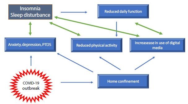
Being forced to stay at home and in many cases working from home, thus reducing social contacts, may have a great impact on daily functioning such as in night sleep and produces alteration of circadian rhythms. Italy was the first European country to impose a total unprecedented lockdown in March 2020. During that month, while people were forced into home confinement, an on-line survey was conducted in 1310Italian residents [25]. It was observed that during home confinement, sleep timing significantly changed, with people going to bed about one ore later and waking up later, spending more time in bed. However, paradoxically, the sleep quality was lower according to the PSQI results [25]. Sleep difficulties, other than correlating with the level of anxiety, were associated with the feeling of elongation of time. Other peculiar data relative to the lockdown were reported, such as people experiencing confusion about what day of the week, day of the month and time of the day it was. After this early study, other studies have confirmed changes in sleep habits during the lockdown period. Some of these confirmed a longer sleep time, particularly among students [26-29]. Lee and co-workers analyzed the sleep pattern of 25217 subjects between 1st January and 29th April 2020 in the US and in16 European countries. They found that the sleeping pattern before and after the country-level lockdown largely differed, but generally the population delayed and slept longer than usual [29]. It is important to note that a longer time spent in bed simply reflects more time spent at home and does not indicate a good quality of sleep, that in fact worsens [25,30]. The stress-sleep link during lockdown periods has been analyzed by a taskforce of the European CBT-I Academy [31]. This taskforce has highlighted that individuals’ sleep habits during lockdown periods are challenged by several factors other than physiological distress. These include alteration of the circadian rhythm due to a reduced exposure to sunlight and reduced physical activity which is known to improve sleep quality [31] (Figure 1).

fig 1

Figure 1: Factors influencing sleep quality during the COVID-19 pandemic.

In working parents, sleep habit may change due to the need to combine work with home-schooling and home administration, that may affect the time spent to sleep. A different situation is that of people living alone, particularly those who have not chosen it. In these individuals home confinement may induce an exacerbation of loneliness and it has been previously established that the lack of social interaction, causing feeling of loneliness, may negatively affect sleep quality [32]. Conversely, it has been reported that during the pandemic those who scored higher on measures of social participation and sense of belonging reported a better quality of sleep [33]. Although common trends have been described, it is clear that effect of confinement measures on sleep quality is not uniform, depending on a variety of individual characteristics. For example, one study has been published showing, unexpectedly, that in one sample COVID-19 lockdown measures worsened sleep quality in pre-pandemic good sleepers, whereas a subset of people with pre-pandemic severe insomnia underwent a clinically meaningful alleviation of symptoms [34]. The concern about negative sleep-related effects of home confinement has prompt the European CBT-I Academy taskforce to provide specific recommendations for the general population, for women and children in family context, for healthcare staff and recommendations for the use of sleep medication [24].

Risk Factors for Sleep Disturbance

Some individuals are more likely to develop sleep problems than others. Those individuals sensitive to stress-related sleep disruption are more likely to develop chronic insomnia [31]. Examined risk factors for COVID-19-related sleep disturbance include age, sex, level of education, occupation and other individual factors [9,31]. It is likely however, that the common means by which sleep is affected is the level of anxiety generated by all pandemic-related concerns, including mainly family’s health, country’s economic stability, physical health and personal economic situation [35]. The effect of gender on sleep disturbance has been extensively investigated. Sleep in woman differs from that of man, mainly for hormonal factors changing during their life span, but also for social factors such as children caring [36,37]. Generally, women report more need to sleep and more insomnia. Sleep disruption is frequently reported by women particularly during pregnancy and the first years of life of children [31,36,37]. As confinement may change habits in the life of the children, this period can be particularly stressful for mothers or other caregivers [31]. Notwithstanding, pre-existing insomnia is a major risk for developing PTSD during the pandemic, particularly in women [31,37]. Many studies have reported that female sex is a major determinant of poor sleep quality [10,11,20]. However, when data were pooled, meta-analysis did not confirm the effect of gender on sleep quality [9,22]. Trakada and colleagues assessed sleep disturbance and risk factors during the lockdown periods in five European countries and in Brazil, in a sample of 1908 participants [38]. Overall the total sleep time was approximately 25 minutes longer than usual but 1in 3 individuals reported sleep disturbance. People with a higher educational level reported the best quality and longer duration of sleep.  Differences in sleep duration were observed among different countries, with the worst quality recorded in Brazil a country experiencing a dramatic outbreak followed by striking political problems [39]. Differences among countries depend on different ways of living and sleeping habits, but also on the socioeconomic status of the country as this can increase the public fear and concern [38]. As for the role of the educational level, differently from this study, other reports showed that people with higher education levels experienced an increased mental stress at the time of the pandemic [18,40]. In a sample of adults in the United States a higher education level was associated with greater concerns about the consequences of COVID-19 (e.g., becoming seriously ill) [40]. Perhaps, a higher education determines greater engagement and interest in health information, but also an increased ability to use information to rationalize risks. The effect of age on sleep disturbance has also been explored. It is now acquired that young adults, without a pre-existing diagnosis, are at great risk to develop sleep disturbance when compared to the general population [9,20,21,25,41]. Other factors have been taken into account as determinants of poor sleep quality. Never, before this pandemic, media have played such a determinant role in providing information and recommending preventive behaviors. However, despite its undisputable usefulness, media, by repeating stressing information, can alter the emotional status, producing anxiety. The predictable overuse of media during lockdowns has been associated with poor sleep quality and day time impairment [25,42]. The over use of digital media before going to bed increases sleep latency, bedtime and wake time [25]. Furthermore, people suffering from insomnia tend to use internet overnight, consequently creating a vicious circle. The lockdowns during the pandemic also caused a reduction in physical activity. As it is known that physical activity improves sleep quality, the reduced activity and the poor quality of sleep are both related to decrease wellbeing during the pandemic [43,44]. Finally, many less obvious factors, belonging to the individual’s personal experiences and life, have been considered as stressors determining sleep disturbance. For example, one study showed that, among American Indians, childhood trauma predicts greater declines in sleep quality associated with the onset of the COVID-19 pandemic [45].

Is Pandemic-related Sleep Disturbance a Transient Problem?

Indeed, the common sense suggests a form of adaptation to this pandemic, as to other negative events that may occur in life. In Germany, a web-based survey was conducted to assess the public mental health burden over a period of 50 days after the COVID-19 outbreak onset. A total of 16245 individuals provided information on sleep disturbances, COVID-19-fear, and level of anxiety [46]. The fear increased with the increasing infection rate but decreased within six weeks to the level before the shutdown, indicating habituation to the threatening situation. The level of anxiety and sleep disturbance proceeded simultaneously with high peaks of infection [46]. Interestingly, one group showed that in France the prevalence of sleep problems significantly decreased during the last weeks of the confinement, and this trend was confirmed one month after the end of the confinement, therefore suggesting that sleep disorders might be transient[47]. However, the possibility of recovering a good sleep pattern largely depends on the type and on the severity of the sleep disorder and is more common in people with mild problems [47]. One study analyzed the effect of sleep habits and social interactions on depression, insomnia and sleepiness, through the COVID-19 pandemic, in a group of patients visiting a specialized sleep clinic in Japan. The authors reported that self-isolation due to COVID-19 had relatively small one-year effects on depression, sleepiness and insomnia in a clinical population [48]. It is therefore likely that as the pandemic is going on, coping strategies have been activated to protect the mental status with favorable consequences on sleep quality.

Sleep Disturbance in Specific Subsets of the Population

Patients with SARS-CoV-2 Infection and COVID-19

Most of the studies on sleep disturbance have been performed in the general population and in health care workers, with fewer studies addressing the problem in patients with SARS-CoV-2 infection/COVID 19. Hartley and co-workers performed an on-line survey in 1777 patients in quarantine for SARS-CoV-2 infection [49]. Forty-seven percent of participants reported a decrease in sleep quality during the quarantine. Factors associated with a reduction in sleep quality by logistic regression were sleep reduction (OR 15.52), going to bed later (OR 1.72), getting up earlier (2.18), an increase in sleep-wake irregularity (OR 2.29), reduced exposure to daylight (OR 1.46) and increased screen use in the evening (OR 1.33) [49]. Liguori and co-workers interviewed a total of 103 patients hospitalized for COVID-19 in order to assess the presence of neurological symptoms. They found that sleep impairment was the most frequent symptom, followed by dysgeusia, headache, hyposmia, and depression [50]. They also reported that women were more commonly affected by daytime somnolence. In addition, sleep impairment was more frequent in patients with more than 7 days of hospitalization [50]. Insomnia was also reported in 12% of patients with COVID-19 during the recovery period after discharge from the hospital [51]. As for other categories, data on patients with COVID-19 were obtained using questionnaires. However, another Italian group assessed sleep using wrist actigraphy in a limited number of patients who had experienced severe respiratory symptoms and who had needed prolonged intensive care unit (ICU). In these patients they reported a lower sleep efficiency and immobility time and a higher fragmentation index compared to those patients who had experienced only mild respiratory symptoms and not requiring ICU stay [52]. A meta-analysis showed that patients with COVID-19 have a higher prevalence of sleep disturbance, 74.8%, compared to the general population and HCW [9]. These data are not surprising as symptoms such as cough, dyspnea, fever and pain may disturb sleep. In addition, drugs used to treat COVID-19, such as oral steroids, may cause insomnia. Furthermore, patients with SARS-CoV2 infection, particularly if hospitalized, show a high prevalence of anxiety and depression that may contribute to impair sleep [53].

Older People

The COVID-19 pandemic has greatly affected the life of the elderly and the association of age with a higher vulnerability to COVID-19 is a subject of major importance [54]. Older adults may experience loneliness due to social distancing and isolation. Moreover, as age is the main factor influencing mortality for Coivid-19, the elderly may be greatly concerned by the risk to be infected, particularly if affected by chronic diseases [54]. With aging the maintenance of circadian rhythms is more difficult, as melatonin level diminishes, and older age is often associated with a reduction in sleep efficiency and continuity [54,55]. Notwithstanding, as most of data on sleep problems during the pandemic have been collected by web survey, studied samples more commonly include young adults, being these the biggest users of the internet, and only few studies included the elderly. Indeed in the general population age is associated with a higher prevalence of sleep disorders during the pandemic [9]. Sleep problems in the elderly correlate with the level of loneliness [56]. This loneliness-sleep disturbance association is especially strong among those people with more COVID-19 related worries or among those with lower resilience [56]. Goodman-Casanova and co-workers in a group of old adults with a diagnosis of moderate dementia, confirmed that those living alone reported greater negative psychological effects and sleeping problems during home confinement [57]. Indeed, further studies are necessary to understand the impact of the pandemic on mental status and sleep quality of the elderly, so that protective strategies can be implemented.

Healthcare Workers

This epidemic is greatly challenging HCW who are particularly vulnerable to the effect of COVID-19 on mental status as well as on physical health. As the emergency started the burden of work has dramatically increased for all healthcare categories and particularly for those directly involved in the management of patients with COVID-19. In addition to common general concerns, the strict contact with patients increases the HCW’s fear of the to be infected or to infect familiars. Moreover, when the pandemic started most of the HCW were unfamiliar with the use of personal protection equipment and were not used to deal with infectious patients, so that their working condition was extremely stressing [58]. As a consequence of accumulated psychological pressure and intense fear of dying, suicides have been also reported among HCW [59]. In this context, the occurrence of sleep disturbance among HCW has been a predictable consequence and has been widely explored since the beginning of the emergency [33,58,60,61]. Ferini-Strambi and co-workers highlight that sleep problems among HCW were common before the pandemic onset, with up to 61% of nurses complaining poor sleep quality [62]. Poor sleep quality reduces day time functioning and mental resources, impairs decision making and reduces concentration [63]. Therefore, in HCW a poor sleep night, particularly if recurrent over time, causes irreversible errors that can threaten patient’s life. Particularly during this pandemic, errors can also threaten the worker’s health, as the risk of infection while working increases. The first large study on medical staff reported insomnia, accompanied by anxiety and depression in more than 30% of frontline workers [64]. Since this and other early observations in China, a large amount of work has been produced and analyzed by a number of meta-analysis [9,65-68]. An early meta-analysis, published in May 2020, included 13 studies with a total of 33062 participants [65]. Anxiety, depression and insomnia were reported with a prevalence rate of 23.2%, 22.8% and 38.9% respectively. The prevalence rate of anxiety and depression appeared to be higher in females. Nurses, that are the larger healthcare workforce and are at close contact with the patients, were more affected than doctors. Conversely, Salari and co-workers reported a pooled prevalence rate of sleep disturbance of 37.8% in nurses and 41.6% in physicians [67]. A more recent meta-analysis including a total of 93 studies on nurses found even higher pooled rates of anxiety and sleep disturbance, 37% and 43%, respectively [65]. Although some small differences among studies, it is clear and expected that the occurrence of sleep disturbance, associated with a high level of physiological stress, among HCW is a worrying issue that may have important social implications. These reports outline the urgency of interventions to improve the psychological wellbeing of health workers.

Conclusion

The COVID-19 pandemic is one of the greatest challenges faced by the world from the beginning of this century, heavily affecting governments, general public and health systems. The pandemic, as well as causing physical problems and deaths, has heavily weakened the population’s mental health, particularly in developed countries where people, since long time, were not used to such kind of catastrophe. The widespread of depression, anxiety, sorrow and worry for the future has been associated invariably with sleep disruption and insomnia. The occurrence of a sleep disorder can further contribute to worsen people’s psychological status in a vicious cycle, that is difficult to break and which can contribute to generate fearsome and irreversible events, including suicide. Sleep disturbance is widespread in all countries parallel with the pandemic and a bidirectional relationship exists between sleep and psychological disorders. In light of these considerations, it is clear that measures must be taken in order to help people to restore their quality of sleep.

Disclosures

The author has no disclosures.

References

  1. WHO (2020) Mental health and psychosocial considerations during COVID-19 outbreak.
  2. Tempesta D, Socci V, De Gennaro L, Ferrara M (2018) Sleep and emotional processing. Sleep Med Rev 40:183-95. [crossref]
  3. Sakurai M, Chughtai H (2020) Resilience against crises: COVID-19 and lessons from natural disasters. Special edition: Orchestration in Contemporary Software Development Ecosystems 29.
  4. Léger D, Bayon V (2010) Societal costs of sleep disturbances. Sleep Med Rev 14:379-89. [crossref]
  5. Grandner MA (2020) Sleep, Health. Sleep Med Clin 15:319-340.
  6. Killgore WDS, Cloonan SA, Taylor EC, Fernandez F, Grandner MA, et al. (2020) Suicidal ideation during the COVID-19 pandemic: The role of insomnia. Psychiatry Res 290:113134. [crossref]
  7. Silva ESME, Ono BHVS, Souza JC (2020) Sleep and immunity in times of COVID-19. Rev Assoc Med Bras 66:143-147. [crossref]
  8. Ibáñez V, Silva J, Cauli O (2018) A survey on sleep assessment methods. PeerJ 6: e4849. [crossref]
  9. Jahrami H, BaHammam HS, Bragazzi NL, Saif Z, Faris MA, et al. (2021) Sleep problems during the COVID-19 pandemic by population: a systematic review and meta-analysis. J Clin Sleep Med 17: 299-313. [crossref]
  10. Zhang F, Shang Z, Ma H, Jia Y, Sun L, et al. (2020) Epidemic area contact history and sleep quality associated with posttraumatic stress symptoms in the first phase of COVID-19. Sci Rep 10: 22463.
  11. Wang C, Pan R, Wan X, Tan Y, Xu L, et al. (2020) Immediate psychological responses and associated factors during the initial stage of the 2019 coronavirus disease (COVID-19) epidemic among the general population in China. Int J Environ Res Public Health 17:1729-132. [crossref]
  12. Zhao X, Lan M, Li H, Yang J (2020) Perceived stress and sleep quality among the non-diseased general public in China during the 2019 coronavirus disease: a moderated mediation model. Sleep Med 77: 339-345
  13. Lin LY, Wang J, Ou-Yang XY, Miao Q, Chen R, et al. (2020) The immediate impact of the 2019 novel coronavirus (COVID-19) outbreak on subjective sleep status. Sleep Med 77: 348-354 [crossref]
  14. Huang, Y and Zhao N (2020) Generalized anxiety disorder, depressive symptoms and sleep quality during COVID-19 epidemic in China: A webbased cross-sectional survey. PsychiatryRes 288: 1129-54. [crossref]
  15. Casagrande M, Favieri F, Tambelli R, Forte G (2020) The enemy who sealed the world: Efects quarantine due to the COVID-19 on sleep quality, anxiety, and psychological distress in the Italian population. SleepMed 75: 12-20. [crossref]
  16. Innocenti P, Puzella A, Mogavero MP, Bruni O, Ferri R, et al. (2020) Letter to editor: COVID-19 pandemic and sleep disorders: a web survey in Italy. Neurol Sci 41:2021-2022. [crossref]
  17. Scarpelli S, Alfonsi V, Mangiaruga A, Musetti A, Quattropani MC, et al. (2021) Pandemic nightmares: Effects on dream activity of the COVID-19 lockdown in Italy. J Sleep Res Pg No: e13300.
  18. Daly M, Sutin AR, Robinson E (2020) Longitudinal changes in mental health and the COVID-19 pandemic: evidence from the UK Household Longitudinal Study. Psychol Med 13: 1-10. [crossref]
  19. Jung S, Kneer J, Krueger T (2020) The German COVID-19 survey on mental health: primary results. MedRxiv.
  20. Beck F, Léger D, Fressard L, Peretti-Watel P, Verger P, et al. (2021) COVID-19 health crisis and lockdown associated with high level of sleep complaints and hypnotic uptake at the population level. J Sleep Res 30:e13119
  21. Kaparounaki CK, Patsali ME, Mousa DV, Papadopoulou EVK, Papadopoulou KKK, et al. (2020) University students’ mental health amidst the COVID-19 quarantine in Greece. Psychiatry Res290:113111. [crossref]
  22. Cénat JM, Blais-Rochette C, Kokou-Kpolou CK, Noorishad PG, Mukunzi JN, et al. (2020) Prevalence of symptoms of depression, anxiety, insomnia, posttraumatic stress disorder, and psychological distress among populations affected by the COVID-19 pandemic: A systematic review and meta-analysis. Psychiatry Res 295:113599. [crossref]
  23. Salari N, Hosseinian-Far A, Jalali R, Vaisi-Raygani A, Rasoulpoor S, et al. (2020) Prevalence of stress, anxiety, depression among the general population during the COVID-19 pandemic: a systematic review and meta-analysis. Global Health 16:57. [crossref]
  24. Partinen M, Bjorvatn B, Holzinger B, Chung F, Penzel T, et al. (2021) Sleep and circadian problems during the coronavirus disease 2019 (COVID‐19) pandemic: the International COVID‐19 Sleep Study (ICOSS). J Sleep Res 30:e13206.
  25. Cellini N, Canale N, Mioni G, Costa S (2020) ense of time and digital media use during COVID-19 lockdown in Italy. J Sleep Res Pg No: e13074. [crossref]
  26. Ong JL, Lau T, Massar SAA, Chong ZT, Ng BKL, et al. (2020) COVID-19 Related Mobility Reduction:heterogenouseffects on physical activity rhythms. Sleep 44:179. [crossref]
  27. Gao C, Scullin MK (2019) Sleep health early in the coronavirus disease 2019 (COVID-19) outbreak in the United States: integrating longitudinal, cross-sectional, and retrospective recall data. Sleep Med 73: 1-10. [crossref]
  28. Wright KP Jr, Linton SK, Withrow D, Casiraghi L, Lanza SM, et al. (2020) Sleep in university students prior to and during COVID-19 Stay-at-Home orders. CurrBiol 30:R797-R798 [crossref]
  29. Lee PH, Marek J, Nálevka P (2021) Sleep pattern in the US and 16 European countries during the COVID-19 outbreak using crowdsourced smartphone data. Eur J Public Health 31: 23-27.
  30. Blume C, Schmidt MH, Cajochen C (2020) Effects of the COVID-19 lockdown on human sleep and rest-activity rhythms. CurrBiol 30: R795-R797. [crossref]
  31. Altena E, Baglioni C, Espie CA, Ellis J, Gavriloff D, et al. Dealing with sleep problems during home confinement due to the COVID‐19 outbreak: Practical recommendations from a task force of the European CBT‐I Academy. J Sleep Res 29:e13052. [crossref]
  32. McHugh JE and Lawlor BA(2013) Perceived stress mediates the relationship between emotional loneliness and sleep quality over time in older adults. Br J Health Psychol 18: 546-555. [crossref]
  33. Xiao H, Zhang Y, Kong D, Li S, Yang N, et al. (2020) The effects of social support on sleep quality of medical staff treating patients with coronavirus disease 2019 (COVID-19) in January and February 2020 in China Med Sci Monit 26:e923549.
  34. Kocevska D, Blanken TF, Van Someren EJW, Rösler L (2020) Sleep quality during the COVID-19 pandemic: not one size fits all. Sleep Med 76:86-88. [crossref]
  35. Statista (2020) Main worries or concerns about the COVID-19 / coronavirus pandemic in the United States, United Kingdom and Germany.
  36. Riemann D, Baglioni C, Bassetti C, Bjorvatn B, Dolenc Groselj L, et al. (2017) European guideline for the diagnosis and treatment of insomnia. J Sleep Res 26: 675-700. [crossref]
  37. Mindell JA and Jacobson BJ (2000) Sleep disturbances during pregnancy. J Obstet Gynecol Neonatal Nurs 29: 590-597. [crossref]
  38. Trakada A, Nikolaidis PT, Andrade MDS, Puccinelli PJ, Economou NT, et al. (2020) Sleep During “Lockdown” in the COVID-19 Pandemic. Int J Environ Res Public Health 17: 9094. [crossref]
  39. The Lancet (2020) COVID-19 in Brazil: “So what?” Lancet395:1461.
  40. Sutin AR, Robinson E, Daly M, Gerend MA, Stephan Y, et al. (2020) Body mass index, weight discrimination, and psychological, behavioral, and interpersonal responses to the coronavirus pandemic. Obesity 28: 1590-1594.
  41. Hyun S, Hahm HC, Wong GTF, Zhang E, Liu CH (2020) Psychological correlates of poor sleep quality among U.S. young adults during the COVID-19 pandemic. Sleep Med 78:51-56. [crossref]
  42. Léger D, Beck F, Fressard L, Verger P, Peretti-Watel P, COCONEL Group (2020) Poor sleep associated with overuse of media during the COVID-19 lockdown. Sleep 43:125. [crossref]
  43. Chouchou F, Augustini M, Caderby T, Caron N, Turpin NA, et al. (2020) The importance of sleep and physical activity on well-being during COVID-19 lockdown: reunion island as a case study. Sleep Med 77: 297-301. [crossref]
  44. Alonso-Martinez AM, Ramirez-Velez R, Garcia-Alonso Y, Izquierdo M, García-Hermoso A (2021) Physical Activity, Sedentary Behavior, Sleep and Self-Regulation in Spanish Preschoolers during the COVID-19 Lockdown. Int J Environ Res Public Health 18:693. [crossref]
  45. John-Henderson NA (2020) Childhood trauma as a predictor of changes in sleep quality in American Indian adults during the COVID-19 pandemic. Sleep Health 6: 718-722 [crossref]
  46. Hetkamp M, Schweda A, Bäuerle A, Weismüller B, Kohler H, et al. (2020) Sleep disturbances, fear, and generalized anxiety during the COVID-19 shut down phase in Germany: relation to infection rates, deaths, and German stock index DAX. Sleep Med 75:350-353. [crossref]
  47. Beck F, Leger D, Cortaredona S, Verger P, Peretti-Watel P, COCONEL Group (2020) Would we recover better sleep at the end of COVID-19? A relative improvement observed at the population level with the end of the lockdown in France. Sleep Med 78:115-119. [crossref]
  48. Ubara A, Sumi Y, Ito K, Matsuda A, Matsuo M, et al. (2020) Self-Isolation Due to COVID-19 Is Linked to Small One-Year Changes in Depression, Sleepiness, and Insomnia: Results from a Clinic for Sleep Disorders in Shiga Prefecture, Japan. Int J Environ Res Public Health 17:8971-76. [crossref]
  49. Hartley S, Colas des Francs C, Aussert F, Martinot C, Dagneaux S, et al. (2020) The effects of quarantine for SARS-CoV-2 on sleep: an online survey. Encephale 3:S53-S59.
  50. Liguori C, Pierantozzi M, Spanetta M, Sarmati L, Cesta N, et al. (2020) Subjective neurological symptoms frequently occur in patients with SARS-CoV2 infection. Brain BehavImmun 88:11-16. [crossref]
  51. Liu HQ, Yuan B, An YW, Chen KJ, Hu Q, et al. (2021) Clinical characteristics and follow-up analysis of 324 discharged COVID-19 patients in Shenzhen during the recovery period. Int J Med Sci 18: 347-355.
  52. Vitale JA, Perazzo P, Silingardi M, Biffi M, Banfi G, et al. (2020) Is disruption of sleep quality a consequence of severe COVID-19 infection? A case-series examination. ChronobiolInt 37:1110-1114.
  53. Liguori C, Pierantozzi M, Spanetta M, Sarmati L, Cesta N, et al. (2021) Depressive and anxiety symptoms in patients with SARS-CoV2 infection.J Affect Disord 278:339-340. [crossref]
  54. Cardinali DP, Brown GM, Reiter RJ, Pandi-Perumal SR(2020) Elderly as a High-risk Group during COVID-19 Pandemic: Effect of Circadian Misalignment, Sleep Dysregulation and Melatonin Administration. Sleep Vigil 26: 1-7.
  55. Welz PS, Benitah SA (2020) Molecular connections between circadian clocks and aging. J Mol Biol 432: 3661-3679.
  56. Grossman ES, Hoffman YSG, Yuval Palgi Y, Shrira A (2021) COVID-19 related loneliness and sleep problems in older adults: Worries and resilience as potential moderators. PersIndivid Dif 168:110371.
  57. Goodman-Casanova JM, Dura-Perez E, Guzman-Parra J, Cuesta-Vargas A, Mayoral-Cleries F (2020) Telehealth home support during COVID-19 confinement for community-dwelling older adults with mild cognitive impairment or mild dementia: survey study. J Med Internet Res 22:e19434. [crossref]
  58. Sheng X, Liu F, Zhou J, Liao R (2020) Psychological status and sleep quality of nursing interns during the outbreak of COVID-19. Nan Fang Yi Ke Da Xue Xue Bao 40:346-350. [crossref]
  59. Montemurro N (2020) The emotional impact of COVID-19: From medical staff to common people. Brain BehavImmun 87: 23-24. [crossref]
  60. Qi J, Xu J, Li BZ, Huang JS, Yang Y, et al. (2020) The evaluation of sleep disturbances for Chinese frontline medical workers under the outbreak of COVID-19. Sleep Med. 72:1-4. [crossref]
  61. Tu ZH, He JW, Zhou N (2020) Sleep quality and mood symptoms in conscripted frontline nurse in Wuhan, China during COVID-19 outbreak: a cross-sectional study. Medicine (Baltimore) 99:e20769. [crossref]
  62. Ferini-Strambi L, Zucconi M, Casoni F, Salsone M (2020) COVID-19 and Sleep in Medical Staff: Reflections, Clinical Evidences, and Perspectives. Curr Treat Options Neurol 22:29. [crossref]
  63. Medic G, Wille M, Hemels ME (2017) Short- and long-term health consequences of sleep disruption. Nat Sci Sleep 9:151-161.
  64. Zhang C, Yang L, Liu S, Ma S, Wang Y, et al. (2020) Survey of insomnia and related social psychological factors among medical staff involved in the 2019 novel coronavirus disease outbreak. Front Psychiatry 11: 306.
  65. Pappa S ,Ntellac V, Giannakas T, Giannakoulis VG, Papoutsi E, et al. (2020) Prevalence of depression, anxiety, and insomnia among healthcare workers during the COVID-19 pandemic: A systematic review and meta-analysis. Brain, Behavior, and Immunity 88:901-907. [crossref]
  66. Al Maqbali M, Al Sinani M, Al-Lenjawi B (2021) Prevalence of stress, depression, anxiety and sleep disturbance among nurses during the COVID-19 pandemic: A systematic review and meta-analysis. J Psychosom Res 141: 110343. [crossref]
  67. Salari N, Khazaie H, Hosseinian-Far A, Ghasemi H, Mohammadi M, et al. (2020) The prevalence of sleep disturbances among physicians and nurses facing the COVID-19 patients:a systematic review and meta-analysis.Global Health.16:92.
  68. Zeng LN, Yang Y, Wang C, Li XH, Xiang YF, et al. (2019) Prevalence of poor sleep quality in nursing staff: a meta-analysis of observational studies. Behav Sleep Med 18: 746-759. [crossref]