Author Archives: rajani

FIG 1

Tissue Infiltration of Tumor-Associated Macrophages: Towards the Identification of Therapeutic Targets

DOI: 10.31038/IMROJ.2021611


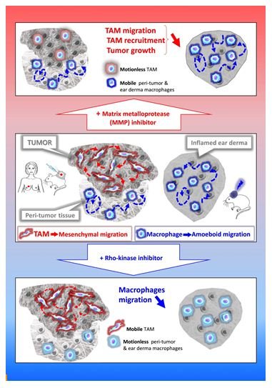
Macrophages are present in all body tissues. They play a key role in the clearance of pathogens, participate in the immune and inflammatory responses, and partake in tissue repair and homeostasis. However, tissue infiltration of macrophages also exacerbates pathological processes, such as cancers [1-4]. Tumor-associated macrophages (TAMs) mostly originate from blood monocytes [5]. They are recruited to the tumor stroma at all stages of cancer progression [2,6] and can represent more than fifty percent of the tumor mass, thus representing by far the most abundant immune cell of the tumor stroma. Their number positively correlates with poor prognoses in most solid cancers as they are involved in several cancer-promoting events such as angiogenesis, lymphangiogenesis, immunosuppression, metastasis formation and resistance to therapies [7,8]. Therefore, the control of TAM infiltration is a current therapeutic strategy against cancers [9-11]. However, many questions regarding the mechanism of TAM tissue migration remain unresolved, which further hinders the development of novel therapeutic approaches.

FIG 1

Cell migration in tissues occurs in three dimensions (3D) that profoundly differs from 2D migration processes [12,13]. Two main mechanistically distinct migration modes have been described in 3D environments: amoeboid and mesenchymal [14]. The amoeboid movement is characterized by rounded, ellipsoid, or moderately elongated cells that form blebs or generate small actin-rich filopodia [15-17]. These cells do not require adhesion to the extracellular matrix (ECM), but rather use a propulsive and pushing migration mode [16,18,19]. This non-directional motility involves acto-myosin contractions and depends on the Rho–ROCK pathway. Cells migrating through the mesenchymal mode adopt an elongated and protrusive morphology [15,17,18]. The movement is directional, involves cell adhesion to the substratum, and requires proteases to degrade the ECM in order to create paths through dense environments. In macrophages, in contrast to the amoeboid movement, mesenchymal migration is not inhibited, but rather stimulated, by treatment with ROCK inhibitors [20,21]. Unlike lymphocytes, neutrophils and monocytes [3,22,23], macrophages share the capacity with only few cell types including tumor cells or immature dendritic cells (DCs) [17,24,25], to use both amoeboid and mesenchymal migration modes in 3D environments. In vitro studies revealed that macrophages tailor their migration mode to the architecture of the surrounding ECM [20,22,26-35]. In vivo in mouse tumors and ex vivo in human breast cancer explants, macrophages use the two migration modes depending on the tissue they infiltrate. TAMs use the protease-dependent mesenchymal migration mode in mouse fibrosarcoma in vivo or human breast cancers ex vivo [20]. In contrast, in non-tumorous tissues such as the tumor periphery or in inflamed ear derma, macrophages use the amoeboid motility in vivo [20]. A chronic treatment with a broad-spectrum inhibitor of matrix metalloproteinases (MMPs) blocked the mesenchymal migration of macrophages, which correlates with a decrease in both TAM infiltration and tumor growth in vivo [20]. These findings strongly suggest that inhibition of TAM motility could be a way to impede their pro-tumor action and urge to identify specific effectors of the TAM mesenchymal migration as new targets in anti-cancer therapy.

Among effectors of TAM mesenchymal migration, MMPs need to be considered. MMP inhibitors have already been used to hamper tumor cell invasiveness by impeding tumor stroma remodeling and cancer cell escape from the primary tumor as well as decreasing angiogenesis [36,37]. Anti-tumor action of MMP inhibitors can now be explained by their action on TAM motility. Batimastat as well as its orally bioavailable derivative Marimastat were the first MMP inhibitors to enter clinical trials more than a decade ago [36]. However, clinical trials in patients with pancreatic, brain, lung or renal cancers were disappointing, essentially because these drugs were only tested in patients with advanced diseases despite the fact that studies in animal models had shown a most effective effect in treating early-stage diseases [38]. In addition, the primarily tested broad-spectrum MMP inhibitors were non-specific and did not differentiate between pro-tumor and anti-tumor MMPs depending on the type of cancer [38]. Thus, the recent knowledge on MMP biology and their differential involvement in tumor progression [39] together with the development of new generation MMP inhibitors [40-42] and the involvement of MMP activity on TAM motility stress the need to reassess the use of such inhibitors in early cancer treatment in combination with other anti-cancer molecules.

Another future strategy to identify new potential therapeutic targets consists in identifying new specific effectors of TAM mesenchymal migration. Therefore, exhaustive approaches to reach a comprehensive understanding of this process will be necessary as recently described in a transcriptomic-based analysis [27]. This strategy leads to the identification of a large number of potential targets and the future challenge will be to validate or invalidate all the potential hits as effective actors of macrophage migration both in vitro and in vivo through functional studies. For such large-scale screening approaches, new cellular tools are needed. Many studies use bone marrow-derived macrophages (BMDMs) from wild-type (WT) and knock-out (KO) mice or macrophage cell lines such as murine Raw 264.7 cells or human U937, HL-60 or THP1 cells for this purpose. All these cell models have several drawbacks such as the use of numerous animals, the limited number of cells and the impossibility to generate stable mutants in primary cultures or the fact that macrophage cell lines are usually distantly related to blood-derived macrophages or BMDMs particularly because they are cancer cells. Expansion of murine hematopoietic precursors that were transiently immortalized through a retroviral-delivery of an estrogen-inducible form of the transcription factor Hoxb8 has been described [43] and validated for the study of hematopoietic cell biology [43-52]. The possibility to use the CRISPR/Cas9 technology in this long-term hematopoietic progenitor cell lines has enabled the creation of new genetically modifiable cell models [45]. During the last few years, ectopic expression of Hoxb8 has been used in several studies mainly focused on DC biology [45,47,52-54], but also to generate surrogate macrophages [43,52,55]. This new cellular tool that combines the unlimited proliferative capacity of conditionally Hoxb8-immortalized hematopoietic progenitor cells with the CRISPR/Cas9 technology represents a powerful tool to genetically manipulate macrophages and explore their functionalities in a broad range of applications [55].

In conclusion, the tissue migration of TAMs emerges as a new therapeutic target to combat cancer diseases and the development of new cellular models to molecularly dissect the mesenchymal migration process should lead to the identification of new leads in anti-cancer therapy in the coming years.

References

  1. Allavena P, Chieppa M, Bianchi G, M Fabbri, G Laskarin, et al. (2010) Engagement of the mannose receptor by tumoral mucins activates an immune suppressive phenotype in human tumor-associated macrophages. Clinical & Developmental Immunology 2010: 547179.
  2. Condeelis J, Pollard JW (2006) Macrophages: obligate partners for tumor cell migration, invasion, and metastasis. Cell 124: 263-266. [crossref]
  3. Friedl P, Weigelin B (2008) Interstitial leukocyte migration and immune function. Nature Immunology 9: 960-969. [crossref]
  4. Yiangou Y, Facer P, Durrenberger P, Iain P Chessell, Alan Naylor, et al. (2006) COX-2, CB2 and P2X7-immunoreactivities are increased in activated microglial cells/macrophages of multiple sclerosis and amyotrophic lateral sclerosis spinal cord. BMC Neurology 6: 12. [crossref]
  5. Lahmar Q, Keirsse J, Laoui D, Kiavash Movahedi, Eva Van Overmeire, et al. (2016) Tissue-resident versus monocyte-derived macrophages in the tumor microenvironment. Biochim Biophys Acta 1865: 23-34. [crossref]
  6. Allavena P, Sica A, Solinas G, Chiara Porta, Alberto Mantovani (2008) The inflammatory micro-environment in tumor progression: the role of tumor-associated macrophages. Critical Reviews in Oncology/Hematology 66: 1-9. [crossref]
  7. Noy R, Pollard JW (2014) Tumor-associated macrophages: from mechanisms to therapy. Immunity 41: 49-61. [crossref]
  8. Ruffell B, Coussens LM (2015) Macrophages and therapeutic resistance in cancer. Cancer Cell 27: 462-472. [crossref]
  9. Mantovani A, Marchesi F, Malesci A, Luigi Laghi, Paola Allavena (2017) Tumour-associated macrophages as treatment targets in oncology. Nature Reviews Clinical Oncology 14: 399-416. [crossref]
  10. Morrison C (2016) Immuno-oncologists eye up macrophage targets. Nature reviews Drug discovery 15: 373-374. [crossref]
  11. Ostuni R, Kratochvill F, Murray PJ, Natoli G (2015) Macrophages and cancer: from mechanisms to therapeutic implications. Trends in Immunology 36: 229-239. [crossref]
  12. Doyle AD, Wang FW, Matsumoto K, Yamada KM (2009) One-dimensional topography underlies three-dimensional fibrillar cell migration. J Cell Biol 184: 481-490. [crossref]
  13. Hooper S, Marshall JF, Sahai E (2006) Tumor cell migration in three dimensions. Methods in Enzymology 406: 625-643.
  14. Van Goethem E, Le Cabec V, Poincloux R, Fabienne Gauffre, Isabelle Maridonneau-Parini (2010) Microenvironment structural constraints impact the 3D migration mode of macrophages and the formation of 3D podosomes. Eur J Clin Invest 40: 43.
  15. Friedl P, Wolf K (2003) Proteolytic and non-proteolytic migration of tumour cells and leucocytes. Biochem Soc Symp 277-85. [crossref]
  16. Lammermann T, Sixt M (2009) Mechanical modes of ‘amoeboid’ cell migration. Current Opinion in Cell Biology 21: 636-644. [crossref]
  17. Sahai E, Marshall CJ (2003) Differing modes of tumour cell invasion have distinct requirements for Rho/ROCK signalling and extracellular proteolysis. Nature Cell Biology 5: 711-719. [crossref]
  18. Fackler OT, Grosse R (2008) Cell motility through plasma membrane blebbing. J Cell Biol 181: 879-884. [crossref]
  19. Paluch EK, Aspalter IM, Sixt M (2016) Focal Adhesion-Independent Cell Migration. Annu Rev Cell Dev Biol 32: 469-490. [crossref]
  20. Gui P, Ben-Neji M, Belozertseva E, Florence Dalenc, Camille Franchet, et al. (2018) The Protease-Dependent Mesenchymal Migration of Tumor-Associated Macrophages as a Target in Cancer Immunotherapy. Cancer Immunology Research 6: 1337-1351. [crossref]
  21. Gui P, Labrousse A, Van Goethem E (2014) Rho/ROCK pathway inhibition by the CDK inhibitor p27kip1 participates in the onset of macrophage 3D-mesenchymal migration. Journal of Cell Science 127: 4009-4023.
  22. Cougoule C, Van Goethem E, Le Cabec V, Fanny Lafouresse, Loïc Dupré, et al. (2012) Blood leukocytes and macrophages of various phenotypes have distinct abilities to form podosomes and to migrate in 3D environments. European Journal of Cell Biology 91: 938-949. [crossref]
  23. Lammermann T, Germain RN (2014) The multiple faces of leukocyte interstitial migration. Seminars in Immunopathology 36: 227-251. [crossref]
  24. Cougoule C, Lastrucci C, Guiet R, Rémi Mascarau, Etienne Meunier, et al. (2018) Podosomes, But Not the Maturation Status, Determine the Protease-Dependent 3D Migration in Human Dendritic Cells. Frontiers in immunology 9: 846. [crossref]
  25. Friedl P, Wolf K (2003) Tumour-cell invasion and migration: diversity and escape mechanisms. Nature Reviews Cancer 3: 362-374. [crossref]
  26. Barros-Becker F, Lam PY, Fisher R, Huttenlocher A (2017) Live imaging reveals distinct modes of neutrophil and macrophage migration within interstitial tissues. Journal of CELL science 130: 3801-3808. [crossref]
  27. Cermak V, Gandalovicova A, Merta L, Jitka Fučíková, Radek Špíšek, et al. (2018) RNA-seq of macrophages of amoeboid or mesenchymal migratory phenotype due to specific structure of environment. Scientific Data 5: 180198. [crossref]
  28. Cougoule C, Le Cabec V, Poincloux R, Talal Al Saati, Jean-Louis Mège, et al. (2010) Three-dimensional migration of macrophages requires Hck for podosome organization and extracellular matrix proteolysis. Blood 115: 1444-1452. [crossref]
  29. Gui P, Labrousse A, Van Goethem E (2014) Rho/ROCK pathway inhibition by CDK inhibitor p27kip1 participates in the onset of macrophage 3D-mesenchymal migration. Journal of Cell Science.
  30. Guiet R, Verollet C, Lamsoul I, Céline Cougoule, Renaud Poincloux, et al. (2012) Macrophage mesenchymal migration requires podosome stabilization by filamin A. The Journal of Biological Chemistry 287: 13051-13062. [crossref]
  31. Jevnikar Z, Mirkovic B, Fonovic UP, Nace Zidar Urban Švajger, et al. (2012) Three-dimensional invasion of macrophages is mediated by cysteine cathepsins in protrusive podosomes. European Journal of Immunology 42: 3429-3441.
  32. Van Goethem E, Guiet R, Balor S, Guillaume M.Charrière, Renaud et al. (2011) Macrophage podosomes go 3D. European Journal of Cell Biology 90: 224-236.
  33. Van Goethem E, Poincloux R, Gauffre F, Isabelle Maridonneau-Parini, Véronique Le Cabec (2010) Matrix architecture dictates three-dimensional migration modes of human macrophages: differential involvement of proteases and podosome-like structures. J Immunol 184: 1049-1061. [crossref]
  34. Lugo-Villarino G, Verollet C, Maridonneau-Parini I, Neyrolles O (2011) Macrophage Polarization: Convergence Point Targeted by Mycobacterium Tuberculosis and HIV. Frontiers in Immunology 2: 43. [crossref]
  35. Verollet C, Souriant S, Bonnaud E, Paul Jolicoeur, Brigitte Raynaud-Messina et al. (2015) HIV-1 reprograms the migration of macrophages. Blood 125: 1611-1622. [crossref]
  36. Hidalgo M, Eckhardt SG (2001) Development of matrix metalloproteinase inhibitors in cancer therapy. Journal of the National Cancer Institute 93: 178-193. [crossref]
  37. Amorosa V, Tebas P (2006) Bone disease and HIV infection. Clinical infectious diseases : an official publication of the Infectious Diseases Society of America 42: 108-114.
  38. Overall CM, Kleifeld O (2006) Tumour microenvironment – opinion: validating matrix metalloproteinases as drug targets and anti-targets for cancer therapy. Nature Reviews CANCER 6: 227-239. [crossref]
  39. Mason SD, Joyce JA (2011) Proteolytic networks in cancer. Trends in Cell Biology 21: 228-237. [crossref]
  40. Devel L, Czarny B, Beau F, Dimitris Georgiadis, Enrico Stura, et al. (2010) Third generation of matrix metalloprotease inhibitors: Gain in selectivity by targeting the depth of the S1′ cavity. Biochimie 92: 1501-1508. [crossref]
  41. Fingleton B (2008) MMPs as therapeutic targets–still a viable option? Seminars in Cell & Developmental Biology 19: 61-68. [crossref]
  42. Tauro M, McGuire J, Lynch CC (2014) New approaches to selectively target cancer-associated matrix metalloproteinase activity. Cancer Metastasis Rev 33: 1043-1057. [crossref]
  43. Wang GG, Calvo KR, Pasillas MP, David B Sykes, Hans Häcker, et al. (2006) Quantitative production of macrophages or neutrophils ex vivo using conditional Hoxb8. Nat Methods 3: 287-293. [crossref]
  44. Lee S, Kivimae S, Szoka FC (2017) Clodronate Improves Survival of Transplanted Hoxb8 Myeloid Progenitors with Constitutively Active GMCSFR in Immunocompetent Mice. Molecular Therapy Methods & Clinical Development 7: 60-73. [crossref]
  45. Hammerschmidt SI, Werth K, Rothe M, Melanie Galla, Marc Permanyer, et al. (2018) CRISPR/Cas9 Immunoengineering of Hoxb8-Immortalized Progenitor Cells for Revealing CCR7-Mediated Dendritic Cell Signaling and Migration Mechanisms in vivo. Frontiers in Immunology 9: 1949. [crossref]
  46. Di Ceglie I, van den Akker GG, Ascone G, Bas Ten Harkel, Hans Häcker, et al. (2017) Genetic modification of ER-Hoxb8 osteoclast precursors using CRISPR/Cas9 as a novel way to allow studies on osteoclast biology. Journal of Leukocyte Biology 101: 957-966. [crossref]
  47. Rosas M, Osorio F, Robinson MJ, Luke C Davies, Nicola Dierkes, et al. (2011) Hoxb8 conditionally immortalised macrophage lines model inflammatory monocytic cells with important similarity to dendritic cells. European Journal of Immunology 41: 356-365. [crossref]
  48. Chu JY, McCormick B, Mazelyte G, Melina Michael, Sonja Vermeren, et al. (2019) HoxB8 neutrophils replicate Fcgamma receptor and integrin-induced neutrophil signaling and functions. Journal of Leukocyte Biology 105: 93-100. [crossref]
  49. Witschi R, Johansson T, Morscher G, Louis Scheurer, Jacqueline Deschamps, et al. (2010) Hoxb8-Cre mice: A tool for brain-sparing conditional gene deletion. Genesis 48: 596-602. [crossref]
  50. Gurzeler U, Rabachini T, Dahinden CA, M Salmanidis, G Brumatti, et al. (2013) In vitro differentiation of near-unlimited numbers of functional mouse basophils using conditional Hoxb8. Allergy 68: 604-613. [crossref]
  51. Zach F, Mueller A, Gessner A (2015) Production and Functional Characterization of Murine Osteoclasts Differentiated from ER-Hoxb8-Immortalized Myeloid Progenitor Cells. PloS one 10: e0142211. [crossref]
  52. Cabron AS, El Azzouzi K, Boss M, Philipp Arnold, Jeanette Schwarz, et al. (2018) Structural and Functional Analyses of the Shedding Protease ADAM17 in HoxB8-Immortalized Macrophages and Dendritic-like Cells. J Immunol 201: 3106-3118. [crossref]
  53. Grajkowska LT, Ceribelli M, Lau CM, Margaret E Warren, Ioanna Tiniakou, et al. (2017) Isoform-Specific Expression and Feedback Regulation of E Protein TCF4 Control Dendritic Cell Lineage Specification. Immunity 46: 65-77. [crossref]
  54. Leithner A, Renkawitz J, De Vries I, Robert Hauschild, Hans Häcker, et al. (2018) Fast and efficient genetic engineering of hematopoietic precursor cells for the study of dendritic cell migration. European Journal of Immunology 48: 1074-1077. [crossref]
  55. Accarias S, Sanchez T, Labrousse A, Myriam Ben-Neji, Aurélien Boyance, et al. (2020) Genetic engineering of hoxb8 immortalized hematopoietic progenitors: a potent tool to study macrophage tissue migration. Journal of cell science.


fig 1

Arsenic Influence Variations in Animals by Its Toxicity

DOI: 10.31038/IJVB.2021514

Abstract

There have been several revisions that have examined the effectiveness of chronic arsenic exposure in animals. Specifically, we aim to investigate the effects of arsenic trioxide on nephrotoxicity, hepatotoxicity, and nephrotoxicity. This section concludes by examining the impact of Arsenic. Taken together, these results suggest that Arsenic is a pollutant to both persons and animals. The conclusions got, in this case, hold globally that we have associated Arsenicals with skin, lung, and bladder cancers of the skin, lung and bladder. Arsenic (As) has both metallic and nonmetallic properties and is known as a metalloid. They contain it in ore, and it crushes rocks of an average density. This suggests complex chemistry, which exists in four separate valences (III, O, III, and V). And I have used it for a wider range of business and farming purposes in many chemical types. The most common arsenic compound used in industry is an arsenic oxide (As2O3), which is used to synthesize other inorganic and biological Weapons. For the prevention of acute pro-monocytic leukemia, arsenic trioxide is also being used. Absorption occurs mainly by ingestion in the small intestine, but through skin contact and respiration, fewer absorption orcs. Nausea, vomiting, stomach pain, extreme diarrhea and peripheral neuropathy were originally associated with acute arsenic poisoning. The multisystem disease may result from chronic arsenic poisoning.

Keywords

Arsenic (As), Arsenic oxide, Chronic poisoning, Neuropathy, Valence, Phosphorylation, Diseases

Introduction

Arsenic is a nonmetallic present mainly inland, atmosphere and in water from natural and human-made causes. It is volatile in biological forms and various states of oxidation (−3, 0, +3, +5). In terms of ecological exposure, toxicology of arsenic is mainly related to mild pentavalent oxidation state. Arsenic ionic form, arsenic acid ionic form, and arsenic compounds. Monomethyl arsonate acid (MMAV) and dimethyl arsenic acid (DMAV) are volatile arsenic-stable methylated mammalian substances and are mainly expelled in the urine. DMAV and sodium salts of MMAV are used as weedkillers.

In the body of animals and vegetable cells, arsenic has been extensively studied for its poisonousness and healing use. It is found in petrifaction and soils ranging varies from 1 million to hundreds of ppm, with an average range value of 2 ppm. In sulfide layers, many metals such as lead, copper sulfides, silver, and iron, arsenic with sulfides are present mainly.

High levels of arsenic were recorded in many areas of the world. A bearable arsenic amount of up to 1100 pg/1 have appeared in drinking water in many places. Arsenic is also present in seawater.

Arsenic is used as an agricultural wood preservative, desiccant, and herbicide and a finishing agent for the goblet manufacturing and copper and lead mixtures. It is predicted as human emissions of arsenic at 41,000 metric tons every year. Worldwide discharges from ordinary resources are estimated at 7800 metric tons every year. Further input of marine arsenic encouraged the study of the accretion and lethal effects of aquatic organisms.

Regarding arsenic, most people think of it as a deadly toxin that has been used for periods. However, most human exposure to arsenic comes from sources such as drinking water, food, dust, and soil, although it is believed that low levels of arsenic are essential nutrients but it as adverse health effects. Although the concentration of exposure in drinking water depends on the determination of the current maximum container level (MCL) of 50 ~ g/1 of arsenic.

It is narrated by [1] Arsenic (AS) is a common contaminant in various parts of the ecosphere. Arsenal and its derivatives are environmentally mobile. Rock decomposition changes arsenic sulfide to arsenic trioxide, which comes in the arsenic environment through the dust cycle or dissolves in rain, rivers, or water table. Chronic contact with organic arsenic can flow to skin, lung, vesica urinaria, and liver cancers.

These metalloids are generally toxic in various chemical forms; therefore, volatile shapes of arsenic (i.e., arenite, arsenate) are more Methylated forms (methyl methacrylate, MMA), dimethyl arsenate (DMA) are moderately examined toxins. Supplementary arsenic types, such as a trimethyl-arsenic oxide (TMAO) and tetra-methyl-arsonium (tetra), are also considered moderately toxic, as is arsenic betaine (ASB), arsenic choline (ASC), and other arsenic sugars (AS) does not show (Fatorini et al., 2006).

Numerous epidemiological revisions in diverse geographical zones have shown that mineral arsenic is a human cancer-causing agent. Though, the process of cancer is not yet known. Research on the absence of carcinogenesis in vivo animal representations [2] or in vitro cell systems is instrumental in clarifying the reasons for arsenic carcinogenesis.

Some studies have reported that arsenic leads to gene enhancement, chromosomal abnormalities, of DNA repair inhibition, decreased DNA activity, and proton gene activation. Earlier, we stated that V79-Cl3 (Chinese hamster cells) undertook early genetic uncertainty or programmed cell death when visible to sodium arsenic (S.A.).

In explanations made during and later dealing, genetic variability was evident in the existence of irregular and aneuploid cells, nonetheless chromosome abnormal cells. As mitotic cells became more sensitive to S.N. disclosure due to direct arsenic action in the mitotic spindle assembly, we later discovered the movement of hereditarily uneven cells that escape apoptosis by cutting mitotic round-up. The cell population was still not genetically stable when tested after two months of cell culture (120 cell generations).

Although there has been much progress in the field of synthetic drugs in recent years, they have had some or other side effects, but it has been suggested that naturopathy with phytochemicals may still be beneficial in many situations [3].

Arsenic is found in the blood as a brain barrier, where it has neurotoxic effects on many assemblies, such as the ganglia. Although the information on the impact of arsenic on the central nervous system (CNS) is incomplete, the basal ganglia appear to be sensitive to the toxins effects, such as 3-nitroprionic acid, succinic dihydroxy dip. If the basal ganglia’s monoamine content can be modified by arsenic exposure, it affects the behaviour. Studies of locomotor activity in rats have been reported to reduce the levels of arsenic trioxide used. Defects in functional study work have also been reported. The data’s inefficiency and the fact that the results of the dose and exposure time have not been examined in previous studies make it difficult to conclude the effects of arsenic on the nervous system in the in vivo model. To research the neurotoxic consequence of arsenic exposure and assess the ace of basal ganglia for roles such as learning, remembrance, and movement, it is essential to determine whether arsenic revelation alters these compound functions.

Arsenic can induce male reproductive toxins such as the dose-dependent reduction in testes and associated genital weight [4]. It can reduce the number of epididymal sperm [5], function, and motility [6]. The general form and the antioxidant, immune system [7]. There is an increase of hormones like Luteinizing hormone (L.H.), follicle-stimulating hormone (FSH), testosterone, and massive depletion of germ cells in testicular tissue [8] Arsenic poisoning. Oxidative stress and the production of reactive oxygen species (ROS) may be a consequence of arsenic exposure [9]. Several protein functions can be modified to produce ROS and bind to arsenic protein thiol groups [10]. Such as arsenic on oxidative stress and DNA damage caused by ROS production [11]; Biswas RPS, 2007.

The International Agency classifies inorganic arsenic for Research on Cancer. Human cancer is known to the U.S. Environmental Protection Agency. This arrangement is based on numerous epidemiological revisions showing arsenic exposure and cancer development. Fowler’s solution (potassium arenite), proficient introduction to inhaled copper smelters, or logically polluted drinking water. Cancers that occur later arsenic inhalation are mainly found in the lungs, although they are first detected on the skin subsequently exposure to arsenic. However, the following studies suggest that people who regularly consume arsenic-contaminated drinking water are more likely to develop internal organ cancer. Tumor sites include the bladder, liver, and kidneys. Even though there is enough indication to classify arsenic as human cancer, it is unclear whether it happens in research laboratory animals. In many studies, animals have no cancer effect after exposure to inorganic arsenic for the rest of their lives after drinking, feeding, or oral hatching. It has been testified in rats, dogs and monkeys. Mice reveal to arsenic in drinking water for 26 weeks did not develop tumors. However, several positive information about arsenic cancer in animal samples. In the 1980s, numerous studies reported transient arsenic cancer in hamsters after intratracheal insulation. This direction has been used to mimic arsenic respiration. In these studies, it includes used of chemicals i.e., arsenic trioxide (As2O3), calcium arsenate (Ca (AsO4) 2), and arsenic trisulfide (As2S3). Tumors were examined once a week for 15 weeks, and tumors were examined during life.

Data Collection

PubMed has conducted a literary search using the Google Scholar and Science-Hub database to identify relevant studies from the past to the present. Summary of studies stated on introduction to arsenic-polluted drinking water.

Using a verified reference list from former review articles, I searched manually for more relevant studies to identify studies that could not be recovered over an electronic record. We only consider peer-review journals and articles published in English. Each selected report critically assessed and summarized the range of relationships between position, design, organ impact, sample size, exposure assessment, arsenic exposure, and development.

Ancient Beneficial Uses of Arsenic

In ancient times arsenic was used as an attenuating representative after Greek doctors such as Hippocrates and Galen popularized its use. With that arsenic complexes are found in the form of solutions, tablets, pastes, and injection forms. Utmost recently, in 1958, the British Pharmacological and Therapeutic Products Handbook, revised by Martindale, listed indications for Fowler’s solution: leukemia, skin conditions. Fowler’s solution is also indicated as a health energizer.

Current Beneficial Uses of Arsenic

Arsenal trioxide (AS2O3), and endosperm based on its structure, is now widely used in patients with severe primality rocambole withdrawal [12]. Arsenal insisted on apoetical. [13]. AIF then affects programmed cell death, resulting in nuclear biochemistry, chromatin condensation, DNA fragmentation, and cell death. Arsenic is often an additive, sometimes mercury and lead [14]. The California Department of Health retail displays 251 products in herbal goods, and 36 products (14%) contain arsenic.

Coal naturally has High Arsenic Levels

As most coal has arsenic content of fewer than 5 milligrams per kilogram, ordinary coal is low in arsenic, which is less harmful to human health. Some coal can, however, comprises up to 35 000 mg per 1 kg of coal [15]. Numerous research studies have shown how indoor air can be polluted by burning high arsenic coal. For instance, P.R. The effects of coal-burning have been investigated on residents of Guizhou Province, China. Coal, water, air, food, urine, and hair samples from local and limited zones were obtained and arsenic examined.

Chronic Arsenic Poisoning

Arsenic poisoning processes include the inhibition of the substitution of cellular enzymes comprising the community of sulfides and phosphate molecules in ‘amino lysis’ [16]. In inhibiting enzymes, trivalent arsenic complexes are more effective, while arsenic lysis is done by pentavalent compounds [16]. Trivalent is a human carcinogen, including mild bronchogenic carcinomas, hepatic angiosarcomas [17], and intradermal carcinomas (Bowen’s disease) [18]. Carcinomas of squamous cells, carcinomas of basal cells, and ‘integrated’ types of skin cancer [16]. Myelogenous leukemia can occur as well [19]. French gold miners are more vulnerable to Hodgkin’s disease. Arsenic Exposed [20]. Flow The repeated use of Fowler solution or arsenic-contaminated drinking water has been associated with lung, liver, bladder, and kidney cancers [17].

Acute Arsenic Poisoning

Overdose can lead to serious reactions such as diarrhea, vomiting, pain, desiccation, and faintness. Nowadays, severe intoxication is very rare in Western European countries; if that happens, it is usually the result of intentional (suicide or homicide) or accidental poisoning. Professional exposure to as is infrequent and usually occurs in the form of arsenic gas. It causes different symptoms due to eating [21]. Exposure often occurs when arsenic gas escapes during transport or production. Minerals or metals containing arsenic are treated with acid. [22,23]. It is associated with gastrointestinal disorders. Cardiac and pulmonary symptoms include hypotension, shock, pulmonary oedema, and cardiac arrest.

Arsenic Exposure

Exposure of arsenic is caused through skin, and by the usage of contaminated drinking water. Arsenic in the diet occurs as comparatively nontoxic organic compounds. Seafood, fish, and algae are rich in biological resources [24]. These organic compounds increase the level of arsenic in the blood but do not change in the urine [25]. Arsenic consumption is higher in solid foods than in liquids, including drinking water [26]. Organic and volatile arsenic compounds may enter the vegetarian diet from farming goods or soils cultivated with arsenic contaminants [27].

Absorption

The gastrointestinal tract is the leading absorption process by an electronic process with a proton gradient (Gonzalez et al., 1997), The normal pH range for arsenic absorption is 5.0, and (Silver et al. (1984) found that the small intestine’ pH is approximately 7.0 after pancreatic bicarbonate secretion. (Ratnayake et al., 2000).

Evolution

Arsenic poisoning changes with its oxidation conditions. Organic types are usually more toxic than in organic matter. Arsenic (AsIII) is 60 times more toxic than arsenate. This latter species is 70 times more toxic than the methylated species, with Monomethyl arsenic acid (MMA) and dimethyl arsenic acid (DMA), the last two forms being considered only moderately toxic [28]. In mammals, arenite was methylated by depletion, arenite methyltransferase [28].

The absorbed arsenic endures hepatic biomethylation and forms Monomethyl arsenic acid and dimethylarcinic acid, which are nontoxic but ultimately harmless [29]. 50% of the dose may be excreted in the urine. In three or five days. The metabolism of dimethylarginine acid (60% –70%) in urine contrasts with Monomethyl arsenic acid [30]. Small amounts of inorganic arsenic do not change. Spectrometry studies of electrothermal nuclear absorption after acute poisoning show high arsenic levels in the kidneys and liver. In the muscles, nervous system, small intestine, and spleen [31]. Although most arsenic was removed from these sites, the remaining keratin remains in tissues, nails, hair, and skin. After about two weeks, arsenic will be dumped on the hair and nails.

The Biological Cycle of Arsenic

In water, mostly arsenic can be found in the form of arsenic or arsenic. There are also methylated arsenic compounds that arise naturally in the environment due to biological activity (Craig et al., 1989). Coal industrial metal smelting (Tomaki et al., 1992) and the semiconductor industry [32] are major sources of pollution and labor exposure to arsenic. Arsenic poisoning depends largely on its oxidation status: arsenic is 100 times more toxic than pentavalent derivatives [33]. The toxicity of arsenic (IAS) is due to the protein binding to sulfhydryl groups [34]. Insoluble arsenide’s can be converted to soluble forms of arsenate and arenite by industrial methods and fecal smelting. The presence of arsenate, arsenic, and methyl-arsenic can be found in exposed individuals [35]. Biological redox reactions alter arsenic and arsenate, and arsenic can be methylated by bacteria, fungi, and algae [36]. Few organisms such as algae and crustaceans focus on arsenic well over the level in the environment. The arsenic biotic cycle in nature is summarized in Figure 1.

fig 1

Figure 1: Arsenic biological cycle in nature.

Arsenic Accumulation in Water Biota

Plankton

Because of phytoplankton’s role in defining the fundamental properties of seawater, very little has been available about the total arsenic in plankton. They report the total arsenic concentration of three zooplankton species from seawater in the Northeast Atlantic Ocean to be 14 to 42 grams g dry weight [37]. I have reported similar or smaller attentions from other waters. Recently, arsenic is directly toxic to zooplankton. However, it is present in top levels in solution. Its effect on phytoplankton species structure (in favor of tolerant species) may be significant aspects of coastal phytoplankton and zooplankton—water. Arsenic data in echinoderms are minimal [38] found a high aggregation of total arsenic (varying depending on the sample’s location) in the lipids of many organisms. A study [37] reported that the asteroids Metaseries Glacier and Asterias Rubens of Portland, Dorset, had a dry mass of 5.8 and 10 grams, respectively.

The total arsenic level in most polycystic is not significant. For example, Neris has been reported to have a g-1 dry weight of 4 to 87 grams of the diverse color element, depending on Madis’s region.

The other thing, discrete data on arsenic are present in other aspects than those discussed above. For example, it is reported by [37], From the North Atlantic Ocean, 72 µg g-1 arsenic weight of telefelony from Southampton waters, but low concentrations (2.8–6.6 g -1 dry weight) in one sponge and two tunicate materials.

The Mechanism of Arsenic Poisoning in Modern Pregnancy Targets

Methods by which exposure to arsenic adversely affects pregnancy are not completely understood; however, some potential methods based on investigational evidence have been proposed. Arsenic occurs as part of organic compounds or volatile compounds in elemental form. The predominance of volatile species in water is [39]. By rapid urination or a continuous methylation procedure that occurs mostly in the liver, volatile arsenic is extracted from the body [40]. An association between arsenic venomousness in swallowing water and contrary effects on pregnancy has been found in numerous epidemiological studies.

Effect of Arsenic on the Skin

Most skin changes occur with chronic exposure. (Lyon et al., 1994), Dermatologic changes are communal, and the primary clinical analysis is often founded on hyperpigmentation, palmar, and solar keratosis. Keratosis appears as a single lump or separate nodule. Arsenic is a non-melanin pigment that can cause basal cell carcinoma in the skin [41].

Although human skin is more susceptible to arsenic poisoning and carcinogenicity, wildfire rat skin models with carcinogenicity do not respond well to arsenic. Mouse skin is sensitive to most chemical cancers; rat skin is not very sensitive to chemical carcinogens.

Arsenic Deficiency in the Nervous System

The main target of many metals’ toxic effects is the nervous system, especially heavy metals such as mercury, lead, and arsenic. Edgy products are many and varied. Peripheral neuropathy is the most common finding that mimics Guillain-Barre disorder with similar electromyographic results [42] Glove at the beginning of neuropathy and storage is sensitive to anesthesia.

Improved oxidative stress is an important component in arsenic-induced neurotoxicity. It is found that the main to cause oxidative damage to ecosystems is due to arsenic exposure by increasing free radical species [1], leading to enlarged lipid peroxidation and protein carbonyl. In addition, GSH levels decreased [1,43]. Glutathione is an important molecule involved in the fight against toxins. Decreases in GSH levels after arsenic exposure are related to higher arsenic interactions with GSH [44].

Because neurotransmitters play a significant role in modulating the behavioral and signaling cascade, several experimental studies have been conducted to investigate the arsenic effect on their levels and metabolic activity [45]. Changes in dopaminergic, cholinergic, serotonergic, and glutamatergic systems have been reported in arsenic-infected rats and mice. Differences in exposure pathways did not result in consistent changes in brain neurotransmitter levels, however, prolonged exposure to arsenic reduced dopamine levels in the striatum and brain [46].

Effect of Arsenic on the Liver

Arsenic treatment involves histological changes in hepatic tissues, including cytoplasmic vacuuming, cell degeneration, and focal necrosis. Accumulation of neutrophils and lymphocytes leads to deterioration of liver tissue and necrosis of the central vein. Similar results in liver pathology in mice confirm our products. In the current study, reduced Hb in the blood and increased bilirubin levels may be due to changes in hematopoietic function caused by arsenic intoxication, which corresponds to [47]. Arsenic compound [48] is a protoplasmic toxin. Arsenic property considered the mechanism for its toxic effect.

The onset of oxidative stress after contact with hepatotoxic agents is an essential factor in primary chronic liver disease associated with fibrosis. Membrane molecules that harm membrane lipids, building blocks of proteins (amino acids), sugars (carbohydrates), and nucleic acids are the major components of chemically persuaded hepatocellular injury. The liver [49] is the site of metabolism of many chemicals, counting mineral arsenic in many animals, including mammals. It acts as an essential antioxidant to reduce GSH in hepatocytes.

Biochemical and structural sign of arenite-induced changes in rat liver also with changes in development of rat has been found. Evidence includes GSH levels, hepatocyte nuclei, an intermittent vacation of hepatocytes, and sinusoidal dilatation in arsenic-fed animals. To recognize the exact mechanisms fundamental arsenic toxicity at the cellular and molecular levels, extensive studies using different doses of sodium arenite, and different lengths of exposure to diverse growing periods should be measured.

Effect of Arsenic on the Kidneys

Kidneys are prone to arsenic-induced damage due to extensive perfusion and increased concentration of excretory compounds in renal tubular cells [50]. Our results confirm increased serum urea, uric acid, and creatinine levels in the excretion of high levels of arsenic metabolism by renal tubular cells, which are more prone to damage and kidney problems.

Arsenic trioxide affects bone marrow, kidneys, and hemoglobin metabolism. [51] Any substance that significantly affects red blood cell values and associated parameters has been shown to affect the bone marrow, kidney, and hemoglobin metabolism.

In the present study, arsenic intoxication induced significant hepatic and renal oxidative stress, resulting in decreased cellular antioxidant enzyme activity, consistent with previous reports [52-56].

Conclusion

Arsenic compounds are toxic substances that can significantly affect the health of animals. Arsenic poisoning has been confirmed to be related to its chemical forms. Although most are known to affect mammals’ signaling pathways, in this evaluation, we associated phosphorylation spots in diverse mammal and nonmammalian species.

As a result, we can assume that the same effects found in mammalian specimens of aquatic life are likely to occur. Also, arsenic has been shown to cause oxidative tension in mammals and nearly marine organisms. Current work shows that animals intoxicated with arsenic trioxide show significant impairment of liver function and kidney function. There are reports that the effects of AS exposure on the development of sensory systems are minimal. It is also likely to cause significant changes in specific biochemical parameters, including the liver and kidneys. There is reliable and consistent evidence to support an optimistic relationship among high levels of inorganic arsenic exposure to drinking water, miscarriage, stillbirth, and low birth weight.

Acknowledgement

I want to give all my credit to my parents and all author’s whose article I have been considered for my publication.

References

  1. Flora SJS, Bhadauria TS, Panta SC, Dhakade RK (2005) Arsenic-induced blood, brain oxidative stress, and reaction to specific thiol chelators in rats. Life Sciences 77: 2324-2337. [crossref]
  2. Caceres DD, Pino P, Montesinos N, Atalah E, Amigo H, et al. (2005) Exposure to inorganic arsenic in drinking water and total urinary arsenic concentration in a Chilean population. Environmental Research 98: 151-159. [crossref]
  3. Tripoli E, La Guardia M, Giammanco S, Di Majo D, Giammanco M (2007) Citrus flavonoids: Molecular structure, biological activity and nutritional properties: A review. Food Chemistry 104: 466-479.
  4. Van Geen A, Radloff K, Aziz Z, Cheng Z, Huq MR et al. (2008) Comparison of arsenic concentrations in simultaneously-collected groundwater and aquifer particles from Bangladesh, India, Vietnam, and Nepal. Applied Geochemistry 23: 3244-3251.
  5. Jana K, Jana S, Samanta PK (2006) Effects of chronic exposure to sodium arsenite on hypothalamo-pituitary-testicular activities in adult rats: possible an estrogenic mode of action. Reproductive Biology and Endocrinology 4: 9. [crossref]
  6. Mukherjee S, Mukhopadhyay P (2009) Studies on arsenic toxicity in male rat gonads and its protection by high dietary protein supplementation. Al Ameen J Med Sci 2: 73-77.
  7. Manna P, Sinha M, Sil PC (2008) Arsenic-induced oxidative myocardial injury: protective role of arjunolic acid. Archives of toxicology 82: 137-149. [crossref]
  8. Guvvala PR, Sellappan S, Parameswaraiah RJ (2016) Impact of arsenic (V) on testicular oxidative stress and sperm functional attributes in Swiss albino mice. Environmental Science and Pollution Research 23: 18200-18210. [crossref]
  9. Shi H, Shi X, Liu KJ (2004) Oxidative mechanism of arsenic toxicity and carcinogenesis. Molecular and Cellular Biochemistry 255: 67-78. [crossref]
  10. Wang TC, John KY, Wang AS, Gurzar (2007) Trivalent arsenicles cause lipid peroxidation, protein carbonization, and oxidative DNA damage in human urothelial cells. Mutat Res 615: 75-86. [crossref]
  11. Balakumar B, Ramanathan K, Kumaresan S, Suresh R (2010) DNA damage by sodium arsenite in experimental rats: ameliorative effects of antioxidant vitamins C and E. Ind J Sci Tech 3: 322-327.
  12. Shen ZX, Chen Zik, Ni JH, X S Li, S M Xiong et al. (1997) Use of Arsenic Trioxide (AS2O3) in the Treatment of Acute Promyelocytic Leukemia (APL): II. Clinical efficacy and pharmacokinetics in rehabilitated patients. Blood 89: 3354-3360. [crossref]
  13. Lorenzo HK, Susin SA, Penninger J, G Kroemer et al. (1999) Apoptosis-Inducing Factor (AIF): The phylogenetically old, caspase-free effect of cell death. Cell Death Difference 6: 516-24. [crossref]
  14. Co RJ (1999) Causes of herbal poisoning, epidemiology, and clinical evaluation. Clin Toxicol 37: 697-708. [crossref]
  15. Ding Z, Zheng B, Long J, Belkin HE, Finkelman RB, et al. Guizhou, in the southwestern province of China. Applied Geochemistry 16: 1353-1360
  16. ATSDR (1990) NVSDR Case Studies in Environmental Medicine. Agency for Toxic Substances and Disease Registry, Atlanta GA, USA.
  17. Bates MN, Smith AH, Hoppenhein-Rich C (1992) Arsenic addiction and internal cancer: A review. Am J Epidemiol 135: 462-476. [crossref]
  18. Gochfeld M, (1995) Chemical agents. Of: Brooks S, Gochfeld M, Herstein J, et al. Environmental Medicine 592-614.
  19. Keldesburg cR, Ward HP (1972) Leukemia in arsenic poisoning. On Intern Medal 77: 935-937. [crossref]
  20. Simonato L, Malin Lynn JJ, Javalad B, Ferro G, Wilde P, et al. Refinery in France. Am J 25: 625-633
  21. De Wolf FA, Edelbrock PM (1994) Neurotoxicity of arsenic and its compounds. Vincennes & Bruin, Vol. Handbook of Clinical Neurology. Elsevier Science 1994: 283-291.
  22. Wilkinson SP, McHugh P, Horsley S, Tubbs H, Louis M, et al. (1975) Br Med J 3: 559-63.
  23. Pullan-James S, Woods SE (2006) Professional arsenic gas exposure. J Not Med Ashok 98: 1998-2001
  24. Edmunds JS, Francisco KR (1987) Arsenic is converted into the marine environment. Experiment 43: 553-557
  25. Bouchet JP, Liegen D, Ruggery M, et al. (1996) Estimating inorganic arsenic exposure to human cancer due to fish consumption. Arch Toxicol 70: 773-778.
  26. Thomas KW, Pellissery ED, Berry M (1999) Demographic Diet and Tap Water Density for Selected Factors for EPA Region v. National Human Exposure Assessment Survey (NHAS). J Expo Anal Environment Epidemiol 9: 402-413.
  27. Tomaki S, Frankenberger WT Jr (1992) Environmental Biochemistry of Arsenic. Rev Environment Condom Toxicol 124: 79-110. [crossref]
  28. Actor KF, Owens G, Davy DE, Naidu R (2005) Arsenic Specification and Toxicity in Biological Systems. Rev Environment Condom Toxicol 184: 97-149. [crossref]
  29. Thompson DJ (1993) A chemical theory for the arsenic methylation in mammals. Chem Biol Interact 88: 89-114. [crossref]
  30. Hoppenhein C, Ferrexio C, Browning SR, Huang B, Peralta C et al. (2003) Birth weight from drinking water, birth weight. Epidemiology 14: 593-602.
  31. Ghosh P, Banerjee M, De Chaudhuri S, Chowdhury R, Das JK, et al. (2007) Comparison of health effects between individuals with and without skin lesions in the population exposed to arsenic through drinking water in West Bengal, India. Journal of exposure science & environmental epidemiology 17: 215-223. [crossref]
  32. Kedelman p (1999) Environmental and Office Appreciation of the Semiconductor: Implications) The Future of the Labor Force and Society. The Environment T Wealth Perspective 6: 291 295.
  33. Knowles FC, Benson AA (1983) Arsenic Biochemistry. Trends Biochem Science. 8: 178 18
  34. Summer AO, Silver s (1978) Microbial transformations of metals. That day. Rev Microbiol 32: 637-672.
  35. Farmer JG, Johnson L (1990) Inorganic Arsenic Exposure Assessment Based on Urine Concentration and Arsenic Specification. Br J Med 47: 342-348.
  36. Hughes m, Pool RK (1989) Metals and microorganisms. Chapman & Hall, London.
  37. Leatherland TM, Burton JD, Kalkin F, McCartney MJ, Morris RJ (1973) Concentrations of certain metals and mercury in pelagic organisms in the waters of the Northeast Atlantic Ocean. Deep Sea Res 20: 679-685.
  38. Vaskovsky VE, Korochenko OD, Kosheleva LP, Lenin VS (1977) Arsenic in marine invertebrate lipid extract. Comp Biochem Physiol 41: 777-784.
  39. International Agency for Research on Cancer (2012) Arsenic, metals, fibers and dust particles. Review of human cancer factors; International Agency for Research on Cancer, World Health Organization: Geneva, Switzerland.
  40. Albiac J, Calvo E, Tapia J, Esteban E (2010) Water quality and nonpoint pollution: Comparative global analysis. Re-thinking Water and Food Security
  41. Abernathy CO, Liu YP, Long Fellow D, et al. (1999) Arsenic: health effects, mechanism of action, and research issues. Environment Health Perspective 107: 593-597. [crossref]
  42. Goddard MJ, Tanheko JL, Dow PC (1992) Massage of chronic arsenic poisoning as a laundry-Guillain-barre syndrome. Electromyography Clin Neurophysiole 32: 419-423. [crossref]
  43. Shila S, Kokilawani V, Subhadra M, Panneerselvam CB (2005) Local brain response in the antioxidant system to ని-lipoic acid in arsenic-mice. Toxicology 210: 25-36. [crossref]
  44. Apocalypse HV, Apocalypse MM (1989) New developments in arsenic poisoning. J Colonel Toxicol 8:1297-1305.
  45. Rodrıguez VM, Jimenez-Capdeville ME, Giordano M (2003) The effects of arsenic exposure on the nervous system. Toxicology Letters 145: 1-18. [crossref]
  46. Krishnan GM, Tripathi N, Dubey SN, Gupta M, Flora SJS. J Toxicol Clin Toxicol 39: 675.
  47. Patrick L (2003) Toxic metals and antioxidants: part II. The role of antioxidants in arsenic and cadmium toxicity. Alternative Medicine Review 8: 106-128. [crossref]
  48. Chattopadhyaya S, Bhumik S, Nagdhadhari A, Dasgupta S (2002) Arsenic changes in vivo, tissue culture, neonatal and adult brain cell growth and apoptosis. Toxicol Let 128: 73-84
  49. Wahr M (2009) Effects of arsenic on maternal and fetal health. Annu Rev Nutter 29: 381-399. [crossref]
  50. Verma RJ, Vasu A, Zayed AA (2004) Arsenic poisoning in rats and its possible improvement. J Environment Science 16: 447-453.
  51. Young NS, Masijevsky J (1997) The Path Physiology of Acquired Aplastic Anemia. New Engine J Med 336: 1365. [crossref]
  52. Wang TC, Jan KY, Wang AS, Gurr JR (2007) Trivalent arsenicals induce lipid peroxidation, protein carbonylation, and oxidative DNA damage in human urothelial cells. Mutation Research 615: 75-86. [crossref]
  53. Apocalyptic HV (1997) Enzymatic methylation of arsenic species and other mechanisms for arsenic poisoning. On Rev Pharmacol Toxicol 37: 397-419. [crossref]
  54. Majumder C (2018) Arsenic (V) removal using activated alumina: Kinetics and modeling by response surface. Journal of Environmental Engineering 144: 04017115.
  55. Nordman R (1994) Alcohol and antioxidant systems. Alcohol 29, 513-522. [crossref]
  56. Arsenic Biological cycle available at (https://images.app.goo.gl/JMbwE1ouWoQRcPuQA)

Influences of COVID-19 on Environment

DOI: 10.31038/IJVB.2021513

Abstract

This study shows positive and negative effects of coronavirus on the environment and also tests how individual mobility has affected by the spread of local diseases and homoeopathic regulations in the epidemic of coronavirus. Meanwhile, solitary confinement can affect intellectual strength and purpose of this study is to quantify the occurrence of sadness, nervousness, and sleep illnesses in the world during incarceration. I associate educated air pollution levels with unusually high atmospheric precipitation levels in different parts of the world. Electricity demand has significantly abridged because of the current COVID-19 epidemic. Administrations worldwide are being forced to cut business operations in comeback to reducing the hazard of coronavirus. This continuing condition because of coronavirus epidemic has altered the lifestyle around the world as people live at home and work at home when possible. Therefore, there is a noteworthy rise in the claim for real estate and also with this; there is a considerable reduction in trade and manufacturing things. That dire circumstance poses novel tasks to the power sector’s practical and economic services, which is why many resources around the world have embarked on a disaster risk management program to address these ongoing challenges/threats. Satellite data has shown declining air pollution levels in many areas of the earth. This review purposes to examine the global COVID-19 conditions worldwide.

Keywords

COVID-19, Electricity demand, Depression, Industrial goal, Pollution

Introduction

Negotiation research spans many disciplines [1] and 2019 Coronavirus (COVID-19) become the leading cause of shortest and unintended mental and public outcomes (M.H.) Not only during each epidemic but also in the upcoming era. This outcome of segregation has assessed throughout earlier epidemics, e.g., a respiratory syndrome (SARS) outburst in the year of 2003 and Ebola in the year of 2014, showing that the influence of M.H. will be extensive, significant, and lifelong [2]. In his report [2] explained that Between the effects of isolation there are severe depressive illnesses, fatigue, tetchiness, stress, low awareness and uncertainty, impaired work routine, stress disorder, severe depression, symptoms of depressive, and sleeplessness. The existing data that could predict M.H. conclusions are contradictory [2], e.g., Stage of development, schooling, gender, and childbearing has considered both [3] and outside [4] to meet psychological problems. Besides, M.H.’s principal stresses during separation sometimes resulted in a period of separation, fear of contagion, hindrance, insufficient purchases, and insufficient evidence [2]. In China, the physical reaction during the breakdown of COVID-19 has widely studied. Findings from China reported an increase in stress during the separation by up to 37% [3] and increased anxiety by up to 35% [5].

As mentioned, the main purpose of the current study to quantify the mental impact of COVID-19 and the corresponding prevention strategies in a nationwide study that has examined an increase in depressive, anxiety, and sleep disorders around the world.

A explained above, this study aims to show the positive and negative effects of COVID-19 on the environment and evaluate how different matters have been affected by the spread of local diseases and homoeopathic regulations during the COVID-19 epidemic.

The Consequence of Psychological Health

The coronavirus 2019 epidemic (COVID-19) and the international outreach programs they have developed have disrupted daily activities.

Negotiation research spans many disciplines [6]. The disease COVID-19 has led to unprecedented prevention in many cities and regions in China [7]. Further studied [8] that School habits are important ways to deal with the mental and physical health problem of young people. During the epidemic, when education is almost restricted to online system and institutions are closed, students lose balance in their lives and show the symptoms of restlessness. The infection of COVID-19 has characterized by respiratory depression, which has prompted Herculean efforts to increase respiratory equipment supply in the United States It has also reported that health care workers, whether directly or partially in contact with the health care system that provides care for people with SARS, experience significant levels of depression supported over one year after the outbreak, showing that the reaction is not just a correction problem [9]. This is unparalleled by people with limited resources and poor health. Communal loneliness measures can lead to separation of an individual in an offensive home, through trauma probable to increase during this period of commercial uncertainty and pressure [6].

Atmosphere Quality

The COVID-19 epidemic has certainly changed the issue, as, for instance, public devote extra time at home and fewer time on the road [10]. This effect has been widely studied [11,12] Low mobility reduces nitrogen oxides, a significant fire product. A total drop in atmospheric NO2 in the spring of 2020 indicates this emission’s result. NO2 is focused in town areas and is easily visible. In the space, satellite descriptions have provided a pure sign of declining population density in current months, encouraging observation on refining air superiority). Negotiation research spans many disciplines [13] Decreases have been severe in areas controlled by diesel automobiles, with more NOx emissions than in other petrol. At the same time, the relief of NO2 satellite imagery has led many to focus on changes in NOx discharges, which means that many factors complicate this reduction of COVID-19. It is reported [14] At the similar time, an analysis of variations in the atmosphere over the past few months, and in the coming months with the decrease (and redistribution) of COVID-19 grounded limitations, will deliver an original, complete understanding of chemicals, including secondary emissions and secondary pollution.

Commercial Outcomes

As a result of the COVID-19 epidemic, the worldwide economy has been collapsed almost overnight [15]. The worldwide economy is severely pretentious by these actions and rises redundancy and shortage. This poses threats to achieving the U.N.’s goal of Sustainable Development Goals (S.D.G. s). The COVID-19 epidemic has taken its toll on the world. Whenever there is a significant shift in economic activity, it will have an impact on the environment. The epidemic faces the tourism industry with an unprecedented challenge. COVID-19 curve strategies such as public closure, public evacuation, home instructions, travel, and travel boundaries have controlled the momentary closing of many hospitality trades and meaningfully abridged businesses’ necessity to continue operating that is exclaimed by [16]. All cafeterias were asked to shut down their shops during the severe condition. The restrictions imposed on travel and accommodation orders issued by the authorities have resulted in a significant reduction in hotel accommodation and revenue. To explore how government assistance can be prioritized in the tourism sector, we align [17] a security market-based approach to mimic the impact of COVID-19 on the hotel, aviation, boat, and rental industries. We see that shock is not a single event, but instead better followed as there is a changing nature.

Negotiation research spans many disciplines [18,19] As per there is no cure up till now, keeping a distance is the best way to overcome the spreading of disease, and many countries set for a country with a strong closure, social restrictions, travel restrictions, unemployment, and home plan have enforced most people to stay indoors, which has affected standard professional operations and condensed demand for power on the countrywide network. Industries have moved to less manual labour or limit their efficiency. The business declined their performance; travel restrictions almost collapsed in the flight business, small companies almost stagnant, schools, universities moving to an online method, and many other parts are embracing domestic policy.

This effect has been widely studied by [20-24]. The catastrophic event has affected the country’s social and economic spheres. Strict closures have halted industrial operations due to staff shortages and limited business due to travel restrictions. Although all community transportation modes are not electric, in many countries and essential parts of all transportation are electricity such as; Streetcar, train and common transportations so, limited traffic has affected the demand for electricity in the transport sector. Most governments and organizations around the world are putting forth effort and money to fight coronavirus disease. Therefore, there may be delays or reductions in funding for many research projects, such as reusable energy programs. On the other hand, this effect has been widely studied [25-27] the epidemic has created several diverse research gaps such as medical emergency management, mental health care [28,29]. Negotiation research spans many disciplines [30-32] economic recovery, and energy subdivision supervision to accomplish the electricity system in such critical situations, etc. Strict closures have halted industrial operations due to staff shortages and limited business due to travel restrictions. All of this has indirectly contributed to reducing pollution in the industrial sector, which positively affects the environment [33-41].

Conclusion

Coronavirus has affected humans and the environment directly or indirectly. It has affected the economy rate of every country, besides this also human health is affected badly. Due to coronavirus, only Air pollution has decreased.

Acknowledgement

I acknowledge my parents and also every author whose research I have considered in my review article.

References

  1. Holmes EA, et al. (2020) Multidisciplinary research priorities for the COVID-19 pandemic: a call for mental health science action. The Lancet Psychiatry.
  2. Brooks SK, Webster RK, Smith LE, Woodland L, Wessely S, et al. (2020) The psychological impact of quarantine and how to reduce it: Rapid review of the evidence. Lancet 395: 912-920.
  3. Taylor MR, Agho KE, Stevens GJ, Raphael B (2008) Factors influencing psychological distress during a disease epidemic: Data from Australia’s first outbreak of equine influenza. M.C. Public Health 8: 347. [crossref]
  4. Hawryluck L, Gold WL, Robinson S, Pogorski S, Galea S, et al. (2004) SARS control and psychological effects of quarantine, Toronto, Canada. Emerg Infect Dis 10: 1206-1212.
  5. Huang Y, Zhao N (2020) Generalized anxiety disorder, depressive symptoms, and sleep quality during COVID-19 outbreak in China: A web-based cross-sectional survey. Psychiatry Res 288: 112954. [crossref]
  6. Lee J (2020). Mental health effects of school closures during COVID-19. The Lancet Child & Adolescent Health 4: 421. [crossref]
  7. Gates B (2020) Responding to Covid-19: a once-in-a-century pandemic? N Engl J Med 382: 1677-1679. [crossref]
  8. Boyer J (2020) COVID-19, medication-assisted treatment, and increased risk for further respiratory depression. American Journal of Psychiatry 177: 636-636. [crossref]
  9. Lee AM, Wong JGWS, McAlonan GM, et al. (2007) Stress and psychological distress among SARS survivors one year after the outbreak. Can J Psychiatry 52: 233-240. [crossref]
  10. Quéré L, et al. (2020) Nat Clim Chang 8: 647-653.
  11. Bauwens M, et al. (2020) Geophys Res Lett 47: 2020GL087978.
  12. Liu, F. et al. (2020) Science Advances 6: 2992.
  13. Weiss M, Bonnel P, Hummel R, Provenza A Manfredi U (2011) Environ Sci Technol  45: 8575-8581.
  14. Le T, et al. (2020) Science 369: 702-706.
  15. UNWTO (2020) UNWTO world tourism barometer. 18.
  16. Bartik AW, Bertrand M, Cullen ZB, Glaeser EL, Luca M (2020) How are small businesses adjusting to COVID-19? Early evidence from a survey (No. w26989). National Bureau of Economic Research.
  17. Karafiath I (1988) Using dummy variables in the event methodology. The Financial Review 23: 351-357.
  18. Chintalapudi N, Battineni G, Amenta F (2020) COVID-19 virus outbreak forecasting of registered and recovered cases after a sixty-day lockdown in Italy: A data-driven model approach. J Microbiol Immunol Infect 53: 396-403. [crossref]
  19. Barkur G, Vibha Kamath GB (2020) “Sentiment analysis of nationwide lockdown due to COVID 19 outbreak: Evidence from India” (in eng). Asian J Psychiatry 51: 102089. [crossref]
  20. Chatterjee K, Chatterjee K, Kumar A, Shankar S (2020) Healthcare impact of COVID-19 epidemic in India: A stochastic mathematical model. Med J Armed Forces India.
  21. Zambrano-Monserrate MA, Ruano MA, Sanchez-Alcalde L (2020) Indirect effects of COVID-19 on the environment. Sci Total Environ 728: 138813. [crossref]
  22. Cao W, et al. (2020) The psychological impact of the COVID-19 epidemic on college students in China. Psychiatry Res 287: 112934. [crossref]
  23. Francis NN, Pegg S (2020) Socially distanced school-based nutrition program under COVID 19 in the rural Niger Delta. The Extractive Industries and Society.
  24. Haleem A, Javaid M, Vaishya R (2020) Effects of COVID 19 pandemic in daily life, (in eng). Curr Med Res Pract.
  25. Jin H, Lu L, Liu J, Cui M (2020) Complex emergencies of COVID-19: management and experience in Zhuhai, China. Int J Antimicrobial Agents 55: 105961. [crossref]
  26. Lau H, et al. (2020) Internationally lost COVID-19 cases. J Microbiol Immunol Infect 53: 454-458. [crossref]
  27. (2012) The American Journal of Emergency Medicine. Wilderness & Environmental Medicine 23: 89.
  28. Roy D, Tripathy S, Kar SK, Sharma N, Verma SK, et al. (2020) Study of knowledge, attitude, anxiety & perceived mental healthcare need in the Indian population during COVID-19 pandemic. Asian J Psychiatry.
  29. Rajkumar RP (2020) COVID-19 and mental health: A review of the existing literature. Asian J Psychiatry 52: 102066. [crossref]
  30. Zhang D, Hu M, Ji Q (2020) Financial markets under the global pandemic of COVID-19. Finance Res Lett 36: 101528. [crossref]
  31. Gallego V, Nishiura H, Sah R, Rodriguez-Morales AJ (2020) The COVID-19 outbreak and implications for the Tokyo 2020 Summer Olympic Games (in eng). Travel Med Infect Dis 34: 101604. [crossref]
  32. Holmes EA, O’Connor RC, Perry VH, Tracey I, Wessely S, et al. (2020) Multidisciplinary research priorities for the COVID-19 pandemic: A call for action for mental health science. Lancet Psychiatry 7: 547-560. [crossref]
  33. Australian energy market operator, quarterly energy dynamics Q1 2020, market insights and WA market operations. AEMO.
  34. Ahmed MZ, Ahmed O, Aibao Z, Hanbin S, Siyu L, et al. (2020) Epidemic of COVID-19 in China and associated Psychological Problems. Asian J Psychiatry 51: 102092. [crossref]
  35. Bates M (2020) Public transport authorities and COVID-19, impact and response to a pandemic. International Association of Public Transport Australia/New Zealand.
  36. “World Markets.”
  37. Kabir M, Afzal MS, Khan A, Ahmed H (2020) COVID-19 pandemic and economical cost; impact forcibly displaced people, (in eng). Travel Med Infect Dis.
  38. Krotkov NA, et al. Atmos Chem Phys 16, 4605-4629 (2016
  39. Martin RV, et al. J Geophys Res 108, 4537 (2003).
  40. Schneider P, van der ARJJ (2012) Geophys Res 117: 16309.
  41. UNESCO. COVID-19 educational disruption and response.
fig 1

Aortitis Associated to with Rheumatoid Arthritis: A Challenginge Rheumatoid Vasculitis Presentation: A Case Report

DOI: 10.31038/IJOT.2021412

Abstract

Rheumatoid Vasculitis (RV) is a rare but serious extra-articular manifestation of Rheumatoid Arthritis (RA). Its varied clinical presentation makes it hard to diagnose and treat. Hereby we describe a case of an aortitis revealing RV which is a rare presentation of a rare complication of RV. A 56-year-old man with rheumatoid arthritis treated with methotrexate presented with fever, chest pain and arthritis. Blood tests revealed inflammatory syndrome associated with cholestasis. The diagnosis of pericarditis associated with aortitis was retained. Cholestasis was mostly due to methotrexate. The patient was treated with cyclophosphamide pulses and high doses of prednisolone. The patient was in complete remission of articular and extra-articular manifestations after two months of treatment.

Keywords

Rheumatoid arthritis, Rheumatoid vasculitis, Aortitis, Immunosuppressive therapy, Case report

Introduction

Rheumatoid Arthritis (RA) is a connective tissue disease predominantly affecting the joints. Extra-articular manifestations develop in up to 40% of cases [1], of which Rheumatoid Vasculitis (RV) is the most serious. The widespread vascular involvement, which effects not only the synovia but also other organs such as the skin, eye and nerves, can be life threatening. Mortality can reach up to 40% within five years of disease onset [2]. Fortunately, RV is a rare complication that occurs in 1-5% of RA patients [3]. It commonly affects small and medium blood vessels [4]. Large vessel vasculitis is unusual during RA. Hereby we describe a case of an aortitis revealing RV which is a rare presentation of a rare complication of RV.

Case Description

Patient Information and Clinical Findings

A 54-year-old man, a North African policeman living in an urban environment with a personal history of smoking and pulmonary tuberculosis in 1991 was diagnosed in 2009 with seropositive and erosive RA associated with Sjögren’s syndrome. He was being treated with methotrexate (25 mg/week). On November 2019, the patient presented to our hospital with fever, fatigue and chest pain that had started one week prior. Physical examination found a high temperature of 38.5°C. Systolic blood pressure was 100 mmHg and diastolic blood pressure was 60 mmHg in both arms. He had tachycardia, with a heart rate of 115 beats per minute. There were no signs of heart failure and respiratory rate was normal. Mobilization of the wrists, elbows and shoulders was painful, with a swollen right wrist. He had a dislocation of the right ulnar styloid. He had rheumatoid nodules on the outer side of both elbows. There were no skin ulcerations no erythema nodosum. The rest of the physical examination was normal.

Diagnostic Assessment

An electrocardiogram showed atrial fibrillation with a heart rate of 115 beats per minutes associated with diffuse ST-segment elevation with upward concavity. Blood tests showed hyper leukocytosis at 11000/mm3, C-reactive Protein (CRP) levels of 117 mg/l, an Erythrocyte Sedimentation Rate (ESR) of 118 mm and cholestasis with increased gamma glutamyl transferase and phosphatase alkaline levels of 199 UI/L (four times the normal rate) and 348 UI/L (five times the normal rate), respectively. Transaminase levels were normal (ALAT level of 35 UI/L and ASAT level of 25 UI/L). A procalcitonin test was negative (<0.5 mg/L). Blood gas analysis was normal (pH level of 7.40, PaO2 level of 90 mmHg, PaCO2 level of 42 mmHg, HCO3- level of 25 mmol/l).

Blood cultures were negative even for fungal agents. A chest X-ray showed a flask-shaped enlarged cardiac silhouette. Transthoracic echocardiography confirmed a non-compressive large posterior pericardial effusion. Abdominal ultrasound was normal. A thoraco-abdominal Computed Tomography (CT) scan showed pericardial effusion with enhancement of the pericardium, compatible with pericarditis, and regular parietal hypodense circumferential concentric thickening of the aortic arch and supra aortic arterial trunk root, confirming aortitis (Figures 1 and 2). CT showed no bubbles in nor around the aorta wall. There was no mural thrombus nor aortic aneurysm nor signs of atherosclerosis. There was emphysema in pulmonary parenchyma but no evidence of active tuberculosis on chest-CT. Positron emission tomography was not perform due to its availability in our country.

fig 1

Figure 1: Thickening of the aorta (red arrow) associated with cardiac effusion (yellow arrow) in a sagittal chest CT.

fig 2

Figure 2: Thickening of the aorta (red arrow) associated with cardiac effusion (yellow arrow) in a frontal chest CT.

A biopsy of the cardiac effusion was not possible due to its posterior location. The sputum test for Koch’s bacillus was negative. Viral hepatitis B and C and syphilis serology were negative. Antinuclear antibodies were positive at 1/1200. Anti-LKM1, anti-CCP and rheumatoid factor levels were respectively of 1/80, 40 IU/ml and 50 IU/ml. Immunoglobulin levels were normal (Immunoglobulin G level of 14.3 g/L, immunoglobulin M level of 2.08 g/L and immunoglobulin A level of 1.8 g/L). Liver biopsy showed peliosis with no sign of auto-immune hepatitis nor auto-immune biliary cholangitis. The diagnosis of RV with large vessel and cardiac involvement was retained. Disease activity was evaluated as moderate by the DAS-28 CRP score.

Therapeutic Intervention

The patient was treated with a high dose of prednisone (60 mg/day) and IV pulses of 1000 mg cyclophosphamide every month for six months. Treatment with methotrexate was stopped. Because of a personal history of pulmonary tuberculosis and the prescription of corticosteroids and cyclophosphamide in an epidemic country of tuberculosis, we administered prophylactic antitubercular therapy. The patient was also by treated with 200 mg/day of amiodarone for the atrial fibrillation.

Follow-Up and Outcomes

The patient was apyretic after day 2 and their heartbeat became normal. Chest pain and articular manifestations decreased and disappeared after one month of treatment. CRP levels decreased to 12 mg/L after steroids and cyclophosphamide pulses. ESR became normal after two months of treatment. Cholestasis disappeared after one and a half months of methotrexate withdrawal.

Investigations of the cholestasis highlighted the iatrogenic involvement of methotrexate, confirmed by a report from the National Drug Safety Department in Charles Nicolle’s Hospital of Tunis.

Cardiac echography showed the disappearance of the cardiac effusion at two months. A chest CT scan showed a significant regression of the vasculitis.

Discussion

The epidemiology of RV is hard to define. Heterogeneous clinical presentation, paucity of specific data to confirm the diagnosis and lack of a unanimous definition of RV are some of the reasons why it is hard to define. Many authors claim that RV can be observed in up to 5% of RA patients [5]. Our patient was 54 years old. The mean age of patients at diagnosis of aortitis in a literature review was 56 ± 15.2 years old [6].

RV commonly affects small and medium sized vessels. The skin is the most commonly damaged organ (90% of patients). The association of aortitis with RV is not widely recognized. However, many authors reported an association of RA with aortitis [6]. Our patient had a lesion typical of vasculitis of the aorta on his chest CT scan. Differential diagnoses were ruled out such as syphilis, tuberculosis or other infectious causes (negative sputum test for Koch’s bacillus, negative serologies of viral hepatitis B and C and syphilis, negative blood cultures for bacteria or fongi).

Systemic erythematous lupus was unlikely. The patient didn’t have any suggestive skin lesions nor pleurisy. The direct coombs test was negative. He didn’t have any cytopenia and proteinuria was negative. Anti-DNAn, anti-Sm, anti-nucleosome, anti-histones were negative. Takayasu’s arteritis was also unlikely. The patient had no upper or lower limbs claudication. He had no dizziness nor ocular symptoms. Blood pressure was normal and symmetrical. Peripheral pulses were present and symmetrical. The lesions of the aorta on CT scan was not suggestive of Takayasu’s Arteritis.

Giant cell arteritis were excluded. The patient had no history of stroke, no headache nor ocular symptoms suggestive of Giant cell arteritis. Temporal artery was normal with a normal and symmetrical pulse. Aortitis was the only manifestation of RV in our patient. Other similar cases have been reported. In half of the cases, aortitis was isolated, with no other features of vasculitis [6]. In our patient, RV was revealed after 11 years of RA onset while the patient was treated with methotrexate and a low dose of corticosteroids. In a review of the literature, RV appeared after a mean disease duration of six years. Rheumatoid nodules were observed in up to half of patients with RV [6].

Our patient had poor articular manifestations with disease activity evaluated as moderate by the DAS-28 score. The concept of ‘burnt out’ disease is described by many authors, consisting of the contrast between benign articular presentation and severe life-threatening RV [4]. This leads us to insist on actively screening this rare but fatal complication. RV typically occurs in long-standing seropositive and erosive RA, especially in males, smokers and patients with rheumatoid nodules or rheumatoid pericarditis [1,5]. Our patient had all these conditions.

There are no randomized controlled studies to guide the management of RV. However, treatment must be guided by the severity of organ involvement. High doses of corticosteroids and cyclophosphamide have been known to be the treatment of severe forms of RV such as aortitis [1,5-9]. Our patient had a good response to high doses of prednisolone and cyclophosphamide. Biotherapy such as TNF inhibitors, rituximab, abatacept and anakinra could be a good alternative [4,10].

Another particularity of our patient is his liver injury. The patient had increased gamma glutamyl transferase and phosphatase alkaline levels with normal transaminases. This cholestasis is mostly due to methotrexate. This was confirmed by the complete normalization of liver enzyme levels after methotrexate withdrawal and the report from the drug safety department. However, our patient had Autoimmune Hepatitis (AIH) antibodies without any histological pattern of AIH and with normal levels of transaminase and immunoglobulins. There was not enough evidence to retain the diagnosis of AIH. Furthermore, surveillance of liver biology is recommended to assess the risk of developing AIH [11-13].

Conclusion

In the past years, the incidence of RV has decreased. Early diagnosis of RA, treat-to-target treatment strategies and the large use of methotrexate and biological molecules has improved the quality of life of RA patients [5]. Better management of the disease has led to a diminishing incidence of RV. However, clinical presentation remains unchanged. The mortality rate remains high, making RV a life-threatening condition that must be screened and treated early and aggressively. In addition, liver injury in RA patients varies from infectious (hepatitis B or C), toxic (paracetamol, methotrexate) and autoimmune.

Data Availability

All data underlying the results are available as part of the article and no additional source data are required.

Consent

Written informed consent for publication of their clinical details and images was obtained from the patient.

Competing Interests

No competing interests were disclosed.

Grant Information

The author(s) declared that no grants were involved in supporting this work.

References

  1. Makol A, Crowson CS, Wetter DA, Sokumbi O, Matteson EL, et al. (2014) Vasculitis associated with rheumatoid arthritis: A case-control study. Rheumatology (Oxford, England) 53: 890-899. [crossref]
  2. Turesson C, O’Fallon WM, Crowson CS, Gabriel SE, Matteson EL, et al. (2002) Occurrence of extrarticular disease manifestations is associated with excess mortality in a community based cohort of patients with rheumatoid arthritis. J Rheumatol 29: 62-67. [crossref]
  3. Cojocaru M, Cohocaru IM, Chico B (2015) New insight into the rheumatoid vasculitis. Romanian J Intern Med Rev Roum Med Interne 53: 128-132. [crossref]
  4. Kishore S, Maher L, Mjithia V (2017) Rheumatoid Vasculitis: A Diminishing Yet Devastating Menace. Current Rheumatology reports 19: 39. [crossref]
  5. Makol A, Matteson EL, Warrington KJ (2015) Rheumatoid vasculitis: An update. Curr Opin Rheumatol 27: 63-70. [crossref]
  6. Genta MS, Genta RM, Gabay C (2006) Systemic rheumatoid vasculitis: A review. Semin Arthritis Rheum 36: 88-98. [crossref]
  7. Kaneko S, Yamashita H, Sugimori Y, Takahashi Y, Kaneko H, et al. (2014) Rheumatoid arthritis-associated aortitis: A case report and literature review. Springer Plus 3: 509. [crossref]
  8. Scott DG, Bacon PA (1984) Intravenous cyclophosphamide plus methylpred­nisolone in treatment of systemic rheumatoid vasculitis. Am J Med 76: 377-384. [crossref]
  9. Nanatsaki E, Mooney J, Scott DGI, Watts RA (2014) Systemic rheumatoid vasculitis in the era of modern Immunosuppressive therapy. Rheumatology (Oxford, England) 53: 145-152. [crossref]
  10. Van der Bijl AE, Allaart CF, Van Vugt J, Van Duinen S, Breedveld FC (2005) Rheumatoid vasculitis treated with infliximab. J Rheumatol 32: 1607-1609. [crossref]
  11. Gatselis NK, Zachou K, Koukoulis GK, Dalekos GN (2015) Autoimmune hepatitis, one disease with many faces: Etiopathogenetic, clinico-laboratory and histological characteristics. World J Gastroenterol 21: 60-83. [crossref]
  12. Weinblatt ME, Tesser JR,Gilliam JH (1982) The liver in rheumatic diseases. Semin in Arthritis and Rheumatism 11: 399-405. [crossref]
  13. Utiyama S, Zenatti K, Nóbrega H, Soares J, Skare T, et al. (2016) Rheumatic disease autoantibodies in autoimmune liver diseases. Immunol Invest 45: 566-573. [crossref]
fig 1

A Novel Strategy for P. falciparum Malaria based on Syk Kinase Inhibitors to Design Triple Artemisininbased Combination Therapies (TACTs) to Counteract Delayed Parasite Clearance (Drug Resistance) Following Standard ACT Treatment

DOI: 10.31038/IMCI.2021411

Background

Since their introduction in 2002, Artemisinin-based combination therapies (ACTs) represent the most effective antimalarial drugs available for the treatment of uncomplicated P. falciparum malaria. ACTs clearly contributed to the 60% decline of malaria mortality rates observed between2000 and 2015, anyway, malaria eradication remains an elusive goal because from 2014 the decline of malaria incidence slowed dramatically, with some areas of the world now showing an increase in the incidence of malaria [1]. Although the precise causes of this trend are not easily definable, artemisinin resistance is considered a contributing factor [2,3].

The major feature of ACTs relies on the combination of two drugs with different mechanisms of action and with markedly different pharmacokinetics: a fast acting/short lived artemisinin derivative combined with a slow acting/long lived standard antimalarial drug such as mefloquine, amodiaquine or piperaquine, commonly defined as “partner drugs”.

The rationale of their antimalarial effectiveness is therefore based on the sequential action of two drugs: i) artemisinin derivatives acting within hours on most of parasite blood stages but relying on the formation of very short-lived radical intermediates (limited compliance); and ii) quinoline-based antimalarial drugs with a prolonged plasma half-life but affected by established forms of resistance. In addition, those partner drugs are prevalently active only on mature parasite stages. No synergistic interactions between the two components of ACTs have been observed, therefore ACTs rely on the sequential action of two different drugs.

Because the contribution of artemisinin to parasite clearance was dominant in the activities of the various ACTs, artesimisinin’s remarkable potency is thought to have obscured the effects of mutations that would normally have rendered the parasite resistant to the partner drugs. While this in effect acted to suppress re-emergence of resistance to piperaquine, mefloquine, and amodiaquine for many years, it also set up situation where the eventual advent of resistance to artemisinin would enable the rapid re-emergence of resistant mutations to the long-lived partner drugs. Indeed, resistance to ACTs is now being observed, and as no alternatives to ACTs are currently available, discovery of strategies for preserving and, possibly, improving the efficacy of the ACTs is increasingly urgent. For this reason, the efficacies of triple combination therapies (TACTs) involving artemisinin plus two former partner drugs are now being explored. [4].

The Emergence of Artemisinin Resistance

In 2009 the first evidence of artemisinin resistance was identified in a small cohort of patients in western Cambodia [5], where a delay in parasite clearance half-time was observed in patients treated with either artesunate monotherapy or artesunate plus mefloquine therapy. Increased recrudescence was observed only in the group treated with artesunate monotherapy, although, artemisinin as a single drug was always characterized by variable rates of treatment failure. It should be also considered that parasite clearance half-time has been demonstrated to be affected by different factors, including: the viability of the parasites detected by microscopy, the level of pre-existing anti-plasmodial immunity and the stage of parasite maturation at time of treatment [6-9].

In 2014 a well-defined mutation in the PF3D7_1343700 kelch propeller domain (‘K13-propeller’) was shown to be strongly associated with delayed parasite clearance in vivo and with drug resistance in vitro [10].

Although complex mechanisms of resistance have been proposed [11], the causal relationships between K13-propeller mutations and artemisinin resistance are still unclear. Interestingly, in a large cohort study, low levels of antibodies directed to P. falciparum antigens correlated both with the prevalence of K13 mutations and with prolonged parasite clearance half time following ACT treatment [12] confirming a substantial role of host immunity on parasite clearance and, possibly, on the deselection of the K13 mutations. In agreement with this htypothesis, 3.4-fold higher pretreatment parasitemia in patients presenting both the K13 (C580Y) mutation and high prevalence of delayed parasite clearance following ACT (dihydropiperaquine-dihydroartemisinine) treatment [13].

Considering that the mechanism of resistance to artemisinin remains unknown and that no unequivocal marker for artemisinin resistance currently exists, the indicator to identify “suspected artemisinin partial resistance” to artemisinin is the proportion of patients who are parasitaemic on day 3 [14]. The discrimination between artemisinin resistance and/or resistance to the partner drug remains, therefore, unprecise.

Clinical ACT Resistance

ACTs are currently the treatment of choice for uncomplicated malaria, anyway, recent reports showed high rates of treatment failure leading the change of treatment policies in SMS countries [14-16] and in some African countries [17]. In general, ACT failure have been reported in areas where both artemisinin resistance and resistance to the partner drug has been shown to be endemic. In conclusion, besides the sub-clinical effects of artemisinin resistance, available ACTs are showing decreased clinical effectiveness in different countries where established resistance to the partner drug was documented. While several new drugs are undergoing clinical development, no replacement for current ACTs will be available for several years, raising serious concern regarding the ability to successfully treat all strains of malaria in the future.

Triple Artemisinin Combination Therapies (TACTs)

To counteract the loss of efficacy of ACT, an apparently promising solution to prevent further diffusion of ACT resistance lies in the development of new TACTs [18,19].

In general, the design of TACTs has focused on either compensating for the insufficient activity of artemisinin adding a third fast-acting drug or compensating for the loss of activity of the slower acting partner drug by adding a related partner drug. Importantly, both strategies have been tested in clinical trials (Table 1).

Table 1: Different strategies for TACTs development

ADDED DRUG

EXPECTED EFFECTS

COMMENTS

1) Short-lived, free radical

drugs acting in parallel

with artemisinin

(methylene blue)

Enhancement of the

effectiveness of the ACT with

the potential to compensate

for artemisinin resistance

Free radical drugs can be

hemolytic in G6PD deficient

subjects. Further studies may be

needed to evaluate the risk

2) Long-lived drugs

currently utilized as

partner drugs

(mefloquine,

amodiaquine, etc)

Enhancement of the

effectiveness of the ACT

compensating for the

resistance to the original

partner drug

Established resistance to all

available partner drugs. Risk of

selecting multi-resistant

parasites (?)

3) Targeting a host enzyme

to prevent the selection

of resistant parasites

(Syk inhibitors)

Fast synergistic action on

artemisinin and delyed effect

on the egress phase to

potentiate the efficacy of both

components of ACTs. [26]

Need of further clinical data to

determine the clinical efficacy in

areas with demonstrated ACT

resistance.

In a test of the former approach, methylene blue, a fast-acting redox compound that is toxic to both intra-erythrocyte and gametocyte forms of P. falciparum, was shown to synergize with artesunate in vitro. While a subsequent clinical study in Burkina Faso of the triple combination of methylene blue plus artesunate plus amodiaquine showed fast parasite clearance, the therapy was compromised by some degree of hemolysis [20-22]. Consequently, this promising TACT may require further investigation to evaluate the risk of severe hemolysis in G6PD deficient individuals. In a recent example of the second approach, mefloquine, a commonly utilized partner drug in ACTs, was added to a dihydroartemisinin plus piperaquine ACT to augment the activity of the slower acting piperaquine [23]. Importantly, the triple combination therapy showed high efficacy in areas of Cambodia, Thailand and Vietnam known to have significant ACT resistance, and more importantly, the TACT was well tolerated. However, because both mefloquine and piperaquine resistant strains are diffuse in the same areas [14], in presence of delayd parasite clearance/artemisinin resistance, the selection of resistant strains to both partner drugs may occur.

Therefore, in our opinion, an ideal TACT, to prevent further resistance, should not only contain a fast-acting drug and a standard partner drug but also a totally novel third component with an orthogonal mechanism of action to which the parasite has never been exposed. Such a triple combination would also include the best properties of the existing ACTs plus the additional benefit of a new anti-malarial drug with a unique but potent mechanism of action.

New TACTs based on Syk Inhibitors

One possible TACT that could meet the above requirements would be comprised of dehydroartemisinin + piperaquine + an inhibitor of the erythrocyte tyrosine kinase, Syk, a red blood cell enzyme that plays a central role in the remodeling of the red cell membrane following P. falciparum invasion. From the early stages of parasite maturation, Syk phosphorylation of band 3 is markedly enhanced by the oxidative stress-induced inactivation of the erythrocyte’s tyrosine phosphatases and oxidative cross-linking of band 3 [24-26]. This elevated tyrosine phosphorylation of band 3 is then known to cause radical destabilization of the membrane, which in turn has been demonstrated to be essential for the effective egress of the merozoites and for the propagation of the infection to new erythrocytes [27]. Thus, treatment of P. falciparum-infected erythrocytes with a Syk kinase inhibitor has been demonstrated to prevent parasite egress from the red blood cell, thereby terminating the parasite’s life cycle.

On the other hand, Syk inhibitors act synergistically in combination with various artemisinin derivatives through the accumulation of redox active hemichromes [28]. Therefore, Syk inhibitors exert at least two relevant effects on infected erythrocytes: they quickly catalyze the activation of artemisinin in its pharmaceutically active radical form and, lately, they inhibit the parasite egress phase.

Our proposal for addition of a Syk kinase inhibitor as the third component of a TACT would successfully contribute to a totally orthogonal mechanism of action that should minimize the emergence of resistant strains to any of the TACT components (Figure 1).

fig 1

Figure 1: Scheme showing the complex mechanism of action of Syk inhibitors and their interactions with ACTs

A clinical trial to assess safety and effectiveness of a TACT composed by dihydroartemisinin, piperaquine and imatinib (a drug with a relevant inhibitory activity on Syk) is currently undergoing in Vietnam (in a site with demonstrated delayed parasite clearance) and Laos (no delayed parasite clearance). Good safety and effectiveness have been observed in a relatively small cohort of patients, the results will be published in short.

Concluding Remarks

  • ACT resistance is a tangible worldwide menace.
  • ACT effectiveness is based on the result of the sequential action of two different drugs. Any factor (artemisinin resistance and/or partner drug resistance, changes of the immune status of patients, etc.) capable to interfere with this mechanism of action may determine treatment failure.
  • New strategies to counterbalance the demonstrated default of both components of ACTs should be rapidly identified and tested.
  • TACTs have been introduced to counteract ACT resistance but the development of multiresistant parasites should be considered very carefully.
  • Human Syk inhibitors, acting on a non-parasite target and potentiating the action of artemisinin should not lead to the selection of resistant strains.

References

  1. World malaria report 2019.
  2. Ashley EA, Dhorda M, Fairhurst RM, Amaratunga C, Lim P, et al. (2014) Spread of artemisinin resistance in Plasmodium falciparum malaria. N Engl J Med 371: 411-23. [crossref]
  3. Imwong M, Suwannasin K, Kunasol C, Sutawong K, Mayxay M, et al. (2017) The spread of artemisinin-resistant Plasmodium falciparum in the Greater Mekong subregion: a molecular epidemiology observational study. Lancet Infect Dis 17: 491-7. [crossref]
  4. van der Pluijm RW, Tripura R, Hoglund RM, Aung Pyae Phyo, Dysoley Lek, et al. (2020) Triple artemisinin-based combination therapies vs artemisinin- based combination therapies for uncomplicated Plasmodium falciparum malaria: A multicentre, open-label, randomized clinical trial. The Lancet 395(10233): 1345-1360 [crossref]
  5. Dondorp AM, Nosten F, Yi P, Das D, Phyo AP, et al. (2009) Artemisinin Resistance in Plasmodium falciparum Malaria. N Engl J Med 361: 455-467. [crossref]
  6. Khoury D, Zaloumis S, Grigg M, Haque A, Davenport M. (2020) Malaria Parasite Clearance: What Are We Really Measuring? Trend in Parasitology. Volume 36 (5): 413-426. [crossref]
  7. Nicholas J. White, (2017) Malaria parasite clearance. Malaria Journal 16: 88.
  8. White NJ. (2011) Determinants of relapse periodicity in Plasmodium vivax malaria. Malar J 11; 10: 297. [crossref]
  9. Hastings, I.M., Hodel, E.M. (2014) Pharmacological considerations in the design of anti-malarial drug combination therapies – is matching half-lives enough? Malar J 13: 62. [crossref]
  10. Ariey F, Witkowski B, Amaratunga C, Beghain J, Langlois AC, et al. (2014) Jan A molecular marker of artemisininresistant Plasmodium falciparum malaria. Nature 2;505(7481): 50-5. [crossref]
  11. Mbengue A, Bhattacharjee S, Pandharkar T, Liu H, Estiu G, et al. (2015) A molecualar mechanism of artemisinin resistance in Plasmodium falciparum malaria. Nature 520(7549): 683-687 [crossref]
  12. Ataíde R, Powell R, Moore K, McLean A, Phyo AP, et al. (2017) Declining Transmission and Immunity to Malaria and Emerging Artemisinin Resistance in Thailand: A Longitudinal Study. J Infect Dis 216(6): 723-731. [crossref]
  13. Pau MC, Pantaleo A, Tsamesidis I, et al. (2019) Clinical impact of the two ART resistance markers, K13 gene mutations and DPC3 in Vietnam. PLoS One 14(4): e0214667. [crossref]
  14. WHO report – https: //www.who.int/docs/defaultsource/ documents/publications/gmp/who-cds-gmp-2018-26-eng.pdf?ua=1
  15. van der Pluijm, R. W., Imwong, M., Chau, N. H., Hoa, N. T., Thuy-Nhien, N. T., et al (2019) Determinants of dihydroartemisinin-piperaquine treatment failure in Plasmodium falciparum malaria in Cambodia, Thailand, and Vietnam: a prospective clinical, pharmacological, and genetic study. The Lancet. Infectious diseases, 19(9), 952-961. [crossref]
  16. Saunders DL, Vanachayangkul P, Lon C. (2014) Dihydroartemisinin-Piperaquine Failure in Cambodia. N Engl J Med 371: 484-485 [crossref]
  17. (2019) Resistance to Artemisinin Combination Therapies (ACTs): Do Not Forget the Partner Drug! Trop. Med Infect Dis 4: 26; [crossref]
  18. White NJ. (2019) Triple artemisinin-containing combination anti-malarial treatments should be implemented now to delay the emergence of resistance. Malar J 18(1): 338.
  19. Rosenthal JP, Are three drugs for malaria better than two? The Lancet 395(10233), 1316-1317.
  20. Zoungrana A, Coulibaly B, Sié A, Walter-Sack I, Mockenhaupt FP, et al. (2008) Safety and efficacy of methylene blue combined with artesunate or amodiaquine for uncomplicated falciparum malaria: a randomized controlled trial from Burkina Faso. PLoS One 3(2). [crossref]
  21. Coulibaly B, Pritsch M, Bountogo M, Meissner PE, Nebié E, et al. (2015) Efficacy and safety of triple combination therapy with artesunate-amodiaquinemethylene blue for falciparum malaria in children: a randomized controlled trial in Burkina Faso. J Infect Dis 211(5): 689-97.
  22. Müller O, Lu G, Jahn A & Mockenhaupt FP. (2019) How worthwhile is methylene blue as a treatment of malaria? Expert Review of Anti-infective Therapy 17(7): 471-473.
  23. van der Pluijm RW et al. (2020) Triple artemisinin-based combination therapies versus artemisinin-based combination therapies for uncomplicated Plasmodium falciparum malaria: a multicentre, open-label, randomised clinical trial. Lancet 395: 1345-60. [crossref]
  24. Bordin B, Ion-Popa F, Brunati AM, Clari G, Low PS. (2005) Effector-induced Syk-mediated phosphorylation in human erythrocytes. Biochimica et Biophysica Acta 1745: 20-28
  25. Pantaleo A, Ferru E, Pau MC, Khadjavi A, Mandili G, et al. (2016) Band 3 Erythrocyte Membrane Protein Acts as Redox Stress Sensor Leading to Its Phosphorylation by p (72) Syk. Oxid Med Cell Longev [crossref]
  26. Pantaleo A, Ferru E, Giribaldi G, Mannu F, Carta F, et al. (2009) Oxidized and poorly glycosylated band 3 is selectively phosphorylated by Syk kinase to form large membrane clusters in normal and G6PD-deficient red blood cells. Biochem J 1;418(2): 359-67.
  27. Pantaleo A, Kesely KR, Pau MC, Tsamesidis I, Schwarzer E, (2017) Syk inhibitors interfere with erythrocyte membrane modification during P falciparum growth and suppress parasite egress. Blood 130(8): 1031-1040. [crossref]
  28. Tsamesidis I, Reybier K, Marchetti G, et al. (2020) Syk Kinase Inhibitors Synergize with Artemisinins by Enhancing Oxidative Stress in Plasmodium falciparum- Parasitized Erythrocytes. Antioxidants 9(8): 753. [crossref]

Incobotulinumtoxin Diluted in Zinc Gluconate Solution for Facial Wrinkles: Randomized Clinical Trial

DOI: 10.31038/JCRM.2021414

Abstract

Background: We describe the results of the application of Incobotulinumtoxin A reconstituted in a solution composed of zinc gluconate 0.02% and we evaluated whether the presence of zinc would increase or not the duration of treatment.

Methods: The sample consisted of 48 female participants, over 50 years old, with no history of chronic gastrointestinal disease or diabetes, and who had not undergone treatment with botulinum toxin less than 6 months ago. All patients answered the questionnaire with socio-demographic and health questions in which the patient’s perception of her quality of life was assessed through validated WHOQOL-Bref questionnaire before and after application.

Results: The results obtained showed that there is no relationship between race, alcoholism, smoking and use of sunscreen with the effect of the toxin in the two study groups. Concerning the variables age and amount of weekly zinc intake, the result of the Manny-Whitney test indicated that there is no significant difference between the groups, p-value>0.05. All patients were evaluated with the frontalis muscle at rest and in movement. In the group of patients who received solubilized incobotulinumtoxine A in sodium chloride, within 14 weeks of application there was a 66% reduction in the effects in patients with frontal wrinkles at rest. In the group of patients who received incobotuliniumtoxin A solubilized in zinc gluconate, within 14 weeks of application, there was a 100% reduction in effects in patients with frontal wrinkles at rest.

Conclusions: The data identified that the patients’ lifestyle habits do not influence the final result of the procedure, and that the duration of the effect is not linked to the dilution of the product with a substance different from the classic dilution with physiological saline solution.

Keywords

Zinc, Botulinum toxins type A, Facial muscles, Efficiency, Side effects, Adverse drug-related reactions, Record of clinical trial: ISRCTN27486491

Introduction

Botulinumtoxin type A is a zinc-dependent endoprotease that acts on vulnerable cells to separate the polypeptides, which are essential for the cation. When the exogenous Zinc (Zn2+) cation is added to the toxin that was removed by soluble chelators, the molecule coats the cation and recovers the activity of catalytic and neuromuscular block. Exogenous Zn2+ can restore activity of the toxin, either when the toxin is free in solution outside the cell or when internalized in cytosol [1]. Because they are zinc-dependent proteases, the question arose whether the intracellular zinc concentration could influence the action of the toxin [2].

Materials and Methods

This is a prospective, double-blind study. The sample size was composed of 48 individuals and was calculated by use of the G-Power program to verify the association between variables. For this purpose, an effect of 0.5 was used, a test power of 80% and a confidence level of 95%. The sample was composed of female participants, over 50 years of age, without a history of chronic gastrointestinal disease or diabetes, and who had not undergone treatment with botulinum toxin less than 6 months ago. Every patient answered the questionnaire with socio-demographic and health questions, in which the patient’s perception of her quality of life was assessed through the validated WHOQOL-bref11 questionnaire before and after application. The study assessed the effectiveness and duration of the effect of botulinumtoxin type A (Incobotulinium) reconstituted in a 0.02% zinc gluconate solution versus dilution in 0.9% saline. The participants were photographed in similar lighting conditions before treatment in weeks 0, 2, 4 and 14 after the procedure.

Discussion

The results obtained showed that there is no relationship between race, alcoholism, smoking and use of sunscreen with the effect of the toxin in the two study groups. Concerning the variables age and amount of weekly zinc intake, the result of the Manny-Whitney test showed that there is no significant difference between the groups, p-value> 0.05. Before checking the duration of the relaxing effect of the frontalis muscle in the application of botulinum toxin solubilized with 0.9% sodium chloride and with zinc gluconate 0.02%, the individualized views of patients and the examining physician regarding the degree of front line wrinkles [3] that each participant presented before the treatment. The result indicated that there was agreement between the patients and the doctor, that is, when the participant looked at herself in the mirror and compared it to the scale of the lines the doctor also found similar results. Another study [4] which also used the same scale of front lines to check the duration of three types of botulinum toxin type A in 180 patients (incobotulinumtoxin A, onabotulinumtoxin A, and abobotulinumtoxin A) considered only the opinion of subjects and did not compare the doctor’s point of view. The beginning of the treatment was defined as the fourteenth day (or second week) after execution of the application, since the effect of botulinum toxin, according to the literature [5] is complete in two weeks, enough time for the observer and patient to find a decrease in frontal muscle activity compared to photographs of two weeks before treatment. The calculation also included the twenty-eighth day and finally the ninety-eighth day (or 14th weeks) after application. All patients were assessed with the frontalis muscle at rest and in motion. In the group of patients who received incobotulinumtoxin A solubilized in sodium chloride, within 14 weeks of application there was a 66% reduction of effects in patients with frontal wrinkles at rest. On the other hand, the patients who were asked to move the frontal muscle (movement) there was a reduction of 75% of the desired effect within 98 days of application. Moving wrinkles seem to tend to return to the initial moment earlier compared to the resting wrinkles. The results of this study are consistent with previous6 research that demonstrated that intramuscular treatment with botulinum toxin type improves the appearance of lines at rest and overall skin quality. One possible explanation for this effect is that muscle weakness eliminates the fold the repetitive fold of the skin and relieves chronic stress applied to overlapping skin, which causes elastin and collagen to be strengthened in these places over time [6]. In the group of patients who received incobotuliniumtoxin A solubilized in gluconate zinc, after 14 weeks of application there was a 100% reduction in the effects on patients with frontal wrinkles at rest. The patients who were asked to move the frontalis muscle there was also a 75% reduction in the effect within 98 days of application, corroborating with a study [7] that compared two presentations of Botulinum Toxin Type A (onabotulinium toxin A and incobotulinium toxin A). There are many myths that involve how to handle the botulinum toxin, many doctors use it only if they are reconstituted on the day of application, however, a study [8] confirmed that Botulinum Toxin Type A is an extremely stable molecule, and vigorous stirring does not impair its potency, even after 6 weeks of dilution. The results of the present study also showed that individuals who consume more zinc-rich foods, regardless of whether they were treated with incobotulinumtoxin A diluted in chloride sodium or zinc gluconate, had no more lasting effect. This variant was not evaluated by the author who described the oral use of zinc to increase the duration of effects of botulinum toxin [2]. This study evaluated whether some factors such as alcoholism, smoking, use of sunscreen and race could influence the results, considering the frontal wrinkles at rest and movement. It is known that wrinkles are also associated with aging, hormonal status, smoking and illness complications [9] and that there is a link between perioral wrinkles and smoking and that smoking is a risk factor for premature skin aging. Studies have proposed that cigarette exposure increases matrix-1 and 3 metalloproteinase activity and decreases the production of pro-collagen in the skin, possibly due to the production of free radicals induced by tobacco [10,11]. However, this study has not demonstrated therapeutic success or shorter duration in current smoking patients who received botulinum toxin applications with both dilutions.

Conclusions

The data identified that the duration of the effect of incobotulinumtoxin A diluted with Zinc gluconate was not superior to dilution with 0.9% sodium chloride. However, the study demonstrated that in patients who drink alcohol and who used botulinum toxin diluted in zinc gluconate 0.02% there seems to be a better efficiency. Another finding was that the habit of not using sunscreen does not influence the effect of Botulinum Toxin duration. Patients with low or high phototypes have the same duration of effect as botulinum toxin.

Acknowledgments

We thank all patients who participated in this study.

Financial Disclosure

None to declare.

Conflict of Interest

All cited authors declare that they have no conflict of interest to disclose.

Informed Consent

All participants agreed to the survey and signed a free and informed consent form.

Author Contributions

Leonardo Bianquini wrote the manuscript; Denise Cantarelli approved the final version; Paula Dellacqua collected data for review; Lúcia Pimassoni did a statistical analysis.

Data Availability

The data supporting the findings of this study are available from the corresponding author upon reasonable request.

References

  1. SIMPSON LL, et al. (2001) The role of zinc binding in the biological activity of botulinum toxin. J Biol Chem 20(29): 27034-41. [crossref]
  2. KOSHY JC, et al. (2012) Effect of dietary zinc and phytase supplementation on botulinum toxin treatments. Journal of Drugs in Dermatology 11(4): 507-512. [crossref]
  3. MERZ AESTHETICS. (2016) Upper face, Disponível. Acesso em:
  4. RAPPL T, et al. (2013) Onset and duration of effect of incobotulinumtoxinA, onabotulinumtoxinA, and abobotulinumtoxinA in the treatment of glabellar frown lines: a randomized, double-blind study. Clinical, Cosmetic and Investigational Dermatology 24(6): 211-9. [crossref]
  5. CHEN JJ; DASHTIPOUR, K. Mar. (2013) Abo-, inco-, ona-, and rima-botulinum toxins in clinical therapy: a primer. Pharmacotherapy 33(3): 304-18. [crossref]
  6. CARRUTHERS, A. Sep. (2016) Repeated OnabotulinumtoxinA treatment of glabellar lines at rest over three treatment cycles. Dermatol Surg 42(9): 1094-101. [crossref]
  7. INUI K (2012) Toxic and nontoxic components of botulinum neurotoxin complex are evolved from a common ancestral zinc protein. Biochem Biophys Res Commun 419(3): 500-4. [crossref]
  8. SHOME D, et al. (2010) Botulinum toxin A: is it really that fragile a molecule? Dermatologic Surgery 36(4): 2106-2110. [crossref]
  9. MANRÍQUEZ JJ, et al. (2014) Wrinkles. BMJ Clin Evid. [crossref]
  10. CHIEN AL, et al. (2016) Perioral wrinkles are associated with female gender, aging, and smoking: development of a gender-specific photonumeric scale. Journal of the American Academy of Dermatology, 74(5): 924-930. [crossref]
  11. Development of the World Health Organization WHOQOL-BREF quality of life assessment. The WHOQOL Group. Psychol Med 1998 28(3): 551-8. [crossref]

Travelers’ Diarrhea in the Era of COVID-19

DOI: 10.31038/JCRM.2021413

Abstract

The COVID 19 pandemic started as a cluster of unexplained pneumonia in Wuhan, China and till now more than 111 million cases have been reported. Due to stringent public health measures, including lockdown strategies, the international travels were tremendously reduced. Hopes rise for end of pandemic as Corona virus vaccinations proved to have high efficacy and the true real-world effectiveness is estimated to be very good. International travels will probably start and the many safety issues should be remembered and emphasized for all travelers and any destination. The most predictable and avoidable travel related illness is infectious diarrhea that may be reduced by simple measures as are hand hygiene, food and water safety and less by antibiotic use or other pharmacologic options.

Keywords

Traveler’s diarrhea, Guidelines, Antibiotics

The COVID-19 pandemic forced tourism to shift from the global to an idyllic local pattern within country’s borders but the Coronavirus vaccination strategies applied all over the world bring the expectation for less travel restrictions.

Travelers’ diarrhea (TD) was the most predictable travel-related illness with rates (30-70%) depending on destination, season, adventurous eating or sexual practices and resulting in unpleasant holiday, hospitalization and eventually prolonged recovery mainly in immunosuppresed patients [1-3].

The etiology is dominated by bacteria (80%-90%), less by viruses and protozoa. Guidelines are published in the Yellow Book by CDC, the International Society of Travel Medicine, providing relevant data, clinical evidence and consensus statements [2,3]. Diarrhea often occurs abruptly and is accompanied by abdominal cramping, fever, nausea, or vomiting. The previous severity definitions based on the number of unformed stools per day were revised by using the relevant criteria of functional impact. This therapeutic approach depending on severity, safety and the effectiveness of treatment classifies TD in: mild (acute diarrhea that is tolerable, is not distressing, and does not interfere with planned activities), moderate (acute diarrhea that is distressing or interferes with planned activities) and severe (acute diarrhea that is incapacitating or completely prevents planned activities). Acute severe diarrhea also includes dysentery (grossly bloody stools) and persistent diarrhea (lasting>2 weeks) [3].

The main exposures, epidemiological entities and etiologies are expressed as:

  • Foodborne outbreaks associated with many food items [noroviruses, nontyphoidal Salmonella, Clostridium perfringens, Bacillus cereus, Staphylococcus aureus, Campylobacter spp, coli pathotypes (enteroaggregative, enterotoxigenic, enteroinvasive), Listeria, Shigella, Yersinia, Cyclospora cayetanensis, Cryptosporidium spp, hepatitis A virus],
  • Waterborne (drinking or swimming) – Campylobacter, Cryptosporidium, Giardia, Shigella, Salmonella, STEC, Plesiomonas shigelloides,
  • Travel to resource-challenged countries – coli (enteroaggregative, enterotoxigenic, enteroinvasive), Shigella, Typhi and nontyphoidal Salmonella, Campylobacter, Vibrio cholerae, Entamoeba histolytica, Giardia, Blastocystis, Cyclospora, Cystoisospora, Cryptosporidium) [3,4].

Any traveler should be advised about the probable exposures, food, water safety and hygiene, and informed about individual and other population consequences related to the travel (dissemination of antimicrobial resistance), the self assessment of disease severity and treatment [3,5].

High-risk destinations (TD incidence >20%) include Africa (exception of South Africa), South and Central America, South and Southeast Asia, Mexico, Haiti, and the Dominican Republic, intermediate-risk destinations (TD incidence 8 to < 20%) include Southern and Eastern Europe, Central and East Asia (including China and Russia), the Middle East (including Israel), South Africa, and Caribbean Islands and low-risk destinations (TD incidence < 8%) include North America, Northern and Western Europe, Australia, New Zealand, Singapore, and Japan [1,6]. Foodborne outbreaks may occur in well developed countries affecting local population and travelers as it happened in Spain in 2019, Listeria monocytogenes was linked to the consumption of domestically produced chilled pork roast from a single manufacturer in the municipality of Sevilla [7].

The incubation period is short for viruses and bacteria (6-72 hours) and much longer for intestinal parasites (1-3 weeks). Untreated bacterial diarrhea usually lasts 3-7 days, viral diarrhea 2-3 days and parasitic enteritis lasts a couple of weeks. Usually, the microbiologic testing is not necessary except for persistent or severe diarrhea in returning travelers (strong recommendation, low/very low level of evidence). Molecular testing may confirm frequent or rare etiologies when needed [2-4]. Klem et al. found that the most frequent long term complication is postinfectious irritable bowel syndrome, 41.9% after enteritis caused by parasites and protozoa, and 13.8% after bacterial infection [8].

There were many studies and meta-analyses upon prophylaxis and treatment options in TD with the conclusion that antibiotics are not to be considered routinely in mild and moderate acute diarrhea [2,3]. No vaccines are available for common enteric pathogens causing TD, except for cholera and typhoid fever. Live attenuated cholera vaccines are recommended to adults, as a single dose given orally with a good efficacy (90% at 10 days and 80% at 3 months). The vaccines are not recommended to immunosuppressed patients, except for asplenic patients or those having chronic kidney disease. The commonly used typhoid fever vaccine is an inactivated Vi capsular polysaccharide vaccine given intramuscularly, in children ≥ 2 years and adults with an efficacy of 50-80%, the booster dose can be given after 2 years after primary vaccination. Both vaccines are recommended only for travelers to high risk regions, unconventional itineraries and housing (Table 1) [2].

Table 1: Treatment options in TD diarrhea [2,3].

Treatment

Grade Practice Recommendation/Level of Evidence

Acute mild diarrhea 1. Oral rehydration (sealed beverages).

2. Antibiotics are not recommended.

3. Self treatment may be considered with Loperamide* or bismuth subsalicylate (BSS)**.

2, 3. Strong recommendation, moderate level of evidence
Acute moderate diarrhea 1. Oral rehydration [sealed beverages, oral rehydration solution (ORS) if dehydration is severe].

2. Loperamide may be considered for use as monotherapy or adjunctive therapy.

3. Antibiotics may be used: azithromycin, fluoroquinolones, rifaximin.

2. Strong recommendation, high level of evidence.

3. Weak recommendation, moderate level of evidence.

Acute severe diarrhea 1. Oral or parenteral rehydration.

2. Antibiotics should be used.

3. Azithromycin is preferred.

4. Fluoroquinolones or rifaximin may be used.

2. Strong recommendation, high level of evidence.

3. Strong recommendation, moderate level of evidence.

4. Weak recommendation, moderate level of evidence.

*Loperamide, 4 mg (2 mg/tb), as soon as possible then 2 mg after each lost stool, maximum 12 mg/day. Not recommended for children <12 years, in febrile or bloody diarrhea [9].
**BSS 524 mg (262 mg/tb) every 30 minutes to 1 hour as needed (maximum of 8 doses/24 hours). Not recommended for children <12 years, pregnant women, travelers taking aspirine or methotrexate [10,11].

Antibiotics in TD

  • Azithromycin may be used to treat moderate TD and should be used in severe TD as a single dose regimen (1,000 mg) or two doses of 500 mg 12 hours apart (better tolerated) or 500 mg/day for 3 days (if no resolution in 24 hours). It is the preferred regimen for invasive and febrile diarrhea and in regions where there are suspected or demonstrated fluoroquinolone resistant coli pathotypes and Campylobacter. Can be given to pregnant women and children.
  • Fluoroquinolones (orally) may be used in moderate noninvasive TD and less in severe TD (weak recommendation, moderate level of evidence). There is some evidence about the emergence of antimicrobial resistance and the risk of dysbiosis beyond the well-known musculoskeletal adverse events that makes the benefit/risk ratio doubtful. Levofloxacin may be used as a single dose of 500 mg or in a 3 day course, ciprofloxacin 750 mg as a single dose or 500 mg in a 3 day course and ofloxacin 400 mg as a single dose or in a 3 day course. The 3 day course is considered when symptoms persist > 24 hours [3,4].
  • Rifaximin may be used in non-invasive moderate and severe TD (weak recommendation, moderate level of evidence). Rifaximin is not recommended in invasive TD (Campylobacter, Salmonella, Shigella, invasive coli). Since it is a non-absorbable oral antibiotic, the safety profile is excellent. In TD rifaximin is given as 200 mg three times daily for three days [9-12].

Antibiotic regimens may be combined with loperamide because the anti-motility action is the fastest then completed by the curative antibiotic treatment and there are no more adverse effects with the combined strategy [3,9]. Doxycycline might be recommended for malaria prophylaxis and was associated with lower TD risk, suggesting bacterial enteropathogen susceptibility similar to previous observations and additional benefit in infection prevention [13].

Some studies showed higher rates of extended-spectrum β-lactamase producing Enterobacteriaceae (ESBL-E) if combined therapy (loperamide and antibiotic) was used in TD [3,14]. Arcilla et al. found that the most important predictors for the acquisition of ESBL-E during international travel were: antibiotic use during travel (adjusted odds ratio 2.69, 95% CI 1.79-4.05), persistent TD after return (2.31, 1.42-3.76), and pre-existing chronic bowel disease (2.10, 1.13-3.90) [5]. Ghandi et al. evaluated the patterns of empiric antibiotic self-treatment in international travelers from US using 31 Global TravEpiNet (GTEN) sites (Centers for Disease Control and Prevention sponsored consortium of clinics that provide pretravel health consultations). Between 2009 and 2018 the rate of antibiotic prescription declined steadily from >75%, mainly for fluoroquinolones showing that doctors and travelers are less prone to antibiotic treatment or prevention [15].

TD Prevention

Beyond hand hygiene, sanitation and food safety recommendations, antimicrobial prophylaxis is not routinely considered in international travelers (strong recommendation, low/very low level of evidence) being prescribed only for travelers at high risk of health-related complications of TD (strong recommendation, low/very low level of evidence). The most commonly recommended antibiotic is rifaximin which has an excellent safety profile [3,4,12].

BSS (two tabs 4 times a day) may be used for prophylaxis and can reduce the incidence of travelers’ diarrhea by almost half, though it should be avoided in children and pregnant women due salicylate side effects (strong recommendation, high level of evidence) [3,10,11]. Regarding probiotics and prebiotics there is insufficient evidence to recommend their use as preventive or treatment measure in TD. A recent systematic Cochrane review found that probiotics may not affect the duration of diarrhea [16].

Foodborne and waterborne infections, may be severe in immunocompromised people. Travelers with liver disease should avoid direct exposure to salt water that may expose them to Vibrio spp., and all immunocompromised hosts should avoid raw seafood. Drug interactions should be evaluated before considering antibiotic prophylaxis or self treatment. Fluoroquinolones and rifaximin have significant interactions with antiviral HIV treatment. Macrolides may have significant interactions with antiviral HIV medication and transplant-related immunosuppressive drugs. Fluoroquinolones and azithromycin in combination with calcineurin inhibitors and mTor inhibitors (tacrolimus, cyclosporine, sirolimus, everolimus) may cause prolonged QT interval [2].

With or without COVID-19 vaccine passports, probably more and more people will travel all over the world in the next years needing protection for the most frequent unpleasant event during the travel, TD. Somehow, a blessing in disguise, the COVID-19 pandemic imposed the highest hygiene rules and probably lower rates of infectious diarrhea in international travelers will be observed.

Author Contributions

Concept and writing of the manuscript (A.R.). The author approved the final version of the manuscript.

Funding

No external financial support was received.

Conflict of Interest

Nothing to declare for the author.

References

  1. Steffen R, Hill DR, DuPont HL (2015) Traveler’s diarrhea: a clinical review. JAMA 313(1):71-80. [crossref]
  2. Centers for Disease Control and Prevention. CDC Yellow Book 2020: Health Information for International Travel. New York: Oxford University Press.
  3. Riddle MS, Connor BA, Beeching NJ, DuPont HL, Hamer DH, et al. (2017) Guidelines for the prevention and treatment of travelers’ diarrhea: a graded expert panel report. J Travel Med 24(suppl_1):S57-S74. [crossref]
  4. Shane AL, Mody RK, Crump JA, et al. (2017) Infectious Diseases Society of America Clinical Practice Guidelines for the Diagnosis and Management of Infectious Diarrhea. Clin Infect Dis 65(12):1963-1973. [crossref]
  5. Arcilla MS, van Hattem JM, Haverkate MR, Bootsma MCJ, van Genderen PJJ, et al. (2017) Import and spread of extended-spectrum β-lactamase-producing Enterobacteriaceae by international travellers (COMBAT study): a prospective, multicentre cohort study. Lancet Infect Dis 17(1):78-85. [crossref]
  6. LaRocque R, Harris J.B. Travelers’ diarrhea: Microbiology, epidemiology, and prevention.
  7. European Centre for Disease Prevention and Control, European Food Safety Authority, 2019. Multi-country outbreak of Listeria monocytogenes sequence type 6 infections linked to ready-to-eat meat products – 25 November 2019.
  8. Klem F, Wadhwa A, Prokop LJ, Sundt WJ, Farrugia G, et al. (2017) Prevalence, Risk Factors, and Outcomes of Irritable Bowel Syndrome After Infectious Enteritis: A Systematic Review and Meta-analysis. Gastroenterology 152:1042–1054. [crossref]
  9. Dunn N., Chika N. Okafor. Travelers Diarrhea. [crossref]
  10. Brum JM, Gibb RD, Ramsey DL, Balan G, Yacyshyn BR (2020) Systematic Review and Meta-Analyses Assessment of the Clinical Efficacy of Bismuth Subsalicylate for Prevention and Treatment of Infectious Diarrhea. Dig Dis Sci, [crossref]
  11. Budisak P, Abbas M (2020) Bismuth Subsalicylate. StatPearls Publishing, [crossref]
  12. Robertson KD, Nagalli S (2020) Rifaximin. StatPearls Publishing, [crossref]
  13. Lago K, Telu K, Tribble D, Ganesan A, Kunz A, et al. (2020) For The Infectious Disease Clinical Research Program TravMil Study Group. Doxycycline Malaria Prophylaxis Impact on Risk of Travelers’ Diarrhea among International Travelers. Am J Trop Med Hyg 103(5):1864-1870. [crossref]
  14. Kantele A, Mero S, Kirveskari J, Lääveri T (2016) Increased Risk for ESBL-Producing Bacteria from Co-administration of Loperamide and Antimicrobial Drugs for Travelers’ Diarrhea. Emerg Infect Dis 22(1):117-120. [crossref]
  15. Gandhi AR, Rao SR, Chen LH, Nelson MD, Ryan ET, et al. (2020) Prescribing Patterns of Antibiotics for the Self-Treatment of Travelers’ Diarrhea in Global TravEpiNet, 2009–2018, Open Forum Infectious Diseases, [crossref]
  16. Collinson S, Deans A, Padua-Zamora A, Gregorio GV, Li C, et al. (2020) Probiotics for treating acute infectious diarrhoea. Cochrane Database of Systematic Reviews, Art. No.: CD003048. [crossref]

Water Intake Before Bed Decreases the Morning Platelet Activity in At-Risk Patients

DOI: 10.31038/JCCP.2021411

Abstract

Background: Platelet activity is a major risk factor for developing thrombosis and cardiovascular and cerebrovascular complications, with almost half of these occurring in the morning.

The aim of our study was to assess the effect of potable water intake on platelet activity in the morning.

Method: Using a VerifyNow system, we evaluated the laboratory parameters and morning platelet activity in a group of (n = 85) outpatients on follow-up in a clinic of internal medicine. All enrolled patients were asked to drink 400 mL of water every evening before bed, with the laboratory investigations repeated the next morning.

Results: In a subgroup of patients with a higher baseline degree of platelet activity, intake of 0.4 liters of water before bed led to a statistically significant (p=0.0002) decrease in platelet activity.

Conclusion: Our data suggest that, in patients at increased risk of cardiovascular and cerebrovascular complications, water intake before bed will decrease platelet aggregation in the morning.

Introduction

Water is an essential component of the human body. Regular water intake has a beneficial impact on the functioning of individual blood cell types [1,2]. The impact of water intake on cell function has not been conclusively determined yet. Platelet activity plays a key role in the development of cardiovascular and cerebrovascular complications potentially progressing to thrombotic events [3,4]. The mortality rates of patients with increased thromboxane levels are 3.5 times higher compared with those with lower thromboxane levels, with an association between the rates of thrombotic events, platelet activity and thromboxane levels documented by a number of studies [5-7]. Platelet activity can be determined by a variety of techniques, one of them being use of the VerifyNow system (ITC, Edison, NJ, USA) [7,8].

Aim of the Study

Our study was designed to show that determine whether or not intake of 400 mL of water before bed has an effect of platelet activity in the morning.

Method

The study was a project conducted from April to August 2018 in a Prague-based clinic of internal medicine. The study protocol was reviewed and approved by the Ethics Committee of the Second Faculty of Medicine in Prague, Charles University, Prague, Czech Republic.

Patients

Of the originally enrolled 100 volunteers, 15 were excluded from the study based on the results of their baseline assessment, making the final number of study participants 85 (51 men, 34 women. Those included had their platelet activity assessed in the morning after an overnight fast using a VerifyNow system and blood drawn for blood count, blood coagulation parameters and for blood biochemistry and first morning urine specimen assessment. The participants were asked to drink the evening before 400 mL of water before bed. The next morning, they presented for a follow-up examination with all the procedures repeated.

The study participants were outpatients ages 33 to 87 years (mean age of 61 ± 14 years) who were on follow-up in a Prague-based clinic of internal medicine. The exclusion criteria were antiplatelet or anticoagulation therapy, a malignancy, hematologic, liver or kidney disease, and/or current laboratory signs of dehydration. Also ineligible for the study were patients with a history of diarrhea and signs of dehydration or disorders of water intake or those with acute infection. Hydration status was determined by measuring specific urine gravity in the patients´ first morning sample. Patient characteristics are shown in Tables 1-6.

Table 1: Anthropometric characteristics of the study group.

Anthropometric characteristics of the study group (n=85)
Age (years)

62 ± 13

Women, n (%)

34 (40)

Body weight (kg)

84.7 ± 19

Height (cm)

174 ± 9.4

BMI (body mass index) (kg/m2)

27.9 ± 5.4

BSA (body surface area) (m2)

1.99 ± 0.24

Table 2: Hematologic parameters of the study group.

Erythrocyte sedimentation rate (mm/h)

27 ± 20

Leukocytes (× 109/L)

6.5 ± 1.9

Hemoglobin (g/L)

141.2 ± 18.2

Hematocrit (L/L)

0.43 ± 0.05

Thrombocytes (× 109/L)

234.8 ± 73.5

Mean platelet volume (fL)

11.07 ± 0.95

Table 3: Biochemical characteristics of the study group.

Creatinine (µmol/L)

74.63 ± 19.34

Urea (mmol/L)

5.5 ± 1.97

Calcium (µmol/L)

2.37 ± 0.09

Phosphorus (mmol/L)

0.96 ± 0.18

Zinc (µmol/L)

18.65 ± 3.21

Magnesium (mmol/L)

0.88 ± 0.17

Sodium (mmol/L)

139.93 ± 2.15

Potassium (mmol/L)

4.18 ± 0.44

Chlorine (mmol/L)

104.46 ± 2.94

AST (aspartate transaminase) (µkat/L)

0.44 ± 0.18

ALT (alanine transaminase) (µkat/L)

0.53 ± 0.38

GGT (gamma-glutamyl transferase) (µkat/L)

0.63 ± 0.58

Albumin (g/L)

41.43 ± 3.105

Total protein (g/L)

70.36 ± 4.23

Specific urine gravity (kg/m3)

1017.2 ± 7.1

Table 4: Values of biological markers as potential risk factors of atherosclerosis.

CRP (mg/L)

3.1 ± 3.8

Glucose (mmol/L)

5.7 ± 1.6

Cholesterol (mmol/L)

4.9 ± 1.2

TG (mmol/L)

1.4 ± 1.0

HDL-cholesterol (mmol/L)

1.3 ± 0.3

LDL-cholesterol (mmol/L)

3.1 ± 0.9

CRP – C-reactive protein; TG – triglycerides; HDL – high-density lipoprotein; LDL – low-density lipoprotein.

Table 5: Characteristics of the study group in terms of comorbidities.

Comorbidities

Incidence, n=85 (100%)

Hypertension

65 (76.5%)

Diabetes mellitus

10 (11.8%)

Dyslipidemia

46 (54.1%)

Vitamin D deficiency

13 (15.3%)

History of peptic ulcer disease

7 (8.2%)

Table 6: Comparison of individual quartiles.

Quartile number

Q1

Q2+Q3

Q4

Number of patients

21

43

21

VerifyNow assay (units)

417.8 ± 42.4

563.4 ± 41.4

642 ± 14.0

Mean platelet volume (fL)

10.9 ± 1.0

11.1 ± 11.2

11.0

BMI (kg/m2)

29.8 ± 5.9

27.5 ± 5.5

26.4 ± 3.9

Platelets (× 109/L)

224.7±75.1

228.4 ± 70.3

260.7 ± 64.4

Fibrinogen

2.9 ± 0.7

3.0 ± 0.5

3.0 ± 0.7

Urea (mmol/L)

5.8 ± 1.8

5.5 ± 1.7

5.3 ± 2.7

Glucose (mmol/L)

5.7 ± 1.2

5.7 ± 1.5

5.6 ± 2.2

Water intake (L)/body weight (kg)

0.007 ± 0.002

0.008 ± 0.005

0.01 ± 0.006

ΔTBX (units)

-59.6 ± 96.2

-11.8 ± 85.1

46.7 ± 63.5

ΔTBX (%)

-15.3 ± 26.1

-2.4 ± 15.5

7.3 ± 10.0

ΔTBX – difference in VerifyNow results before and after measurement.

VerifyNow System

Platelet activity was assessed using the VerifyNow system (ITC, Edison, NJ, USA). The results of the test indicate the amount of thromboxane B2-(TBX) mediated activation of GP IIb/IIIa receptors involved in platelet aggregation. It is a turbidometric-based optical detection system utilizing a single-use cartridge with microbeads and fibrinogen-coated thrombocyte agonists to measure platelet-induced aggregation. The assay determines the capacity of activated platelets to bind to fibrinogen expressed as the ratio of platelets aggregated on fibrinogen-coated microbeads to the number of activated GPIIb/IIIa receptors. The intensity of the detected optical signal is proportionate to thromboxane (platelet) activity. The manufacturer´s specification for the coefficient of variation is ≤ 10%. Generally, it is a readily available assay with a closed system [9]. The assay, performed within 5 minutes of blood sampling, is employed in cardiac surgery to predict the risk of postoperative bleeding [7].

Statistical Analysis

The results are presented as means ± standard deviation with quantitative data and as percentage with qualitative data. For statistical analysis of data, the two-sample T-test, Mann-Whitney non-parametric test, analysis of variance (ANOVA) and the χ2 test in frequency tables were used. With values that do not have Gaussian distribution, logarithmic transformation was used.

Results

Division of the enrolled volunteers by their baseline values assigned 21 patients to the first quartile (Q1) with VerifyNow assay units below 471, 43 patients to the intermediate quartiles (Q2+Q3) with 472–619 units, and 21 patients to the fourth quartile (Q4) with 620 units and over.

No statistically significant differences between the quartiles were noted in leukocyte count, erythrocyte sedimentation rate, mean platelet volume (MPV), fibrinogen levels, urea, glycemia, and water intake.

Compared with the Q4 group, patients in Q1 had a significantly higher BMI (p=0.03).

Compared with the Q4 group, Q1 patients had a significantly lower platelet count (p=0.03) and their water intake to kg body weight was significantly lower (p=0.047). Platelet activity in the morning after drinking 400 mL of water the night before was significantly lower in Q4 patients compared with the Q1 group. The difference between platelet activity values (ΔTBX) was significantly greater in Q4 patients compared with the Q1 group both in absolute figures (p=0.0002) and percentage (p=0.001). This finding suggests that, in patients with high baseline platelet activity, drinking of 400 mL of water before bed does affect platelet activity.

Discussion

As the main component of the human body, water plays a critical role in almost all life processes. Potable water is absorbed from the gastrointestinal tract within 5 minutes of intake and becomes detectable in blood cells. Assuming an average consumption of 2 liters of water per day and the biological half-life of water in a healthy individual of 10 days, 99% of water in the body gets completely exchanged within some 50 days [10]. The scientific literature addressing the effects of alcoholic drinks, of tea and coffee as the most popular beverages, and of sugar-sweetened beverages on individual organ systems drinks is more abundant compared with that examining the effect of water intake [11-18].

Potable water is generally considered the best liquid for body hydration and maintenance of water homeostasis in a healthy population [19-21]. Regular consumption of sufficient amounts of drinking water has been conclusively shown to play a major role in the prevention of malignancies [urinary bladder cancer, colorectal cancer), urinary tract infection, nephrolithiasis, obesity, constipation, migraine, bronchial asthma, skin disorders and depression [22-27]. Earlier studies showed that intake of 300 ml of potable water after a 12-hour fast had a significant impact on the biochemical parameters of blood, i.e., total protein, urea, bilirubin, total cholesterol, triglycerides, uric acid, aspartate transaminase (AST), gamma-glutamyl transferase (GGT) and lactate dehydrogenase [28].

A prospective study enrolling 20,000 patient with coronary heart disease investigated the importance of regular daily consumption of more than 1 liters of water. The male arm of the study showed an almost 54% reduction in cardiovascular mortality without a significant effect in the female arm with only a 13% decrease in cardiovascular mortality [29]. Water intake has multiple effects on the body and affects the proper functioning of cellular elements, coagulation factors, hydration, viscosity and other hemodynamic parameters as well as the incidence of cardiovascular complications [1,2,30].

Regular water intake exerts a beneficial effect of hormonal activity while reducing the levels of hs-CRP as a marker of inflammation and a risk factor for cardiovascular disease [1,31]. Likewise, regular consumption of 2 liters of water has an effect on the erythrocytes, the cell components of blood, significantly raising mean hemoglobin concentrations (MCH), mean corpuscular hemoglobin concentration (MCHC) while significantly decreasing mean platelet volume and, indirectly, also blood coagulation and hemodynamic parameters [1]. Adequate hydration is crucial for hemoglobin synthesis, transformation of deoxygenated hemoglobin to its oxidized form and its allosteric conformation [2,30].

Thrombocytes play a key role in platelet aggregation and pathophysiology of thrombotic complications. Platelet size (mean platelet volume, MPV) is an independent risk factor for cardiovascular disease [28,32-35]. Patients with a higher MPV who had had a myocardial infarction or myocardial revascularization have a poorer prognosis or are at increased risk of stent restenosis. The mechanism and causal relationship between MPV and increased incidence of thrombotic complications experienced by coronary heart disease patients have not been clearly established to date [32]. Likewise, we have been unable to demonstrate a correlation with MPV and platelet activity. Results of meta-analyses document a higher incidence of cardiovascular events in the period around morning awakening. The risk is 40% higher for myocardial infarction, 29% for sudden cardiac death, and 49% for stroke [36]. The morning incidence of cardiovascular and cerebrovascular events is affected by several factors including sympathetic system activity, increased blood pressure and heart rate, cardiac output, peripheral resistance and platelet aggregation [37].

A correlation between high thromboxane levels and incidence of cardiovascular and cerebrovascular events has been reported by several studies [5-7]. Our data have demonstrated the impact of consumption of 400 mL of water before bed on the morning platelet activity in a subgroup of patients with enhanced platelet activity and, hence, increased thromboxane levels. The implication is that intake of 400 mL of water before bed in at-risk patients will significantly reduce platelet activity as a major risk factor for thrombotic complications. Similar results were reported by Maehashi suggesting that water intake before bed will attenuate morning platelet activity [38].

Study Limitations

A major limitation of our study is the relatively small number of participants. Further studies are definitely warranted to obtain conclusive evidence of the clinical importance of regular water intake on the incidence of cardiovascular and cerebrovascular events and/or patient prognosis.

Conclusion

Intake of water and its impact on the individual functions of the components of blood and organ system, hemostasis and blood coagulation parameters is a most intricate and intertwined process. Physiological functions associated with water metabolism are capable of making up for single changes in relatively small amounts of fluids without critically affecting their function. Regarding the incidence of cardiovascular and cerebrovascular events in at-risk patients, intake of 400 mL of water before bed significantly reduces platelet activity. Our data suggest that regular consumption of water by at-risk patients may help reduce the risk of cardiovascular and cerebrovascular events in the morning hours. However, this hypothesis needs to be supported and/or confirmed by further studies.

Referenes

  1. HK Kim, SH Kim, JK Ryu (2017) Changes in the Blood Components Caused by Water Intake. Korean J Clin Lab Sci 49: 227-232.
  2. Colombo MF, Rau DC, Parsegian VA (1992) Protein solvation in allosteric regulation: a water effect on hemoglobin. Science 256: 655-659. [crossref]
  3. Roth GA, Huffman MD, Moran AE, Feigin V, Mensah GA, et al. (2015) Global and regional patterns in cardiovascular mortality from 1990 to 2013. Circulation 132:1667- 1678. [crossref]
  4. Oksak GA, Golovanova IA (2017) Contribution of mortality from cardiovascular disease to overall mortality. Wiad Lek 70: 449-455. [crossref]
  5. Eikelboom JW, Hirsh J, Weitz JI, Johnston M, Yi Q, et al. (2002) Aspirin-resistant thromboxane biosynthesis and the risk of myocardial infarction, stroke, or cardiovascular death in patients at high risk for cardiovascular events. Circulation 105: 1650-1655. [crossref]
  6. Patrono C, Ciabattoni G, Pugliese F, Pierucci A, Blair IA, et al. (1986) Estimated rate of thromboxane secretion into the circulation of normal humans. J Clin Invest 83: 590-594. [crossref]
  7. Renda G, De Caterina R (2012) Measurements of thromboxane production and their clinical significance in coronary heart disease. Thromb Haemost 108: 6-8. [crossref]
  8. Paniccia R, Priora R, Liotta AA, Abbate R (2015) Platelet function tests: a comparative review. Vasc Health Risk Manag 11: 133-148. [crossref]
  9. Smith JW, Steinhubl SR, Lincoff AM, J C Coleman, T T Lee, et al. (1999) Rapid platelet-function assay: an automated and quantitative cartridge-based method. Circulation 99: 620-625. [crossref]
  10. Peronnet F, Mignault D, du SP, Vergne S, Le BL, et al. (2012) Pharmacokinetic analysis of absorption, distribution and disappearance of ingested water labeled with D(2)O in humans. Eur J Appl Physiol 112: 2213-2222. [crossref]
  11. Imamura F, O’Connor L, Ye Z, Mursu J, Hayashino Y, et al. (2016) Consumption of sugar sweetened beverages, artificially sweetened beverages, and fruit juice and incidence of type 2 diabetes: systematic review, meta-analysis, and estimation of population attributable fraction. Br J Sports Med 50: 496-504. [crossref]
  12. Jayalath VH, de Souza RJ, Ha V, Mirrahimi A, Blanco-Mejia S, et al. (2015) Sugar-sweetened beverage consumption and incident hypertension: a systematic review and meta-analysis of prospective cohorts. Am J Clin Nutr 102: 914-921. [crossref]
  13. Tang J, Zheng JS, Fang L, Jin Y, Cai W, et al. (2015) Tea consumption and mortality of all cancers, CVD and all causes: a meta-analysis of eighteen prospective cohort studies. Br J Nutr 114: 673-683. [crossref]
  14. Crippa A, Discacciati A, Larsson SC, Wolk A, Orsini N (2014) Coffee consumption and mortality from all causes, cardiovascular disease, and cancer: a dose-response meta-analysis. Am J Epidemiol 180: 763-775. [crossref]
  15. Zhang X, Albanes D, Beeson WL, van den Brandt PA, Buring JE, et al. (2010) Risk of colon cancer and coffee, tea, and sugar-sweetened soft drink intake: pooled analysis of prospective cohort studies. J Natl Cancer Inst 102: 771-783. [crossref]
  16. Huxley R, Lee CM, Barzi F, Timmermeister L, Czernichow S, et al. (2009) Coffee, decaffeinated coffee, and tea consumption in relation to incident type 2 diabetes mellitus: a systematic review with meta-analysis. Arch Intern Med 169: 2053-2063. [crossref]
  17. Wu JN, Ho SC, Zhou C, Ling WH, Chen WQ, et al. (2009) Coffee consumption and risk of coronary heart diseases: a meta-analysis of 21 prospective cohort studies. Int J Cardiol 137: 216-225. [crossref]
  18. Ashima KK, Graubard B (2017) A prospective study of water intake and subsequent risk of all-cause mortality in a national cohort. Am J Clin Nutr 105: 212-220. [crossref]
  19. Food and Nutrition Board (2004) Panel on Dietary Reference Intakes for Electrolytes and Water. Dietary Reference Intakes for water, potassium, sodium, chloride, and sulfate. Washington (DC): National Academy Press.
  20. EFSA Panel on Dietetic Products, Nutrition, and Allergies (NDA) (2010) Scientific opinion on Dietary Reference Values for water. EFSA J 8: 1459.
  21. Valtin H (2002) “Drink at least eight glasses of water a day.” Really? Is there scientific evidence for “8 x 8”? Am J Physiol Regul Integr Comp Physiol 283: R993-1004. [crossref]
  22. Lappalainen R, Mennen L, van Weert L, and Mykkanen H (1993) Drinking water with a meal: a simple method of coping with feelings of hunger, satiety and desire to eat. Eur J Clin Nutr 47: 815-819. [crossref]
  23. Rolls BJ, Bell EA, and Thorwart ML (1999) Water incorporated into a food but not served with a food decreases energy intake in lean women. Am J Clin Nutr 70: 448-455. [crossref]
  24. Rolls BJ, Castellanos VH, Halford JC, Kilara A, Panyam D, et al. (1998) Volume of food consumed affects satiety in men. Am J Clin Nutr 67: 1170-1177. [crossref]
  25. Stookey JD (2001) Energy density, energy intake and weight status in a large free-living sample of Chinese adults: exploring the underlying roles of fat, protein, carbohydrate, fiber and water intakes. Eur J Clin Nutr 55: 349-359. [crossref]
  26. Anti M, Pignataro G, Armuzzi A, Valenti A, Iascone E, et al. (1998) Water supplementation enhances the effect of high-fiber diet on stool frequency and laxative consumption in adult patients with functional constipation. Hepatogastroenterology 45: 727-732. [crossref]
  27. McCord H (2001) Are you drinking too much water? Prevention 53: 62-63.
  28. Benozzi SF, Unger G, Campion A, Pennacchiotti LG (2018) Fasting conditions: Influence of water intake on clinical chemistry analytes. Biochem Med (Zagreb) 28: 010702. [crossref]
  29. Chan J, Knutsen SF, Blix GG, Lee JW, and Fraser GE (2002) Water, other fluids, and fatal coronary heart disease. The Adventist Health Study. Am J Epidemiol 155: 827-833. [crossref]
  30. Bulone D, San Biagio PL, Palma-Vittorelli MB, Palma MU (1993) The role of water in hemoglobin function and stability. Science 259: 1335-1336. [crossref]
  31. Soriano-Guillén L, Hernández-García B, Pita J, Domínguez-Garrido N, Del Río-Camacho G, et al. (2008) High-sensitivity C-reactive protein is a good marker of cardiovascular risk in obese children and adolescents. Eur J Endocrinol 159: R1-4. [crossref]
  32. Chu SG, Becker RC, Berger PB, Bhatt DL, Eikelboom JW, et al. (2010) Mean platelet volume as a predictor of cardiovascular risk: a systematic review and meta-analysis. J Thromb Haemost 8: 148-56. [crossref]
  33. Burr ML, Holliday RM, Fehily AM, Whitehead P (1992) Haematological prognostic indices after myocardial infarction: evidence from the diet and reinfarction trial (DART) Eur Heart J 13:166-170. [crossref]
  34. Martin JF, Bath PM, Burr ML (1991) Influence of platelet size on outcome after myocardial infarction. Lancet 338: 1409-1411. [crossref]
  35. Cattela F, Healy D, Lawson JA, Fitzgerald GA (1986) 11-Dehydrothromboxane B2 a quantitative index of thromboxane A2 formation in the human circulation. PNAS 83: 5861-5865.
  36. Elliot WJ (2001) Cyclic and circadian variations in cardiovascular events. Am J Hypertens 14: 291S-295S. [crossref]
  37. Weber AM, Fodera MS (2004) Circadian Variations in Cardiovascular Disease: Chronotherapeutic Approaches to the Management of Hypertension. Rev Cardiovasc Med 5: 148-155. [crossref]
  38. Maehashi N, Sano H, Iwata S, Rin K, Ito Y (1996) Effect of a Water Intake on the Morning Surge of Platelet Activity. 276A ABSTRACTS-Poster JACC February 1996.

Clinical and Microbiological Outcomes of Er:YAG Laser Used as an Adjunct in Non-Surgical Periodontal Therapy: A Randomized Clinical Trial

DOI: 10.31038/JCRM.2021412

Abstract

Introduction: Er:YAG laser has the potential to reach areas of the periodontal pocket that are not accessible with traditional scaling and root planing (SRP). However the clinical and microbiological additional benefits of Er:YAG lasers as an adjunct to conventional SRP are not well documented.

Objective: Evaluate the efficacy of Er:YAG laser as an adjunct in non-surgical periodontal therapy (NSPT).

Materials and methods: A prospective randomized, controlled, split mouth single blinded clinical trial was performed on eleven participants diagnosed with moderate to severe periodontitis with probing depths (PD) ≥ 5 mm. One quadrant received SRP with ultrasonic and hand instrumentation only, while in the other quadrant Er:YAG was used in addition to SRP. Clinical data were collected at baseline, 6 weeks and 3 months following SRP+/-Er:YAG. Microbial samples were collected, analyzed numerically, and cultured for a panel of periodontal pathogens at baseline and 3 months.

Results: In sites with 5 mm probing depths or more, the SRP group had an initial mean PD of 5.9 ± 1.1 mm which decreased to 4.5 ± 1.7 mm at 6 weeks and remained the same at 3 months. The SRP+Er:YAG group had an initial mean PD of 6.0 ± 1.4 mm that decreased to 4.6 ± 2.0 mm and to 4.6 ± 1.8 mm at 6 weeks and 3 months respectively. The SRP group had an initial mean BOP of 92.7% which decreased to mean of 64.6% and 61.5% at the 6 weeks and 3 months respectively. The SRP+Er:YAG group had an initial mean BOP of 80.9%. BOP decreased to 58.4% and 43.8% at 6 weeks and 3 months respectively. There were no statistically significant differences between the SRP and SRP+Er:YAG group in clinical parameters and bacterial counts. There was an overall decrease in the percentage of the subgingival cultivable microflora at 3 months in both groups. There were no statistically significant differences between the SRP and SRP+Er:YAG group with respect to clinical changes, bacterial counts, and cultivability profiles.

Conclusion: Within the study limitations, the use of Er:YAG laser when used as an adjunct to SRP conferred no additional clinical and microbial benefits.

Introduction

Er:YAG (erbium-doped yttrium aluminum garnet) laser is one of the most frequently used lasers in non-surgical and surgical periodontal therapy. Er:YAG lasers emit infrared light at 2940 nm which is highly absorbed by water molecules, resulting in instant vaporization. Er:YAG laser light is well absorbed by all biological tissues with high water content and it can be used for soft tissue ablation and bacterial killing [1]. Er:YAG laser light is also absorbed by hydroxyapatite, thus making it suitable for ablation and calculus removal [2]. These properties are advantageous in periodontal therapy where removal of plaque and calculus is traditionally done by scaling and root planing (SRP).

Er:YAG is a suitable choice for effective calculus removal. Its wavelength and the use of water spray surface coolant effectively remove smear layers, calculus, and necrotic cementum from root surfaces without inducing tissue damage. The use of Er:YAG laser has demonstrated better wound healing, decreased swelling and scarring, less pain and better patient tolerance compared to conventional non- surgical and surgical methods [3].

Several other studies have shown improved periodontal clinical parameters after laser therapy. Schwarz et al. showed that laser therapy resulted in significant reduction in bleeding on probing and gains in clinical attachment levels compared to scaling and root planning alone. The clinical attachment gain in the laser group was maintained for more than 2 years [4]. Ando et al. concluded from an in vitro study that the Er:YAG laser has bactericidal effect at low energy levels [5] and Ishikawa et al. reported that Er:YAG laser may provide an antimicrobial advantage compared to conventional mechanical periodontal therapy [1]. The laser degrades and removes bacterial endotoxins without producing a smear layer [1]. However, a recent review of the literature was inconclusive regarding the clinical effects and microbiological outcomes of Er:YAG lasers when used as an adjunct to conventional scaling and root planing [3].

While scaling and root planing results in significant reduction of tissue inflammation [6], several studies have demonstrated that there is a limitation in the efficacy of curettes during scaling and root planing in periodontal pockets that are 5 mm or more [7,8]. Thus, there is clearly a need for developing effective methods of plaque and calculus removal in deep pockets and hard-to access locations. In addition, the evaluation of Er-YAG laser as an adjunct to SRP is needed, as the two treatments are not mutually exclusive.

The purpose of this study was to evaluate the efficacy of Er:YAG laser as an adjunct to scaling and root planing in non-surgical periodontal therapy on probing depth, clinical attachment levels, gingival bleeding and microbial colonization patterns.

Materials and Methods

Enrollment of Participants

This prospective randomized, controlled, split mouth single blinded clinical trial was conducted at Tufts University School of Dental Medicine (TUSDM). The protocol was approved by the Tufts University Medical Center Institutional Review Board (IRB #12321). Participants were patients enrolled for treatment in the Post-graduate Periodontology Clinic at TUSDM.

Potential participants were informed about the study and provided with ample time to ask any questions on study participation. Patients who decided to participate then provided informed. A copy of the informed consent form (ICF) was given to the participant. Demographic information was collected and a medical history was obtained.

Inclusion Criteria

To qualify for the study, each subject had to have all single rooted permanent maxillary and mandibular teeth from the central incisors to the second premolars, a recent diagnosis of moderate to severe chronic periodontitis [8] and a full mouth and vertical bite-wing series of diagnostic radiographs exposed at TUSDM within 6 months preceding entry of the study.

Exclusion Criteria

Participants who did not speak or write English were excluded from the study. Participants who had received mechanical debridement or any other professional periodontal therapy within 6 months prior to entering the study, who presented with significant chronic oral soft tissue pathologies, who presented with fixed or removable appliances partial dentures, who were current smokers, who ordinarily required prophylactic antibiotics prior to dental procedures, or who had taken systemic antibiotic medications within the previous 6 were excluded from the study.

Participants with uncontrolled chronic systemic conditions or diseases such as diabetes mellitus (HbA1C>7%), with immunological disorders, with known drug allergies or known adverse effects following the use of oral hygiene products, and who might be pregnant or lactating were excluded from the study.

Also, teeth with Miller grade III mobility or teeth with hopeless prognosis [9] indicated for extraction were excluded from the study.

Clinical Measurements

Using radiographs and the results of a complete periodontal examination, the diagnosis of generalized moderate to severe chronic periodontitis was confirmed, The examiner (I.K.) was previously calibrated to >90% accuracy in repeat probing depth measurements using the UNC probe [10]. Clinical measurements were taken at baseline and at 6 weeks and 3 months post treatment. The examiner was blind to all treatment assignments.

Two quadrants in each subject with single rooted teeth that demonstrated periodontal probings ≥ 5 mm were selected for the trial. A computer-generated randomization list was used to determine which quadrant would receive scaling and root planing only and which quadrant would receive SRP supplemented with the Er:YAG laser therapy.

Microbial Sampling and Transport

Before treatment, baseline microbial samples were obtained on single rooted, non-furcated teeth with 5-9 mm probing depths and bleeding on probing. The deepest two sites in each quadrant were chosen for microbial sampling giving a total of 4 microbial samples per participant. These same sites would be sampled again for microbial analysis post- treatment at the scheduled 3 month follow-up visit.

After air drying and isolation with cotton rolls and careful removal of supra-gingival plaque, one sterile, absorbent paper point, (Johnson & Johnson, East Windsor, NJ), was advanced into each of the selected periodontal sites for 10 seconds [11]. After removal, the two paper points per quadrant were pooled together in a glass vial of 2.0 mL anaerobically prepared viability medium Gothenburg anaerobic (VMGA) III transport medium [10]. Subgingival samples were then transported within 24 hours for microbial analysis to the Oral Microbiology Testing Service (OMTS) Laboratory located at the Temple University Kornberg School of dentistry. All laboratory microbiologic procedures were performed by OMTS personnel who were unaware of the participants’ overall health status, clinical diagnosis, and site specific conditions or that participants were part of a clinical trial [10].

Putative periodontal pathogens examined for in this study include Aggregatibacter actinomycetemcomitans, Porphyromonas gingivalis, Tannerella forsythia, Prevotella intermedia/nigrescens, Parvimonas micra, Fusobacterium nucleatum, Campylobacter rectus, Streptococcus constellatus, Streptococcus intermedius, Staphylococcus aureus, Enterococcus faecalis, Gram-negative enteric rods/pseudomonads, and Candida species [10]. The specimen vials were heated to 35°C [10,12]. The plaque samples were dispersed from the paper points using a vortex mixer for 45 seconds in Möller’s viability medium Gothenburg I anaerobic dispersion solution comprised of pre-reduced, anaerobically sterilized 0.25% tryptose, 0.25% thiotone E peptone, and 0.5% sodium chloride. The same medium was used to create a 10-fold dilutions of the bacterial suspensions [10]. Then, 0.1 mL dilution aliquots were spread with a sterile bent-glass rod onto non- selective enriched Brucella blood agar (EBBA) primary isolation plates [10]. EBBA was comprised of 4.3% Brucella agar supplemented with 0.3% bacto-agar, 5% defibrinated sheep blood, 0.2% hemolyzed sheep red blood cells, 0.0005% hemin, and 0.00005% menadione. EBBA plates were incubated at 35°C for 7 days in an anaerobic chamber containing 85% N2, 10% H2, and 5% CO2, EBBA plates were used to determine the composition of predominant cultivable microbial species using established OMTS methods [10,11].

Total anaerobic viable counts were made on non-selective EBBA primary isolation plates. Porphyromonas gingivalis, Tannerella forsythia, Prevotella intermedia/nigrescens, Parvimonas micra, Fusobacterium nucleatum, Campylobacter rectus, Streptococcus constellatus, Streptococcus intermedius, Staphylococcus aureus, Enterococcus faecalis were distinguished on EBBA using established microbial identification OMTS protocols [10]. Aggregatibacter actinomycetemcomitans, Gram-negative enteric rods, pseudomonads, and Candida species were quantitated on selective trypticase soy-bacitracin-vancomycin (TSBV) agar plates that were incubated at 35°C for 3 days in air plus 5% CO2 [10]. The proportional recovery of each test species was ascertained for each individual by calculating the percentage of positive test species colony forming units relative to total subgingival anaerobic viable counts as determined on non- selective EBBA primary isolation plates [10].

Treatment

Scaling and root planing was performed with an ultrasonic scaler (Varios 360 LUX Model: NE149) and hand scalers and Gracey curettes under local anesthesia using 2% lidocaine + 1:100 000 epinephrine.

Er:YAG laser treatment was delivered with AdvErL Evo Er:YAG Laser for Dentistry (Morita Model: MEY-1-A), with PS600TS tip using the manufacturer’s recommended settings (70 mJ, 25 pulses per second, PPS). The laser tip was inserted into the periodontal pockets measured prior treatment with PD ≥ 5 mm and was moved repeatedly in an apico-coronal direction without touching the root for 10 seconds.

Participants were given a prescription of 0.12% chlorhexidine (Peridex) mouthwash to use for 6 weeks after treatment. One operator (A.A) performed all scaling and root planing and Er:YAG laser treatments.

Statistical Analysis

Descriptive statistics (means and standard deviations for continuous items, counts and percentages of categorical items) were calculated.

Differences in probing depth reduction or attachment gain between laser treatment and control groups were analyzed with a nested mixed effects linear regression model. Normality of the data was assessed graphically. Statistical significance between the two groups’ gingival bleeding index was determined with a generalized estimating equation model. Differences in microbial load was investigated with the Wilcoxon signed-rank test. P-values less than 0.05 were considered statistically significant. The software Stata 13.1 (StataCorp LLC, College Station, TX) was used for the analysis.

Results

Seventeen study volunteers were screened and 11 participants (6 males and 5 females) were enrolled in this prospective, randomized single-blinded clinical trial. All 11 study participants complied fully with all study procedures and follow-up visits. The mean age of the study population was 48 ± 16 years. Table 1 shows the demographic background of the study population.

Table 1: Demographics of the study population.

Parameters

 
Age (years, mean ± SD)

48 ± 16

Gender n (%)
·         Male

6 (54.6%)

·         Female

5 (45.5%)

Race n (%)
·         Native Americans

3 (27.2%)

·         Asian

3 (27.2%)

·         Black/African American

2 (18.18%)

·         White/Non- Hispanic

3 (27.2%)

Total

11

Clinical parameters (PD, CAL and BOP) were improved after treatment in both the SRP and SRP+Er:YAG laser group (Table 2). However, there were no significant differences between the SRP and the SRP + Er:YAG groups at 3 months (P = 0.08).

Table 2: Mean probing depths (PD, mm) , clinical attachment levels (CAL, mm), and bleeding on probing (BOP, %) of sites with baseline probing depth of ≥ 5 mm in SRP only (Control) and SRP + Er:YAG (Test) groups at baseline, 6 weeks and 3 months for all study participants.

Time

Baseline 6 Weeks

3 Months


SRP Only (Mean ± SD)

SRP + Er:YAG laser (Mean ± SD) SRP Only (Mean ± SD) SRP + Er:YAG laser (Mean ± SD) SRP Only (Mean ± SD)

SRP + Er:YAG laser (Mean ± SD)

Mean PD

5.9 ± 1.1

6.0 ± 1.4 4.5 ± 1.7 4.6 ± 2.0 4.7 ± 1.6

4.6 ± 1.8

Mean CAL

6.2 ± 1.3

6.3 ± 1.5 4.9 ± 2.5 5.0 ± 2.4 5.3 ± 2.3

5.0 ± 2.2

Mean BOP

92.7%

80.9% 64.6% 58.4% 61.5%

43.8%

SD=Standard deviation.

Microbiological data was expressed as percentage of the cultivable microflora before and 3 months after SRP or SRP+Er:YAG treatment. A. actinomycetemcomitans, Enteric gram negative rods, E. faecalis, S. aureus and Candida species were not detected in any participant at baseline or at the 3 months follow up visit in either the test and control groups in all participants. Porphyromonas gingivalis was detected in one participant only in both treatment groups (Table 3). When comparing baseline and 3 months data, there was an overall reduction in the percentage of cultivable microflora for Porphyromonas gingivalis, Tannerella forsythia, Prevotella intermedia, Fusobacterium nucleatum and Parvimonas micra in both SRP and SRP+Er:YAG laser groups. There was a slight increase in Campylobacter rectus and Streptococcus constellatus in the SRP group and in Streptococcus intermedius in the SRP+Er:YAG laser group from baseline to 3 months follow up in one participant only. However no statistically significant differences in percentage of the cultivable microflora were found when comparing SRP and SRP-Er:YAG lase treated groups. Since the data is not normally distributed, the Wilcoxon signed-rank test was used. There was no statistically significant difference in the change in any bacteria between baseline and 3 months follow up (P > 0.05) (Table 4).

Table 3: Comparison between the mean percentage in the cultivable microflora between baseline and 3 months for the SRP and SRP+Er:YAG groups.

table 3

Table 4: Comparison between the difference in the mean of the cultivable microflora between baseline and 3 months for the SRP and SRP+Er:YAG groups.

Bacteria/Time

SRP*

(mean ± SD)

(Median/IQR)

SRP + Er:YAG *

(mean ± SD)

(Median/IQR)

P value

A. actinomycetemcomitans (Aa)

Not detected

Not detected

Porphyromonas gingivalis (Pg)

0.2 ± 0.8

0/0

0.2 ± 0.9

0/0

0.32

Tannerella forsythia (Tf)

2.4 ± 9.6

0/2.1

0.6 ± 2.6

0/0.7

0.42

Prevotella intermedia (Pi)

0.7 ± 6.4

2/5.4

4.7 ± 7.9

2/10

0.33

Fusobacterium nucleatum (Fn)

3.8 ± 8.2

4.2/6.5

3.9 ± 9.8

3/9

0.32

Parvimonas micra (Pm)

0.3 ± 8.4

-0.9/11.7

2.4 ± 6.3

0.1/8.3

0.53

Campylobacter rectus (Cr)

-0.009 ± 0.03**

0/0

0.00 ± 0.00

0/0

0.32

Streptococcus constellatus (Sc)

-0.66 ± 2.2**

0/0

0.00 ± 0.00

0/0

0.32

Streptococcus intermedius (Si)

0.3 ± 0.9

0/0

-1.8 ± 6.03**

0/0

0.16

Enteric gram negative rods

Not detected

Not detected

Enterococcus faecalis (Ef)

Not detected

Not detected

Staphylococcus aureus

Not detected

Not detected

Candida species

Not detected

Not detected

SD=Standard deviation IQR: Inter-quartile range.
*Difference between baseline and 3 months data from 11 participants.
**Negative value indicates an increase from baseline to 3 months.

Discussion

The current literature is equivocal regarding the efficacy of Er:YAG laser as part of non-surgical periodontal therapy. The present prospective randomized single-blinded clinical trial was designed to better understand the efficacy of Er:YAG laser therapy on clinical periodontal parameters as well as microbial recolonization patterns in patients diagnosed with chronic moderate to severe chronic periodontitis.

Er:YAG laser in combination with SRP and ultrasonic instrumentation effectively reduced PD by 1.4 mm from baseline to 3 months in sites with PD ≥ 5 mm. This is comparable to results in a literature review by Cobb, who reported an average PD reduction of 1.29 mm in 4-6 mm sites [13]. However, we did not find statistically significant differences between SRP and SRP+Er:YAG treated pockets after 6 weeks and 3 months. Periodontal inflammation as measured by BOP also decreased in both of our treatment groups at 6 weeks and 3 months; however there was no significant difference between the SRP and SRP+Er:YAG groups. These observations are in line with previous studies [14-19].

Differences in the outcomes of published clinical trials on the effect of Er:YAG laser may be attributable to the various treatments the control groups have received: no treatment, SRP alone, ultrasonic scaling alone or a combination of SRP and ultrasonic; and whether the laser was used alone or in combination with other treatment modalities. A number of studies tested Er:YAG laser against SRP alone, and found no significant Er:YAG laser effect in terms of PD reduction at 3, 6 or 12 months [14-18]. On the other hand, Schwarz et al. [4] reported superior outcomes with Er:YAG laser alone compared to SRP in a randomized controlled split mouth clinical trial. Recently, Zhou et al. found statistically significant effect of Er:YAG laser when compared to SRP alone, albeit the effect was judged clinically minimally important by the authors [20]. Compared to ultrasonic debridement alone, adjunct application of Er:YAG laser induced significant PD reduction 12 months after treatment [19].

In comparison, our approach was to use the common standard of care (SRP combined with ultrasonic scaling) as control, and add Er:YAG laser as an adjunct. Findings presented here indicate that Er:YAG laser does not confer additional benefits in terms of pocket reduction or elimination of inflammation when used in combination with SRP and ultrasonic scaling. An important factor to be considered when comparing studies is various laser energy and pulse settings. In our study we applied 70 mJ at 25 Hz, following manufacturer’s recommendations. Other investigations employed different laser sources and different pulse settings: Schwarz et al. used 160 mJ at 10 Hz [4] and Zhou et al. used 50-100 mJ at 15-30 Hz [20].

We found an overall decrease of the subgingival microbial load at 3 months compared to baseline but that decrease was not significant. This is consistent with other in literature where the total number of the subgingival microbiota was reduced after therapy but the gram-negative species returned to baseline 3 – 6 months post therapy [21,22]. The literature is inconsistent when microbial outcomes were measured. Some studies have shown that there was no significant difference in terms of microbial outcomes between the SRP only group and SRP+Er:YAG laser groups [14,18,19]. One study found significantly greater reduction in A. actinomycetemcomitans, Porphyromonas gingivalis, Prevotella nigrescens and Tannerella forsythia at 6 and 12 months in the SRP + Er:YAG laser group [16]. In that study, patients were enrolled in a rigorous plaque-control program prior to laser treatment.

One of the limitations of our present study is that bacterial samples were collected only after 3 months. By that time the microbial load may have returned to its pre-treatment baseline [21]. Also, using bacterial cultures only enables us to detect live bacteria. Utilizing more sensitive approaches in the microbial analysis might have enabled us to detect more cultivable microflora. It cannot be excluded that the split mouth design might have resulted in translocation of bacteria between the test and control quadrants that influenced the clinical and microbial results. Increasing the sample size might have permitted us to detect treatment effects that did not reach statistical significance. Also, longer follow up periods could have added to our knowledge on the long-term efficacy of laser therapy.

Contradictory data in the literature may be due to heterogeneity in study design such as the different methods used for bacterial sampling and detection, the time it took for samples to be analyzed after they were obtained, different equipment and settings, the method and efficacy of SRP, the uneven root surface topography, the thickness of the subgingival biofilm and the timing and duration of laser therapy. It appears that both the laser type and settings impact the outcomes achieved in different studies. Further studies are needed to establish appropriate settings and the use of water coolant for periodontal nonsurgical therapy with the laser as monotherapy or as an adjunct to SRP [24-26].

Although patient-centered outcomes were not measured in this study, several participants reported less post-treatment sensitivity on the Er:YAG laser treated side. This coincides with studies that have shown significant reduction in dentin hypersensitivity following Er:YAG application [27,28]. Likewise, bleeding during instrumentation appeared to be less on the Er:YAG laser treated sites, suggestive of Er:YAG laser’s hemostatic properties, which may lead to improved post-operative patient experience, particularly in those taking anticoagulants [2]. It is worth noting that none of the participants reported adverse outcomes. Thus, for future considerations, an evaluation of the post-treatment experience by the participants may add value to the results.

Conclusion

Scaling and root planing with or without Er:YAG laser treatment reduced periodontal inflammation as measured by probing depth reduction, gain in clinical attachment and decreased bleeding on probing. Within the limitations of this study, the use of Er:YAG laser as an adjunctive therapy to scaling and root planing in patients diagnosed with moderate to severe chronic periodontitis did not provide clinical and microbial benefit over a combination of ultrasonic and hand instrumentation.

Acknowledgements

Thomas Rams, DDS, MHS, PhD, Professor – Director of Oral Microbiology Testing Service Laboratory, Department of Periodontology and Oral Implantology, Kornberg School of Dentistry.

Conflict of Interest

The authors declare that they have no conflict of interests in this study.

References

  1. Ishikawa I, A. Aoki, Takasaki AA (2004) Potential applications of Erbium:YAG laser in periodontics. J Periodontal Res 39(4): 275-85. [crossref]
  2. Aoki A, et al. (2004) Lasers in nonsurgical periodontal therapy. Periodontol 2000, 36: 59-97. [crossref]
  3. Cobb CM (2006) Lasers in periodontics: a review of the literature. J Periodontol 77(4): 545-64. [crossref]
  4. Schwarz F, et al. (2001) Periodontal treatment with an Er: YAG laser compared to scaling and root planing. A controlled clinical study. J Periodontol 72(3): 361-7. [crossref]
  5. Ando Y, et al. (1996) Bactericidal effect of erbium YAG laser on periodontopathic bacteria. Lasers Surg Med 19(2): 190-200. [crossref]
  6. Caton J, et al. (1989) Effects of personal oral hygiene and subgingival scaling on bleeding interdental gingiva. J Periodontol 60(2): 84-90. [crossref]
  7. Waerhaug J (1978) Healing of the dento-epithelial junction following subgingival plaque control. I. As observed in human biopsy material. J Periodontol 49(1): 1-8. [crossref]
  8. American Academy of Periodontology Task Force Report on the Update to the 1999 Classification of Periodontal Diseases and Conditions (2015) J Periodontol 86(7): 835-8. [crossref]
  9. McGuire MK (1991) Prognosis versus actual outcome: a long-term survey of 100 treated periodontal patients under maintenance care. J Periodontol 62(1): 51-8. [crossref]
  10. Polson AM, et al. (1997) Multi-center comparative evaluation of subgingivally delivered sanguinarine and doxycycline in the treatment of periodontitis. I. Study design, procedures, and management. J Periodontol 68(2): 110-8. [crossref]
  11. Rams TE, Degener JE, van Winkelhoff AJ (2014) Antibiotic resistance in human chronic periodontitis microbiota. J Periodontol 85(1): 160-9. [crossref]
  12. Rams TE, Listgarten MA, Slots J (1996) Utility of 5 major putative periodontal pathogens and selected clinical parameters to predict periodontal breakdown in patients on maintenance care. J Clin Periodontol 23(4): 346-54. [crossref]
  13. Cobb CM, McCawley TK, Killoy WJ (1992) A preliminary study on the effects of the Nd:YAG laser on root surfaces and subgingival microflora in vivo. J Periodontol 63(8): 701-7. [crossref]
  14. Yilmaz S, et al. (2013) Evaluation of the clinical and antimicrobial effects of the Er:YAG laser or topical gaseous ozone as adjuncts to initial periodontal therapy. Photomed Laser Surg 31(6): 293-8. [crossref]
  15. Lopes BM, et al. (2010) Clinical and microbiologic follow-up evaluations after non-surgical periodontal treatment with erbium:YAG laser and scaling and root planing. J Periodontol 81(5): 682-91. [crossref]
  16. Rotundo R, et al. (2010) Lack of adjunctive benefit of Er:YAG laser in non-surgical periodontal treatment: a randomized split-mouth clinical trial. J Clin Periodontol 37(6): 526-33. [crossref]
  17. Badran Z, et al. (2012) Clinical outcomes after nonsurgical periodontal therapy with an Er:YAG laser device: a randomized controlled pilot study. Photomed Laser Surg 30(7): 347-53. [crossref]
  18. Yilmaz S, et al. (2012) Er:YAG laser versus systemic metronidazole as an adjunct to nonsurgical periodontal therapy: a clinical and microbiological study. Photomed Laser Surg 30(6): 325-30. [crossref]
  19. Sanz-Sanchez I, et al. (2015) Microbiological effects and recolonization patterns after adjunctive subgingival debridement with Er:YAG laser. Clin Oral Investig
  20. Zhou X, et al. (2019) Efficacy of Er:YAG laser on periodontitis as an adjunctive non-surgical treatment: A split-mouth randomized controlled study. J Clin Periodontol 46(5): 539-547. [crossref]
  21. Haffajee AD, et al. (1997) Clinical and microbiological features of subjects with adult periodontitis who responded poorly to scaling and root planing. J Clin Periodontol 24(10): 767-76. [crossref]
  22. Slots J, et al. (1979) Periodontal therapy in humans. I. Microbiological and clinical effects of a single course of periodontal scaling and root planing, and of adjunctive tetracycline therapy. J Periodontol 50(10): 495-509. [crossref]
  23. Schwarz F, et al. (2003) Clinical evaluation of an Er:YAG laser combined with scaling and root planing for non-surgical periodontal treatment. A controlled, prospective clinical study. J Clin Periodontol 30(1): 26-34. [crossref]
  24. Mousques T, Listgarten MA, Phillips RW (1980) Effect of scaling and root planing on the composition of the human subgingival microbial flora. J Periodontal Res 15(2): 144-51. [crossref]
  25. Magnusson I, et al. (1984) Recolonization of a subgingival microbiota following scaling in deep pockets. J Clin Periodontol 11(3): 193-207. [crossref]
  26. Schwarz F, et al. (2003) In vivo and in vitro effects of an Er:YAG laser, a GaAlAs diode laser, and scaling and root planing on periodontally diseased root surfaces: a comparative histologic study. Lasers Surg Med 32(5): 359-66. [crossref]
  27. Schwarz F, et al. (2002) Desensitizing effects of an Er:YAG laser on hypersensitive dentine. J Clin Periodontol 29(3): 211-5. [crossref]
  28. Ehlers V (2012) Clinical comparison of gluma and Er:YAG laser treatment of cervically exposed hypersensitive dentin. Am J Dent 25(3): 131-5 [crossref]
Featured Image2

A Comparative Study on Effect of Low Dose Ketamine versus Dexamethasone on Intraoperative Nausea and Vomiting during Cesarean Section under Spinal Anesthesia

DOI: 10.31038/JCRM.2021411

Abstract

Background and objectives: Intra Operative Nausea and Vomiting (IONV), is distressing for patients, obstetricians, anesthetists; and may increase the risk of visceral injury during surgery by involuntary uncontrolled abdominal movements. In this study, we aimed to compare the antiemetic efficacy of low dose Ketamine versus Dexamethasone to decrease the incidence of IONV during Cesarean Section (C/S) under spinal anesthesia.

Materials and methods: This study was performed on 135 full term parturient women, aged between 18 and 40 years; candidate for cesarean section under spinal anesthesia at operation room. The parturient were allocated randomly to three groups using randomized blocking method. Group I (n = 45) received 8 mg Dexamethasone with total syringe volume of 5 ml, while group II (n = 45) received 20 mg Ketamine with total syringe volume of 5ml, and control group III (n = 45) received 5 ml normal saline. During the intraoperative period, the number of nausea, retching and vomiting episodes were recorded by an anesthetist who was blinded to the drug administered to the patient. Intraoperative hypotension and Bradycardia were recorded. The patient was also requested to report the symptoms of nausea, vomiting and shivering that may occur at the intervals. Vomiting was managed by Metoclopramide 10 mg slowly IV. A standardized surgical technique was used in all cesarean sections. Finally the statistical analysis was done using Statistical Package for Social Science version 21 program (SPSS21).

Results: This study, performed on 135 parturient women divided into three groups with 45 members in each one (first group received Dexamethasone, second and third groups were Ketamine and Placebo receivers in respect). Average age was 30.35 ± 5.94 years in Dexamethasone group, 29.97 ± 6.18 years in Ketamine group, and 29.6 ± 6.03 in control group(P = 0.840). In this study there was statistically insignificant increase in the rate of successful prevention of IONV (P = 0.062) and shivering (P = 0.550) using preoperative Ketamine and Dexamethasone. But there was statistically significant decrease in the rate of IONV in Ketamine group when compared to Dexamethasone group. The studied groups were comparable as regard to mean arterial blood pressure and heart rate, there was no statistically significant difference between groups during the study period. Also intraoperatively, there was no statistically significant difference in hypotensive episodes (P = 0.885), total Ephedrine administered (the average amount in those who suffered hypotension and received Ephedrine) (P = 0.623), and bradycardia (P = 0.146) between the studied groups. There was statistically significant decrease in the incidence of bradycardia in Ketamine group when compared to Dexamethasone group.

Conclusion: This study showed that there was no statistically significant decrease difference between groups receiving Dexamethasone and low dose Ketamine compared to control group during the operation period regarding the decrease rate of nausea and vomiting, hypotension and shivering.

Introduction

Minor side effects such as nausea and vomiting have been seen in more than 66% of cases [1-3]. Sudden contractions of Diaphragm during labor, which is unpleasant for patients can occur due to manipulation and abdominal traction. IONV (Intra Operative Nausea and Vomiting) is distressing for patients, obstetricians, anesthetists; and may increase the risk of visceral injury during surgery by involuntary uncontrolled abdominal movements [4-6]. Many factors may contribute to this high rate of IONV on the patients undergoing abdominal surgery under regional anesthesia: psychological changes due to anxiety, sympathetic block and the resultant hypotension secondary to spinal anesthesia, probably are the most important factors. Hypotension can induce the emetic symptoms by leading to cerebral hypo perfusion [7-9]. In addition, the chance of nausea and vomiting during labor is supposedly increased due to hormonal changes and increased internal pressure of uterus [10]. Considering the Causes of nausea and vomiting and its side effects; improvement of these conditions can have a significant effect on maternal treatment at the time of cesarean section. Prevention of hypotension or prescription of some kind of effective drugs is therefore important for the prevention of IONV. Dexamethasone, Ondansetron, ketamine, Pethidine and tramadol are some of the several drugs which are being used for prevention and treatment of the mentioned unwanted event [4,11-18]. Numerous studies have shown the effect of Dexamethasone in reducing nausea and vomiting [2,4,11]. It is assumed that Ketamine induced rising of blood pressure can reduce the incidence of spinal induced hypotension and consequently reduce nausea and vomiting [7,17,19]. In this study, we aimed to compare the efficacy of low dose ketamine versus Dexamethasone on IONV during C/S under spinal anesthesia.

Patients and Methods

This study is a type of randomized controlled, prospective, double blind clinical trial. The statistical population of the present study were patients referred to Alevi Educational and Medical Center of Ardabil city of Iran, with an indication for cesarean section during the year 2018. This study was performed on 135 ASA I–II female patients, between 18 and 40 years of age undergoing a planned C/S under spinal anesthesia and included three groups: Dexamethasone group, ketamine group and Placebo group, which were the recipients of sole normal saline. Sample volume is based on volume calculation sample for RCT studies with significance level of 0.05% and power of 80%; considering 60% = P1 in Placebo group and 28% = P2 in Dexamethasone group, 38 patients were counted for each group. In the same way, the sample volume is based on the volume calculation sample for RCT studies with significance level of 0.05 and power of 80%; and in terms of P1 = 60% in placebo group and P3 = 22% In the low dose ketamine group, 27 people for each group was calculated. And in terms of 20% possible loss, 7 people were added to Dexamethasone group and 18 patients were added to low dose ketamine group; thus 45 patients were enrolled in the present study to receive each drug for prevention of intraoperative nausea and vomiting.

After obtaining approval from the clinical research ethics committee of medical university with the code: IR.arums.rec.1396.230 and IRCT number: 20150808023559N18, all participants provided a written informed consent. 135 Patients were randomly divided into three groups using simple sampling method and block method (AABBCC) as follow: Ketamine group (including 45 patients receiving 20 mg Ketamine with total syringe volume of 5 ml), Dexamethasone group (including 45 patients receiving 8 mg Dexamethasone with total syringe volume of 5 ml) and control group (including 45 patients who received 5 ml of normal saline). All 3 syringes were similar in terms of color (content of all 3 syringes were colorless) and volume (all 3 syringes contained 5 ml of liquid substance). All three drugs were separately in syringes with specific codes of A,B,C on them. The three syringes were given by the second anesthetist to the anesthetist who was unaware of the content of the syringe and would administer it in a double-blind fashion before surgical incision and after spinal anesthesia. We excluded patients with, preeclampsia, eclampsia, psychiatric disorder, gastrointestinal disease, drug allergies, infection, diabetes, glaucoma, epileptic patients, and those on antiemetic agent in the last 24 hs. All parturient received 1000 ml Ringer solution IV over 30 min through 18 or 20 G IV lines before spinal anesthesia. Patients evaluated with standard monitoring including electrocardiogram, noninvasive arterial blood pressure measurement and pulse oximetry. Spinal anesthesia performed using a 25-gauge spinal needle in sitting position, through the L3-4, or L4-5 interspace.2.5 ml of 0.5% heavy Marcaine (Marcaine Spinal Heavy Ampule 0.5%, Astrazeneca) was administered to the subarachnoid space. Parturient were moved to supine position with operation bed turned 20 degree left lateral tilt to decrease the Aortocaval compression caused by the uterus and reducing hypotension after spinal anesthesia. The parturient were allocated randomly to three groups, ketamine group (n = 45) received 20 mg Ketamine with total syringe volume of 5 ml, Dexamethasone group (n = 45) received 8 mg Dexamethasone diluted in 5 ml normal saline and control group (n = 45) received 5 ml normal saline. the three syringes were given by the second anesthetist to the anesthetist who was unaware of the content of the syringe and would administer it in a double-blind fashion slowly IV over1 min before surgical incision. Face mask oxygenation used to all patients at a rate of 5 L/min, the level of sensory blockage was evaluated before the surgical incision. We excluded Patients with insufficient level of analgesia who were in need of general anesthesia. Estimated fluid deficits and maintenance requirements were replaced with Ringer’s solution IV, Intraoperative hypotension (MABP less than 20% of the basal reading) was managed by increasing the infusion rate of Ringer solution with injecting increments of 10 mg Ephedrine IV and if hypotension persisted another bolus Ephedrine was given. Bradycardia (HR < 50 beat/min) managed by Atropine (0.5 mg). After delivery of the baby, routine use of 10 units Oxytocin IV plus 20 units slow infusion if necessary were given to all parturient to enhance uterine contraction. We recorded the incidence of hypotension and increments of Ephedrine. During the intraoperative period, nausea, retching and vomiting episodes were recorded by an anesthetist who was blinded to the type of drug administered to the patient, questioning the patient in frequent intervals about the emetic symptoms. The patient was also requested to report any symptoms that may occur. Vomiting was managed by Metoclopramide 10 mg slowly IV, and IV analgesic (Fentanyl 50 microgram) was injected and the amount was recorded if required after delivery of the baby. A standardized surgical technique was used in all cesarean sections by Pfannenstiel incision. Nausea and Vomiting, evaluated through the following scoring table: Score 1: No nausea and vomiting, score 2: Only nausea, score 3: one to two episodes of nausea and vomiting, score4: Nausea and vomiting more than twice. Patients’ shivering score was evaluated according to the following shivering table: score0: No shivering observed, score 1: goose pumps and bristling, score 2: shivering localized to the group of body muscles, score 3: shivering in different bunches of body muscles score 4: shivering involves movements of the entire body.

The statistical analysis was done using SPSS program (statistical package for social science) version 21, Anova, Chi Square and Fisher Tests. All the tests were considered meaningful at the level of 0.05. All information and the answers were confidential.

Results

This study performed on 135 parturient women divided into three groups with 45 members in each one (first group received Dexamethasone, second and third groups were Ketamine and Placebo receivers in respect). There was no statistically significant differences in age between the three groups (P = 0.840). In the analysis of educational background, it was observed that 29 patients in Dexamethasone group (64.4%), 29 patients in ketamine group (64.4%) and 26 patients in control group (57.8%) were undergraduate. Anthropometric findings were observed and there was no significant difference in BMI among Dexamethasone, Ketamine and control groups (P = 0.246). ASA evaluated in 6 levels and it was observed that the majority of the patients in three groups were at I-ASA level (P = 0.165). All pregnancies were full term. None of the patients in the three groups had history of previous cardiac disease. None of them had history of cigarette smoking. Anesthetic level was adequate in all patients and sensory block was at T4 level. There was statistically insignificant increase in the rate of successful prevention of IONV in Ketamine group when compared to Dexamethasone group (P = 0.062). The studied groups were comparable as regard to systolic blood pressure, diastolic blood pressure, mean arterial blood pressure and heart rate, there was no statistically significant difference between groups at different spectrum of time during the study period (before surgical incision up to 75 minutes after operation). After analyzing the data in all three groups using Repeat measurement test regarding Systolic blood pressure there was significant difference between three groups. There were significant differences regarding diastolic blood pressure, mean arterial blood pressure and heart rate in each group during the different periods of time throughout the operation. The highest rate of bradycardia occurred in Dexamethasone group with 6 patients (13.3%) (P = 0.146). After two-by-two analyzing of the data in the study groups, it was observed that the incidence of bradycardia in Ketamine group was significantly lower than Dexamethasone group (P = 0.049). while there was no significant differences between Ketamine group compared with control group (P = 0.091), and Dexamethasone group compared with control group (P = 0.748).

Intraoperative hypotension was evaluated in three groups (MABP less than 20% of basic level) and the group with the most hypotensive cases was Dexamethasone group with 28 cases (62/2%); there were 26 patients (57/8%) in each of Ketamine and placebo groups. Analysis of results showed that this finding is not statistically significant.

Regarding shivering, it was observed that shivering occurred in 1 patient at Dexamethasone group with grade 1(2/2%) and 2patients in control group (1 patient in grade 1 and 1 patient in grade 2) (4/4%) and none of the patients in Ketamine group had shivering in three groups statistically was insignificant (P = 0.550).

The need for Ephedrine administration was analyzed and observed that in 32 patients of Dexamethasone group (71/1%), 31 patients of ketamine group (68/9%) and 32 patients of Control group (71/1%) single dose of Ephedrine was administered which was statistically insignificant.

There were no significant differences between three groups regarding the amount of received Ephedrine.

The average amount of received Ephedrine in Dexamethasone group was calculated 6.13 ± 16.22 mg, it was 18 ± 12/17 mg in ketamine group and 18/22 ± 48/12 mg in Control group (P = 0.623).

There was statistically significant increase in the rate of successful prevention of only nausea in Ketamine group when compared to Dexamethasone group (P = 0/023). The incidence of IONV was statistically insignificant when compared Ketamine group versus control group (P = 0/180) and Dexamethasone group versus control group (P = 0/335).

Regarding shivering, it was observed that there was single case of shivering with grade 1(2/2%) and 2 patients with grade 4(4/4%) in Dexamethasone group, and 1 patient with grade 1 and 1 patient with grade 2 in control group whereas no patient with shivering had been recorded in Ketamine group. There were statistically insignificant differences among three groups considering shivering (P = 0/550).

Discussion

In this study, 135 patients were divided in to three groups with 45 members in each (First group was recipient of Dexamethasone, second group was recipients of low dose Ketamine and the third group was placebo).

In the study performed by Ahmed Hassanein and Eissa Mahmoud [19] in Egypt in 2014 on 135 patients candidate for cesarean section, Aiming to determine the effects of low dose Ketamine versus Dexamethasone on intraoperative nausea and vomiting during C/S; The incidence of nausea and vomiting in Dexamethasone group (28/8%) and Ketamine group (22/2 %) (51/1%) were significantly lower than the control group.

But in the present study there was statistically insignificant difference in low dose ketamine group and dexamethasone group compared to the control group in terms of nausea and vomiting in the study performed by Kalani et al. [20] on 120 patients aiming the Effect of Ondansetron and Dexamethasone on nausea and Vomiting under Spinal Anesthesia, it was reported that in the 1st and 5th minutes after surgery, the incidence of nausea and vomiting, was 0% in ondansetron group and 10/2%, 13/3% respectively in Dexamethasone group, and it was observed that incidence of nausea and vomiting in ondansetron group was significantly lower.

In another study by Shalu et al. [21] titled “the Efficacy of Intravenous Dexamethasone in Prolonging the Duration of Spinal Anesthesia in Elective Cesarean Section” performed on 60 patients; Patients were divided into two groups.

The first group received Dexamethasone and the second group received Normal saline. It was observed that there were no statistically significant differences between the two groups in terms of nausea and vomiting which was identical to the findings of the present study.

In the study performed by Behdad et al. [22] on “Effects of Intravenous Ketamine during Spinal Anesthesia” in 60 Pregnant Women Undergoing Cesarean Section in 2013, the first group received 30 mg Ketamine with 1 mg Midazolam and the second group received Midazolam alone.

The statistical differences in nausea and vomiting in the first group Compared to the second group, was significantly high.

In the study performed by Modir et al. [23] under the title of “Prophylactic efficacy of Dexamethasone, Ketamine and Dexmedetomidine against intra- and postoperative nausea and vomiting under spinal anesthesia on 140 Pregnant woman undergoing cesarean section”; Patients were divided into 4 groups.

First group received normal saline, 2nd group received Dexamethasone, the 3rd group received Ketamine and the 4th group received Dexmedetomidine.

It was observed that all three drugs regarding nausea and vomiting are significantly effective compared to the control group and the incidence of nausea and vomiting is significantly less in Dexmedetomidine group compared to others.

In the present study the incidence of hypotension was insignificant in different groups (P = 0/885) while in the study of Hassanein et al. [19] there was no difference in Ephedrine administration (the average amount in those who suffered hypotension and received ephedrine), between Ketamine and dexamethasone groups, while the control group showed a significant high dose of total Ephedrine administration when compared to Ketamine and Dexamethasone groups (8 ± 3.2 versus 4.5 ± 2.1 in Ketamine group, and 5.3 ± 2 in Dexamethasone group), and a significant high rate of hypotensive episodes when compared to ketamine group (84.4% versus 64%).

In the study of Shalu et al. [21] heart rate, systolic blood pressure, diastolic blood pressure and arterial oxygen saturation in the Dexamethasone group had no significant difference with the control group; Which is in line with the results of our study. In the study of Kalani et al. [20] there were no significant differences between groups in mentioned items.

In Behdad et al. [22] study it was observed that Ketamine failed to make any significant changes to patients’ vital signs. In Modir et al. [23] the lowest heart rate (in normal range) and blood pressure was in Dexmedetomidine group and showed a significant difference compared to other three groups. But in our study, heart rate hadn’t any significant difference between Ketamine group and Dexamethasone group. In the study of Solhpour et al. [24] with the title of “ Comparison of prophylactic use of Meperidine, Meperidine plus Dexamethasone, and Ketamine plus Midazolam in preventing of shivering during spinal anesthesia” None of the four groups of this study showed any statistically significant difference in vital signs. Reviewing these studies, it was observed that Ketamine and Dexamethasone can’t make any significant changes in vital signs in a single dose and the majority of studies showed that vital signs were relatively more stable with these two drugs compared to the control group. In current study there wasn’t any significant difference regarding shivering among three groups. while in the study of Solhpour et al. [24] administration of combined Dexamethasone with Meperidine, could reduce the incidence of shivering. There was no significant differences among the four groups in nausea and vomiting. It also showed that Ketamin can reduce the incidence of nausea and vomiting while Dexamethasone can’t. The current study is in line with this finding.

Conclusion

This study showed that nausea and vomiting and bradycardia significantly decreased in low dose Ketamine group compared to Dexamethasone group while there weren’t any differences in hypotension and shivering between the two groups. There was no statistically significant difference between groups receiving Dexamethasone and low dose Ketamine compared to control group during the operation period regarding the decrease rate of nausea and vomiting, hypotension and shivering.

Acknowledgements

This article is derived from my final PhD thesis Which has been submitted to research deputy of Ardabil University of Medical Science and translated by me for the Journal of Clinical Research and Medicine. I would like to express my gratitude to my parents and my sisters who gave me the needed emotional support to persevere through what seemed like a totally overwhelming and never ending task.

References

  1. Balki M, Carvalho JC (2005) Intraoperative nausea and vomiting during cesarean section under regional Int J Obstet Anesth 14(3): 230-241. [crossref]
  2. Rodseth RN, Gopalan PD, Cassimjee  HM,  Goga  S  (2010)  Reduced  incidence of postoperative nausea and vomiting in black South Africans and its utility for a modified risk scoring Anesth Analg 110(6): 1591-1594. [crossref]
  3. Griffiths JD, Gyte GML, Paranjothy S, Brown HC, Broughton HK, et al. (2012) Interventions for preventing nausea and vomiting in women undergoing regional anaesthesia for caesarean section. Cochrane Database Syst Rev 9: CD007579. [crossref]
  4. Voigt M, Fröhlich CW, Hüttel C (2013) Prophylaxis of intra- and postoperative nausea and vomiting in patients during cesarean section in spinal Med Sci Monit 19: 993-1000. [crossref]
  5. Ngan Kee WD, Khaw KS, Ng FF (2004) Comparison of phenylephrine infusion regimens for maintaining maternal blood pressure during spinal anesthesia for caesarean Br J Anaesth 92(4): 469-74. [crossref]
  6. Cunningham FG, leveno KJ, Bloom SL (2010) Williams obstetrics and Gynecology 2010. 23th ed USA: McGraw-Hill, pp: 544-77.
  7. Miller RD, Cohen NH, Eriksson LI, Fleisher LA, WienerKronish JP, Young Miller’s Anesthesia.8th ed. USA. Churchill Livingstone. 2015.
  8. Apfel CC, Roewer N (2003) Risk assessment of postoperative nausea and Int Anesthesiol Clin 41: 13-32. [crossref]
  9. Morgan EJ, Mikhail MS, Murray J (2006) Nonvolatile anesthetic agents, clinical anesthesiology. 4th , vol 1. Lange Medical Books/McGraw-Hill.
  10. Rasooli S, Moslemi F, Khaki A (2014) Effect of subhypnotic doses of propofol and midazolam for nausea and vomiting during spinal anesthesia for cesarean section. Anesthesiol Pain Med 4(4): e19384. [crossref]
  11. Kalani N, Zabetian H, Sanie MS, Deylami M, Radmehr M, et (2017) The Effect of Ondansetron and Dexamethasone on Nausea and Vomiting under Spinal Anesthesia. World Journal of Plastic Surgery 6(1): 88-93.
  12. Pecora FST, Malbouisson LM, Torres MLA  (2009)  Supplemental  oxygen  and the incidence of perioperative nausea and vomiting in cesarean sections under subarachnoid Rev Bras Anestesiol 59: 558-569. [crossref]
  13. Habib AS, Itchon-Ramos N, Phillips-Bute BG, Gan TJ (2006) Transcutaneous acupoint electrical stimulation with the Relief Band for the prevention of nausea and vomiting during and after cesarean delivery under spinal anesthesia. Anesth Analg 102: 581-584. [crossref]
  14. Rowbotham DJ (2005) Recent advances in the nonpharmacological management of postoperative nausea and Br J Anaesth 95: 77-81. [crossref]
  15. Demirhan A, Tekelioglu YU, Akkaya A, Ozlu T, Yildiz I, et al. (2013) Antiemetic effects of dexamethasone and ondansetron combination during cesarean sections under spinal African Health Sciences 13(2): 475-482. [crossref]
  16. Voigt M, Fröhlich CW, Hüttel C, Kranke P, Mennen J, et al. (2013) Prophylaxis of intra- and postoperative nausea and vomiting in patients during cesarean section in spinal anesthesia. Medical Science Monitor: International Medical Journal of Experimental and Clinical Research 19: 993-1000. [crossref]
  17. Rahmanian M, Leysi M, Hemmati AA, Mirmohammadkhani M (2015) The Effect of Low-Dose Intravenous Ketamine on Postoperative Pain Following Cesarean Section with Spinal Anesthesia: A Randomized Clinical Oman Medical Journal 30(1): 11-16. [crossref]
  18. Behdad S, Hajiesmaeili MR, Abbasi HR, Ayatollahi V, Khadiv Z, et al. (2013) Analgesic Effects of Intravenous Ketamine during Spinal Anesthesia in Pregnant Women Undergone Caesarean Section; A Randomized Clinical Anesthesiology and Pain Medicine 3(2): 230-233. [crossref]
  19. Hassanein A, Mahmoud E (2015) Effect of low dose ketamine versus dexamethasone on intraoperative nausea and vomiting during cesarean section under spinal anesthesia. Egyptian Journal of Anaesthesia 31: 59-63.
  20. Kalani N, Zabetian H, Sanie MS, Deylami M, Radmehr M, et (2017) The Effect of Ondansetron and Dexamethasone on Nausea and Vomiting under Spinal Anesthesia. World J Plast Surg 6(1): 88-93. [crossref]
  21. Shalu PS, Ghodki PS (2017) To Study the Efficacy of Intravenous Dexamethasone in Prolonging the Duration of Spinal Anesthesia in Elective Cesarean Section. Anesth Essays Res 11(2): 321-325. [crossref]
  22. Behdad S, Hajiesmaeili MR, Abbasi HR, Ayatollahi V, Khadiv Z, et al. (2013) Analgesic Effects of Intravenous Ketamine during Spinal Anesthesia in Pregnant Women Undergone Caesarean Section; A Randomized Clinical Anesth Pain Med 3(2): 230-3. [crossref]
  23. Modir H, Moshiri E, Kamali A, Shokrpour M, Shams N (2019) Prophylatic efficacy of dexamethasone, ketamine and dexmedetomidine against intra- and postoperative nausea and vomiting under spinal Formos J Surg 52: 17-23.
  24. Solhpour A, Jafari A, Hashemi M, Hosseini B, Razavi S, et (2016) A comparison of prophylactic use of meperidine, meperidine plus dexamethasone, and ketamine plus midazolam for preventing of shivering during spinal anesthesia: a randomized, double-blind, placebo-controlled study. J Clin Anesth 34: 128-35. [crossref]