Author Archives: rajani

fig 1

Pestivirus Infection Does Not Affect Prevalence of Brucella melitensis and Encephalitozoon cuniculi in Small Ruminants of the State of Nuevo Leon, Mexico

DOI: 10.31038/IJVB.2021512

Abstract

Background: Members of the Pestivirus genus cause diseases associated with immunosuppression in cattle, which are involved in important economic losses. Similarly, Brucella melitensis is an intracellular bacterium of goats and sheep, whose infection significantly impacts on livestock production, whereas, Encephalitozoon cuniculi is an intracellular parasite of small ruminants, causing a subclinical disease, which worsens if they are immunosuppressed. In the present study, susceptibility to B. melitensis and E. cuniculi in small ruminants infected with Pestivirus was investigated.

Methods: Two hundred goat serological samples were obtained in production units proportionally distributed in 4 rural development districts (RDD) in the state of Nuevo León, Mexico. For the serological identification of Pestivirus, B. melitensis, and E. cuniculi, ELISA, Rose Bengal test, and carbon immunoassay methods were respectively used.

Results: Twenty five animals selected from each RDD were positive for Pestivirus. In addition, B. melitensis and E. cuniculi seroprevalence was 96% and 97.5%, respectively, regardless of Pestivirus infection.

Conclusion: Pestivirus does not influence animals susceptibility to B. melitensis and E. cuniculi infection. In addition, B. melitensis and E. cuniculi prevalence in the state of Nuevo Leon, Mexico was demonstrated.

Keywords

Small ruminants, Seroprevalence, Antibodies, Pestivirus, Brucella melitensis, Bovine viral diarrhea, Encephalitozoon cuniculi, Microsporidia

Introduction

Members of the Pestivirus genus of the Flaviviridae family are the causative agents of bovine viral diarrhea, border disease, and classic swine fever [1]. These are some of the most common ailments associated with immunosuppression of cattle, which increases secondary and opportunistic infections [2]. Infected animals show marked temporary immunosuppression resulting from leukocytes viral infection [3]. In these cells, viruses replicate, leading to genetic changes that eventually cause their adaptation and interspecies interaction. Pestivirus infections persist and disseminate within domestic and wild artiodactyls, causing significant economic losses [4].

These viruses cross placenta during pregnancy, inducing embryo death and abortions, reproductive disorders such as fetal mummification, stillbirths, congenital disabilities, and malformations [5]. By colonizing the fetus during early gestational development, viruses cause persistent infection, characterized by immunological tolerance [6]. However, the disease is often subclinical and is only revealed by the presence of specific antibodies [7]. However, if the infection occurs in the first third of pregnancy before the immune system develops, the fetus will become persistently infected and produce a high viral load without developing an immune response [8]. Such animals (infected with non-cytopathic viruses) are weak and more prone to infections of the digestive and respiratory tract. If these animals are infected with another cytopathic type-strain, they develop a disease called mucosal disease with 100% mortality [9].

Pestiviruses have a worldwide distribution, whose prevalence varies among countries and regions, becoming an essential factor in virus transmission between animal species. In Mexico, there is a 33.6% prevalence of Pestivirus in cattle [10], whereas globally its prevalence ranges from 2% to 25% [11]. However, studies on goats or sheep in Mexico have not been developed to date. Some control programs against Pestiviruses are based on the application of biosecurity measures in livestock, which involve early evaluation of the herd searching for signs of infection, implementation of a vaccination system where the disease is present, determination and elimination of persistently infected animals [12], and periodic monitoring of the herds [8]. Control and eradication models depend on the prevalence and control laws of each country and region.

In addition, Encephalitozoon cuniculi is an opportunistic and obligated intracellular parasite, known to be a pathogen to different species, including small ruminants [13]. Generally, the infection is subclinical, however, in immunosuppressed animals, it is common to find nephritis, non-suppurative encephalitis, and vasculitis, among other affections [14]. The transmission of this intracellular agent is by ingestion or inhalation of the spores in urine or feces.

On the other hand, Brucella melitensis is a Gram-negative intracellular bacterium, endemic of sheep and goats, which is highly contagious to humans. Brucella affects the mammary gland, uterus, and testicles, causing abortions in pregnant females, and orchitis in males [15]. This disease causes an economic impact on livestock production, especially in the countries in which ovine and caprine production is essential [16].

In the present study, prevalence of B. melitensis and E. cuniculi in goats and sheep of Northeast Mexico, infected with Pestivirus was investigated.

Methods

Study Area and Animals Source

This study was developed with serum samples from 200 small ruminants (goats) proportionally distributed in the rural development districts (RDD) Anahuac, Apodaca, Montemorelos, and Galeana of the state of Nuevo Leon, Mexico. Animal serum samples were evaluated for the presence of B. melitensis and E. cuniculi antibodies by the Rose Bengal test and the carbon immunoassay (CIA) method respectively.

Detection of Antibodies against Pestivirus

A competitive ELISA (INgezim BVD Compac, Ingenasa, Madrid, Spain) was used to detect Pestivirus specific antibodies against NS2-3 antigen (p80/p125 inactivated viral antigens) in goats and sheep. Sensitivity and specificity of the test are 95% and 92.0%, respectively, according to the manufacturer.

Detection of Antibodies to B. melitensis

The hemagglutination Rose Bengal test was used to detect B. melitensis antibodies in serum [17]. Antigen stained with Rose Bengal was used and buffered at a low pH; 25 μl of serum samples were placed in a plate, along with 25 μl of antigen. Mixture was then stirred for approximately 4 minutes until hemagglutination was observed; if there was no evidence of agglutination, it was considered as unfavorable.

Detection of antibodies to E. cuniculi

CIA method was performed according to manufacturer’s instructions (Medicago, Uppsala, Sweden). It consists of using E. cuniculi complete spores (killed by heat) suspended with carbon contained in the kit, and exposing them to serum samples; presence of antibodies will be visualized as agglutination under optical microscopy at 40X [18].

Results

As shown in Figure 1a, 50.5% ± 0.6% of animals from RDDs were positive for Pestivirus, whereas seroprevalence for B. melitensis and E. cuniculi was 97% ± 2.5% and 96.5% ± 1.9%, respectively. In Pestivirus-free animals, B. melitensis and E. cuniculi seroprevalence was 96% ± 5.65% and 97% ± 3.8% respectively (Figure 1b).

fig 1

Figure 1: Seroprevalence of Pestivirus, B. melitensis, and E. cuniculi in RDD goats positive (a) and negative (b) for Pestivirus infection.

Discussion

Pestivirus infection causes immunosuppression, affecting IFN-γ and IL-2 production, phagocytosis and elimination of microorganisms, lymphocytes, and MHC regulation, which may reactivate some microorganisms already present or allow other opportunists to impair the infection [8,9,20].

The aim of the present study was to evaluate the role of Pestivirus infection on B. melitensis and E. cuniculi prevalence, potentially impairing the development of disease in small ruminants. We expected that those individuals infected with Pestivirus would be more susceptible to infection by Brucella and Encephalitozoon. However, there was no difference on the prevalence of these microorganisms in control animals not infected with Pestivirus. This can be explained by not considering additional environmental or management factors such as malnutrition and hygiene conditions in pens.

B. melitensis and E. cuniculi co-infection was present in goats of the state of Nuevo Leon, Mexico, with a combined seroprevalence of animals infected and not infected with Pestivirus of about 97%, which is very high, compared with that of other states such as Veracruz (18.18% seroprevalence) [21], and with results from other countries, including Nigeria (9.6% seroprevalence) [22] and Colombia (1.2 % seroprevalence) [23]. The high prevalence of E. cuniculi should be taken into consideration, since this disease is not endemic of Nuevo Leon or even Mexico. There are no studies reporting seroprevalence or cases of this microorganism in small ruminants. However, presence of microsporidia in ruminants has been reported [24]; Juránková et al. [24] showed no significant difference between Enterocytozoon bieneusi prevalence of 17.5% and 13.33% in bovine herds positive and negative for Pestivirus respectively. Despite there are no reports to date about co-infections with E. cuniculi, these data indicated the potential of microsporidia to use ruminants as hosts [25].

The first report of specific antibodies against E. cuniculi in ruminants showed that were 43.6% seropositivity of Slovakia cows [26], in which immunohistochemical tests showed the presence of spores, suggestive of E. cuniculi in the placenta, brain, liver, myocardium, kidneys, and lungs of aborted fetuses. These findings are compatible with those observed in fetuses aborted by microorganisms of the Brucella genus, although they corresponded to E. cuniculi. This is one of the main reasons why the family Brucella was considered within the organisms to study in this investigation [26,27].

Pestivirus infection can maintain an immunosuppression status in animals because of the decrease in CD4 + and CD8 + lymphocytes in peripheral blood, as observed in HIV infections [20]. Other reports of Pestivirus-induced immunosuppression, resulted in the presence of opportunistic pathogens such as Neospora caninum, Mycoplasma bovis, and Salmonella typhimurium in ruminants [28,29].

Conclusion

We have demonstrated that Pestivirus infection is not a predisposing factor to acquire other opportunist intracellular microorganisms, since B. melitensis and E. cuniculi were prevalent in small ruminants in the absence of the virus. In addition, a significant prevalence of 97% for B. melitensis was observed, whereas E. cuniculi seroprevalence was 96.5% in goats of the state of Nuevo Leon, Mexico, that were infected with Pestivirus. Similarly, in Pestivirus-free animals, prevalence of B. melitensis and E. cuniculi was 96% and 97% respectively. Furthermore, we believe this is the first report on E. cuniculi seroprevalence in small ruminants in Mexico.

References

  1. Isken O, Langerwisch U, Schonherr R, Lamp B, Schroder K, et al. (2014) Functional characterization of bovine viral diarrhea virus nonstructural protein 5a by reverse genetic analysis and live cell imaging. J Virol 88: 82-98. [crossref]
  2. Otachel-Hawranek J (2004) Reproductive losses in dairy cattle infected with BVD-MD virus – A field study. Bull Vet Inst Pulawy 48: 355-359.
  3. Brewoo JN, Haase CJ, Sharp P, Schultz RD (2007) Leukocyte profile of cattle persistently infected with bovine viral diarrhea virus. Vet Immunol Immunopathol 115: 369-374. [crossref]
  4. Tao J, Liao J, Wang Y, Zhang X, Wang J, Zhu G (2013) Bovine viral diarrhea virus (BVDV) infections in pigs. Vet Microbiol 165: 185-189. [crossref]
  5. Ross CE, Dubovi EJ, Donis RO (1986) Herd problem of abortions and malformed calves attributed to bovine viral diarrhea. J Am Vet Med Assoc 188: 618-619. [crossref]
  6. Peterhans E, Jungi TW, Schweizer M (2003) BVDV and innate immunity. Biologicals 31: 107-112. [crossref]
  7. Letellier C, Kerkhofs P, Wellemans G, Vanopdenbosch E (1999) Detection and genotyping of bovine diarrhea virus by reverse transcription-polymerase chain amplification of the 5’ untranslated region. Vet Microbiol 64: 155-167. [crossref]
  8. Lanyon SR, Hill FI, Reichel MP, Brownlie J (2014) Bovine viral diarrhoea : Pathogenesis and diagnosis. Vet J 199: 201-209. [crossref]
  9. Rajput MK, Darweesh MF, Park K, Braun LJ, Mwangi W, et al. (2014) The effect of bovine viral diarrhea virus (BVDV) strains on bovine monocyte-derived dendritic cells (Mo-DC) phenotype and capacity to produce BVDV. Virol J 11: 1-15. [crossref]
  10. Sánchez-Castilleja YM, Rodríguez Diego JG, Pedroso M, Cuello S (2012) Simultaneidad serológica de Neospora caninum con Brucella abortus y los virus de la rinotraqueítis infecciosa bovina y diarrea viral bovina en bovinos pertenecientes al Estado de Hidalgo, México. Rev Salud Anim 34: 95-100.
  11. Bachofen C, Bollinger B, Peterhans E, Stalder H, Schweizer M (2013) Diagnostic gap in Bovine viral diarrhea virus serology during the periparturient period in cattle. J Vet Diagn Invest 25: 655-661. [crossref]
  12. Dias NL, Fonseca Júnior AA, Oliveira AM, Sales ÉB, Alves BRC, et al. (2014) Validation of a real time PCR for classical swine fever diagnosis. Vet Med Int 2014: 171235-171239. [crossref]
  13. Morsy EA, Salem HM, Khattab MS, Hamza DA, Abuowarda MM (2020) Encephalitozoon cuniculi infection in farmed rabbits in Egypt. Acta Vet Scand 62: 1-11.
  14. Webster JD, Miller MA, Vemulapalli R (2008) Encephalitozoon cuniculi-associated placentitis and perinatal death in an alpaca (Lama pacos). Vet Pathol 45: 255-258. [crossref]
  15. Ganter M (2015) Zoonotic risks from small ruminants. Vet Microbiol 181: 53-65. [crossref]
  16. Juste RA, Leginagoikoa I, Villoria M, Minguijon E, Elguezabal N, et al. (2013) Control of brucellosis and of respiratory Small Ruminant Lentivirus infection in small ruminants in the Basque country, Spain. Small Rumin Res 110:115-119.
  17. Ismael AB, Swelum AAA, Mostafa SAH, Alhumiany ARA (2016) Latex agglutination using the periplasmic proteins antigen of Brucella melitensis is a successful, rapid, and specific serodiagnostic test for ovine brucellosis. Int J Immunopathol Pharmacol 29: 480-487. [crossref]
  18. Boot R, Hansen AK, Hansen CK, Nozari N, Thuis HC (2000) Comparison of assays for antibodies to Encephalitozoon cuniculi in rabbits. Lab Anim 34: 281-289. [crossref]
  19. Ridpath J (2010) The contribution of infections with bovine viral diarrhea viruses to bovine respiratory disease. Vet Clin Food Anim Pract 26: 335-348. [crossref]
  20. Chase CC, Thakur N, Darweesh MF, Morarie-Kane SE, Rajput MK (2015) Immune response to bovine viral diarrhea virus—looking at newly defined targets. Anim Health Res Rev 16: 4-14. [crossref]
  21. Román-Ramírez DL, Martínez-Herrera DI, Villagómez-Cortés JA, Peniche-Cardeña AEDeJ, Morales-Álvarez F, et al. (2017) Epidemiología de la brucelosis caprina en la Zona Centro del Estado de Veracruz. Gac Med Mex 153: 26-30.
  22. Olufemi OT, Dantala DB, Shinggu PA, Dike UA, Otolorin GR, et al. (2018) Seroprevalence of brucellosis and associated risk factors among indigenous breeds of goats in Wukari, Taraba State, Nigeria. J Pathog 2018: 1-5. [crossref]
  23. Tique V, Daza E, Álvarez J, Mattar S (2010) Seroprevalence of Brucella abortus and ocurrence of Brucella melitensis in goats and sheep of Cesar and Sucre. Rev UDCA actual divulg cient 13: 133-139.
  24. Juránková J, Kamler M, Kovařčík K, Koudela B (2013) Enterocytozoon bieneusi in bovine viral diarrhea virus (BVDV) infected and noninfected cattle herds. Res Vet Sci 94:100-104. [crossref]
  25. Kašičková D, Sak B, Kváč M, Ditrich O (2007) Detection of Encephalitozoon cuniculi in a new host – Cockateel (Nymphicus hollandicus) using molecular methods. Parasitol Res 101: 1685-1688. [crossref]
  26. Halánová M, Letková V, MacÁk V, Štefkovič M, Halán M (1999) The first finding of antibodies to Encephalitozoon cuniculi in cows in Slovakia. Vet Parasitol 82: 167-171. [crossref]
  27. Ocholi RA, Kwaga JKP, Ajogi I, Bale JOO (2005) Abortion due to Brucella abortus in sheep in Nigeria. Rev Sci Tech.; 24: 973-979. [crossref]
  28. Arcangioli MA, Duet A, Meyer G, Dernburg A, Bézille P, et al. (2008) The role of Mycoplasma bovis in bovine respiratory disease outbreaks in veal calf feedlots. Vet J 177: 89-93. [crossref]
  29. Duong MC, Alenius S, Huong LTT, Björkman C (2008) Prevalence of Neospora caninum and bovine viral diarrhoea virus in dairy cows in Southern Vietnam. Vet J 175: 390-394. [crossref]

Commentary: Postoperative Pain Management Strategies in Hip Arthroscopy

DOI: 10.31038/IJAS.2021211

Abstract

Hip arthroscopy is a rapidly growing field due to its significant diagnostic and therapeutic value in treating a variety of hip disorders. Due to the lack of standardized protocol for pain management in these patients, adequate control of postoperative pain continues to be challenging. Several techniques have been employed to find a regimen that is effective at reducing postoperative pain, narcotic consumption and cost to the patient and healthcare system. The purpose of this article is to provide a review of important conclusions from the previous paper “Postoperative Pain Management Strategies in Hip Arthroscopy” and report on possible implications of the article.

Recent literature supports the use of a multi-modal approach to managing postoperative pain in patients undergoing hip arthroscopy. When a pre-and postoperative analgesic regimen is used in combination with peripheral nerve block or intraoperative anesthetic injection, patients experience less pain and postoperative narcotic consumption. Postoperative pain scores and opioid consumption are similar between the different techniques. However, postoperative complications are less in those receiving Intra-Articular (IA) injection or Local Anesthetic Infiltration (LAI) compared to peripheral nerve blocks.

Recent studies suggest that intraoperative techniques such as IA injection or LAI used in conjunction with a pre-and postoperative analgesic regimen may be the safest and most effective multi-modal strategy for reducing postoperative pain in these patients. In addition, omitting the use of peripheral nerve block may lead to decreased anesthesia procedural fees and operating room turnover time, resulting in decreased cost to the patient and increased efficiency of the facility.

Introduction

Hip arthroscopy is gaining popularity among orthopedic surgeons due to its significant diagnostic and therapeutic value in the management of common hip disorders such as Femoroacetabular Impingement (FAI), labral pathology, loose body, snapping hip, septic arthritis, synovial disorders and gluteus tendon tears. Despite the recent increased prevalence of hip arthroscopy to treat hip pathology, postoperative pain management in these patients continues to be challenging for orthopedic surgeons. Unfortunately, there is a lack of standardized protocols for postoperative pain management in hip arthroscopy, likely due to the paucity of comparative high-quality studies exploring the efficacy of different techniques.

The purpose of Postoperative Pain Management Strategies in Hip Arthroscopy [1] was to provide an up to date comprehensive review of current literature regarding postoperative pain management techniques in patients undergoing hip arthroscopy. In addition, it provides a source that orthopedic surgeons can reference to determine which hip arthroscopy pain management technique, or combination thereof, is best for their practice and patients. The purpose of this commentary is to provide a rapid review of the most important conclusions of Postoperative Pain Management Strategies in Hip Arthroscopy [1] and report on possible implications of the article.

Important Conclusions

Several recent studies have proven the effectiveness of oral medications such as acetaminophen, gabapentin and cyclobenzaprine for management of pain in patients undergoing major orthopedic surgery [2-4]. However, these drugs have not been extensively studied in patients undergoing hip arthroscopy. In contrast, Celecoxib has been extensively studied in hip arthroscopy and has proven to be an efficacious oral analgesic and nonsteroidal anti-inflammatory (NSAID) due to its high oral bioavailability, rapid absorption, and selective cyclooxygenase (COX)-2 inhibition [5]. In two randomized controlled trials, preoperative celecoxib resulted in significantly lower Visual Analogue Scale (VAS) pain scores at 1, 12 and 24 hours postoperatively compared to placebo [5,6]. In addition, patients receiving celecoxib also spent less time in the Post-Anesthesia Care Unit (PACU) compared to placebo [5,6].

There has been growing interest surrounding the use of peripheral nerve blocks such as Lumbar Plexus Block (LPB), Femoral Nerve Block (FNB) and Fascia Iliaca Block (FIB) in the management of postoperative pain in patients undergoing hip arthroscopy. Patients undergoing LPB had statistically, but not clinically significant lower VAS pain scores in the PACU compared to those with general anesthesia only, or placebo [7,8]. However, patients receiving LPB required less postoperative narcotics, anti-emetics and ketorolac than the control group [7]. Patients who receive FNB tend to require less intraoperative and postoperative narcotics compared to those who receive general anesthesia alone or placebo [9,10]. Patient reported pain scores were lower in patients receiving FNB compared to general anesthesia alone, however time in PACU was higher in those receiving FNB [10]. Importantly, patient satisfaction was higher and time to discharge was lower in patients receiving FNB compared to those who received IV morphine for pain [11]. Patients who received FIB during hip arthroscopy had fairly good overall pain scores in PACU (3.85/10) [12].

Although peripheral nerve blocks are effective at reducing postoperative pain, they are not without complication. Peripheral nerve blocks have the potential for intravascular injection, iatrogenic nerve injury, postoperative falls, infection, hematoma and rebound pain after discharge [13-15]. In addition, peripheral nerve blocks require specialized equipment and an anesthesiologist to perform them, resulting in increased cost to the patient and hospital.

When compared to FNB, local anesthetic Intra-articular (IA) injections have proven to be just as effective. Child et al. [14] reported no significant difference in patient reported pain scores at 1, 3 and 6 weeks postoperatively compared to FNB. Importantly, the occurrence of postoperative falls in the IA injection group was significantly lower (5 vs 19, p <0.001). There was also a lower rate of postoperative peripheral neuritis in the IA injection group compared to the FNB group (2 vs 26 p < 0.001). Therefore, IA injection with local anesthetic provides a valuable alternative to FNB for postoperative pain control, due to significantly less complications associated with the procedure and similar pain scores as FNB. In patients who received preoperative celecoxib with acetaminophen plus IA injection with morphine and clonidine, there was a significant reduction in postoperative narcotic consumption in PACU compared to patients who received oral analgesics only. However, pain scores were similar between the two groups and there was no significant difference in time to discharge [16]. Therefore, an IA injection with clonidine and morphine may reduce complications associated with postoperative opioid consumption such as respiratory depression and dependency, while providing adequate analgesia to patients undergoing hip arthroscopy.

Recently, Local Anesthetic Infiltration (LAI) has become popular among orthopedic surgeons as an efficacious alternative to more expensive procedures such as peripheral nerve blocks. In a study conducted by Philippi et al. [17], patients who received intraoperative LAI requested fewer rescue postoperative femoral nerve blocks compared to the non-LAI group. However, there was no significant difference in opioid consumption in PACU between the groups (p=0.740) [17]. When compared to FIB, patients who received LAI had clinically significant less pain following surgery. In addition, average morphine consumption was twice as low in the LAI group, resulting in less nausea and vomiting compared to the FIB group within 24 hours postoperatively [18]. Interestingly, when LAI was compared with IA injection, patients who received LAI required significantly more rescue medication compared to IA injection group (2.33 mg vs 0.57 mg, p = 0.036). However, VAS pain scores were not statistically different between groups at 1 and 2 hours postoperatively [19]. Therefore, LAI is an effective procedure that offers similar pain management outcomes as peripheral nerve blocks, without the risk of intravascular injection, iatrogenic nerve injury, postoperative falls and higher cost to the patient and hospital. Additionally, LAI is performed intraoperatively by the surgeon, without the need for ultrasound guidance which could decrease operating room turnover time.

Implications

The findings in recent literature support the use of a multi-modal approach to managing postoperative pain in patients undergoing hip arthroscopy. When a multi-modal approach consisting of a pre-and postoperative analgesic regimen combined with peripheral nerve block or intraoperative anesthetic injection is employed, patients experience less pain and postoperative narcotic consumption. Postoperative pain scores and opioid consumption are similar between the different techniques. However, postoperative complications are less in those receiving IA injection or LAI compared to peripheral nerve blocks. In addition, peripheral nerve blocks have the potential for intravascular injection, iatrogenic nerve injury, and require highly trained anesthesiologists resulting in higher costs associated with the procedure. IA injection and LAI are quick procedures performed intraoperatively by the orthopedic surgeon. Use of these intraoperative techniques could increase efficiency of the operating room and decrease cost to the patient and hospital by decreasing turnover time and avoiding anesthesia procedural fees. Therefore, a multi-modal approach consisting of a pre-and post-operative analgesic regimen in combination with IA injection or LAI may be the optimal strategy to manage postoperative pain and increase cost effectiveness of hip arthroscopy.

Conflict of Interest

Collin LaPorte, Michael Rahl declare no conflicts of interest. Olufemi Ayeni is part of a speaker’s bureau for Conmed, outside of the submitted work. Travis Menge reports consulting fees from Smith & Nephew, and research support/grants from Stryker, DJO, and Smith & Nephew, outside of the submitted work.

References

  1. LaPorte C, Rahl MD, Ayeni OR, Menge TJ (2019) Postoperative Pain Management Strategies in Hip Arthroscopy. Current Reviews in Musculoskeletal Medicine 12: 479-485. [crossref]
  2. Schug SA, Sidebotham DA, McGuinnety M, Thomas J, Fox L (1998) Acetaminophen as an Adjunct to Morphine by PatientControlled Analgesia in the Management of Acute Postoperative Pain. Anesthesia & Analgesia 87: 368-372. [crossref]
  3. Han C, Li X, Jiang H, Ma JX, Ma XL (2016) The use of gabapentin in the management of postoperative pain after total hip arthroplasty: a meta-analysis of randomised controlled trials. Journal of Orthopaedic Surgery and Research 11: 79. [crossref]
  4. Witenko C, Moorman-Li R, Motycka C, Duane K, Hincapie-Castillo J, et al. (2014) Considerations for the appropriate use of skeletal muscle relaxants for the management of acute low back pain. P & T: a peer-reviewed. Journal for Formulary Management 39: 427-435. [crossref]
  5. Kahlenberg CA, Patel RM, Knesek M, Tjong VK, Sonn K, et al. (2017) Efficacy of Celecoxib for Early Postoperative Pain Management in Hip Arthroscopy: A Prospective Randomized Placebo-Controlled Study. Arthroscopy: The Journal of Arthroscopic and Related Surgery 1180-1185. [crossref]
  6. Zhang Z, Zhang Z, Zhu W, et al. (2014) Efficacy of celecoxib for pain management after arthroscopic surgery of hip: a prospective randomized placebo-controlled study. European Journal of Orthopaedic Surgery & Traumatology 24: 919-923.
  7. Schroeder KM, Donnelly MJ, Anderson BM, Ford MP, Keene JS (2013) The Analgesic Impact of Preoperative Lumbar Plexus Blocks for Hip Arthroscopy. A Retrospective Review. HIP International 23: 93-98. [crossref]
  8. YaDeau JT, Tedore T, Goytizolo EA, et al. (2012) Lumbar plexus blockade reduces pain after hip arthroscopy: a prospective randomized controlled trial. Anesthesia and Analgesia 115: 968-972.
  9. Dold AP, Murnaghan L, Xing J, et al. (2014) Preoperative Femoral Nerve Block in Hip Arthroscopic Surgery: A Retrospective Review of 108 Consecutive Cases. The American Journal of Sports Medicine 144-149.
  10. Xing JG, Abdallah FW, Brull R, Oldfield S, Dold A, et al. (2015) Preoperative Femoral Nerve Block for Hip Arthroscopy: A Randomized, Triple Masked Controlled Trial. The American Journal of Sports Medicine 43: 2680-2687. [crossref]
  11. Ward JP, Albert DB, Altman R, et al. (2012) Are Femoral Nerve Blocks Effective for Early Postoperative Pain Management after Hip Arthroscopy? Arthroscopy: The Journal of Arthroscopic and Related Surgery 28: 1064-1069.
  12. Purcell RL, Nappo KE, Griffin DW, McCabe M, Anderson T, et al. (2018) Fascia iliaca blockade with the addition of liposomal bupivacaine vs. plain bupivacaine for perioperative pain management following hip arthroscopy. Knee Surgery Sports Traumatology Arthroscopy 26: 2536-2541. [crossref]
  13. Stein BE, Srikumaran U, Tan EW, Freehill MT, Wilckens JH (2012) Lower-Extremity Peripheral Nerve Blocks in the Perioperative Pain Management of Orthopaedic Patients. J Bone Joint Surg Am 94: e167. [crossref]
  14. Childs S, Pyne S, Nandra K, et al. (2017) The Effect of Intra-articular Cocktail versus Femoral Nerve Block for Patients Undergoing Hip Arthroscopy. Arthroscopy 33: 2170-2176.
  15. Louw A, Diener I, Butler DS, Puentedura EJ (2013) Preoperative Education Addressing Postoperative Pain in Total Joint Arthroplasty: Review of Content and Educational Delivery Physiother Theory Pract 29: 175-194. [crossref]
  16. Cogan CJ, Knesek M, Tjong VK, et al. (2016) Assessment of Intraoperative Intra-articular Morphine and Clonidine Injection in the Acute Postoperative Period After Hip Arthroscopy. Orthopaedic Journal of Sports Medicine 4: 2325967116631335.
  17. Philippi MT, Kahn TL, Adeyemi TF, Nair R, Kahlenberg C, et al. (2018) Extracapsular local infiltration analgesia in hip arthroscopy: a retrospective study. Journal of Hip Preservation Surgery 5: 60-65. [crossref]
  18. Garner M, MSc, Alsheemeri Z, MRCS, Sardesai, et al. (2016) A Prospective Randomized Controlled Trial Comparing the Efficacy of Fascia Iliaca Compartment Block Versus Local Anesthetic Infiltration After Hip Arthroscopic Surgery. Arthroscopy: The Journal of Arthroscopic and Related Surgery 33: 125-132.
  19. Baker JF, McGuire CM, Byrne DP, Hunter K, Eustace N, et al. (2011) Analgesic control after hip arthroscopy: a randomised, double-blinded trial comparing portal with intra-articular infiltration of bupivacaine. Hip international: the journal of Clinical and Experimental Research on Hip Pathology and Therapy 21: 373-377. [crossref]
fig 2

Retrospective Google Trends Analysis to Evaluate Possible COVID-19 Outbreak Onset in Italy

DOI: 10.31038/PEP.2021212

Abstract

Background: Due to the delayed communication by Chinese authorities and International bodies, it is difficult to settle when COVID-19 pandemic has started. Italy has been the first country outside Asia to experience the spreading of SARS-CoV-2 among general population, but it is possible that some patients had already developed the infection, before the first Italian official case was confirmed at the end of February.

Methods: We have performed a specific analysis from 1st August 2019 to 29th February 2020 on Google Trends, which is a publicly available tool that compares the volume of Internet searches concerning specific queries in different areas and periods. The analysis was retrospectively extended up to 5-years in order to study the seasonality of Google Trends’ search volumes in relation to potential COVID-19 symptoms.

Results: Our analyses concerning researchers on the Internet support the evidence that the outbreak onset in Italy could be set some weeks before the first confirmed case, maybe before flights closure between Italy and China imposed at the end of January 2020.

Conclusions: Internet-acquired data might represent a preliminary real-time surveillance and alert tool for healthcare systems to plan the most appropriate responses in case of health emergency such as COVID-19 pandemic.

Keywords

COVID-19; Symptoms; Internet; Web; Searches; Google trends

Introduction

The huge amount of searches run through Google creates trends data that can be analyzed by a specific function named “Google Trends” (GT), a publicly available tool that compares the volume of Internet searches concerning specific queries in different areas and periods [1]. Individuals affected by any clinical condition frequently use search engines, such as Google, to look for terms related to their diseases, possible causes and symptoms [2]. In this view, Google Trends can provide indirect approximations of the burden and symptoms of several diseases, so that they have been used for preliminary epidemiological surveillance purposes [2]. Google Trends can integrate and lead up to traditional surveillance systems in early stage detection of seasonal or annual outbreaks of infectious (i.e. influenza, scarlet fever, HIV) and non-infectious (i.e. cancer, epilepsy) diseases, presenting specific search patterns in different parts of the world [2].

Google Trends had positively been associated with the disease prevalence in many COVID-19 studies [3]. Accordingly, researchers hypothesized that this kind of “digital epidemiology” could come up with valuable insights into the spread of viral infections. We have specifically applied this methodology to evaluate the onset of COVID-19 outbreak in Italy, the first country in Europe to experience the spreading of coronavirus SARS-COV2. Italy was also the first country to impose a nationwide lockdown since Wuhan outbreak (February, 2020). Several clinical and epidemiological studies have been presented on the prevalence of COVID-19, but it is possible that some patients had already developed the infection although it was not specifically diagnosed before the first official case, confirmed in Italy at the end of Febraury [4]. Overall, the coronavirus activity has been associated with specific seasonal patterns in relation to other viral diseases such as influenza [3]. The aim of this work was to predict, through Google Trends, the amount of searches referring to COVID-19 related symptoms in Italian population that can be inferred from Internet-based searching before the first COVID-19 confirmed case in an Italian native patient.

Materials and Methods

We have used the publicly available tool “Google Trends” to determine the amount of searches concerning COVID-19 related symptoms from March 2015 to August 2020 performed by Italian users of Google engine. Search queries were ranged simultaneously into three blocks (most common, less common and severe) as listed by WHO [5]. The search was performed in Italian language to take into account only data belonging to people living in Italy. The first block was related to ‘most common symptoms’ and included: fever (in Italian: ’febbre’), tiredness (in italian: ’spossatezza’), and dry cough (in italian: ’tossesecca’). The second block concerned ‘less common symptoms’ corresponding to: rash(in italian: ’eruzione cutanea’), taste (in italian: ’gusto’), headache (in italian: ’mal di testa’), sore throat (in italian: ’mal di gola’), smell (in italian: ’olfatto’). The third block concerning ‘severe symptoms’ included: loss of voice (in italian: ‘afasia’), chest pain (in italian: ’dolore al petto’), muscles pain (in italian: ’dolori muscolari’), shortness of breath (in italian: ’fiatocorto’) [5,6].

Google Trends tool uses a fraction of searches for a specific term (‘keyword’ or ‘search term’) and automatically standardizes the data for the total number of searches gradually presenting them as comparative search volumes (ranging from 0 to 100), in order to compare variations of different search terms across time series and queries (topics in which the word was searched) [2]. Search volumes about COVID-19 symptoms were extracted from July 2015 to August 2020. The selection of the retrospective 5-years did not represent a random selection as it is bound by the extraction limits of the GT tool. Indeed, trends for periods equal or less than 5 years, are collected by days. This method allows for greater evidence than the monthly-based analysis. Scores, recorded per each day, are based on the absolute search volume for each term and day, being related to the absolute search volume on Google on the same day. Subsequently, GT was adjusted for the annual rate variation (provided by Italian Institute for Statistics, ISTAT) for the age groups showing the highest probability to use Internet (14-74 years old). Thus, for statistical purposes, the terms were aggregated by mean estimator to assess researches concerning COVID-19 ‘most common’, ‘less common’, and ‘severe’ symptoms performed by Italian Internet users.

The study includes three statistical analyses:

  1. Main Analysis: the primary objective was to assess the amount of searches referring to COVID-19 related symptoms that can be present in Italian population before the first COVID-19 officially confirmed case in Italy;
  2. Exploratory analysis: the objective was to assess the peak of terms related to COVID-19 symptoms during the pandemic period;
  3. Adherence analysis: the objective was to assess the extent to which the Internet user’s research behavior corresponded to Google trends queries related to the COVID-19 symptoms.

Main Analysis

As main analysis, an interrupted time series analysis (ITS) was used to examine the effect of coronavirus on Google searches for terms describing symptoms potentially related to COVID-19. Google Trends data were seasonally adjusted and analyzed by using auto-regressive integrated moving average (ARIMA) modelling. The implementation of the exposition was very clear with a ban on searches of symptoms terms throughout Italy across six months from August 2019 (estimated time when the virus was circulating yet) to February 2020 (the month before the first COVID-19 confirmed case in Italy, which actually occurred at the end of February) . As “control group”, we used Internet-based searches that presented the same characteristics of the exposures during continuous period (from March 2015 to July 2019), in order to evaluate the trend changes to the breaking point (F-value test). A model stratified by calendar months was adopted to control seasonality effects. The method includes a bootstrap model by default, which runs 250 replications of the main model with randomly drawn samples. A trimmed mean F-value (10 percent removed) is reported and a boot strapped p-value was derived from it. As exploratory analysis, a generalized linear models (GLM) was adapted to assess the trend peaks of epidemics.

Exploratory Analysis

The exploratory analysis was performed to study the seasonality of Google Trends’ search volume in Italy about potential COVID-19 from August 2019 to August 2020, and evaluate possible differences in relative search volumes for ‘most common’, ‘less common’ and ‘severe’ symptoms across different months, adjusted by years and during the last year since the pandemic. According to the date of the first infected with COVID-19 in Wuhan, the month of December 2019 was considered as reference month. The results were presented as rate ratio and 95% confidence intervals (CIs). Finally, cycle plots were built to show the GLM results and their monthly trends. The vertical positions of the inserted subseries plots indicate the average searches per month. The subseries plot was made up considering monthly trends fit of the y-variable (response variable) and its confidence band; the horizontal axis shows the mean y-value over the considered time interval.

Adherence Analysis

An adherence score, stratified by symptoms’ type, was provided to describe the degree to which Internet users correctly searched terms matching the study topic. The score was computed as the total of the queries that met the study objective on the total of the queries for each type of symptoms (ex: researches related to the ‘superenalotto’ topic are considered not adhering to the objective of the study).

SAS and R studio software have been used for data processing and statistical analyses. Results have been considered statistically significant if p<0.05.

Results

The results are presented in three sub-sections (Most common symptoms, Less common symptoms, Serious symptoms). Then the main analysis and the exploratory analysis are described for each category of symptoms:

Most Common Symptoms

The ‘most common’ symptoms had a positive and significant variation in the exposure group (Internet users’ searches from 1st August 2019 to 29th February 2020) than the control group (p<0.001; F-value=1.69). The Google Trends plot of key terms from February 2018 to April 2019 versus search volumes from February 2019 to April 2020 showed how the interest was considerably higher during the COVID-19 pandemic compared to the peak of previous annual flu outbreak (Figure 2A).

The exploratory analysis (under α=0.05)–performed using as reference the month of December, 2019 adjusted by year–showed a significant increased probability from 2019 to 2020 concerning the search volumes in January 2020 (p=0.018;OR=1.67; CI=1.09-2.55), February 2020 (p=0.003;OR=1.91; CI=1.26-2.91), March 2020 (p=0.002; OR=1.96, CI=1.29-2.99), June 2020 (p=0.007; OR=1.78; CI=1.68-2.71) and July 2020(p=0.008; OR=1.75; CI=1.15-2.67), and confirmed the peak between the end of February 2020 and the beginning of March 2020 (Figure 1A).

Less Common Symptoms

The ‘less common’ symptoms showed a positive and significant variation in the exposure group (users research from 1st August 2019 to 29th February 2020) than the control group (p<0.001; F-value=1.63). The Google Trends plot of key terms from February 2018 to April 2019 versus search volumes from February 2019 to April 2020 showed how the interest was considerably higher during the COVID-19 pandemic compared to the peak of previous year flu outbreak (Figure 2B).

The exploratory analysis (under α=0.05)-performed using as reference the month of December 2019 adjusted by year-showed a significant decreasing probability from 2019 to 2020 concerning the search volumes in January 2020(p=0.005; OR=0.62; CI=0.39-1.00), April 2020 (p=0.034; OR=0.60; CI=0.37-0.96), May 2020 (p=<0.001; OR=0.38; CI=0.22-0.65), June 2020 (p=<0.001; OR=0.32, CI=0.19-0.56) and July 2020 (p=<0.001; OR=0.34; CI=0.19-0.58) and reported the peak between the end of February and the beginning of March 2020 (Figure 1B).

Severe Symptoms

The ‘severe’ symptoms showed a positive and significant variation in the exposure group (Internet users’ searches from August 2019 to February2 020) than the control group (p<0.001; F-value=0.54). The Google Trends plot of key terms from February 2018 to April 2019 versus search volumes from February 2019 to April 2020 showed how the interest was considerably higher during the COVID-19 pandemic compared to the peak of previous year flu outbreak (Figure 2C).

The exploratory analysis (under α=0.05) performed using as reference month December 2019adjusted by year, showed a significant increased probability from 2019 to 2020 concerning the search volumes in February (p=0.048; OR=1.34; CI=1.00-1.78), March 2020(p=<0.001; OR=1.82, CI=1.31-1.54) and April 2020 (p=0.018; OR=1.42; CI=1.06-1.91) and reported the peak between the end of February 2020 and the beginning of March 2020 (Figure 1C).

fig 1

Figure 1: Cycle plot by monthly average GT search for Most common symptoms (A), Less common symptoms (B) and Serious symptoms (C). *Subseries shows the spline fit of search terms in each month.

fig 2

Figure 2: Interrupted time series of GT search for Most common symptoms (A), Less common symptoms (B) and Serious symptoms (C).

Discussion

In the last decade, growing evidence has been made available that Google Trends analyses may be a reliable tool for providing estimates of awareness about many diseases and treatments, which are parallel to real-world epidemiology of diseases and drug use data. This study is the first analysis concerning web search behaviours related to the coronavirus outbreak, both in quantitative and qualitative terms, aimed at assessing the time of COVID-19 onset in Italy. Additional objective of the study was to evaluate and possibly validate the epidemiological reliability of Google Trends in different non-clinical settings, for less common, most common and severe symptoms attributable to COVID-19.

Our findings confirmed how the virus may have been spreading in Italy some weeks before the first Italian native case was officially detected. Indeed, the GT symptom terms potentially related to COVID-19 (based on 250 bootstrap simulations) increased significantly in the exposure group (searches performed from 1st August 2019 to 29th February 2020) compared to the control group (users’ trends of the 5-year time series). Moreover, Google Trends for ‘less common’ ‘and ‘most common’ symptoms presented higher significant association (F-value=1.68 and 1.63, respectively) than severe symptoms (F-value=0.58) considering the exposures as reference group (August 2019 to February 2020). The reasons of these differences could be explained by the fact that ‘common’ terms (such as ‘less’ and ‘most’) considered in this analysis represent a kind of basic noisy as they are very similar to flu-like symptoms than terms used in searches concerning severe ones (such as loss of voice, chest pain, muscles pain and shortness of breath). It is also possible that the virus was initially carried by one or more people with negligible symptoms (mostly related to ‘most common’ and ‘less common’ GT term symptoms) some weeks before the outbreak.

The exploratory analysis of the results reinforces the thesis that the virus could be present in Italy several weeks before the lockdown (March 5th 2020): the ‘less common’ symptoms are more significant in the month of December 2019 than March 2020, which appears to be the peak of the pandemic (Figure 1). As highlighted in other studies [7,8], the ‘less common’ symptoms, such as loss of taste and loss of smell, are the most frequent clinical symptoms (about 90% of cases) in COVID-19 patients. Very recently, a young football player living nearby Lodi (the city where the first official Italian native case was coming from) has been proposed as the possible first documented case, as he showed SARS-COV-2 antibodies (identified on subsequent serum analyses) and severe COVID-related symptoms requiring admission to Intensive Care Unit at the beginning of February 2020, namely three weeks before the hospital admission of the first official Italian native case.

Furthermore, the temporal distribution of web-data seems consistent with the clinical trend of the pandemic: relative search volumes for ‘less common’ (Figure 2A), ‘most common’ (Figure 2B) and ‘severe’ symptoms (Figure 2C) in the period 2019-2020 were positively associated and presented a similar monthly sinusoidal pattern as previously shown in clinical studies evaluating the COVID-19 spread in Italy [9]. This was in line with the trend of hospitalizations in Italy recorded in the same months [9]. Also the spreading of the coronavirus by number of infections in the months of June, July and August 2020 is estimated to be higher than December 2019 (Figure 1B).

Nevertheless, our study has some limitations: the main one is that search volumes of Google Trends are frequently found to be increased in case of conditions with large media coverage or, at least, during periods characterized by a higher burden of disease, so that they are gaining attractivity in surveillance studies on several epidemiologically relevant diseases [10]. This is the case, for example, of coronavirus symptoms, which were the focus of large media coverage in the last months. Another limitation could be that search trends might be produced by people other than patients with COVID-19, who are nevertheless interested about this topic. Furthermore, available data are clearly limited to Google users, and are related to the possibility to use a computer with Internet access, as well as by computer literacy and skills. Therefore, a non-representative sampling bias might have occurred due to different factors, such as age, disability, income or preferred search engine [11]. To overcome this problem, the adherence analysis confirms the consistency between the terms analyzed in the study and the topics related to Covid-19. Google Trends queries related to the terms analyzed during the exposure period were highly adherent to the objective of the study (Table 1: ‘most common’ score=91.9; ‘less common’ score=82.4; ‘severe’ score=91.9). Despite this, in some cases, search biases may be found such as in the case of fever, sore throat, headache, loss of smell or taste and loss of voice (Table 1).

Table 1: Adherence scores of Google trends queries by types and terms related to COVID-19 symptoms during the exposure period (August 2019 to February 2020).

Type

Term Query* Query Rate StudyObjective Adherence Score
Most Common

Fever

Influenza 2020 sintomi

High Yes

91,9%

Influenza 2020 durata

High Yes
Codici superenalotto la febbre del sabato sera High

No

Influenza senz afebbre 2020

High Yes
Dopo quanto tempo fa effetto la tachipirina High

Yes

Tiredness

spossatezza cause

60%

Yes

Drycough

Selentus sciroppo tosse secca

High Yes

Sedativo tossesecca

130%

Yes

Tossesecca e grassa

90% Yes
Aereosoltossesecca 70%

Yes

Sciroppo per tossesecca

60%

Yes

Less Common

Rashon skin

Eruzionecutanea o rash High

Yes

82,4%

Taste

Perditaolfatto e gusto 100%

Yes

Hedache

Mal di testa elodie

High No
Mal di testa pre ciclo High

Yes

Mal di testa tutti i giorni 160%

Yes

Tachipirina 1000

130% Yes
Svegliarsi con il mal di testa 130%

Yes

Sore throat

Nenuco mal di gola

High No
Okitask High

Yes

Rimedio naturale mal di gola

180%

Yes

Rimedi naturali per il mal di gola

110% Yes
Mal di gola e raffreddore 100%

Yes

Smell

Smell

100% Yes
Olfatto 86%

Yes

Smelltraduzione 19%

No

Perditaolfatto

8% Yes
Olfattocane 5%

Yes

Severe

Lossofspeach

Afasiasinonimo

High

No

91,9%
Afaisa primaria progressiva 200%

Yes

Afasia motoria

180%

Yes

Chestpain

Dolore in mezzo al petto

130% Yes
Dolere al petto cause 60%

Yes

Dolore petto e schiena

40%

Yes

Muscules pain

Tachipirina dolori muscolari 70%

Yes

Shortnessofbreath

Fiato corto cause

100% Yes
Fiato corto e tosse 83%

Yes

Fiato corto cuore 65%

Yes

Conclusions

This study provides additional evidence for seasonality of COVID-19 by using Google Trends. In light of our results, we have proposed a method for the right use of Google Trends to predict the pandemic’s trend. This method can serve as a baseline standard to ensure methodological understanding and reproducibility for researchers who choose to use the tool in the future for other countries or regions. In fact, a future approach could be to compare the results between countries or regions and investigate possible correlations with environmental conditions [11]. Internet-acquired data might represent a preliminary real-time surveillance tool and an alert for the care systems to plan the most appropriate resources in specific periods in case of health emergency such as epidemics or pandemics [2]. However, our results support the evidence that the beginning of the outbreak in Italy were probably seeded weeks before the first detection and possibly before the first COVID-19 patient detected and also before the flights closure between Italy and China were suspended at the end of January 2020. As a future perspective, COVID-19 related to Google Trends might be validated with external clinical data sets.

Author Contributions

Conceptualization, A.F. and P.P.; methodology, A.F.; software, A.F.; validation, P.A., P.P. and M.C.; formal analysis, A.F.; investigation, A.F.; resources, A.M.; data curation, P.P.; writing—original draft preparation, A.F and P.P.; writing—review and editing, G.I.; visualization, P.A.; supervision, A.M.; project administration, A.M.; All authors have read and agreed to the published version of the manuscript.

Funding

This research received no external funding.

Conflicts of Interest

The authors declare no conflict of interest.

References

  1. Nuti SV, Wayda B, Ranasinghe I (2014) The use of Google Trends in health care research: a systematic review. PLoS ONE, e109583. [crossref]
  2. Freyer Dugas A, Jalalpour M, Gel Y (2013) Influenza Forecasting With Google Flu Trends. PLoS ONE, [crossref]
  3. Walker A, Hopkins C, Surda P (2020) Use of Google Trends to investigate loss-of-smell-related searches during the COVID-19 outbreak2020. Int Forum Allergy Rhinol 839-847. [crossref]
  4. Porcheddu R, Rubino S (2020) Similarity in Case Fatality Rates (CFR) of COVID-19/SARS-COV-2 in Italy and China. J Infect Dev Ctries 125-128. [crossref]
  5. Guo Y, Yan Y (2020) The Origin, Transmission and Clinical Therapies on Coronavirus Disease 2019 (COVID-19) Outbreak – An Update on the Status. Mil Med Res [crossref]
  6. Ciaffi J, Meliconi R (2020) Google trends and COVID-19 in Italy: could we brace for impact?. Intern Emerg Med 1-5.
  7. Lopez J, Cummins S (2018) The use of controls in interrupted time series studies of public health interventions. Int J Epidemiol 2082-2093. [crossref]
  8. Lee Y, Min P (2020) Prevalence and Duration of Acute Loss of Smell or Taste in COVID-19 Patients. J Korean Med Sci, e174. [crossref]
  9. Distante C, Piscitelli P, Miani A (2020) Covid-19 Outbreak Progression in Italian Regions: Approaching the Peak by the End of March in Northern Italy and First Week of April in Southern Italy. Int J Environ Res Public Health [crossref]
  10. Nuti , S.V, Wayda B, Ranasinghe I (2013) The Use of Google Trends in Health Care Research: A Systematic Review. PLoS ONE, e109583. [crossref]
  11. Setti L, Passarini F (2020) Searching for SARS-COV-2 on Particulate Matter: A Possible Early Indicator of COVID-19 Epidemic Recurrence. Int J Environ Res Public Health 2986. [crossref]
fig 1

Effects of a Symbiotic on the Quality of Life of Elderly Patients with Breast Cancer: a Randomized Controlled Pilot Trial

DOI: 10.31038/CST.2021613

Abstract

Background: A number of studies have confirmed the beneficial effects of prebiotics and probiotics on several physical and psychological health outcomes. The present study aims to evaluate the feasibility, tolerance and preliminary results of a symbiotic, composed by oryzalose and lactobacillis plantarum on sleep quality and psycho-physical stress in a group of elderly patients affected by hormonal dependent breast cancer.

Methods: A total of 40 patients with hystologically proven breast cancer were randomly assigned to group A (intervention) or group B (placebo). Pittsburgh Sleep Quality Index (PSQI), Short Form Health Survey (SF-36) and HADS (Hospital Anxiety and Depression Scale) were submitted to all participants, provided an informed consent, at the enrolment and 6 months later.

Results: After treatment, the group A showed the highest SF-36 physical functioning and vitality score (P=0,01), the lowest bodily pain score (P=0,01) compared to the placebo control group. Group A achieved a significant improvement in the quality of life for all SF-36 domains. Moreover, the intake of symbiotic led to a significant improvement of several PSQI subscales (sleep quality, sleep disturbances, daytime dysfunction). There was a non-significant increased rate of participants classified as good sleepers. Decreased levels of anxiety and depression were observed in group A, but the difference was not significant.

Conclusion: According to our results, a combination of oryzalose and a probiotic could significantly improve both physical and psychological outcomes in a population of elderly breast cancer patients, with excellent safety profiles and optimal compliance.

Keywords

Quality of life, Psychological distress, Cancer, Microbiome, Oncobiotic

Introduction

The human microbiota is gaining more and more attention in the pathogenesis and management of several cancers, including breast cancer, within a new frame of science defined oncobiotic. As regards breast cancer, microbiome seems to be relevant for at least five reasons: the impact of dysbiosis on immune competence [1], systemic inflammation [2], hormonal milieu through the so called “estrobolome” [3-5], the emotional balance (psychobiotic) [6] and breast tissue microbial composition [7]. In fact, several studies have shown that breast tissue has a distinct microbiome with particular species enriched, and somewhat related to the gut bacteria through a gut-breast axis [8-9]. The question remains whether the microbiome plays a causal role in breast carcinogenesis or is an epiphenomenon; accordingly, probiotic treatment may be protective against the incidence of cancer and at least some of cancer related side effects [10]. The microbiome can also interfere with pharmacodynamics and efficacy of some anticancer treatment protocols, including chemotherapy and immunotherapy [11-14]. In addition, achieving benefits in terms of QoL has become increasingly important in cancer treatment, with the traditional endpoint of survival deemed insufficient as the only treatment outcome [15]. Immune dysfunction leading to inflammation is the underlying mechanism that affects the patient physically and emotionally, which also has an indirect impact on social functioning [16]. Inflammation is a hall-mark of cancer as it is associated with the microenvironment of almost all tumor sites [17]. Persistent and localized inflammation can lead to the leaking of pro-inflammatory cytokines into circulation and trigger a systemic inflammatory cascade [18]. There is a consistent relationship between increasing systemic inflammation and worsening of all QoL parameters, such as global health, physical and social functioning, fatigue, pain [19]. Increased inflammation in the central nervous system also triggers behavioural co-morbidities, including depression, anxiety, fatigue, cognitive disturbances, and neuropathic pain. In the present study, we aim to study the feasibility and tolerance of a symbiotic supplement (Superbran) composed by a prebiotic molecule (Oryzalose, a polysaccharide derived from enzymatically treated rice bran with an extract of the shiitake mushrooms), in association with Lactobacillum Plantarum, a probiotic with proven efficacy in activating the cytokine TRAIL (Tumor Necrosis Factor-Related Apoptosis Inducing Ligand), gamma-amino butyric acid (GABA) and anthocyanin in a population of elderly breast cancer patients. The secondary endpoint of this pilot randomized controlled trial is to evaluate the effects of this supplement on quality of sleep and quality of life of a sample of elderly breast cancer patients, compared to placebo. The health-related quality of life (QoL) of cancer patients includes the subjective perception of symptoms, as well as physical, emotional, social and cognitive functions, and the side effects of hormonal treatments [20].

Materials and Methods

This is a parallel, randomized, double-blind and placebo-controlled trial carried out at Fondazione Policlinico Universitario A. Gemelli IRCCS, Center for Integrative Oncology, in Rome and at International Institute of Psychoneuroendocrineimmunology (PNEI) in Milan. A total of forty (40) non-metastatic female patients, over the age of 65 (median age: 71 years, range 65-83), with histologically proven hormone-sensitive breast cancer (ER+ and/or PR+), undergoing adjuvant hormonal therapy (aromatase inhibitors) were recruited from May 2018 to December 2019. Only three (3) of total 40 patients did not finish the study due to previous comorbidities. Twenty (20) patients were randomly assigned to the intervention group (A) and twenty (20) to the placebo group (B), matched by age and performance status. Informed consent was obtained from all the patients. The eligibility criteria were as follows: histologically proven hormonal responsive breast cancer, no ongoing corticosteroids therapy due to their immunosuppressive effects, and no concomitant treatment with other immunomodulating agents, such as interferons, interleukins and monoclonal antibodies. In both arms, supplement and placebo, supplied by PneiPharma (Milan, Italy), were administered orally in a three times/day dose for six months. At the enrolment, patients were asked to collect venous blood at 0 (baseline) and 6 months later, in the morning after an overnight fast. In each blood sample, we counted lymphocytes, monocytes and some lymphocyte subpopulations, including TH lymphocytes (CD4), cytotoxic T lymphocytes (CD8), T reg (CD4+CD25+), NK cells (CD16+CD56). Data were reported as mean ± SE, and statistically analyzed by the Chiquare test, the Student’s test, and the coefficient of correlation, as appropriate. Moreover, we measured patients’ symptoms of depression and anxiety using the Hospital Anxiety and Depression Scale (HADS), including 14 items rated on a 4-point Likert-type scale (higher scores indicate more severe symptoms). The PSQI (Pittsburgh Sleep Quality Index Malay Version) is a standardized, self-administered questionnaire that evaluates retrospective sleep quality and disturbances within the past month. It includes 19 items forming seven subscales: (1) sleep quality (1 item), (2) sleep latency (2 items), (3) sleep duration (1 item), (4) sleep efficiency (3 items), (5) sleep disturbance (9 items), (6) sleep medication (1 item), and (7) daily dysfunction (2 items). The PSQI was evaluated following the original scoring system. Each component has a score ranging from 0 to 3. The scores of seven components will be added up to get a total PSQI score ranging from 0 to 21. Respondents with an overall score above 5 are classified as ‘poor sleepers’, while those with a score of 5 or below are classified as ‘good sleepers’. The SF-36 (Short Form Survery) measures 8 QOL domains which are dichotomized in physical (functioning, physical role limitations, pain, general health) and mental health (vitality, social functioning, emotional role limitations and emotional/mental health) [21]. Item scores were converted to a scale of 0–100 points; the domain scores were derived by averaging individual items within the subscale; and physical and mental health composite scores were derived by averaging the four component domains of each one. Higher values are indicative of better QOL.

All the questionnaires were administered at 0 (baseline), 3 and 6 months in all the participants.

Results

The patients both in the intervention and in the placebo group tolerated well the treatment, did not report any remarkable side effect and only three drop out was recorded. The clinical characteristics of the evaluable patients are reported in Table 1. A clear relief from asthenia was achieved by patients enrolled in the intervention group (83%).

Table 1: Subjects’ characteristics.

GROUP A

GROUP B (PLACEBO)

Age, Years, Mean (SD)

70 (65-78)

73 (67-83)

Height (m)

1.57

1.61

Weight (kg)

70,64

73,86

Improvements in QOL (SF-36) scores were reported at 3 and 6 months (Table 2), particularly in physical functioning, role limitations and pain, while the components of mental health QOL that improved more significantly were vitality and social functioning. Tables 3 and 4 report the results of groups A and B, respectively, for each domain of the SF-36.

Table 2: The SF-36 score of the 2 groups at baseline, at 3 and 6 months.

GROUP A

GROUP B

P*

SF-36 (WEEK 0)

50.3 ± 11.24

49.6 ± 9.3

 0.001

SF-36 (WEEK 12)

53.3 ± 7.03

46.5 ± 11.24

 0.001

SF-36 (WEEK 24)

55.4 ± 8.1

47.69 ± 10.8

 0.001

Table 3: SF-36 domains in group A.

SF-36

0 month 3 month

6 months

Physical functioning

40.0 [20.0; 60.0]

47.5 [43.0; 65.0]*

52.5 [45.0; 61.0]*

Physical role

25.0 [00.0; 50.0]

30.5 [02.0; 120.0]*

37.5 [00.0; 100.0]

Bodily pain

31.0 [22.0; 41.0]

38.5 [38.0; 42.0]*

41.5 [41.0; 50.0]*

General health

48.5 [22.0; 77.0]

50.0 [42.0; 72.0

52.0 [43.0; 62.0]

Vitality

27.5 [15.0; 40.0]

35.0 [30.0; 40.0]*

45.0 [40.0; 60.0]*

Social function

50.0 [25.0; 75.0]

53.5 [50.0; 65.0]

55.5 [50.0; 75.0]

Emotional role

33.3 [00.0; 66.7]

33.3 [00.0; 100.0]

33.3 [00.0; 100.0]

Mental health

60.0 [28.0; 76.0]

66.0 [48.0; 78.0]*

72.0 [55.0; 80.0]*

*Statistically significant (p=0.001).

Table 4: SF-36 domains in group B.

SF36

0 month 3 months

6 months

Physical functioning

38.0 [20.0; 60.0]

40.5 [43.0; 65.0]

40.5 [45.0; 61.0]

Physical role

27.0 [00.0; 50.0]

30.5 [02.0; 120.0]

31.5 [00.0; 100.0]

Bodily pain

29.0 [22.0; 41.0]

30.5 [38.0; 42.0]

33.5 [41.0; 50.0]

General health

50.5 [22.0; 77.0]

52.0 [42.0; 72.0]

52.0 [43.0; 62.0]

Vitality

30.5 [15.0; 40.0]

33.0 [30.0; 40.0]

34.0 [40.0; 60.0]

Social function

47.0 [25.0; 75.0]

47.5 [50.0; 65.0]

46.5 [50.0; 75.0]

Emotional role

31.3 [00.0; 66.7]

33.3 [00.0; 100.0]

32.3 [00.0; 100.0]

Mental health

58.0 [28.0; 76.0]

60.0 [48.0; 78.0]

61.0 [55.0; 80.0]

There were no significant differences between group A and group B in mean HADS-A or HADS-D scores at baseline or during follow-up. However, after three months of follow-up there was a trend towards a reduction in the mean HADS–A score in group A compared to group B, resulting in a significant difference in mean change: -0.9 (-1.8, – 0.01) in group A versus 0.5 (-0.4 to 1.4) in group B, p = 0.02. Moreover, after 6 months of follow-up the HADS-D scores remained stable in group A, but tended to increase in group B, resulting in a significant difference in the variation of score mean during this period: 0.05 (-0.8,0.9) in group A versus 1.0 (0.3 – 1.8) in group B (p = 0.03) (Table 5). Significant improvements were also observed in the PSQI score of the both study groups but in group A the difference was statistically significant (p= 0.002) (Figure 1).

Table 5: Mean HADS score in the 2 groups before and after oryzalose.

GROUP A

GROUP B

HADS-A (WEEK 0)

6.5 (5.46 to 7.4)

6.0 (6.7 to 7.5)

HADS-D (WEEK 0)

6.1 (5.4 to 6.7)

6.0 (5.5 to 6.5)

HADS-A (WEEK 12)

6.3 (5.8 to 6.8)

6.2 (6.8 to 7.6)

HADS-D (WEEK 12)

5.9 (5.3 to 6.5)

6.0 (5.8 to 6.8)

HADS-A (WEEK 24)

6.0 (5.5 to 6.5)

6.3 (6.7 to 7.3)

HADS-D (WEEK 24)

5.7 (5.3 to 6.4)

6.2 (5.6 to 6.6)

fig 1

Figure 1: Mean PSQI score in the 2 groups.

Discussion

Rice bran oryzalose exerts immunomodulating effects, which include upregulation of natural killer (NK) cell activity, increase of phagocytic cell functions, modulation of cytokines production and promotion of T and B lymphocyte proliferation [22]. The remaining components that are resistant to digestion serve as prebiotics for the gut microbiota, which induces anti-inflammatory and immunomodulatory effects and influence behavioral changes across the gut-brain axis. Among the large number of natural agents derived from plants and employed in the integrative management of cancer patients, oryzalose is extremely promising, due to its effectiveness in improving the clinical status of patients [23-26]. QoL improvements (sleep, appetite, digestion, physical activity, anxiety and pain), as well as reduced adverse effects during cancer therapy, have been reported in several studies [27-35]. Supplementating with oryzalose (400 mg/die) for three months also significantly enhanced the QoL scores of healthy elderly adults in a randomized controlled trial [36]. Clinical research on the effects of oryzalose in cancer patients is still in its early stage [37-39], and most of the trials have several limitations, unclear risks of bias, non-validated QoL measurements [40,41]. Furthermore, none of these trials attempted to rule out the impact of placebo in QoL results. The gut microbiota is achieving increasing attention as a powerful regulator of quality of life, sleep and psychological outcomes in cancer patients; moreover, microbiome composition may be modulated by diet, exercise, behaviours, xenobiotics and probiotics [42-46]. Among the most studied and widely used probiotics, Lactobacillus plantarum is an excellent candidate for supplementation, due to its resistance to many classes of antibiotics and anti-inflammatory properties [47]. In this study, we aimed at assessing the effect of the prebiotic oryzalose in association with the probiotic Lactobacillum Plantarum in the management of cancer-related side effects and quality of life of breast cancer patients undergoing hormonal therapy. Several limitations of our study require consideration. First, we carried out this study in only two academic cancer center, in a sample of patients with limited racial and ethnic diversity; therefore, our findings cannot be generalized to other more heterogeneous populations. In addition, the short-term follow-up of the enrolled patients could be considered as another limitation of the study. On of the major strenghs of the study is the advanced age of the population enrolled (over 65), who are usually excluded from clinical trials, despite being the most affected by the disease, due to the complexity of clinical issues [48]. Despite this, our drop out rate and participants compliance to the protocol were excellent, showing high profiles of safety for the compound under investigation. The results of this feasibility trial will inform the planning of a larger clinical trial for definitive conclusions.

Conclusion

This study showed an excellent compliance to the protocol of treatment and, as preliminary results, improved quality of life in terms of physical functioning, pain, vitality and psychological well-being in elderly breast cancer patients in the treatment arm compared to placebo. Further similar studies with longer follow-up periods in breast cancer patients are warranted in order to explore the impact of symbiotics and other modulators of patients’ microbiome on cancer-related symptoms and quality of life, even in elderly populations due to the high adherence and safety profile of the prebiotic and probiotic treatment.

Conflict of Interest

The authors declare they have no competing interests.

References

  1. Britti MS, Roselli M, Finamore A, Merendino N, Mengheri E (2006) Regulation of immune response at intestinal and peripheral sites by probiotics. Biologia 61: 735-740.
  2. Francescone R, Hou V, Grivennikov, SI (2014) Microbiome, Infammation, and Cancer. Cancer J 20: 181-189. [crossref]
  3. Kwa M, Plottel CS, Blaser MJ, Adams S (2016) The Intestinal Microbiome and Estrogen Receptor-Positive Female Breast Cancer. J Natl Cancer Inst [crossref]
  4. Guo M, Liu T, Li P, Wang T, Zeng C, et al. (2019) Association Between Metabolic Syndrome and Breast Cancer Risk: An Updated Meta-Analysis of Follow-Up Studies. Front Oncol [crossref]
  5. Baker JM, Al-Nakkash L, Herbst-Kralovetz MM (2017) Estrogen-gut microbiome axis: Physiological and clinical implications. Maturitas 103: 45-53. [crossref]
  6. Valles-Colomer M, Falony G, Darzi Y, Tigchelaar EF, Wang J, et al. (2019) The neuroactive potential of the human gut microbiota in quality of life and depression. Nat Microbiol 4: 623-632. [crossref]
  7. Costantini L., Magno S., Albanese D, Claudio Donati, Romina Molinari, et al. (2018) Characterization of human breast tissue microbiota from core needle biopsies through the analysis of multi hypervariable 16S-rRNA gene regions. Scientifica Reports
  8. Katherine MH , James AF, Larry JF, Ursel MES, Daniel LB, et al. (2011) Characterization of the diversity and temporal stability of bacterial communities in human milk. PLos One [crossref]
  9. Raul CR, Carmen C, Kirsi L, Seppo S, Erika Isolauri, et al. (2012) The human milk microbiome changes over lactation and is shaped by maternal weight and mode of delivery. J. Clin. Nutr 96: 544-551. [crossref]
  10. Bhatt AP, Redinbo MR, Bultman SJ (2017) Te role of the microbiome in cancer development and therapy: Microbiome and Cancer. CA Cancer J. Clin 67: 326-344. [crossref]
  11. Gopalakrishnan V, Spencer CN, Nezi L, Reuben A, Andrews MC, et al. (2018) Gut microbiome modulates response to anti-PD-1 immunotherapy in melanoma patients. Science 359: 97-103. [crossref]
  12. Routy B, Le Chatelier E, Derosa L, Duong CPM, Alou MT, et al. (2018) Gut microbiome influences efficacy of PD-1-based immunotherapy against epithelial tumors. Science 359: 91-97. [crossref]
  13. Roy S, Trinchieri G (2017) Microbiota: a key orchestrator of cancer therapy. Nat Rev Cancer 17: 271-285. [crossref]
  14. McQuade JL, Daniel CR, Helmink BA, Wargo JA (2019) Modulating the microbiome to improve therapeutic response in cancer. Lancet Oncol
  15. Lissoni P, Messina G, Lissoni A, Franco R (2017) The psychoneuroendocrine-immunotherapy of cancer: Historical evolution and clinical results. J Res Med Sci [crossref]
  16. Lissoni P, Rovelli F (2012) Principles of psychoneuroendocrinoimmunotherapy of cancer.Immunotherapy 4:77-86. [crossref]
  17. Messina G, Lissoni P, Bartolacelli E, Magotti L, Clerici M, et al. (2010)Relationship between psychoncology and psychoneuroendocrinoimmunology (PNEI): enhanced T-regulatory lymphocyte activity in cancer patients with self-punishement, evaluated by Rorschach test. In Vivo, 24: 75-78. [crossref]
  18. McSorley ST, Dolan RD, Roxburgh CSD, McMillan DC, et al. (2017) How and why systemic inflammation worsens quality of life in patients with advanced cancer. Expert Rev Qual. Life Cancer Care 2: 167-175.
  19. Laird BJA, McMillan DC, Fayers P, Fearon K, Kaasa S, et al. (2013) The systemic inflammatory response and its relationship to pain and other symptoms in advanced cancer. Oncol 18: 1050-1055. [crossref]
  20. Liang OS, Pak SC, Micalos PS, Emily Schupfer , Rob Zielinski, et al. (2020) Rice brain arabinoxylan compound and quality of life of cancer patients: study protocol for a randomized pilot feasibility trial. Contemporary Clinical Trials Communications [crossref]
  21. Hays RD, Sherbourne CD, Mazel RM. (1993) The RAND 36-Item Health Survey 1.0. Health Econ 2: 217-227. [crossref]
  22. Mantovani A, Allavena P, Sica A, Balkwill F (2008) Cancer-related inflammation. Nature 454: 436-444.
  23. Hajtò T, Horvath l, Baranyai L, Kuzma M, Perjesi P (2016) Can the EGFR inhibitors increase the immunomodulatory effects of standardized plant extracts (mistletoe lectin and arabinoxylan) with clinical benefit? Case report of a patient with lung adenocarcinoma. Clin Case Rep Rev 2: 456-459.
  24. Meshitsuka KA (2013) case of stage IV hepatocellular carcinoma treated by KM900. Biobran and psychotherapy has presented significant good results. Pers Med Universe (Japanese Ed.) 1: 46-48.
  25. Elsaid AF, Fahmi RM, Shaheen M, Ghoneum M (2020) The enhancing effects of Biobran/ MGN – 3, an arabinoxylan rice bran, on healthy old adults’ health-related quality of life: a randomized, double-blind, placebo-controlled clinical trial. Life Res 29: 357-367. [crossref]
  26. Masood AI, Sheikh R, Anwer RA (2013) “BIOBRAN MGN-3”; Effect of reducing side effects of chemotherapy in breast cancer patients. Prof Med J 20: 13-16.
  27. Colotta F, Allavena P, Sica A, Garlanda C, Mantovani A (2009) Cancer-related inflammation, the seventh hallmark of cancer: links to genetic instability. Carcinogenesis 30: 1073-1081. [crossref]
  28. McAllister SS, Weinberg RA (2014) The tumour-induced systemic environment as a critical regulator of cancer progression and metastasis. Cell Biol 16: 717 -727. [crossref]
  29. Jaffer, R. G. Wade, T. (2010) Gourlay. Cytokines in the systemic inflammatory response syndrome: a review. HSR Proc. Intensive Care Cardiovasc. Anesth 2: 161 -175.
  30. Santos JC, Pyter LM (2018) Neuroimmunology of behavioral comorbidities associated with cancer and cancer treatments. Immunol 9. [crossref]
  31. Ghoneum M, Agrawal S (2011) Activation of human monocyte-derived dendritic cells in vitro by the biological response modifier arabinoxylan rice bran (MGN-3/Biobran). Int J Immunopathol Pharmacol 24: 941-948. [crossref]
  32. Ghoneum M, Gollapudi S (2003) Modified arabinoxylan rice bran (MGN – 3/ Biobran) sensitizes human T cell leukemia cells to death receptor (CD95)-induced apoptosis. Lett 201: 41-49. [crossref]
  33. Pérez Martínez A, Valentín J, Fernández J, Hernández-Jiménez E, López-Collazo E, et al. (2015) Arabinoxylan rice bran (MGN – 3/ Biobran) enhances natural killer cell-mediated cytotoxicity against neuroblastoma in vitro and in vivo. Cytotherapy 17: 601 -612. [crossref]
  34. Ghoneum M (2016) From bench to bedside: the growing use of arabinoxylan rice bran (MGN – 3/ Biobran) in cancer immunotherapy. Austin Immunol
  35. Mendis M, Leclerc E (2016) Simsek. Arabinoxylans, gut microbiota and immunity. Carbohydr Polym 139: 159 -166. [crossref]
  36. Ooi SL, McMullen SL, Golombick T, Pak SC (2018) Evidence-based review of BioBran/MGN-3 arabinoxylan compound as a complementary therapy for conventional cancer treatment. Canc. Ther 17: 165 -178. [crossref]
  37. Kawai T (2004) One case of a patient with umbilical metastasis of recurrent cancer (Sister Mary Joseph’s Nodule, SMJN) who has survived for a long time under immunomodulatory supplement therapy. Clin Pharmacol Ther 14: 281-288.
  38. Kaketani (2014) A case where an immunomodulatory food was effective in conservative therapy for progressive terminal pancreatic cancer. Pharmacol. Ther 14: 273-279.
  39. Okamura Y (2004) The clinical significance of modified arabinoxylan from rice bran (BioBran/ MGN – 3) in immunotherapy for cancer. Clin Pharmacol Ther 14: 289-294. [crossref]
  40. Markus J, Miller A, Smith M (2006) Orengo. Metastatic hemangiopericytoma of the skin treated with wide local excision and MGN – 3. Dermatol Surg 32: 145-147. [crossref]
  41. Hajto T, Baranyai L, Kirsch A, Kuzma M, Perjési P (2015) Can a synergistic activation of pattern recognition receptors by plant immunomodulators enhance the effect of oncologic therapy? Case report of a patient with uterus and ovary sarcoma , Case Rep. Rev 1: 235 -238.
  42. Adlercreutz H (1990) Western diet and Western diseases: Some hormonal and biochemical mechanisms and associations. Scand. J. Clin. Lab. Investig. Suppl 201: 3-23. [crossref]
  43. Keku TO, Dulal S, Deveaux A, Jovov B, Han X (2015) The gastrointestinal microbiota and colorectal cancer. Am J Physiol Gastrointest Liver Physiol [crossref]
  44. Turnbaugh PJ, Hamady M, Yatsunenko T, Cantarel BL, Duncan A, et al. (2009) A core gut microbiome in obese and lean twins. Nature 457: 480-484. [crossref]
  45. Mikó E, Kovács T, Sebő É, Tóth J, Csonka T, et al. (2019) Microbiome-Microbial Metabolome-Cancer Cell Interactions in Breast Cancer-Familiar, but Unexplored. Cells [crossref]
  46. Parida S, Sharma D (2019) The Microbiome-Estrogen Connection and Breast Cancer Risk. Cells [crossref]
  47. Petrovics, Szigeti G, Hamvas S, Máté A, Betlehem J, Hegyi G (2016) Controlled pilot study for cancer patients suffering from chronic fatigue syndrome due to chemotherapy treated with BioBran (MGN-3- Arabinoxylane) and targeted radiofrequency heat therapy. Eur J Integr Med 8: 29-35.
  48. Sedrak M, Freedman R, Cohen H, Muss H, et al. (2020) Older adult participation in cancer clinical trials: a systematic review of barriers and interventions. Ca Cancer J Clin 71.

Hypoadrenocorticism in a Kitten

DOI: 10.31038/IJVB.2021511

Abstract

This case describes how a 7-month-old, female, intact kitten was diagnosed with hypoadrenocorticism and fully recovered after treatment with fludrocortisone acetate. The cat showed signs of weight loss, severe weakness, and anorexia. Clinical findings included severe dehydration, lethargy, and moderate hypothermia. Blood examinations showed severe azotemia, hypernatremia, hypochloremia and hyperkalemia. Hypoadrenocorticism was diagnosed on the basis of low cortisol concentration during hospitalization. The cat had a full recovery after being treated with on daily dosage of fludrocortisone acetate and prednisolone; and is still well after one year. We believe this is the first case describing hypoadrenocorticism in a kitten younger than 12 months. This case demonstrates the success of fludrocortisone acetate as the treatment, using the level of cortisol concentration as an index; and that evaluating the cortisol concentration is a good method to monitor the change of hypoadrenocortism; and that hypoadrenocorticism could be reversed with a good treatment.

Keywords

Cortisol, Feline, Fludrocortisone acetate, Hypoadrenocorticism, Kitten

Introduction

Hypoadrenocorticism, also known as Addison’s disease (AD), is a severe or total deficiency of cortisone. AD is well-described in dogs. Its estimated prevalence is 0.3% to 1.1% [1] and is generally diagnosed at the age between 3 months and 14 years [2,3]. Confirmation of the AD diagnosis is often by ACTH stimulating test and its reading of the cortisol levels in blood [2,4-6]. However, testing the basal plasma cortisol level could be an easier, more reliable and less costly method than ACTH stimulating test [7]. AD is rarely reported in cats. Up to date, approximate 40 cases have been reported [8-18]. In cats, primary AD is less likely to be found, even rarer in cats younger than 12 months. The majority of patients are shorthair domestic cats [8,11,13]; and the onset age is between 1.5 to 14 years old (median 5 years old) [13]. There is no clear evidence showed the morbidity with sex, age and breeding in cats [5]. Some case reports showed that a few factors, such as corticosteroid or/and megestrol acetate withdrawal, neoplastic infiltration, immune-mediated problem, and trauma could contribute to primary or secondary AD [8,14,15,19].

Basically, an excellent outcome of AD can be achieved by using medicines, with a post-diagnosis life expectancy of up to 70 months [20]. There are two protocols to treat AD, using either the combination of methyprednisolone and DOCP or the combination of fludrocortisone acetate and prednisone/prednisolone [2,5,13]. Reports stated a consecutive treatment by using the second protocol can keep the cats alive over than one year without clinical signs [12,13,16,18]. The case aims to describe an AD occurred in a very young kitten and the good outcome after diagnosis and treatment as, based on the authors’ knowledge, no report has described AD in a kitten age under 1 year old.

Case Description

A 7 months old, female intact, and mixed breed shorthair cat was referred to a small animal clinic with a body weight of 1.6 kg and a body condition score of 3/9. The owner said the kitten showed progressive lethargy, unwilling to move, intermittent vomiting and diarrhea 3 weeks ago. The cat had no record of using exogenous steroid or mestrol acetate. After performing the physical examination (Day 0), the patient also showed clinical signs of progressive weight loss, hypothermia (36.5°C), anorexia, 10%-12% dehydration, and mucous membranes were pale and dry. Thoracic auscultation, abdomen palpation and neurological examination, blood pressure, heart rate, and respiratory rate were within normal range. The FIV/FeLV/FCoV (Speed Trio, Virbac) and the feline distemper kit (FPV Ag Test Kit, Bionote Anigen) displayed negative results.

Serum biochemistry revealed electrolyte abnormalities, severe azotaemia, and dehydration (Tables 1 and 2). Urinalysis showed isosthenuria (specific gravity 1.011), and mild proteinuria (1+). Abdominal radiography and ultrasonography returned no specific finding. Based on above findings, taking account of especially the values of electrolytes, this case could be either acute kidney injury (AKI) or AD.

On Day 0, the initial treatments included intravenous (IV) fluid therapy. The cat received 100 mL of 0.9% normal saline subcutaneously and 0.9% normal saline was administered at 120 mL/kg/day to correct the dehydration. Two mL of 10% calcium-gluconate IV and 1 IU of regular insulin SC were administered as a cardioprotective agents because of the severe hyperkalemia. To avoid hypoglycemia, 1 mL of 10% glucose was injected by IV slowly. 200 mg lanthanum carbonate was given twice daily PO for hyperphosphatemia. Eight hours later, the potassium concentration had become normal. After another injection of 1 mL of 10% glucose, the patient’s appetite, spirit started to improve at midnight, and its body temperature gradually returned to 38.4°C. On Day 1, the potassium concentration increased again. The order was the same as Day 0. On Day 2, the potassium and Inorganic phosphate returned to the normal range so cardioprotective agents and lanthanum carbonate were stopped. The kitten presented polyuria/polydipsia. The haematological results showed mild anemia. The BUN was slightly higher and the electrolytes (sodium and chloride) were slightly lower. The other biochemical values were normal. At this point, the kitten had been rescued from emergency situation.

Consequently, IV fluid was changed to 60 mL of 0.9% saline subcutaneous once daily, On Day 3, the owner asked the kitten to be discharged. We prescribed the same treatment as Day 2. One week later, on Day 10, the patient’s condition worsened again and was re-hospitalized. It showed anorexia, lethargy, and severe dehydration. Based on the previous data, we considered the possibility of a rare disease, Addisonian crisis. Haematological and biochemical findings showed mild non-regenerative anemia, severe azotemia, and electrolytes abnormal (Tables 1 and 2). The cat was administered 0.9% normal saline at 120 mL/kg/day and dosed 0.02 mg/kg fludrocortisone acetate and 0.5 mg/kg prednisolone oral once daily as mineralocorticoid and glucocorticoid supplements. On Day 15, the value of cortisol concentration (1.1 ug/dL) became lower than the reference value. On Day 26, the value of cortisol concentration came up to the normal range (2.7 ug/dL) so we decided to tapered and then stopped the oral drugs. On Day 332, the patient was spayed and its cortisol concentration was normal (2.7 ug/dL) (Table 3). The cat has been very well since its discharged a year ago.

Table 1: Serial monitoring of hematological parameters in the kitten.

Parameter

unit Day 0 Day 2 Day 8 Day15 Day21 Day 213

RI

RBC

M/μL

9.98 7.26 7.82 5.58 5.68

6.54-12.20

HCT

%

36.1 27.0 29.2 21.0 23.5 25.6

30.3-52.3

HGB

g/dL

13.3 11.4 10.3 7.6 7.7 8.8

9.8-16.2

MCV

fL

36.2 37.2 37.3 37.6 41.4 41.0

35.9-53.1

MCH

pg

13.3 15.7 13.2 13.6 13.6 14.1

11.8-17.3

MCHC

g/dL

36.8 42.2 35.3 36.2 32.8 34.4

28.1-35.8

RDW

%

29.0 24.9 29.9 25.5 33.8 22.4

15.0-27.0

RETIC

K/μL

3.0 2.9 25.8 4.5 67.6 5.6
RETIC-HGB

pg

15.7 14.8

3.0-50.0

WBC

K/μL

12.02 17.01 11.70 11.20 8.46 10.48

2.87-17.02

NEU

K/μL

9.65 12.49 10.16 7.38 4.92 5.34

1.48-10.29

LYM

K/μL

1.98 3.38 0.92 3.03 2.26 3.14

0.92-6.88

MONO

K/μL

0.31 0.29 0.54 0.33 0.70 0.25

0.05-0.57

EOS

K/μL

0.01 0.27 0.00 0.35 0.44 1.67

0.17-1.57

BASO

K/μL

0.07 0.58 0.08 0.11 0.14 0.08

0.01-0.26

PLT

K/μL

574 272 813 285 994 275

151-600

MPV

fL

18.2 17.8 16.7 16.9 16.2 17.6

11.4-21.6

RBC: Red Blood Cell; HCT: Haematocrit; HGB: Hemoglobin; MCV: Mean Cell Volume; MCH: Mean Cell Hemoglobin; MCHC: Mean Corpuscular Hemoglobin Concentration; RDW: Red Cell Distribution Width; RETIC: Reticulocyte; RETIC-HGB: Reticulocyte Hemoglobin; WBC: White Blood Cell; NEU: Neutrophil; LYM: Lymphocyte; MONO: Monocyte; EOS: Eosinophil; BASO: Basophil; PLT: Platelet; MPV: Mean Platelet Volume.

Table 2: Serial monitoring of the serum biochemistry in the kitten.

parameter

Unit initial 8 h Day 1 Day 2 Day 3 Day 10 Day 13 Day 17 Day 21 Day 22 Day 23 Day 24 Day 25 Day 26 Day 213

RI

Glucose

mg/dL

227 44 141 156 161 148 116 74-159
Total protein

g/dL

10.3 8.1 10.2 7.2 7.9 7.2 7.5 6.7 7.5 7.2

5.7-8.9

Albumin

g/dL

4.1 3.8 4.5 3.4 3.2

2.2-4.0

BUN

mg/dL

>130 62 40 110 42 37 42 30 28 26 31 41

16-36

Creatinine

mg/dL

Over 1.9 1.7 Over 1.5 1.5 2.5 1.9 1.9 2.2 1.9 3.3

0.8-2.4

ALT

U/L

13 80 22 25

12-130

ALP

U/L

17 25 18

14-111

Inorganic phosphorus

mg/dL

14.0 6.3 5.4 >16.1 6.0 5.4 6.8

3.1-7.5

Calcium

mg/dL

10.9 10.0

7.8-11.3

Potassium

mmol/L

8.1 4.7 7.1 4.4 4.3 7.5 4.3 3.5 4.4 4.1 4.4 4.1 4.0 5.3 4.7

3.5-5.8

Sodium

mmol/L

133 140 143 148 146 132 160 160 153 154 156 154 155 162 158

150-165

Sodium

/potassium

16 30 20 34 34 18 37 45 35 38 35 37 39 31 34
Chlorine

mmol/L

98 106 108 109 111 100 123 120 117 117 119 119 116 118 127

112-129

BUN: Blood Urea Nitrogen; ALT: Alanine Aminotransferase; ALKP: Alkaline Phosphatase.

Table 3: Serial monitoring of the serum cortisol concentration.

parameter

unit Day 15 Day 26 Day 332 RI
cortisol μg/dL 1.1 2.7 2.7

1.7-4.2

Discussion

We believe this is the first report case of AD in an intact kitten younger than 1 years old. A report stated that the disease all occurred in neutered adults cats over than 1 years old and the mean age was 5.8±3.7 and the range was between 1.5 and 14 [13]. Some researchers have stated that this disease can occur in 1 years old, neutered cats [10], in 3-6 years old cats [12,16,18] and in cats older than 8 years old [14,15,21,22]. Our study advances the knowledge that AD can occur in an intact kitten younger than 1 year old.

Whether to stop fludrocortisone acetate after the clinical signs have disappeared has been a controversial issue. Some researchers believe treatment of AD is lifelong because it cannot be reversed [2,12,16,18,23]. A report mentioned that a cat suffered from AD again after the treatment was changed from twice daily to once daily [12]. In fact, the decision of prescribing a consecutive treatment is based on how good the clinical signs and electrolyte are [2,18,23]. One case pointed out the cortisol concentration became to normal 40 days after the AD treatment by using prednisolone was stopped [21]. We believe to use fludrocortisone acetate could achieve the same result as the last case based on the change of the basal cortisol concentration in our case during the treatment. After Day 26, the cortisol concentration and other biochemical values have been normal. Treatment on the kitten stopped after Day 26. On Day 332, the cortisol concentration was still normal, indicating that the kitten had recovered from Addisonian crisis. The mechanism behind the kitten’s recovery from AD is unknown.

In conclusion, this is the first case which describes AD in a kitten younger than 12 months old. Secondly, the case shows that evaluating the values of cortisol concentration is a good method to monitor the change of AD. Thirdly, fludrocortisone acetate is also a good method to treat AD. And finally, AD could be reversed after a good treatment.

Acknowledgement

We thank Dr. Pingchih Teng for his kindly suggestions during we wrote this report.

References

  1. Bellumori TP, Famula TR, Bannasch DL, Belanger JM, Oberbauer AM (2013) Prevalence of inherited disorders among mixed-breed and purebred dogs: 27,254 cases (1995–2010). Journal of the American Veterinary Medical Association 242: 1549-1555. [crossref]
  2. Greco DS (2007) Hypoadrenocorticism in small animals. Clinical Techniques in Small Animal Practice 22: 32-35. [crossref]
  3. Peterson ME, Kintzer PP, Kass PH (1996) Pretreatment clinical and laboratory findings in dogs with hypoadrenocorticism: 225 cases (1979-1993). Journal-American Veterinary Medical Association 208: 85-91. [crossref]
  4. Boag AM, Catchpole B (2014) A review of the genetics of hypoadrenocorticism. Topics in Companion Animal Medicine 29: 96-101. [crossref]
  5. Duesberg C, Peterson ME (1997) Adrenal disorders in cats. Veterinary Clinics of North America: Small Animal Practice 27: 321-347. [crossref]
  6. Van Lanen K, Sande A (2014) Canine hypoadrenocorticism: pathogenesis, diagnosis, and treatment. Topics in Companion Animal Medicine 29: 88-95. [crossref]
  7. Lennon EM, Boyle TE, Hutchins RG, Friedenthal A, Correa MT et al. (2007) Use of basal serum or plasma cortisol concentrations to rule out a diagnosis of hypoadrenocorticism in dogs: 123 cases (2000–2005). Journal of the American Veterinary Medical Association 231: 413-416. [crossref]
  8. Berger SL, Reed JR (1993) Traumatically induced hypoadrenocorticism in a cat. Journal of the American Animal Hospital Association 29: 337-339.
  9. Brain PH (1997) Trauma-induced hypoadrenocorticism in a cat. Australian Veterinary Practitioner 27: 178-181.
  10. Hock CE (2011) Atypical hypoadrenocorticism in a Birman cat. The Canadian Veterinary Journal 52: 893-896. [crossref]
  11. Johnessee JS, Peterson ME, Gilbertson SR (1983) Primary hypoadrenocorticism in a cat. Journal of the American Veterinary Medical Association 183: 881-882.
  12. Kasabalis D, Bodina E, Saridomichelakis MN (2012) Severe hypoglycaemia in a cat with primary hypoadrenocorticism. Journal of Feline Medicine and Surgery 14: 755-758. [crossref]
  13. Peterson ME, Greco DS, Orth DN (1989) Primary hypoadrenocorticism in ten cats. Journal of Veterinary Internal Medicine 3: 55-58. [crossref]
  14. Romine JF, Kozicki AR, Elie MS (2016) Primary adrenal lymphoma causing hypoaldosteronism in a cat. Journal of Feline Medicine and Surgery Open Reports 2: 2055116916684409. [crossref]
  15. Rudinsky AJ, Clark ES, Russell DS, Gilor C (2015) Adrenal insufficiency secondary to lymphocytic panhypophysitis in a cat. Australian veterinary journal 93: 327-331. [crossref]
  16. Sicken J, Neiger R (2013) Addisonian crisis and severe acidosis in a cat: a case of feline hypoadrenocorticism. Journal of Feline Medicine and Surgery 15: 941-944. [crossref]
  17. Tasker S, MacKay AD, Sparkes AH (1999) A case of feline primary hypoadrenocorticism. Journal of Feline Medicine and Surgery 1: 257-260. [crossref]
  18. Woolcock AD, Ward C (2015) Successful treatment of a cat with primary hypoadrenocorticism and severe hyponatremia with desoxycorticosterone pivalate (DOCP). The Canadian Veterinary Journal 56: 1158-1160. [crossref]
  19. Chastain CB, Graham CL, Nichols CE (1981) Adrenocortical suppression in cats given megestrol acetate. American Journal of Veterinary Research 42: 2029-2035. [crossref]
  20. Myers N C, Bruyette DS (1994) Feline adrenocortical diseases: II. Hypoadrenocorticism. In Seminars in Veterinary Medicine and Surgery (Small Animal) 9:144-147. [crossref]
  21. Smith SA, Freeman LC, Bagladi-Swanson M (2002) Hypercalcemia due to iatrogenic secondary hypoadrenocorticism and diabetes mellitus in a cat. Journal of the American Animal Hospital Association 38: 41-44. [crossref]
  22. Stonehewer J, Tasker S (2001) Hypoadrenocorticism in a cat. Journal of Small Animal Practice 42: 186-190. [crossref]
  23. Meeking S (2007) Treatment of acute adrenal insufficiency. Clinical Techniques in Small Animal Practice 22: 36-39.

Experimental Studies and First Retrospective Clinical Data Suggest a Possible Benefit of CBD in COVID-19

DOI: 10.31038/JPPR.2021412

Abstract

SARS-CoV-2 damages human cells and organs by multiple mechanisms. Intriguingly, preclinical studies have demonstrated that cannabidiol (CBD) may interact in many ways with virus entry and cell stress on one hand, and with inflammatory mechanisms affecting the lung and other organs on the other. A number of very recent in vitro and in silico studies demonstrate that CBD may be able to affect a high number of different proteins that are involved in the infection process, among them the Glucose Regulated Protein 78, heme oxygenase 1 (HO1), the virus-specific protease SARS-CoV-2 Mpro and apelin. Furthermore, a number of animal studies confirmed independently the anti-inflammatory and organ protective properties of CBD. As there is still no optimal treatment known, highly purified magisterial phyto-CBD has been included to a standard therapy for COVID-19 as an adjunct anti-inflammatory drug. A retrospective analysis of data of 30 patients hospitalised for COVID-19 and who received adjuvant low dose CBD (up to 300 mg/day), show a more pronounced reduction of virus load, normalisation of lymphocyte counts and of other abnormal laboratory parameters when compared to a non-matched group of patients who did not receive CBD.

Keywords

Cannabidiol; CBD; Concomitant treatment; COVID-19; Drug repurposing; SARS-CoV-2

Introduction

Infections with SARS-CoV-2 as well as fatality rates continue to increase despite of efforts to limit the pandemic. Even with the availability of vaccines, the virus will never go away. Mutations cause variants of the virus which may have an influence on infection mechanisms and on the efficacy of vaccines. Therefore, continued research on effective and well tolerated treatments as complementary strategy is mandatory. Since the beginning of 2020, a large number of drugs and combinations have been repurposed for COVID-19. They target widely differing mechanisms related to the infection process and/or to the innate response of the human organism. Many articles have argued also an eventual role of cannabidiol (CBD) and of other cannabinoids in the infection with SARS-CoV-2, but potential inhibitory effects of CBD on virus entry and cell stress have – to the best of our knowledge – never been summarised. In addition, no treatment results have been published so far despite that pharmaceutical grade phyto-CBD received marketing authorisation in the United States already in June 2018 and in the European Community in September 2019. Furthermore, CBD is freely available since many years for magisterial prescription in Austria and Germany; in some cases it is also reimbursed by the social insurance. Based on preclinical studies which are briefly summarised below, highly purified, magisterial phyto-CBD was added to a standard treatment for patients suffering from COVID-19. Observations were compared indirectly to a cohort of patients who did not receive CBD.

Preclinical Data Suggest a Potential Benefit of Cannabidiol in COVID-19

Cannabidiol (CBD) may interfere with the attack of SARS-CoV-2 on host cells by multiple mechanisms. The primary target of SARS-CoV-2 is the membrane-bound angiotensin-converting enzyme 2 receptor (ACE2), whereby the spike protein (S) functions as “door opener”. ACE2 is expressed by most cell types, although in varying densities. Recently, 13 of 22 cannabis extracts high in CBD were shown to down-regulate ACE2 receptor expression and ACE2 protein levels in artificial 3D models of oral, airway, and intestinal human tissues [1]. Theoretically, this would limit SARS-CoV-2 virus entry and disease progression. Unfortunately, pure CBD was not included in this study, and other phyto-compounds may have contributed to the effects. Co-localised with ACE2 is another enzyme, the membrane-bound transmembrane protease serine subtype 2 (TMPRSS2), known to activate in vitro and in vivo a wide range of viruses such as influenza and corona viruses including SARS-CoV-2 [2]. This enzyme is found specifically in cells of the secretory epithelium of airways, and cleaves (primes) the spike protein of SARS-CoV-2, thus facilitating fusion with the host cell. As extracts high in CBD inhibited also TMPRSS2 in the study mentioned above, this may reduce virus invasion further [1]. Apart from TMPRSS2, SARS-CoV-2 can utilise other proteases for priming as well, namely cathepsin B and L (CatB/L) and furin. As a multiplicity of cleavage mechanisms increases the efficacy of infection by SARS-CoV-2, the simultaneous inhibition of proteases used by the virus would enhance the effectiveness of a blockade of cell invasion. Once attached to the receptors, fusion with the host cell membrane occurs as next step; further viral uptake is mediated by endocytosis. Another protein, supposed to act as co-receptor to facilitate the binding of SARS-CoV-2 to the host cell, is the Glucose Regulated Protein 78 (GRP78) [3,4]. GRP78 is a highly conserved protein normally residing inside the cell where it controls the correct folding of proteins. Under stress conditions such as virus multiplication, the expression of GRP78 is considerably increased, and GRP78 is actively translocated from the endoplasmic reticulum (ER) to the cell surface where it acts as co-receptor for spike protein. Indeed, levels of GRP78 were found to be significantly increased in COVID-19 patients [5]. In an in vitro model of cadmium (Cd)-induced neuronal toxicity, a low concentration of CBD (1 µM) significantly prevented the GRP78 increase and ER stress [6]. A reduced expression of GRP78 reduces also its translocation to the cell membrane and availability as co-receptor. Another protein potentially targeted by CBD is the virus-specific protease SARS-CoV-2 Mpro, (also known as nsp5 or 3CLpro) which cleaves the continuous viral polypeptide, generating non-structural proteins. Based on in silico and in vitro molecular docking studies it was found that CBD as well as other cannabinoids bind strongly to SARS-CoV-2 Mpro, resulting in a stable conformation [7]. In this study, CBD was the most potent out of five phyto-cannabinoids (cannabidiol, cannabidiolic acid, delta-9-tetrahydrocannabinol, delta-9-tetrahydrocannabinolic acid, cannabinol), and even more potent than the reference drugs lopinavir, chloroquine and remdesivir. Intriguingly, CBD may be able to bind also to the spike protein as has been demonstrated in a recent in silico study [8]. A further peptide possibly playing more than just one role in early infection is apelin. Apelin is a natural, ubiquitous anti-inflammatory peptide with vasodilatory and positive inotropic activities; it interferes with ACE2 [9]. With viral infection, apelin levels decrease. CBD (5 mg i.p./kg b.w., every other day for three injections), almost normalised levels of apelin in a mouse model where acute respiratory distress syndrome (ARDS) was induced by intranasal administration of polyinosinic:polycytidylic acid [Poly(I:C)], a synthetic analogue of double stranded RNA. In parallel, this reduced also symptoms of ARDS. As apelin serves as well as substrate for ACE2 it may compete with the binding of viruses [10]. The final step in the life cycle of CoV is viral shedding. Viruses can egress the infected cells by many ways; for SARS-CoV-2, an unconventional mechanism via lysosomal vesicles has been proposed recently [11]. However, it is currently unknown whether this represents the only and exclusive mechanism or not. Therefore, it is worth mentioning that in two different models, bacterial- and cancer cells [12,13], CBD was able to inhibit the release of exosomes and microvesicles in vitro. Another cannabinoid, the closely related delta-9-tetrahydrocannabinol (THC) decreased extracellular vesicles in the blood of macaques infected with Simian Immunodeficiency Virus (SIV) in a pharmacological dose of 0.18 mg/kg [14]. Furthermore, reduced plasma HIV-1 RNA viral loads have been observed in HIV-infected subjects with a heavy consumption of cannabis [15]. Taking all these observations together, it may be speculated that CBD could have an influence on the formation of virus-filled lysosomes and/or on the release of extracellular vesicles, although this still needs to be investigated. It should be remembered that CBD is a highly lipophilic substance which interferes with a wide range of membrane-bound receptors, ion channels and other targets [16]. Finally, lymphopenia, particularly of T-lymphocytes, is a well known characteristic of COVID-19. As natural killer cells play an important role in the immune response to virus infections, the observation that low doses of 2.5 mg CBD i.p./kg produced a significant increase in total numbers of NK- and NKT-cells in rats is particularly noteworthy [17].

CBD Likely Protects Cells and Organs Against SARS-CoV-2 Induced Damages In Vivo

Once the virus has hijacked the cell, viral RNA is released into the cytoplasm; transcription and replication starts whereby the virus uses extensively the machinery of the host for synthesising and assembling viral proteins. As has been mentioned, the infection causes oxidative stress of the endoplasmic reticulum (ER), the site of protein synthesis. CBD significantly prevented the ER stress and GRP78 increase in an in vitro model [6]. Also induced by oxidative stress is heme oxygenase 1 (HO1), a cytoprotective enzyme regulated by the nuclear transcription factor Nrf2 of which CBD is an indirect agonist via the peroxisome proliferator-activated receptor gamma (PPARg) pathway. HO1 degrades heme, generating biliverdin/bilirubin, iron/ferritin, and carbon monoxide. It plays a critical role in the prevention of vascular inflammation and survival of endothelial cells. CBD (6 and 10 µM) increases in vitro Nrf2 and the expression of HO1, therefore mitigating the generation of ferritin [18,19]. Infections with viruses or bacteria induce the production of highly reactive oxygen species (ROS) in the mitochondria. As a result, lipid- and protein-peroxide products are formed which induce a strong inflammatory response which may end up in a cytokine release syndrome (CRS), also called “cytokine storm”, even in the absence of (further) viral replication. CBD in low to moderate concentrations has demonstrated anti-inflammatory and immune-modulating properties in many models as has been reviewed recently [20]. It is cytoprotective, reduces oxidative cell stress by ROS, and protects against the cytokine release syndrome (CRS), whereby CBD acts as antioxidant via enzymatic as well as via non-enzymatic mechanisms (as radical scavenger). It stimulates on one hand the transcription of cytoprotective proteins by activating, although weakly, the nuclear factor Nrf2, and downregulates on the other the transcription of pro-inflammatory cytokines by inhibiting NFκB [21]. Consequently, this reduces the release of inflammatory cytokines such as IL-6, TNFa and IFNg, as well as the release of LDH which is a marker of cellular damage.

Different Models Show that CBD Reduces the Inflammation of Airways and Protects Against Acute Respiratory Distress Syndrome (ARDS)

The endocannabinoid system (ECS) also plays a role in the immune-pathogenic response to viral infections. When mice were infected with respiratory syncytial virus (RSV) it was observed that the infection of airways significantly induced the expression of CB1 receptors in lung cells. Activation of CB1 receptors with JZL184, a selective indirect agonist, decreased immune cell influx and cytokine/chemokine production, and alleviated lung damage [22]. In another animal model, the “one lung-injury” model, inhibition of fatty acid amide hydrolase (FAAH) attenuated lung injury and improved ventilation [23]. Interference of CBD with FAAH indirectly increases levels of anandamide (AEA), a CB1 agonist, which may have protective effects against lung injury. This therapeutic potential of CBD for airway inflammation has been reviewed recently [24]. When ARDS was induced in mice by intranasal application of synthetic RNA, a low dose of CBD (5 mg/kg i.p., every other day for a total of three doses) downregulated the level of pro-inflammatory cytokines and improved clinical symptoms of ARDS [21]. In other murine models of lung injury, CBD (20 mg i.p./kg) reduced lipopolysaccharide (LPS)-induced acute pulmonary inflammation[25,26]. Finally, in a mouse model of allergic asthma induced with ovalbumin, CBD (5 or 10 mg i.p./kg) improved lung mechanics, and decreased collagen fibre content in the airways, as well as the inflammatory and remodelling processes[27,28]. A particularly vulnerable group are patients with pulmonary arterial hypertension. Although uncommon with COVID-19, it is worth to mention that CBD (10 mg/kg/day) was able to reduce monocrotaline-induced pulmonary arterial hypertension in two animal models [29,30]. As lung injury in COVID-19 may be increased by hypoxic ischemic brain damage, brain-protective properties of CBD are of further importance.

CBD Reduces Neuroinflammation

Meanwhile, it has been reported repeatedly that SARS-CoV-2 shows brain-neurotropism. Several animal models have demonstrated that CBD may protect from neuroinflammation. CBD (5 mg i.p./kg) daily from days 1 to 7 post-infection demonstrated anti-inflammatory effects in a viral model of multiple sclerosis [31]. Treatment of U373-MG glial cells with low concentrations of CBD (0.5 µM) can enhance the secretion of the neuroprotective neurotrophin (NTF3) and the expression of insulin-like growth factor 1- (IGF-1) genes [32]. In addition, a large number of hypoxia-ischemia models have demonstrated that low doses of CBD (between 0.1 and 5 mg/kg) significantly reduced brain damage, neonatal hypoxia-ischemia induced myelination disturbances, haemodynamic impairment and functional deficits even if CBD was applied hours to days after the hypoxic event [33]. Intriguingly, brain hypoxemia, often silent, occurs also with COVID-19, potentially inducing long lasting sequelae even after remission. Furthermore, CBD mitigates in vivo widely differing forms of cardiomyopathies as has been shown in various animal models including ischemia/reperfusion arrhythmias, myocardial infarction, autoimmune myocarditis or diabetic cardiomyopathy (reviewed recently by [34,35]). This includes also a mouse model of doxorubicin-induced cardiotoxicity [36]. In most models, very low to moderate CBD doses between 0.05 and 10 mg/kg have been used. In short, based on a number of preclinical in vitro and in vivo studies, a benefit of CBD in protecting organs and in limiting the progression and severity of COVID-19 may be expected. Hypothetically, CBD could mitigate COVID-19 on two levels, the infection of cells by SARS-CoV-2, and the protection of host cells and organs against stress and overshooting inflammation (“cytokine storm”). Based on this, we included low dose, adjuvant CBD to the standard treatment for COVID-19 in our hospital where magisterial CBD is routinely used since many years, in a number of conditions and in accordance with relevant regulations.

Methods

Patients have been referred to our hospital by their treating physicians during the second wave of the pandemic in Austria, between September and November 2020. At admission, diagnosis of SARS-CoV-2 infection or COVID-19 respectively was confirmed by real time reverse transcriptase–polymerase-chain-reaction (RT-PCR), CT or X-ray imaging and included also routine laboratory tests. For the majority of PCR-test, the cycle threshold- (ct) value was also available. Patients received immediately oxygen as required and a standard treatment consisting of dexamethason 6 mg/d for 10 days, zinc-orotate 40 mg/d and vitamin C, 500 mg/d for the duration of their stay in the hospital. Patients who were severely ill, needing intubation, unable to swallow or to cooperate have been excluded. CBD was administered orally as a supportive, anti-inflammatory treatment, starting with twice 100 mg CBD/day during the first week, followed by 300 mg/day for the next two weeks. Capsules, each containing 100 mg, have been prepared by a local pharmacy. A limited amount of CBD (magisterial phyto-CBD, purity >99.8%) has been provided, free of charge, by Trigal Pharma GmbH, Vienna, Austria. At discharge, patients received an aliquot of CBD capsules for the remaining period. The local ethics committee had consented to the use of CBD. Other treatments including antibiotics were administered as needed. Patients also continued to receive their usual medication in case of concomitant disorders. All patients with a laboratory result before discharge and at least one further test result at admission were included in the analysis. Assessment was retrospectively; a cohort of 30 patients who received CBD was compared to 24 patients who received the same standard care except CBD as unmatched control group. None of the patients had a history of SARS-CoV-2 vaccination.

Results

Patient characteristics are summarised in Table 1. Patients of the CBD-group were younger (mean age 65.6 years versus 79.5 years), and the percentage of men was higher (57% vs. 42%). Comorbidities were frequent (CBD-group: 50% versus 79% in control patients); arterial hypertension was the most frequent concomitant disorder noted with 66.7% and 68.4% respectively. Sex and age are widely accepted risk factors for the course of COVID-19, with female sex and younger age favouring a better prognosis. The mean duration between onset of disease and hospitalisation was comparable (6.6 versus 6.9 days), as was the duration of hospitalisation (8.7 vs. 9.0 days).

Table 1: Patient characteristics.


Patients with CBD

Patients without CBD
  Male Female All Male Female

All

N

17

13 30 10 14

24

Mean age (y)

63.8

67.8 65.57 72.6 84.43

79.5

Age range (y)

42-90

47-85 42-85 52-90 67-96

52-96

≤50 years

4

1 5

0

51-60

3

3 6 3 0

3

61-70

5

2 7 1 0

1

71-80

3

6 9 2 4

6

>80 years

2

1 3 4 10

14

Number of patients with at least one comorbidity

15

19

Disease onset to hospitalisation (d)

6.9

6.6

Duration of stay in the hospital (d)

8.7

9.0

d – days; y – years.

Among the laboratory parameters which have been reported repeatedly as markers for COVID-19, the number of lymphocytes, the level of lactate dehydrogenase (LDH), C-reactive protein (CRP), ferritin and interleukin 6 (IL-6) have been analysed more closely as they may reflect the hypothetical mechanism of CBD. Results are summarised below (Table 2) and are presented as the number of patients with an abnormal value of the respective parameter at admission, and a normal test result at discharge, out of the total number of patients with values available for analysis.

Table 2: Patients with abnormal laboratory values at admission and normal results at the last control in the hospital (n/N total).

Parameter (normal range)

All with CBD

All without CBD

PCR negative at discharge*

88.5% (23/26)

52.2% (12/23)

Lymphocytes (1.100-4.500/µl)

76.5% (13/17)

31.3% (5/16)

CRP (<0.50 mg/dl)

18.5% (5/27)

0% (0/21)

LDH (≤ 250 U/L)

30.8% (4/13)

0% (0/20)

Ferritin (30-400 ng/ml)

17.6% (3/17)

0% (0/7)

IL-6 (≤ 7pg/ml)

65.0% (13/20)

66.7% (6/9)

*Includes patients with a cycle threshold (ct)-value >30.0 (transmission considered to be unlikely); patients with a ct-value above 30.0 at admission or missing ct-values have been excluded.

As can be seen, the greatest differences concern the reduction of the infectiousness (ct-value, 88.5% vs. 52.2%), the normalisation of lymphocyte counts (76.5% vs. 31.3%), CRP-value (18.5% vs. 0%), LDH (30.8% vs. 0%) and ferritin (17.6% vs. 0%). This suggests an enhanced virus clearance, although results must be seen with caution due to the retrospective evaluation, the low number of patients and heterogeneity of groups. No adverse reactions occurred with concomitant CBD. In summary, a number of preclinical data suggest that CBD could have a broad-spectrum of beneficial properties in combating infections with SARS-CoV-2, by interfering with the attachment of SARS-viruses, reducing intracellular stress, boosting lymphocyte counts and alleviating inflammation. Preliminary observations in patients with COVID-19 could eventually support experimental results. However, our data on patients infected with SARS-CoV-2 are still very limited, and for many reasons they must be interpreted with caution. For a conclusive demonstration of the effectiveness of CBD in COVID-19, randomised controlled clinical trials would be necessary.

Author Contributions

RL: supervision, medical treatment, review,

MK: medical treatment,

SNS: medical treatment,

GN: consulting physicians, conceptualisation, writing the manuscript.

Funding

None

Competing Interests

Authors declare no potential conflict of interest.

GN acts as independent consultant.

References

  1. Wang B, Kovalchuk A, Li D, Rodriguez-Juarez R, Ilnytskyy Y, et al. (2020) In search of preventative strategies: Novel anti- inflammatory high-CBD Cannabis sativa extracts modulate ACE2 expression in COVID-19 gateway tissues. Aging (Albany NY) 12: 22425-22444. [crossref]
  2. Stopsack KH, Mucci LA, Antonarakis ES, Nelson PS, Philip W Kantoff, et al. (2020) TMPRSS2 and COVID-19: Serendipity or ppportunity for intervention?. Cancer Discov 10:1-4. [crossref].
  3. Rangel HR, Ortega JT, Maksoud S, Pujol FH, Serrano ML, et al. (2020) SARS-CoV-2 host tropism: An in silico analysis of the main cellular factors. Virus Research [crossref]
  4. Ibrahim IM, Abdelmalek DA, Elshahat ME, Elfiky AA (2020) COVID-19 spike-host cell receptor GRP78 binding site prediction. Journal of Infection 80: 554-562. [crossref]
  5. Sabirli R, Koseler A, Goren T, Turkcuer I, Ozgur Kurt (2020) High GRP78 levels in Covid-19 infection: A case-control study. Life Sciences [crossref]
  6. Branca JJV, Morucci G, Becatti M, Carrino D, Carla Ghelardini, et al. (2019) Cannabidiol protects dopaminergic neuronal cells from Cadmium. Int J Environ Res Public Health [crossref]
  7. Raj V, Park JG, Cho KH, Choi P, Taejung K, et al. (2021) Assessment of antiviral potencies of cannabinoids against SARS-CoV-2 using computational and in vitro approaches. Int J Biol Macromol 168: 474-485.
  8. Bank S, Basak N, Girish GV, De SK, Smarajit Maiti (2020) In-silico analysis of potential interaction of drugs and the SARS-CoV-2 spike protein. Research Square.
  9. Chen LJ, Xu R, Yu HM, Chang Q, Chang ZJ (2015) The ACE2/Apelin signaling, microRNAs, and hypertension. Int J Hypertension. [crossref]
  10. Salles EL, Khodadadi H, Jarrahi A, Ahluwalia M, et al. (2020) Cannabidiol (CBD) modulation of apelin in acute respiratory distress syndrome. J Cell Mol Med 0:1-4.
  11. Ghosh S, Dellibovi-Ragheb TA, Kerviel A, Pak E, Qi Qiu, et al. (2020) β-Coronaviruses use lysosomes for egress instead of the biosynthetic secretory pathway. Cell 183:1520-1535. [crossref]
  12. Kosgodage US, Matewele P, Awamaria B, Kraev I, Purva Warde, et al. (2019) Cannabidiol is a novel modulator of bacterial membrane vesicles. Front Cell Infect Microbiol. 9. [crossref]
  13. Kosgodage US, Mould R, Henley AB, Nunn AV, Geoffrey W G, et al. (2018) Cannabidiol (CBD) is a novel inhibitor for exosome and microvesicle (EMV) release in cancer. Front Pharmacol [crossref]
  14. Lyu Y, Kopcho S, Mohan M, Okeoma CM (2020) Long-term low-dose delta-9-tetrahydrocannbinol (THC) administration to simian immunodeficiency virus (SIV) infected rhesus macaques stimulates the release of bioactive blood extracellular vesicles (EVs) that induce divergent structural adaptations and signaling cues. Cells [crossref]
  15. Milloy MJ, Marshall B, Kerr T, Richardson L, Silvia Guillemi, et al. (2015) High-intensity cannabis use associated with lower plasma human immunodeficiency virus-1 RNA viral load among recently infected people who use injection drugs. Drug Alcohol Rev 34: 135-140. [crossref]
  16. Nahler G, Jones T, Russo EB (2019) Cannabidiol and contributions of major hemp phytocompounds to the “Entourage Effect”; possible mechanisms. J Altern Complement Integr Med 5.
  17. Ignatowska-Jankowska B, Jankowski M, Glac W, Swiergiel AH (2009) Cannabidiol-induced lymphopenia does not involve NKT and NK cells. J Physiol and Pharmacol 60: 99-103. [crossref]
  18. Böckmann S, Burkhard H (2020) Cannabidiol promotes endothelial cell survival by heme oxygenase-1-mediated autophagy. Cells [crossref]
  19. Schwartz M, Böckmann S, Hinz B (2018) Up-regulation of heme oxygenase-1 expression and inhibition of disease-associated features by cannabidiol in vascular smooth muscle cells. Oncotarget 9: 34595-34616. [crossref]
  20. Nichols JM, Kaplan BLF (2020) Immune responses regulated by cannabidiol. Cannabis Cannabinoid Res 5:12-31. [crossref]
  21. Khodadadi H, Salles EL, Jarrahi A, Chibane F, Vincenzo Costigliola, et al. (2020) Cannabidiol modulates cytokine storm in acute respiratory distress syndrome induced by simulated viral infection using synthetic RNA. Cannabis and Cannabinoid Research 5:197-201. [crossref]
  22. Tahamtan A, Tavakoli-Yaraki M, Shadab A, Rezaei F, Sayed M M, et al. (2018) The role of cannabinoid receptor 1 in the immunopathology of Respiratory Syncytial Virus. Viral Immunol 31: 292-298. [crossref]
  23. Yin H, Li X, Xia R, Yi M, Yan Cheng , et al. (2019) Post treatment with the fatty acid amide hydrolase inhibitor URB937 ameliorates one-lung ventilation-induced lung injury in a rabbit model. J Surg Res 239: 83-91. [crossref]
  24. Costiniuk CT, Jenabian MA. (2020) Acute inflammation and pathogenesis of SARS-CoV-2 infection: Cannabidiol as a potential anti-inflammatory treatment?. Cytokine and Growth Factor Reviews 53: 63-65. [crossref]
  25. Ribeiro A, Ferraz-de-Paula V, Pinheiro ML, Vitoretti LB, Domenica P MS, et al. (2012) Cannabidiol, a non-psychotropic plant-derived cannabinoid, decreases inflammation in a murine model of acute lung injury: Role for the adenosine A2A receptor. Eur J Pharmacol 678: 78-85. [crossref]
  26. Ribeiro A, Almeida VI, Costola-de-Souza C, Ferraz-de-Paula V, Pinheiro M L, et al. (2015) Cannabidiol improves lung function and inflammation in mice submitted to LPS-induced acute lung injury. Immunopharmacol Immunotoxicol 37: 35-41. [crossref]
  27. Vuolo F, Petronilho F, Sonai B, Ritter C, Jaime E C H, et al. (2015) Evaluation of serum cytokines levels and the role of cannabidiol treatment in animal model of asthma. Mediators of Inflammation. [crossref]
  28. Vuolo F, Abreu SC, Michels M, Xisto DG, Natália G B, et al. (2019) Cannabidiol reduces airway inflammation and fibrosis in experimental allergic asthma. Eur J Pharmacol 843: 251-259. [crossref]
  29. Lu X, Zhang J, Liu H, Ma W, Yu L, et al. (2020) Cannabidiol attenuates pulmonary arterial hypertension by normalizing the mitochondrial function in vascular smooth muscle cells.
  30. Sadowska O, Baranowska-Kuczko M, Gromotowicz-Popławska A, Biernacki M, Aleksandra Kicman, et al. (2020) Cannabidiol ameliorates monocrotaline-induced pulmonary hypertension in rats. Int J Mol Sci [crossref]
  31. Mecha M, Feliu A, Inigo PM, Mestre L, Carrillo FJS, et al. (2013) Cannabidiol provides long-lasting protection against the deleterious effects of inflammation in a viral model of multiple sclerosis: A role for A2A receptors. Neurobiology of Disease 59: 141-150.[Crossref].
  32. Miandashti N, Safaralizadeh R, Hosseinpourfeizi MA, Mahdavi M (2019) Neuroprotective effect of cannabidiol on NTF-3 and IGF-1 genes expression. Indian Journal of Traditional Knowledge 18:739-743.
  33. Martinez-Orgado J, Villa M, del Pozo A (2021) Cannabidiol for the treatment of neonatal hypoxic-ischemic brain injury. Pharmacol 11. [crossref]
  34. Kicman A, Toczek M (2020) The effects of cannabidiol, a non-intoxicating compound of cannabis, on the cardiovascular system in health and disease. Int J Mol Sci [crossref]
  35. Garza-Cervantes JA, Ramos-González M, Lozano O, Jerjes-Sánchez C, García-RG (2020) Therapeutic applications of cannabinoids in cardiomyopathy and heart failure. Oxidative Medicine and Cellular Longevity.
  36. Hao E, Mukhopadhyay P, Cao Z, Erdelyi K, Eileen H, et al. (2015) Cannabidiol protects against doxorubicin-induced cardiomyopathy by modulating mitochondrial function and biogenesis. Molecular Medicine 21: 38-45. [crossref]
fig 2

A Retrospective Comparison of the Modified Kakita Method and the Modified Cattel-Warren Anastomosis

DOI: 10.31038/JCRM.2020345

Abstract

Objective: This study compared the perioperative outcomes from patients who underwent pancreaticojejunostomy in pancreaticoduodenectomy via the modified Kakita method anastomosis (KMA) or the modified Cattell-Warren anastomosis (CWA).

Summary of background data: We retrospectively evaluated 43 consecutive patients who underwent pancreaticoduodenectomy between January 2006 and December 2012.

Methods: The modified CWA was exclusively performed before December 2009, and the modified KMA was exclusively performed after January 2010. To evaluate their simplicity and safety, we compared the perioperative outcomes for the patients who underwent CWA (n = 22) and the patients who underwent KMA (n = 21).

Results: Pancreatic fistula was significantly less frequent in the KMA group, compared to in the CWA group (4.8% vs. 36.3% respectively, p = 0.021). In addition, the rate of all surgical complications decreased after the introduction of KMA at our institution.

Conclusions: The results of this retrospective study appear to indicate that KMA is a simpler and safer technique, compared to CWA, for pancreaticojejunostomy in pancreaticoduodenectomy.

Keywords

Kakita method anastomosis, Pancreaticoduodenectomy, Pancreaticojejunostomy, Pancreatic fistula

Manuscript Summary

The major finding(s) from the study: Our results indicate that the modified Kakita technique provided a significantly lower frequency of pancreatic fistula, as well as non-significant reductions in other postoperative complications.

What the findings add to existing knowledge: We conclude that the modified Kakita technique may be simpler and more effective than the modified Cattel-Warren technique.

What is already known in the field: U-sutures may reduce shear forces at the fragile pancreatic parenchyma, and subsequently reduce the incidence of pancreatic fistula.

What should change as a result: We will perform the modified Kakita method anastomosis.

Introduction

The history of pancreaticojejunostomy in pancreaticoduodenectomy has been described throughout the literature, with Whipple et al. reporting the first cases of pancreaticoduodenectomy in 1935 [1]. Whipple also introduced pancreaticojejunostomy with complete one-stage reconstruction in 1946 [2]. However, in 1943, Cattell stated that pancreaticoenteric anastomosis was indispensable, and maintained that leakage of the pancreatic juice accounted for many postoperative complications and deaths among patients who underwent pancreaticoduodenectomy [3]. Therefore, Cattell recommended direct anastomosis of the pancreatic duct and jejunum in patients with a main pancreatic duct that had a sufficient diameter. However, for smaller pancreatic ducts, Cattell recommended the use of a “necrosing suture”, whereby the pancreatic duct was ligated and the cut surface of the pancreas was covered with the jejunal wall. Unfortunately, pancreaticoduodenectomy has historically had high rates of complications and operative mortality, which were often related to suture failure during pancreaticojejunostomy in pancreaticoduodenectomy. However, some high-volume institutions have reported mortality rates of <5% for pancreaticoduodenectomy [4-7], although the postoperative morbidity rates remain high, ranging from 30% to 50% [6-13].

Pancreatic fistula is a well-known complication of pancreaticoduodenectomy, with rates of 2–20% being recently reported [7-17]. To address this issue, several different anastomotic techniques have been used to minimize the incidence of pancreatic fistula [15,16]. These techniques include the modified Cattell-Warren anastomosis (CWA) 3, Peng’s method [16], Blumgart’s method [17], invaginating the pancreatic stump into the jejunal stump [18], and the modified Kakita method anastomosis (KMA) [19,20]. In this retrospective study, we compared the perioperative outcomes for pancreaticoduodenectomy among patients who underwent the CWA and KMA procedures.

Methods

Patients

Between January 2006 and December 2012, 43 consecutive patients underwent pancreaticoduodenectomy with pancreaticojejunostomy in the Department of Gastroenterological Surgery at Tomei Atugi Hospital, and were entered into our prospective database. The modified CWA method was exclusively performed before December 2009, and the modified KMA method was exclusively performed after January 2010. Using a before-after cohort design, we compared the perioperative outcomes for the CWA (n = 22) and the KMA (n = 21) groups.

Surgical Technique and Postoperative Management

All surgical procedures were performed by or under the supervision of experienced pancreatic surgeons. Most patients underwent subtotal stomach-preserving pancreaticoduodenectomy (SSpPD), which involves resection of the pyloric ring and preservation of >95% of the stomach, although some patients underwent conventional pancreaticoduodenectomy with distal gastrectomy. Reconstruction was performed using a modified Child’s technique for both SSpPD and conventional pancreaticoduodenectomy. The anastomosis was performed (in order of preference) between the jejunum and pancreas, bile duct, and stomach. Drain tubes (8-mm silicone tubes) ware placed at the ventral and dorsal sides of the pancreaticojejunostomy. Oral fluids were started at 72 h after the surgery, and oral intake was started at approximately 5 days after surgery, except in cases with postoperative complications, such as delayed gastric emptying. All abdominal drains were removed at day 7 after the surgery if the drainage fluid was clear, did not exceed 300 mL per 24 h, and contained a concentration of amylase that was <3-fold greater than the serum concentration. Second-generation cephem antibiotics were administered immediately before surgery and every 3 h during surgery, with continuation until day 3 after the surgery. In cases that contracted an infectious disease, the antibiotics were changes as necessary; octreotide was not routinely used.

The Modified KMA Technique

The pancreatic duct and jejunal mucosa were joined in an end-to-side fashion, using eight absorbable interrupted sutures (PDSII 5/0, ETHICON) via the duct-to-mucosa anastomosis. All patients who underwent KMA had a 4-Fr to 6-Fr polyvinyl catheter inserted into the main pancreatic duct for external drainage. The unique aspect of this modified KMA technique is the approximation of the pancreatic parenchyma to the jejunal seromuscular layer, using five or six non-absorbable interrupted penetrating sutures (Prolene 3/0, ETHICON) [19,20].

The Modified CWA Technique

The modified CWA was performed after a small incision was made at the antimesenteric side of the jejunal loop. Monofilament absorbable interrupted sutures (PDSII 3/0, ETHICON) were placed using an atraumatic needle, beginning at the posterior surface of the pancreas. The dorsal capsule of the pancreas was sutured to the seromuscular layer of the jejunum, and then the central portion of the anastomosis was completed as a duct-to-mucosa anastomosis, using interrupted sutures (PDSII 5/0, ETHICON). Finally, monofilament absorbable interrupted sutures (PDSII 3/0, ETHICON) were placed at the anterior surface of the pancreas [3]. All patients who underwent CWA had a 4-Fr to 6-Fr polyvinyl catheter inserted into the main pancreatic duct for external drainage.

Data Collection and Evaluation Parameters

We retrospectively reviewed our institution’s database to obtain the following case-specific information: age, sex, preoperative biliary drainage, diagnosis, medical history, preoperative laboratory findings (serum glutamic oxaloacetic transaminase, bilirubin, alkaline phosphatase, albumin, creatinine, lipase, amylase, hemoglobin, white cell count, C-reactive protein, and partial thromboplastin time), body mass index, pancreatic texture, operative time, intraoperative blood loss, number and type of postoperative local and systemic complications, and mortality. Postoperative morbidity was defined as any postoperative surgical or non-surgical complication. Postoperative pancreatic fistula (POPF) was diagnosed and graded based on the International Study Group on Pancreatic Fistula guidelines. The all-inclusive definition was a drain output of any measurable fluid volume on or after postoperative day 3, with amylase concentration of >3-fold higher than the serum amylase concentration. Three different grades of POPF (grades A, B, C) were defined according to the clinical signs of infection and/or a necessary change in the clinical management [21]. A fistula of grade B (fistula requiring any therapeutic intervention) or higher was considered clinically significant.

Statistical Analysis

Consecutive data were expressed as median (range) and were analyzed using the Mann-Whitney U test. Inter-group differences in numerical data were evaluated using the χ2 test (with Yates correction) or Fisher’s exact test when the n-value was <5. All statistical analyses were performed using Ystat2013 (Microsoft Excel), and differences with a p-value of <0.05 were considered statistically significant.

Results

Patient Characteristics

This study evaluated 22 patients who underwent CWA and 21 patients who underwent KMA; their characteristics are shown in Table 1. However, there were no significant differences in age or sex when we compared the two groups. In the CWA group, the pathological diagnoses were pancreatic cancer in 10 patients, cholangiocarcinoma in 9 patients, and intraductal papillary mucinous neoplasms in 3 patients. In the KMA group, the pathological diagnoses were pancreatic cancer in 10 patients, cholangiocarcinoma in 9 patients, and cystic intraductal papillary mucinous neoplasm in 2 patients. When we compared the two groups, no significant differences were observed for pancreatic texture (hard/soft), mean operative time, or intraoperative blood loss.

Table 1: Patient characteristics.

Modified Cattell-Warren anastomosis (n=22)

Modified Kakita method anastomosis (n=21) p-value
Age (years) 69 (56–86) 65 (32–84) 0.518
Sex (male/female) 16:06 14:07 0.92
Diagnosis

Pancreatic cancer

10 10 0.886
        IPMN* 3 2 1
        Cholangiocarcinoma 9 9 0.857
Pancreatic texture

Hard pancreas

9 10 0.892
        Soft pancreas 13 11 0.892
Duration of operation

(min)

580 520 0.345
Estimated blood loss

(mL)

978 933 0.5

IPMN: intraductal papillary mucinous neoplasm

Postoperative Complications

The types and frequencies of the postoperative complications are shown in Table 2. Pancreatic fistula occurred significantly less frequently in the KMA group, compared to in the CWA group (4.8% vs. 36.3%, p = 0.021), and one case of pancreatic fistula-related hemorrhage was observed in the CWA group. When we compared the specific incidences of pancreatic fistulas, grade B or C fistula was recognized in one case for the KMA group, compared to 7 cases for the CWA group, with latent presentation of a pancreatic fistula in one case. In the case with latent presentation of the pancreatic fistula, the drainage fluid amylase concentration was not elevated during the postoperative period, although the fistula was diagnosed via computed tomography after the drain was removed (Table 3). In addition, we observed a noticeable, although not significant, difference in the frequency of surgical complications after the introduction of KMA (23.8% after KMA vs. 45% after CWA; p = 0.242). Furthermore, the KMA group experienced fewer morbidities, although this difference was also not statistically significant (52.3% vs. 68.1%, p = 0.597). No cases of in-hospital mortality were observed for either group.

Table 2: Postoperative complication.

Modified Cattell-Warren anastomosis(n=22)

Modified Kakita method anastomosis (n=21)

p-value

Surgical complications

10 (45.4%)

5 (23.8%)

0.242

Wound infection

2 (9.0%)

3 (14.4%)

0.664

Intra-abdominal abscess

4 (18.2%)

1 (4.8%)

0.344

Chylous ascites

1 (4.5%)

1 (4.8%)

1

Anastomotic hemorrhage

1 (4.5%)

0

1

     Delayed gastric emptying

2 (9.1%)

1 (4.8%)

1

Hemorrhage of pseudoaneurysm

1 (4.5%)

0

1

Pancreatic fistula

8 (36.3%)

1 (4.8%)

0.021

Non-surgical complications

2 (9.0%)

5 (23.8%)

0.24

Enteritis

1 (4.5%)

2 (9.5%)

0.606

Deep venous thrombosis

0

1 (4.8%)

0.488

Respiratory events

0

3 (14.4%)

0.107

Catheter-associated infections

1 (4.5%)

1 (4.8%)

1

Total surgical and nonsurgical complications

12 (54.6%)

10 (47.6%)

0.649

Mortality

0

0

Table 3: Comparison of pancreatic fistula incidence for Cattell-Warren and Kakita method anastomosis.

Modified Cattell-Warren method anastomosis (n=22)

Modified Kakita anastomosis p-value (n=21)

p-value

No pancreatic fistula or

Grade A

14

20

0.0448

Grade B and Grade C

7

1

0.0448

Latent pancreatic fistula

1

0

1

Pancreatic fistula

8

1

0.0212

Comparing the Drainage Fluid Amylase Concentrations and Duration of Drain Insertion

When we compared the two groups, no significant differences were observed in the median drainage fluid amylase concentration in the CWA and KMA groups (CWA: 98 IU/L; range, 2–83,900 IU/L; KMA: 45 IU/L; range, 6–1,036 IU/L) (Figure 1). The drainage fluid amylase concentration exceeded 1,000 IU/L in 4 cases (3 cases in the CWA group and one case in the KMA group) on or after postoperative day 3. When we compared the duration of drain insertion for both groups, no significant difference in the median duration was observed (CWA: 16 days; range, 7–94 days; KMA: 14 days; range, 7–57 days) (Figure 2).

fig 1

Figure 1: Amylase concentrations in the drainage fluid for all cases.

No significant difference was observed when we compared the median amylase concentrations in the drainage fluids from the Cattell-Warren anastomosis (CWA) group (median, 98 IU/L; range, 2–83,900 IU/L) and the Kakita method anastomosis (KMA) group (median, 45 IU/L; range, 6–1,036 IU/L). P=0.088 via the Mann-Whitney U-test.

fig 2

Figure 2: Duration of drain insertion for all cases.

No significant difference was observed when we compared the median duration of drain insertion for the Cattell-Warren anastomosis (CWA) group (median, 16 days; range, 7–94 days) and the Kakita method anastomosis (KMA) group (median, 14 days; range, 7–57 days). P=0.501 via the Mann-Whitney U-test.

Discussion

The techniques that are used for reconstruction of the pancreatic stump after pancreaticoduodenectomy are closely related to the incidence of postoperative complications, mortality, and reduced quality of life. Pancreatic fistula is a well-known complication of pancreaticoduodenectomy, with rates of 2–20% being recently reported [7,8,10,12,14-17,20,22,23]. In many institutions, several different surgical procedures, such as Blumgart anastomosis, have been used to minimize the incidence of pancreatic fistula. Among these procedures, CWA is the most well-known procedure, and has been commonly used for a long period of time. In contrast, KMA is a relatively simple technique, and many surgeons in Japan perform KMA in pancreas-jejunum anastomosis. In this retrospective study, we found that KMA appeared to be a simpler and safer technique for pancreaticojejunostomy, compared to CWA. Moreover, the KMA technique significantly reduced the frequency of pancreatic fistula, with non-statistically significant reductions for other postoperative complications.

Patient age and intraoperative blood loss have been identified as perioperative risk factors for pancreatic fistula. In addition, soft pancreatic texture, pancreatic duct size, and pancreatic juice output have been reported to be predictive factors for pancreatic fistula [24,25]. In the present study, we observed similar trends within both groups, although there were no significant differences when we compared the risk and predictive factors between the two groups.

In CWA, multiple sutures are placed tangentially through the pancreatic capsule, which may create shear forces at the fragile pancreatic parenchyma. Furthermore, the knot-tying may cause the sutures to cut through the pancreas, and the use of multiple sutures is known to cause pancreatic microleakage during the knot-tying [17]. Therefore, it has been speculated that the use of too many sutures and/or too aggressive knot-tying may cause ischemia and necrosis of the pancreatic stump. In contrast, KMA uses only five or six non-absorbable interrupted penetrating sutures to approximate the pancreatic parenchyma to the jejunal seromuscular layer. Thus, this technique reduces the total number of sutures, avoids placing unnecessary shear forces on the fragile pancreatic parenchyma, and avoids some of the complicated manipulations that are required for other surgical techniques [19]. Furthermore, the KMA technique can help to reduce the risk of suture failure as a result of necrosis and ischemia.

Various previous studies have compared different anastomosis techniques, such as pancreaticojejunostomy versus pancreaticogastrostomy [25], Blumgart anastomosis versus modified CWA [17] or versus the Kakita type anastomosis [26], pancreaticojejunostomy with the invagination technique (dunking) versus duct-to-mucosa pancreaticojejunostomy [27], or binding anastomosis [28]. In addition, prospective randomized trials have compared pancreaticojejunostomy to pancreaticogastrostomy, and found that both procedures provided similar incidences of pancreatic fistula [25,29,30]. Similarly, a comparison of the invagination method and pancreatic duct jejunum anastomosis found no difference in the incidence of pancreatic fistula [6]. However, Blumgart anastomosis was associated with a lower incidence of pancreatic fistula, compared to the modified CWA (4% vs. 13%, respectively) [17] or to Kakita type anastomosis (2.5% vs. 36%, respectively) [26]. Similarly, the recessed method has been reported to provide a low incidence of pancreatic fistula [27], and Peng et al. have reported pancreatic leakage rates of 0% using a complex three-layer dunking anastomosis [29,31,32], although this procedure is technically difficult. Interestingly, the Blumgart and “dunking” invagination techniques use U-sutures [33,34], and these techniques provide relatively low complication rates. Therefore, U-sutures may reduce shear forces at the fragile pancreatic parenchyma, and subsequently reduce the incidence of pancreatic fistula. Similarly, the KMA method attempts to reduce the shear force in a manner that is similar to that performed with U-sutures.

Unfortunately, despite various techniques having been developed to manage the pancreatic remnant after pancreaticoduodenectomy, none of these techniques are associated with clearly superior outcomes. Thus, it is important to preserve the pancreatic capsule and to avoid bleeding from the pancreatic parenchyma during pancreaticojejunostomy, which can affect hemostasis in that tissue. Therefore, it is important to use surgical and suturing techniques that preserves as much of the parenchyma as possible (by not placing unnecessary shearing force on the pancreas).

This study has several limitations. First, because it is a retrospective single-center study, there are limitations regarding the generalizability of our data. In addition, over the course of 6 years, there is a possibility that the postoperative management may have changed slightly. Furthermore, it is impossible to completely exclude the potential effect of confounders (e.g., surgical standards and perioperative management), although it is unlikely that these factors strongly influenced the incidence of pancreatic fistula and suture insufficiency. Nevertheless, our results indicate that KMA was a simple and safe technique for reducing the incidence of pancreatic fistula and leakage rates after pancreaticojejunostomy.

References

  1. Whipple AO, Parsons WB, Mullins CR (1935) TREATMENT OF CARCINOMA OF THE AMPULLA OF VATER. Ann Surg 102(4): 763-779 [crossref]
  2. Whipple AO (1946) Observations on radical surgery for lesions of the pancreas. Surg Gynecol Obstet 82: 623-631 [crossref]
  3. Cattell RB (1949) The surgical treatment of carcinoma of the pancreatoduodenal area. Ann R Coll Surg Engl 4(4): 197-205 [crossref]
  4. Poon RT, Lo SH, Fong D, Fan ST, Wong J (2002) Prevention of pancreatic anastomotic leakage after pancreaticoduodenectomy. Am J Surg 183(1): 42-52 [crossref]
  5. Shapiro TM (1975) Adenocarcinoma of the pancreas: a statistical analysis of biliary bypass vs Whipple resection in good risk patients. Ann Surg 182(6): 715-721 [crossref]
  6. Bassi C, Falconi M, Molinari E, Mantovani W, Butturini G. et al. (2003) Duct-to-mucosa versus end-to-side pancreaticojejunostomy reconstruction after pancreaticoduodenectomy: results of a prospective randomized trial. Surgery 134(5): 766-771 [crossref]
  7. Buchler MW, Friess H, Wagner M, Kulli C, Wagener V. et al. (2000) Pancreatic fistula after pancreatic head resection. Br J Surg 87(7): 883-889 [crossref]
  8. Takano S, Ito Y, Watanabe Y, Yokoyama T, Kubota N. et al. (2000) Pancreaticojejunostomy versus pancreaticogastrostomy in reconstruction following pancreaticoduodenectomy. Br J Surg 87(4): 423-427 [crossref]
  9. Birkmeyer JD, Siewers AE, Finlayson EV, Stukel TA, Lucas FL (2002) et al. Hospital volume and surgical mortality in the United States. N Engl J Med 346(15): 1128-1137 [crossref]
  10. Yeo CJ, Cameron JL, Lillemoe KD, Sauter PK, Coleman J. et al. (2000) Does prophylactic octreotide decrease the rates of pancreatic fistula and other complications after pancreaticoduodenectomy? Results of a prospective randomized placebo-controlled trial. Ann Surg 232(3): 419-429 [crossref]
  11. Grobmyer SR, Rivadeneira DE, Goodman CA, Mackrell P, Lieberman MD (2000) Pancreatic anastomotic failure after pancreaticoduodenectomy. Am J Surg 180(2): 117-120 [crossref]
  12. Ohwada S, Ogawa T, Kawate S, Tanahashi Y, Iwazaki S. et al. (2001) Results of duct-to-mucosa pancreaticojejunostomy for pancreaticoduodenectomy Billroth I type reconstruction in 100 consecutive patients. J Am Coll Surg 193(1): 29-35 [crossref]
  13. Shyr YM, Su CH, Wu CW, Lui WY (2003) Does drainage fluid amylase reflect pancreatic leakage after pancreaticoduodenectomy? World J Surg 27(5): 606-610 [crossref]
  14. Strasberg SM, Drebin JA, Mokadam NA, Green DW, Jones KL, et al. (2002) Prospective trial of a blood supply-based technique of pancreaticojejunostomy: effect on anastomotic failure in the Whipple procedure. J Am Coll Surg 194(6): 746-758; discussion 759-760 [crossref]
  15. Rault A, SaCunha A, Klopfenstein D, Larroude D, Epoy FN, et al. (2005) Pancreaticojejunal anastomosis is preferable to pancreaticogastrostomy after pancreaticoduodenectomy for longterm outcomes of pancreatic exocrine function. J Am Coll Surg 201(2): 239-244 [crossref]
  16. Peng SY, Wang JW, Li JT, Mou YP, Liu YB, et al. (2004) Binding pancreaticojejunostomy–a safe and reliable anastomosis procedure. HPB (Oxford) 6(3): 154-160 [crossref]
  17. Kleespies A, Rentsch M, Seeliger H, Albertsmeier M, Jauch KW. et al. (2009) Blumgart anastomosis for pancreaticojejunostomy minimizes severe complications after pancreatic head resection. Br J Surg 96(7): 741-750 [crossref]
  18. Bugiantella W, Rondelli F, Mariani L, Longaroni M, Federici MT, et al. (2014) Pancreatico-jejunal anastomosis with invaginating jenunal “J”-loop. Preliminary report of a new technique. Int J Surg 12 Suppl 1: S87-90
  19. Kakita A, Yoshida M, Takahashi T (2001) History of pancreaticojejunostomy in pancreaticoduodenectomy: development of a more reliable anastomosis technique. J Hepatobiliary Pancreat Surg 8(3): 230-237
  20. Kakita A, Takahashi T, Yoshida M, Furuta K (1996) A simpler and more reliable technique of pancreatojejunal anastomosis. Surg Today 26(7): 532-535 [crossref]
  21. Bassi C, Dervenis C, Butturini G, Fingerhut A, Yeo C, et al. (2005) Postoperative pancreatic fistula: an international study group (ISGPF) definition. Surgery 138(1): 8-13 [crossref]
  22. Kanda M, Fujii T, Kodera Y, Nagai S, Takeda S, et al. (2011) Nutritional predictors of postoperative outcome in pancreatic cancer. Br J Surg 98(2): 268-274 [crossref]
  23. Lai EC, Lau SH, Lau WY (2009) Measures to prevent pancreatic fistula after pancreatoduodenectomy: a comprehensive review. Arch Surg 144(11): 1074-1080 [crossref]
  24. Kollmar O, Moussavian MR, Bolli M, Richter S, Schilling MK (2007) Pancreatojejunal leakage after pancreas head resection: anatomic and surgeon-related factors. J Gastrointest Surg 11(12): 1699-1703 [crossref]
  25. Bassi C, Falconi M, Molinari E, Salvia R, Butturini G, et al. (2005) Reconstruction by pancreaticojejunostomy versus pancreaticogastrostomy following pancreatectomy: results of a comparative study. Ann Surg 242(6): 767-771, discussion 771-763 [crossref]
  26. Fujii T, Sugimoto H, Yamada S, Kanda M, Suenaga M et al. (2014) Modified Blumgart anastomosis for pancreaticojejunostomy: technical improvement in matched historical control study. J Gastrointest Surg 18(6): 1108-1115 [crossref]
  27. Berger AC, Howard TJ, Kennedy EP, Sauter PK, Bower-Cherry M, et al. (2009) Does type of pancreaticojejunostomy after pancreaticoduodenectomy decrease rate of pancreatic fistula? A randomized, prospective, dual-institution trial. J Am Coll Surg 208(5): 738-747; discussion 747-739 [crossref]
  28. Peng S, Mou Y, Cai X, Peng C (2002) Binding pancreaticojejunostomy is a new technique to minimize leakage. Am J Surg 183(3): 283-285 [crossref]
  29. Yeo CJ, Cameron JL, Maher MM, Sauter PK, Zahurak ML, et al. (1995) A prospective randomized trial of pancreaticogastrostomy versus pancreaticojejunostomy after pancreaticoduodenectomy. Ann Surg 222(4): 580-588; discussion 588-592 [crossref]
  30. Duffas JP, Suc B, Msika S, Fourtanier G, Muscari F, et al. (2005) A controlled randomized multicenter trial of pancreatogastrostomy or pancreatojejunostomy after pancreatoduodenectomy. Am J Surg 189(6): 720-729 [crossref]
  31. Peng SY, Mou YP, Liu YB, Su Y, Peng CH, et al. (2003) Binding pancreaticojejunostomy: 150 consecutive cases without leakage. J Gastrointest Surg 7(7): 898-900 [crossref]
  32. Peng SY, Wang JW, Lau WY, Cai XJ, Mou YP, et al. (2007) Conventional versus binding pancreaticojejunostomy after pancreaticoduodenectomy: a prospective randomized trial. Ann Surg 245(5): 692-698 [crossref]
  33. Chen XP, Huang ZY, Lau JW, Zhang BX, Zhang ZW, et al. (2014) Chen’s U-suture technique for end-to-end invaginated pancreaticojejunostomy following pancreaticoduodenectomy. Ann Surg Oncol 21(13): 4336-4341 [crossref]
  34. Kennedy EP, Yeo CJ. (2011) Dunking pancreaticojejunostomy versus duct-to-mucosa anastomosis. J Hepatobiliary Pancreat Sci 18(6): 769-774 [crossref]

COVID-19 is a Nightmare of 2020

DOI: 10.31038/JPPR.2021411

Introduction

According to the World Health Organization (WHO), viral illnesses maintain to emerge and constitute a critical trouble to public health. In the ultimate twenty years, numerous viral epidemics including the excessive acute respiration syndrome coronavirus (SARS-CoV) in 2002 to 2003, and H1N1 influenza in 2009, had been recorded. Most recently, the Middle East respiration syndrome coronavirus (MERS-CoV) became first diagnosed in Saudi Arabia in 2012. In a timeline that reaches the existing day, an endemic of instances with unexplained low breathing infections detected in Wuhan, the biggest metropolitan place in China’s Hubei province, became first mentioned to the WHO Country Office in China, on December 31, 2019. Published literature can hint the start of symptomatic people lower back to the start of December 2019. As they have been not able to discover the causative agent, those first instances have been categorized as “pneumonia of unknown etiology.” The Chinese Center for Disease Control and Prevention (CDC) and nearby CDCs prepared an extensive outbreak research program. The etiology of this infection became attributed to a unique virus belonging to the coronavirus (CoV) family. On February 11, 2020, the WHO Director-General, Dr. Tedros Adhanom Ghebreyesus, introduced that the disorder resulting from this new CoV changed into a “COVID-19,” that is the acronym of “coronavirus disorder 2019”. In the beyond twenty years, extra CoVs epidemics have happened [1]. SARS-CoV provoked a large-scale epidemic starting in China and regarding dozen nations with about 8000 instances and 800 deaths, and the MERS-CoV that started out in Saudi Arabia and has about 2,500 instances and 800 deaths and nonetheless reasons as sporadic instances.

This new virus appears to be very contagious and has quick unfold globally. In a assembly on January 30, 2020, in step with the International Health Regulations (IHR, 2005), the outbreak changed into declared through the WHO a Public Health Emergency of International Concern (PHEIC) because it had unfold to 18 nations with 4 nations reporting human-to-human transmission. An extra landmark happened on February 26, 2020, because the first case of the disorder, now no longer imported from China, changed into recorded withinside the United States (US). Initially, the new virus was called 2019-nCoV. Subsequently, the task of experts of the International Committee on Taxonomy of Viruses (ICTV) termed it the SARS-CoV-2 virus as it is very similar to the one that caused the SARS outbreak (SARS-CoVs). The CoVs have become the major pathogens of emerging respiratory disease outbreaks. They are a large family of single-stranded RNA viruses (+ssRNA) that can be isolated in different animal species. For reasons yet to be explained, these viruses can cross species barriers and can cause, in humans, illness ranging from the common cold to more severe diseases such as MERS and SARS. Interestingly, these latter viruses have probably originated from bats and then moving into other mammalian hosts — the Himalayan palm civet for SARS-CoV, and the dromedary camel for MERS-CoV — before jumping to humans. The dynamics of SARS-Cov-2 are currently unknown, but there is speculation that it also has an animal origin. The capability for those viruses to develop to emerge as an epidemic global appears to be a critical public fitness risk. Concerning COVID-19, the WHO raised the chance to the CoV epidemic to the “very high” level, on February 28, 2020. On March 11, because the quantity of COVID-19 instances outdoor China has expanded thirteen instances and the quantity of nations concerned has tripled with greater than 118,000 instances in 114 international locations and over 4,000 deaths, WHO declared the COVID-19 an epidemic. World governments are at paintings to set up countermeasures to stem feasible devastating effects. Health companies coordinate records flows and troubles directives and hints to exceptional mitigate the effect of the chance. At the equal time, scientists round the sector paintings tirelessly, and records approximately the transmission mechanisms, the scientific spectrum of disease, new diagnostics, and prevention and healing techniques are unexpectedly developing. Many uncertainties stay in regards to each the virus-host interplay and the evolution of the pandemic, with precise connection with the instances while it’s going to attain its peak.

At the moment, the healing techniques to address the contamination are handiest supportive, and prevention aimed toward decreasing transmission withinside the network is our first-rate weapon. Aggressive isolation measures in China have caused a modern discount of cases. In Italy, in geographic areas of the north, initially, and eventually in the course of the peninsula, political and fitness government are making high-quality efforts to include a surprise wave this is seriously trying out the fitness system. In the midst of the crisis, the authors have selected to apply the “Statpearls” platform because, in the PubMed scenario, it represents a completely unique device that can permit them to make updates in real-time. The aim, therefore, is to gather statistics and medical proof and to offer an outline of the subject so one can be constantly updated.

Etiology

CoVs are positive-stranded RNA viruses with a crown-like look beneathneath an electron microscope (coronam is the Latin time period for crown) because of the presence of spike glycoproteins at the envelope. The subfamily Orthocoronavirinae of the Coronaviridae own circle of relatives (order Nidovirales) classifies into 4 genera of CoVs: Alphacoronavirus (alphaCoV), Betacoronavirus (betaCoV), Deltacoronavirus (deltaCoV), and Gammacoronavirus (gammaCoV). Furthermore, the betaCoV genus divides into 5 sub-genera or lineages. Genomic characterization has proven that in all likelihood bats and rodents are the gene reassets of alphaCoVs and betaCoVs. On the contrary, avian species appear to symbolize the gene reassets of deltaCoVs and gammaCoVs. Members of this massive own circle of relatives of viruses can motive respiratory, enteric, hepatic, and neurological sicknesses in one-of-a-kind animal species, together with camels, cattle, cats, and bats. To date, seven humans CoVs (HCoVs) — able to infecting humans — had been diagnosed. Some of HCoVs had been diagnosed withinside the mid-1960s, whilst others had been most effective detected withinside the new millennium.

In general, estimates advise that 2% of the populace are wholesome vendors of a CoV and that those viruses are answerable for approximately 5% to 10% of acute respiration infections [2].

  • Common human CoVs: HCoV-OC43, and HCoV-HKU1 (betaCoVs of the A lineage); HCoV-229E, and HCoV-NL63 (alphaCoVs). They can motive not unusual place colds and self-restricting higher respiration infections in immunocompetent individuals. In immunocompromised topics and the elderly, decrease respiration tract infections can occur.
  • Other human CoVs: SARS-CoV, SARS-CoV-2, and MERS-CoV (betaCoVs of the B and C lineage, respectively). These motive epidemics with variable medical severity offering respiration and extra-respiration manifestations. Concerning SARS-CoV, MERS-CoV, the mortality prices are as much as 10% and 35%, respectively.

Thus, SARS-CoV-2 belongs to the betaCoVs category. It has spherical or elliptic and frequently pleomorphic form, and a diameter of about 60–one hundred forty nm. Like different CoVs, it’s far touchy to ultraviolet rays and heat [3]. In this regard, even though excessive temperature decreases the replication of any species of virus. Currently, the inactivation temperature of SARS-CoV-2 have to be properly elucidated. It appears that this virus may be inactivated at approximately 27°C. On the contrary, it is able to face up to the bloodless even beneath 0°C. Furthermore, those viruses may be successfully inactivated through lipid solvents together with ether (75%), ethanol, chlorine-containing disinfectant, peroxyacetic acid, and chloroform besides for chlorhexidine. In genetic terms, Chan [4] have demonstrated that the genome of the brand new HCoV, remoted from a cluster-affected person with abnormal pneumonia after traveling Wuhan, had 89% nucleotide identification with bat SARS-like-CoVZXC21 and 82% with that of human SARS-CoV. For this reason, the brand-new virus changed into known as SARS-CoV-2-. Its single-stranded RNA genome consists of 29891 nucleotides, encoding for 9860 amino acids. Probably, numerous SARS-CoV-2 exist. Although the SARS-CoV-2 origins aren’t totally understood, genomic analyses propose that SARS-CoV-2 in all likelihood advanced from a stress discovered in bats. The ability amplifying mammalian host, intermediate among bats and humans, is, however, now no longer known. Since the mutation withinside the authentic stress ought to have immediately brought on virulence closer to humans, it isn’t always sure that this middleman exists.

Transmission

Because the primary instances of the COVID-19 ailment had been related to direct publicity to the Huanan Seafood Wholesale Market of Wuhan, the animal-to-human transmission turned into presumed as the primary mechanism [5,6]. Nevertheless, next instances had been now no longer related to this publicity mechanism. Therefore, it turned into concluded that the virus may also be transmitted from human-to-human, and symptomatic human beings are the maximum common supply of COVID-19 unfold. Because of the opportunity of transmission earlier than symptoms, and for this reason folks that continue to be asymptomatic may want to transmit the virus, isolation is the great manner to comprise this epidemic. As with different breathing pathogens, inclusive of flu and rhinovirus, the transmission is assumed to arise via breathing droplets (particles >5-10 μm in diameter) from coughing and sneezing. Aerosol transmission is likewise viable in case of protracted publicity to accelerated aerosol concentrations in closed spaces [7]. Analysis of statistics associated with the unfold of SARS-CoV-2 in China appears to suggest that near touch among people is necessary. Of note, pre-and asymptomatic people may also make a contribution to up 80% of COVID-19 transmission. The unfold, in fact, Is mainly restricted to own circle of relatives members, healthcare professionals, and different near contacts (6 feet, 1.8 meters). Concerning the length of infection on gadgets and surfaces, a examine confirmed that SARS-CoV-2 may be determined on plastic for up to two-three days, chrome steel for up to two-three days, cardboard for up to at least one day, copper for as much as four hours. Moreover, evidently infection is better in extensive care units (ICUs) than widespread wards and SARS-Cov-2 may be determined on floors, pc mice, trash cans, and sickbed handrails in addition to in air as much as four meters from patients.

Based on facts from the primary instances in Wuhan and investigations performed through the China CDC and neighbourhood CDCs, the incubation time might be commonly inside three to 7 days (median 5.1 days, just like SARS) and up to two weeks because the longest time from contamination to signs became 12.5 days (95% CI, 9.2 to 18) [8]. This fact additionally confirmed that this novel epidemic doubled approximately each seven days, while the fundamental replica number (R0-R naught) is 2.2. In different words, on average, every affected person transmits the infection to an extra 2.2 individuals. Of note, estimations of the R0 of the SARS CoV epidemic in 2002-2003 had been about three. It has to be emphasised that these statistics is the end result of the primary reports. Thus, in addition research are had to recognize the mechanisms of transmission, the incubation instances and the scientific course, and the length of infectivity.

Epidemiology

Data furnished via way of means of the WHO Health Emergency Dashboard document 3,679,499 showed instances of COVID-19, inclusive of 254,199 deaths. Of note, 6.90 of instances had been deadly (as of 6:32 pm CEST, 7 May 2020). To date, there are instances in 215 Countries. Considering case comparison, in Europe there are 1,626,037 showed instances; Americas 1,542,829; Eastern Mediterranean 235,398; Western Pacific; South-East Asia 82,852; Africa 35,470. The maximum deadly instances had been recorded withinside the US (65,197) accompanied via way of means of UK (30,076), and Italy (29,684).

The maximum up to date supply for the epidemiology of this rising pandemic may be observed at the subsequent reasserts:

The WHO Novel Coronavirus (COVID-19) Situation Board. The Johns Hopkins Center for Systems Science and Engineering web website online for Coronavirus 
Global Cases COVID-19, which makes use of brazenly public reassets to song the unfold of the epidemic.

Pathophysiology

CoVs are enveloped, positive-stranded RNA viruses with nucleocapsid. For addressing pathogenetic mechanisms of SARS-CoV-2, its viral shape, and genome need to be considerations. In CoVs, the genomic shape is prepared in a +ssRNA of about 30 kb in length — the biggest recognised RNA viruses — and with a 5′-cap shape and 3′-poly-Atail. Starting from the viral RNA, the synthesis of polyprotein 1a/1ab (pp1a/pp1ab) withinside the host is realized. The transcription works via the replication-transcription complex (RCT) prepared in double-membrane vesicles and through the synthesis of subgenomic RNAs (sgRNAs) sequences. Of note, transcription termination takes place at transcription regulatory sequences, positioned among the so-referred to as open analyzing frames (ORFs) that paintings as templates for the manufacturing of subgenomic mRNAs. In the abnormal CoV genome, as a minimum six ORFs may be present. Among these, a frameshift among ORF1a and ORF1b courses the manufacturing of each pp1a and pp1ab polypeptides which are processed with the aid of using virally encoded chymotrypsin-like protease (3CLpro) or primary protease (Mpro), in addition to one or papain-like proteases for generating sixteen non-structural proteins (nsps) [9]. Apart from ORF1a and ORF1b, different ORFs encode for structural proteins, inclusive of spike, membrane, envelope, and nucleocapsid proteins. And accent protein chains. Different CoVs gift unique structural and accent proteins translated with the aid of using committed sgRNAs.

Pathophysiology and virulence mechanisms of CoVs, and consequently additionally of SARS-CoV-2 have hyperlinks to the feature of the nsps and structural proteins. For instance, studies underlined that nsp is capable of block the host innate immune response. Among features of structural proteins, the envelope has a essential function in virus pathogenicity because it promotes viral meeting and release. However, a lot of those features (e.g., the ones of nsp 2, and 11) have now no longer but been described. Among the structural factors of CoVs, there are the spike glycoproteins composed of subunits (S1 and S2). Homotrimers of S proteins compose the spikes at the viral surface, guiding the hyperlink to host receptors [10]. Of note, in SARS-CoV-2, the S2 subunit — containing a fusion peptide, a transmembrane area, and cytoplasmic area — is extraordinarily conserved. Thus, it can be a goal for antiviral (anti-S2) compounds. On the contrary, the spike receptor-binding area provides handiest a 40% amino acid identification with different SARS-CoVs. Other structural factors on which studies ought to always cognizance is the ORF3b that has no homology with that of SARS-CoVs and a secreted protein (encoded through ORF8), that is structurally distinct from the ones of SARS-CoV. In global gene banks including GenBank, researchers have posted numerous Sars-CoV-2 gene sequences. This gene mapping is of essential significance permitting researchers to hint the phylogenetic tree of the virus and, above all, the popularity of traces that fluctuate consistent with the mutations. According to latest research, a spike mutation, which likely befell in past due November 2019, precipitated leaping to humans. In particular, Angeletti [11] as compared the Sars-Cov-2 gene series with that of Sars-CoV. They analyzed the transmembrane helical segments withinside the ORF1ab encoded 2 (nsp2) and nsp3 and located that function 723 affords a serine in place of a glycine residue, even as the placement 1010 is occupied through proline in place of isoleucine. The count of viral mutations is fundamental for explaining capacity disorder relapses. The studies might be had to decide the structural traits of SARS-COV-2 that underlie the pathogenetic mechanisms. Compared to SARS, for example, preliminary medical information display much less more breathing involvement, despite the fact that because of the dearth of large information, it isn’t feasible to attract definitive medical information.

The pathogenic mechanism that produces pneumonia appears to be specifically complex. Clinical and preclinical studies will need to give an explanation for many factors that underlie the unique medical displays of the disease. The information to date to be had appear to signify that the viral contamination is able to generating an immoderate immune response withinside the host. In a few cases, a response takes location which as an entire is categorised a ‘cytokine storm’. The impact is significant tissue harm with dysfunctional coagulation. Just some time ago, Italian researched added the time period of MicroCLOTS [12] (microvascular COVID-19 lung vessels obstructive thromboinflammatory syndrome) for underlying the lung viral harm related to the inflammatory response and the microvascular pulmonary thrombosis. While numerous cytokines which include the tumor necrosis thing α (TNF-α), IL-1β, IL-8, IL-12, interferon-gamma inducible protein (IP10), macrophage inflammatory protein 1A (MIP1A), and monocyte chemoattractant protein 1 (MCP1) are implicated withinside the pathogenic cascade of the disease, the protagonist of this typhoon is interleukin 6 (IL-6). IL-6 is produced more often than not through activated leukocytes and acts on a big variety of cells and tissues. It is capable of sell the differentiation of B lymphocytes, promotes the boom of a few classes of cells, and inhibits the boom of others. It additionally stimulates the manufacturing of acute-section proteins and performs a vital position in thermoregulation, in bone preservation and withinside the capability of the principal worried system. Although the primary position performed through IL-6 is pro-inflammatory, it could additionally have anti-inflammatory effects [13]. In turn, IL-6increases in the course of inflammatory diseases, infections, autoimmune disorders, cardiovascular diseases, and a few forms of cancer. It is likewise implicated withinside the pathogenesis of the cytokine launch syndrome (CRS) this is an acute systemic inflammatory syndrome characterised through fever and a couple of organ dysfunction. IL-6 isn’t always the handiest protagonist at the scene. It turned into proved, for instance, that the binding of SARS-CoV-2 to the Toll-Like Receptor (TLR) induces the discharge of pro-IL-1β that is cleaved into the energetic mature IL-1β mediating lung inflammation, till fibrosis.

Histopathology

Tian [14] And others pronounced histopathological records received at the lungs of sufferers who underwent lung lobectomies for adenocarcinoma and retrospectively discovered to have had the contamination on the time of surgery. Apart from the tumors, the lungs of both ‘accidental’ instances confirmed edema and crucial proteinaceous exudates as massive protein globules. The authors additionally pronounced vascular congestion blended with inflammatory clusters of fibrinoid fabric and multinucleated massive cells and hyperplasia of pneumocytes. More recently, Zhang [15] executed a postmortem transthoracic needle lung biopsy in an affected person who died of COVID-19. Immunostaining confirmed diffuse alveolar harm and a vital alveolar expression of viral antigens. In autopsies on COVID-19 cases, the authors provided an in-depth image of the histological styles in lung and extrapulmonary tissues. This image turned into characterised through capillary congestion, necrosis of pneumocytes, hyaline membrane, interstitial edema, pneumocyte hyperplasia, and reactive atypia. Platelet-fibrin thrombi in small arterial vessels have been the expression of intravascular coagulopathy. Moreover, withinside the lung they located infiltrates expressed as macrophages in alveolar lumens and lymphocytes withinside the interstitium. In summary, much like SARS and MERS, excessive COVID-19 lung harm turned into manifested in phrases of Diffuse Alveolar Disease (DAD) with excessive capillary congestion. Again, numerous findings have been suggestive for vascular dysfunction [16], in lung and different tissues.

History and Physical

The medical spectrum of COVID-19 varies from asymptomatic or paucisymptomatic bureaucracy to medical situations characterised with the aid of using respiration failure that necessitates mechanical air flow and assist in an ICU, to multiorgan and systemic manifestations in phrases of sepsis, septic shock, and more than one organ disorder syndromes (MODS). In one of the first reviews at the disease, Huang [17] Illustrated that patients (n. 41) suffered from fever, malaise, dry cough, and dyspnea. Chest automated tomography (CT) scans confirmed pneumonia with extraordinary findings in all cases. About a 3rd of those (13, 32%) required ICU care, and there have been 6 (15%) deadly cases. The case research of Li [7] posted withinside the New England Journal of Medicine (NEJM) on January 29, 2020, encapsulates the primary 425 instances recorded in Wuhan. Data imply that the patients’ median age turned into fifty-nine years, with various 15 to 89 years. Thus, they pronounced no medical instances in kids under 15 years of age. There had been no large gender differences (56% male). On the contrary, in different reviews there’s a decrease incidence withinside the woman gender. Clinical and epidemiological facts from the Chinese CDC and concerning 72,314 case records (confirmed, suspected, diagnosed, and asymptomatic instances) had been shared withinside the Journal of the American Medical Association (JAMA) [18], offering a primary crucial instance of the epidemiologic curve of the Chinese outbreak. There had been 62% showed instances, consisting of 1% of instances that had been asymptomatic, but were laboratory-positive (viral nucleic acid test). Furthermore, the general case-fatality rate (on showed instances) became 2.3%. Of note, the deadly instances had been basically aged sufferers, mainly the ones elderly ≥ eighty years (approximately 15%), and 70 to seventy-nine years (8.0%). Approximately half (49.0%) of the crucial sufferers and laid low with pre-existing comorbidities which include cardiovascular disease, diabetes, persistent breathing disease, and oncological diseases, died. While 1% of sufferers had been elderly nine years or younger, no deadly instances passed off on this group.

The authors of the Chinese CDC file divided the medical manifestations of the sickness through there severity:

  • Mild sickness: non-pneumonia and moderate pneumonia; this passed off in 81% of cases.
  • Severe sickness: dyspnea, breathing frequency ≥ 30/min, blood oxygen saturation (SpO2) ≤ 93%, PaO2/FiO2 ratio or P/F [the ratio between the blood pressure of the oxygen (partial pressure of oxygen, PaO2) and the percentage of oxygen supplied (fraction of inspired oxygen, FiO2)] < 300> 50% inside 24 to 48 hours; this passed off in 14% of cases.
  • Critical sickness: breathing failure, septic shock, and/or a couple of organ dysfunction (MOD) or failure (MOF); this passed off in 5% of cases.

Data available from reviews and directives supplied via way of means of fitness coverage agencies, permit dividing the medical manifestations of the ailment in step with the severity of the medical pictures. The COVID-19 might also additionally gift with mild, moderate, or intense illness. Among the intense medical manifestations, there are intense pneumonia, ARDS, sepsis, and septic surprise. The medical direction of the ailment appears to are expecting a positive fashion withinside the majority of patients. In a percent nevertheless to be described of cases, after approximately per week there’s a surprising worsening of medical situations with hastily worsening respiration failure and MOD/MOF. As a reference, the standards of the severity of respiration insufficiency and the diagnostic standards of sepsis and septic surprise may be used.

Uncomplicated (Slight Illness)

These sufferers commonly gift with signs and symptoms of a higher respiration tract viral infection, together with slight fever, cough (dry), sore throat, nasal congestion, malaise, headache, muscle pain, or malaise. New lack of flavor and/or smell, diarrhea, and vomiting are commonly observed. Signs and signs and symptoms of a extra severe disease, which include dyspnea, aren’t gift.

Moderate Pneumonia

Respiratory signs and symptoms which include cough and shortness of breath (or tachypnea in children) are gift with out symptoms and symptoms of extreme pneumonia.

Severe Pneumonia

Fever is related to extreme dyspnea, breathing distress, tachypnea (> 30 breaths/min), and hypoxia (SpO2 < 90% on room air). However, the fever symptom should be interpreted cautiously as even in extreme types of the disease, it could be mild or maybe absent. Cyanosis can arise in children. In this definition, the prognosis is clinical, and radiologic imaging is used for aside from complications.

Acute Respiratory Distress Syndrome (ARDS)

The prognosis calls for scientific and ventilatory criteria. This syndrome is suggestive of a severe new-onset breathing failure or for worsening of an already diagnosed breathing picture. Different types of ARDS are outstanding primarily based totally at the diploma of hypoxia. The reference parameter is the PaO2/FiO2, or P/F ratio:

  • Mild ARDS: two hundred mmHg < PaO2/FiO2 ≤ three hundred In not-ventilated sufferers or in the ones controlled via non-invasive ventilation (NIV) via way of means of the use of tremendous end-expiratory pressure (PEEP) or a non-stop tremendous airway pressure (CPAP) ≥ five cm H2O.
  • Moderate ARDS: one hundred mmHg < PaO2/FiO2 ≤ two hundred
  • Severe ARDS: PaO2/FiO2 ≤ one hundred

When PaO2 isn’t Always Available, a Ratio SpO2/FiO2 ≤ 315 is Suggestive of ARDS

Chest imaging applied consists of chest radiograph, CT scan, or lung ultrasound demonstrating bilateral opacities (lung infiltrates > 50%), now no longer absolutely defined through effusions, lobar, or lung collapse. Although in a few cases, the scientific state of affairs and ventilator facts may be suggestive for pulmonary edema, the number one respiration starting place of the edema is confirmed after the exclusion of cardiac failure or different reasons consisting of fluid overload. Echocardiography may be useful for this purpose [19].

Sepsis

According to the International Consensus Definitions for Sepsis and Septic Shock (Sepsis-3), sepsis represents a life-threatening organ disorder as a result of a dysregulated host reaction to suspected or demonstrated infection, with organ disorder. The scientific pics of sufferers with COVID-19 and with sepsis are specially serious, characterised through a huge variety of symptoms and symptoms and signs of multiorgan involvement. These symptoms and symptoms and signs encompass respiration manifestations inclusive of excessive dyspnea and hypoxemia, renal impairment with decreased urine output, tachycardia, altered intellectual status, and practical changes of organs expressed as laboratory statistics of hyperbilirubinemia, acidosis, excessive lactate, coagulopathy, and thrombocytopenia. The reference for the assessment of multiorgan harm and the associated prognostic importance is the Sequential Organ Failure Assessment (SOFA) rating, which predicts ICU mortality primarily based totally on lab outcomes and scientific statistics. A pediatric model of the rating has additionally acquired validation [20].

Septic Shock

In this scenario, that is related to elevated mortality, circulatory, and cellular/metabolic abnormalities including serum lactate stage more than 2 mmol/L (18 mg/dL) are present. Because sufferers commonly be afflicted by persisting hypotension in spite of quantity resuscitation, the management of vasopressors is needed to keep a median arterial pressure (MAP) ≥ 65 mmHg [20].

The Peculiar History of This New Disease

In a few sufferers, the medical records of this ailment happen with specific characteristics. It foresees that the affected person manifests specially fever, which isn’t always very conscious of antipyretics, and a nation of malaise. A dry cough is regularly associated. After 5-7 days, older sufferers with already impaired lung characteristic start to revel in shortness of breath and improved breathing rate. In extra fragile sufferers, however, dyspnea can also additionally already seem on the onset of symptoms. On the alternative hand, in more youthful topics and in people who do now no longer have primary breathing impairments or different comorbidities, dyspnea can also additionally seem later. In those sufferers experiencing worsening inflammatory-triggered lung injury, there may be a lower in oxygen saturation.

The situation is clearly notable because, for sufferers who’re paucisymptomatic and barely hypoxic, the primary healing technique is oxygen remedy. Although this approach is effective, the worsening of respiration failure can also additionally arise in a few sufferers. With the power preserved, the following step, consistent with logic, is the NIV. This remedy has a fast achievement with the aid of using growing the P/F. In a few sufferers, however, there’s a sudden, sudden worsening of scientific conditions. Patients fall apart below the operator’s eyes and require fast intubation and invasive mechanical air flow. However, after 24-48 hours the affected person may have a fast development with a boom in P/F. Operators are consequently tempted to continue with weaning. But very often, after a preliminary achievement, there’s a brand new worsening of respiration conditions, consisting of two require a brand new invasive remedy. Therefore, mechanical air flow has additionally been counselled for 1-2 weeks.

Evaluation

Most nations are making use of a few kind of scientific and epidemiologic facts to decide who ought to have trying out performed. In the US, standards were evolved for humans beneath investigation (PUI) for COVID-19. According to the U.S. CDC, maximum sufferers with showed COVID-19 have evolved fever and/or signs and symptoms of acute respiration illness (e.g., cough, trouble breathing). If someone is a PUI, it’s far advocated that practitioners at once installed area contamination manage and prevention measures. Initially, they advocate trying out for all different reassets of respiration contamination. Additionally, they advocate the usage of epidemiologic elements to help in choice making. There are epidemiologic elements that help withinside the choice on who to test. This consists of absolutely everyone who has had near touch with a affected person with laboratory-showed COVID-19 inside 14 days of symptom onset or a records of journey from affected geographic areas (currently China, Italy, Iran, Japan, and South Korea) inside 14 days of symptom onset [21,22].

Diagnosis

Molecular Test

The WHO recommends gathering specimens from each the top breathing tract (naso-and oropharyngeal samples) and decrease breathing tract along with expectorated sputum, endotracheal aspirate, or bronchoalveolar lavage. The series of BAL samples need to most effective be achieved in automatically ventilated sufferers as decrease breathing tract samples appear to stay superb for a extra prolonged period. The samples require garage at 4 tiers celsius. In the laboratory, amplification of the genetic fabric extracted from the saliva or mucus pattern is thru a opposite polymerase chain reaction (RT-PCR), which entails the synthesis of a double-stranded DNA molecule from an RNA mold. Once the genetic cloth is sufficient, the quest is for the ones quantities of the genetic code of the CoV which can be conserved. The probes used are primarily based totally at the preliminary gene series launched through the Shanghai Public Health Clinical Center & School of Public Health, Fudan University, Shanghai, China on Virological.Org, and next confirmatory assessment through extra labs. If the take a look at end result is positive, it’s miles encouraged that the take a look at is repeated for verification. In sufferers with showed COVID-19 diagnosis, the laboratory assessment ought to be repeated to assess for viral clearance previous to being launched from observation. The availability of checking out will range primarily based totally on which united states someone lives in with growing availability happening almost daily.

Serology

Despite the several antibody exams designed, so far serologic analysis has obstacles in each specificity and sensitivity. Again, outcomes from extraordinary exams vary. A CDC studies on a take a look at advanced through the United States Vaccine Research Center on the National Institutes of Health is ongoing. Of note, this takes a look at appears to have a specificity better than 99% with a sensitivity of 96%. Nevertheless, similarly studies are wanted for elucidating numerous elements of the matter. In particular:

  • If IgG antibodies will offer immunity from destiny SARS-CoV-2 infection.
  • On the protecting titer of antibodies.
  • On the period of the protection.

Serologic, however, will have a crucial position in broad-primarily based totally surveillance.

Laboratory Examinations Concerning Laboratory Examinations

  • In the early degree of the disease, an everyday or reduced general white blood mobileular rely (WBC) and a reduced lymphocyte rely may be Interestingly, lymphopenia seems to be a poor prognostic factor.
  • Increased values of liver enzymes, lactate dehydrogenase (LDH), muscle enzymes, and C-reactive protein may be
  • Unless a bacterial overlap, an everyday procalcitonin cost is found.
  • The improved neutrophil-to-lymphocyteratio (NLR), derived NLR ratio (d-NLR) [neutrophil count divided by the result of WBC count minus neutrophil count], and platelet-to-lymphocyte ratio, may be the expression of the inflammatory storm. The correction of those indices is an expression of a positive
  • Increased D-dimer
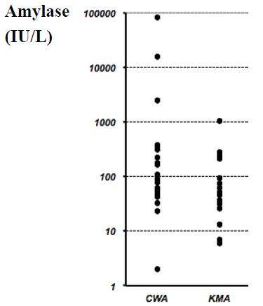
  • In essential patients, D-dimer valueis increased, blood lymphocytes decreased persistently, and laboratory changes of multiorgan imbalance (excessive amylase, coagulation disorders, etc.) are found [23].

Imaging

Chest X-ray Exam

Since the ailment manifests itself as pneumonia, radiological imaging has a essential function withinside the diagnostic process, management, and follow-up. Standard radiographic exam (X-ray) of the chest has a low sensitivity in figuring out early lung modifications and withinside the preliminary ranges of the ailment. At this stage, it may be absolutely negative. In the greater superior ranges of infection, the chest X-ray exam usually suggests bilateral multifocal alveolar opacities, which generally tend to confluence as much as the entire opacity of the lung. Pleural effusion may be associated.

Chest Computed Tomography

Given the excessive sensitivity of the technique, chest computed tomography (CT), especially excessive-decision CT (HRCT), is the technique of preference withinside the have a look at of COVID-19 pneumonia, even withinside the preliminary stages. Several non-particular HRCT findings and styles may be found. Most of those findings can also be discovered in different lung infections, including Influenza A (H1N1), CMV, SARS, MERS, streptococcus, and Chlamydia, Mycoplasma. The maximum not unusual place findings are multifocal bilateral “floor or floor glass” (GG) regions related to consolidation regions with patchy distribution, especially peripheral/subpleural and with extra involvement of the posterior areas and decrease lobes. The “loopy paving” sample may be additionally observed. This latter locating is characterised with the aid of using the presence of GG regions with superimposed interlobular septal thickening and intralobular septal thickening. It is a non-particular locating that may be detected in one-of-a-kind conditions. Other findings are the “reversed halo sign” that is a focal place of GG delimited with the aid of using a peripheral ring with consolidation, and the locating of cavitations, calcifications, lymphadenopathies, and pleural effusion.

Lung Ultrasound

Ultrasound method can permit comparing the evolution of the disease, from a focal interstitial sample up to “white lung” with proof frequently of subpleural consolidations.

It ought to be finished in the first 24 hours withinside the suspect and each 24/48 hours and may be beneficial for affected person follow-up, desire of the putting of mechanical ventilation, and for indication of inclined positioning. The most important sonographic functions are:

  • Pleural traces frequently thickened, irregular, and discontinuous till it nearly seems discontinuous; subpleural lesions may be visible as small patchy consolidations or nodules.
  • B traces. They are frequently motionless, coalescent, and cascade and may waft as much as the rectangular of “White lung”.
  • They are maximum glaring withinside the posterior and bilateral fields specially withinside the decrease fields; the dynamic air bronchogram in the consolidation is a manifestation of sickness evolution.
  • Perilesional pleural effusion.
  • In summary, throughout the direction of the disease, it’s miles feasible to become aware of the primary section with focal regions of constant B traces, a section of numerical boom of the traces B as much as the white lung with small subpleural thickenings, and in addition development till proof of posterior consolidations.

Treatment/Management

There isn’t any particular antiviral remedy encouraged for COVID-19, and no vaccine is presently available. The remedy is symptomatic, and oxygen remedy represents step one for addressing breathing impairment. Non-invasive (NIV) and invasive mechanical ventilation (IMV) can be important in instances of breathing failure refractory to oxygen remedy. Again, extensive care is wanted to address complex styles of the disease. Concerning ARDS remedy, amassing understanding at the pathophysiology of lung damage, have progressively triggered clinicians to check techniques for managing breathing failure.

As Gattinoni [24] Suggested, COVID-19-caused ARDS (CARDS) isn’t always a “Typical” ARDS. This element of the sickness is of essential significance and has probable negatively affected the healing technique withinside the early tiers of the pandemic. Indeed, notwithstanding at starting of the pandemic, early IMV became postulated because the higher approach for addressing CARDS, in COVID-19 pneumonia the standard ARDS respiration mechanics offering decreased lung compliance (i.e., cappotential to stretch and enlarge lungs) can’t be found. On the contrary, in CARDS, excellent pulmonary compliance may be demonstrated. As a consequence, and in assessment to what became to start with believed, NIV will have a key function in CARDS therapy.

O2 Fast Challenge

In an affected person with a SpO2 < 93> 28-30/min, or dyspnoea, the management of oxygen through a 40% Venturi masks have to be performed. After a five to ten mins reassessment, if the scientific and instrumental photo has stepped forward the affected person keeps the remedy and undergoes a re-assessment inside 6 hours. In case of failure improvement, or new worsening, the affected person undergoes a non-invasive remedy, if now no longer contraindicated.

HFNO and Non-invasive Ventilation

In regards to HFNO or NIV, the experts’ panel, factors out that those tactics done via way of means of structures with appropriate interface becoming do now no longer create great dispersion of exhaled air, and their use may be taken into consideration at low danger of airborne transmission.

HFNO

Because this technique has a more danger of aerosolization, it need to be utilized in terrible strain rooms.

Suggested Approaches to Control HFNO

  • Indication: while it’s far hard to hold SpO2 > 92% and/or now no longer stepped forward dyspnoea thru general
  • Setting: 30-forty L/min and FiO2 50-60%; alter consistent with medical
  • Switch to NIV if the symptomatology isn’t stepped forward after 1 hour with flow > 50 L/min and FiO2>70%.
  • HFNO also can be used for CPAP breaks (among CPAP cycles) and for assisted fibreoptic tracheal intubation in seriously sick
  • Contraindication to HFNO: hypercapnic patient.
  • Non-invasive air flow and Continuous Positive Airway Pressure NIV/CPAP has a key position in coping with COVID-19-related breathing
  • Suggested methods for acting NIV/CPAP:
  • Interface: Helmet is desired for minimizing the chance of aerosolization. In the case of NIV with face mask (full-face or oronasal), the usage of expiratory valve included and non-tubes with exhalation port, and insert an antimicrobial clear out out at the expiratory valve is recommended.

Setting

  • Continuous Positive Airway Pressure (CPAP): begin with 8-10 cmH2O and FiO2 60%
  • NIV (e.g., Pressure guide ventilation, PSV): begin with PEEP five cmH2O checking the tolerance of the affected person and produce to 8-10 cmH2O, FiO2 60%, PS 8-10 cmH2O • Management: do now no longer make many adjustments withinside the first 24 hours; after as a minimum 4-6 hours, if stabilized, detach for optimum 1 hour and permit the consumption of small portions of fluids; during the night, NIV continuously.

Intubation and Protecting Mechanical Air Flow

Special precautions are vital all through intubation. The method needs to be performed through a professional operator who makes use of non-public protecting equipment (PPE) together with FFP3 or N95 masks, protecting goggles, disposable robe lengthy sleeve raincoat, disposable double socks, and gloves. If possible, fast series intubation (RSI) need to be performed. Preoxygenation (100% O2 for five minutes) need to be performedviathe non-stop wonderful airway pressure (CPAP) method. Heat and moisture exchanger (HME) need to be located among the masks and the circuit of the fan or among the masks and the air flow balloon [25].

Mechanical air flow has to be with decrease tidal volumes (four to six ml/kg expected frame weight, PBW) and decrease inspiratory pressures, accomplishing a plateau pressure (Pplat) < 28 to 30 cm H2O. PEEP ought to be as excessive as feasible to hold the using pressure (Pplat-PEEP) as little as feasible (< 14 cm H2O). Moreover, disconnections from the ventilator should be averted for stopping lack of PEEP and atelectasis. Finally, the usage of paralytics isn’t always advocated except PaO2/FiO2 < 150> 12 hours in line with day, and the usage of a conservative fluid control method for ARDS sufferers without tissue hypoperfusion (robust recommendation) are emphasized [26].

Other Therapies

Among different healing strategies, despite the fact that systemic corticosteroids for the remedy of viral pneumonia or acute breathing misery syndrome (ARDS) have been now no longer recommended, in extreme CARDS those capsules are generally used (e.g., methylprednisolone 1 mg/Kg die). Unselective or beside the point management of antibiotics ought to be avoided, despite the fact that a few facilities advocate it. Although no antiviral remedies were approved, numerous procedures were proposed inclusive of lopinavir/ritonavir (400/a hundred mg orally each 12 hours). Nevertheless, a latest randomized, controlled, open-label trial tested no advantage with lopinavir/ritonavir remedy in comparison to conventional care [27]. Preclinical research recommended that remdesivir (GS5734) — an inhibitor of RNA polymerase with in vitro pastime towards more than one RNA viruses, together with Ebola — may be powerful for each prophylaxis and remedy of HCoVs infections [28]. This drug changed into undoubtedly examined in a rhesus macaque version of MERS-CoV infection [29]. Alpha-interferon (e.g., five million gadgets through aerosol inhalation two times according to day) changed into additionally used. Chloroquine (500 mg each 12 hours), and hydroxychloroquine (two hundred mg each 12 hours) have been proposed as immunomodulatory therapy. Of note, in a non-randomized trial, Gautret [30] confirmed that hydroxychloroquine turned into appreciably related to viral load discount till viral disappearance and this impact turned into greater with the aid of using the macrolides azithromycin. In vitro and in vivo studies, indeed, have proven that macrolides may also mitigate infection and modulate the immune system [31]. In particular, those tablets may also result in the downregulation of the adhesion molecules of the molecular surface, decreasing the production of pro-inflammatory cytokines, stimulating phagocytosis with the aid of using alveolar macrophages, and inhibiting the activation and mobilization of neutrophils. However, similarly research is wanted for recommending using azithromycin, by myself or related to different tablets inclusive of hydroxychloroquine, outdoor of any bacterial overlaps [32]. Again, interest should be paid with the concomitant use of hydroxychloroquine with azithromycin because the affiliation can cause a better danger of QT c program language period prolongation and cardiac arrhythmias. Chloroquine also can set off QT prolongation. Because COVID-19 sufferers have a better prevalence of venous thromboembolism and anticoagulant remedy is related to decreased ICU mortality, it’s miles recommended that sufferers ought to obtain thrombo-prophylaxis [33]. Moreover, within side the case ofknown thrombophilia or thrombosis, complete therapeutic-depth anticoagulation (e.g., enoxaparin 1 mg/kg two times daily) is indicated.

In Italy, a wonderful research led via way of means of the Istituto Nazionale Tumori, Fondazione Pascale di Napoli is targeted on using tocilizumab similarly to conventional therapies. It is a humanized IgG1 monoclonal antibody, directed towards theIL-6 receptorand usually used withinside the remedy of rheumatoid arthritis, juvenile arthritis, massive mobileular arthritis, Castleman’s syndrome, and for handling toxicity because of immune checkpoint inhibitors. Moreover, withinside the US, a Phase 2/3, randomized, double-blind, placebo-managed have a look at on sarilumab this is some other anti-IL-6R antibody, is ongoing [34,35]. When the disorder outcomes in complicated medical images of MOD, organ characteristic assist further to respiration assist, is mandatory. Extracorporeal membrane oxygenation (ECMO) for sufferers with refractory hypoxemia regardless of lung-protecting air flow ought to advantage attention after a case-by-case analysis. It may be recommended for people with bad outcomes to susceptible role air flow.

Prevention

Preventive measures are the cutting-edge method to restrict the unfold of cases. Because an endemic will growth so long as R0 is more than 1 (COVID-19 is 2.2), manage measures need to cognizance on lowering the price to much less than 1.

Preventive techniques are centered at the isolation of sufferers and cautious contamination manage, which includes suitable measures to be followed all through the prognosis and the supply of scientific care to an inflamed patient [35]. For instance, droplet, contact, and airborne precautions need to be followed all through specimen collection, and sputum induction need to be avoided.

  • The WHO and different businesses have issued the subsequent well-known recommendations:
  • Avoid near touch with topics stricken by acute breathing
  • Wash your fingers frequently, specifically after touch with infected people or their environment.
  • Avoid unprotected touch with farm or wild animals.
  • People with signs of acute airway contamination ought to hold their distance, cowl coughs or sneezes with disposable tissues or garments and wash their fingers.
  • Strengthen, in particular, in emergency medicinal drug departments, the utility of strict hygiene measures for the prevention and manipulate of infections.
  • Individuals which might be immunocompromised ought to keep away from public gatherings.

The maximum crucial method for the populous to adopt is to often wash their fingers and use transportable hand sanitizer and keep away from touch with their face and mouth after interacting with a probable infected environment. Healthcare employees being concerned for inflamed people need to make use of touch and airborne precautions to encompass PPE inclusive of N95 or FFP3 masks, eye protection, gowns, and gloves to save you transmission of the pathogen. Meanwhile, clinical studies are developing to expand a coronavirus vaccine. In current days, China has introduced the primary animal tests, and researchers from the University of Queensland in Australia have additionally introduced that, after finishing the three-week in vitro study, they’re shifting directly to animal testing. Furthermore, withinside the U.S., the National Institute for Allergy and Infectious Diseases (NIAID) has introduced that a segment 1 trial has begun for a singular coronavirus immunization in Washington state.

Differential Diagnosis

The signs of the early ranges of the sickness are nonspecific. Differential prognosis ought to consist of the opportunity of an extensive variety of infectious and non-infectious (e.g., vasculitis, dermatomyositis) not unusual place breathing disorders.

  • Adenovirus
  • Influenza
  • Human metapneumovirus (HmPV)
  • Parainfluenza
  • Respiratory syncytial virus (RSV)
  • Rhinovirus (not unusual place cold)

For suspected cases, speedy antigen detection, and different investigations have to be followed for comparing not unusual place respiration pathogens and non-infectious conditions. The Mayo Clinic proposed a COVID-19 self-evaluation device designed for setting up a ability candidate for a COVID-19 diagnostic test (https://www.Mayoclinic.Org/covid-19-self-evaluation-device).

Prognosis

Preliminary records indicate the suggested demise charge tiers from 1% to 2% pending at the take a look at and country. The majority of the fatalities have come about in sufferers over 50 years of age. Young youngsters look like mildly inflamed however might also additionally function a vector for extra transmission.

Complications

Long time period headaches amongst survivors of contamination with SARS-CoV-2 having clinically massive COVID-19 disorder aren’t but available. The mortality prices for instances globally stay among 1% to 2%. Follow-up research will make clear the quantity of the sequelae on organ functions, which include respiratory, renal, cardiovascular, in addition to psychological/psychiatric, and associated with persistent ache problems.

Deterrence and Patient Education

b

Patients and households have to acquire coaching to:

  • Maintaining correct social distance is obligatory for stopping the unfold of the disease.
  • Strict private hygiene measures (fingers wash) are essential for the prevention and manage of this contamination.
  • Avoid near touch with topics stricken by acute breathing
  • People with signs and symptoms of acute airway contamination have to preserve their distance, cowl coughs or sneezes with disposable tissues or clothes, and wash their fingers.

Immunocompromised sufferers have to keep away from public publicity and public gatherings. If an immunocompromised man or woman ought to be in a closed area with more than one people present, along with a assembly in a small room; masks, gloves and private hygiene with antiseptic cleaning soap need to be undertaken with the aid of using the ones in near touch with the individual. In addition, earlier room cleansing with antiseptic dealers need to be undertaken and executed earlier than publicity. However, thinking about the risk worried to those individuals, publicity need to be averted except a meeting, institution event, etc. Is a real emergency.

References

  1. Perlman S, Netland J (2009) Coronaviruses post-SARS: update on replication and pathogenesis. Nat Rev Microbiol 7: 439-450. [crossref]
  2. Chan JF, To KK, Tse H, Jin DY, Yuen KY (2013) Interspecies transmission and emergence of novel viruses: lessons from bats and birds. Trends Microbiol 21: 544-555. [crossref]
  3. Chen Y, Liu Q, Guo D (2020) Emerging coronaviruses: Genome structure, replication, and pathogenesis. J Med Virol 92: 418-423. [crossref]
  4. Chan JF, Kok KH, Zhu Z, Chu H, To KK, et al. (2020) Genomic characterization of the 2019 novel human-pathogenic coronavirus isolated from a patient with atypical pneumonia after visiting Wuhan. Emerg Microbes Infect 9: 221-236. [crossref]
  5. Guo ZD, Wang ZY, Zhang SF, Li X, Li L, et al. (2020) Aerosol and Surface Distribution of Severe Acute Respiratory Syndrome Coronavirus 2 in Hospital Wards, Wuhan, China, 2020. Emerging Infect Dis 126: 1583-1591. [crossref]
  6. Lauer SA, Grantz KH, Bi Q, Jones FK, Zheng Q, et al. (2020) The Incubation Period of Coronavirus Disease 2019 (COVID-19) From Publicly Reported Confirmed Cases: Estimation and Application. Ann Intern Med 172: 577-582. [crossref]
  7. Li Q, Guan X, Wu P, Wang X, Zhou L, et al. (2020) Early Transmission Dynamics in Wuhan, China, of Novel Coronavirus-Infected Pneumonia. N Engl J Med 382: 1199-1207.
  8. Bauch CT, Lloyd-Smith JO, Coffee MP, Galvani AP (2005) Dynamically modeling SARS and other newly emerging respiratory illnesses: past, present, and future. Epidemiology 16: 791-801. [crossref]
  9. Lei J, Kusov Y, Hilgenfeld R. (2018) Nsp3 of coronaviruses: Structures and functions of a large multi-domain protein. Antiviral Res 149: 58-74. [crossref]
  10. 
Song W, Gui M, Wang X, Xiang Y (2018) Cryo-EM structure of the SARS coronavirus spike glycoprotein in complex with its host cell receptor ACE2. PLoS Pathog. [crossref]
  11. Angeletti S, Benvenuto D, Bianchi M, Giovanetti M, Pascarella S, et al. (2020) COVID-2019: The role of the nsp2 and nsp3 in its pathogenesis. J Med Virol 92: 584-588. [crossref]
  12. Ciceri F, Beretta L, Scandroglio AM, Colombo S, Landoni G, et al. (2020) icrovascular COVID-19 lung vessels obstructive thromboinflammatory syndrome (MicroCLOTS): an atypical acute respiratory distress syndrome working hypothesis. Crit Care Resusc 22: 95-97. [crossref]
  13. Conti P, Ronconi G, Caraffa A, Gallenga CE, Ross R, et al. (2020) Induction of pro- inflammatory cytokines (IL-1 and IL-6) and lung inflammation by Coronavirus-19 (COVI-19 or SARS- CoV-2): anti-inflammatory strategies. J Biol Regul Homeost Agents 34: 327-331. [crossref]
  14. Tian S, Hu W, Niu L, Liu H, Xu H, et al. (2020) Pulmonary Pathology of Early-Phase 2019 Novel Coronavirus (COVID-19) Pneumonia in Two Patients With Lung Cancer. J Thorac Oncol 15: 700-704. [crossref]
  15. Zhang H, Zhou P, Wei Y, Yue H, Wang Y, et al. (2020) Histopathologic Changes and SARS-CoV-2 Immunostaining in the Lung of a Patient With COVID-19. Ann Intern Med 172: 629- 632.
  16. Menter T, Haslbauer JD, Nienhold R, Savic S, Hopfer H, et al. (2020) Post-mortem examination of COVID19 patients reveals diffuse alveolar damage with severe capillary congestion and variegated findings of lungs and other organs suggesting vascular dysfunction. Histopathology 77: 198-209. [crossref]
  17. Huang C, Wang Y, Li X, Ren L, Zhao J, et al. (2020) Clinical features of patients infected with 2019 novel coronavirus in Wuhan, China. Lancet 395: 497-506.
  18. Wu Z, McGoogan JM. (2020) Characteristics of and Important Lessons From the Coronavirus Disease 2019 (COVID-19) Outbreak in China: Summary of a Report of 72 314 Cases From the Chinese Center for Disease Control and Prevention. JAMA 323: 1239-1242. [crossref]
  19. Kogan A, Segel MJ, Ram E, Raanani E, Peled-Potashnik Y, et al. (2019) Acute Respiratory Distress Syndrome following Cardiac Surgery: Comparison of the American-European Consensus Conference Definition versus the Berlin Definition. Respiration 97: 518-524. [crossref]
  20. Singer M, Deutschman CS, Seymour CW, Shankar-Hari M, Annane D, et al. (2016) The Third International Consensus Definitions for Sepsis and Septic Shock (Sepsis-3). JAMA 315: 801-810. [crossref]
  21. Seymour CW, Kennedy JN, Wang S, Chang CH, Elliott CF, et al. (2019) Derivation, Validation, and Potential Treatment Implications of Novel Clinical Phenotypes for Sepsis. JAMA 321: 2003-2017.
  22. Matics TJ, Sanchez-Pinto LN (2017) Adaptation and Validation of a Pediatric Sequential Organ Failure Assessment Score and Evaluation of the Sepsis-3 Definitions in Critically Ill Children. JAMA Pediatr 171
  23. Yang AP, Liu JP, Tao WQ, Li HM (2020) The diagnostic and predictive role of NLR, d-NLR and PLR in COVID-19 patients. Int Immunopharmacol
  24. Gattinoni L, Coppola S, Cressoni M, Busana M, Rossi S, Chiumello D (2020) COVID-19 Does Not Lead to a “Typical” Acute Respiratory Distress Syndrome. Am J Respir Crit Care Med 201: 1299-1300. [crossref]
  25. Hui DS, Chow BK, Lo T, Tsang OTY, Ko FW, et al. (2019) Exhaled air dispersion during high-flow nasal cannula therapy versus CPAP via different masks. Eur Respir J
  26. Wu CN, Xia LZ, Li KH, Ma WH, Yu DN, et al. (2020) High-flow nasal-oxygenation-assisted fibreoptic tracheal intubation in critically ill patients with COVID-19 pneumonia: a prospective randomised controlled trial. Br J Anaesth
  27. Cao B, Wang Y, Wen D, Liu W, Wang J, et al. (2020) A Trial of Lopinavir-Ritonavir in Adults Hospitalized with Severe Covid-19. N Engl J Med 382: 1787-1799. [crossref]
  28. Gordon CJ, Tchesnokov EP, Feng JY, Porter DP, Götte M (2020) The antiviral compound remdesivir potently inhibits RNA-dependent RNA polymerase from Middle East respiratory syndrome coronavirus. J Biol Chem 295: 4773-4779. [crossref]
  29. de Wit E, Feldmann F, Cronin J, Jordan R, Okumura A, et al. (2020) Prophylactic and therapeutic remdesivir (GS-5734) treatment in the rhesus macaque model of MERS- CoV infection. Proc Natl Acad Sci USA 117: 6771-6776.
  30. Gautret P, Lagier JC, Parola P, Hoang VT, Meddeb L, et al. (2020) Hydroxychloroquine and azithromycin as a treatment of COVID-19: results of an open-label non- randomized clinical trial. Int J Antimicrob Agents [crossref]
  31. Zarogoulidis P, Papanas N, Kioumis I, Chatzaki E, Maltezos E, et al. (2012) Macrolides: from in vitro anti-inflammatory and immunomodulatory properties to clinical practice in respiratory diseases. Eur J Clin Pharmacol 68: 479-503. [crossref]
  32. Mercuro NJ, Yen CF, Shim DJ, Maher TR, McCoy CM, et al. (2020) Risk of QT Interval Prolongation Associated With Use of Hydroxychloroquine With or Without Concomitant Azithromycin Among Hospitalized Patients Testing Positive for Coronavirus Disease 2019 (COVID-19). JAMA Cardiol
  33. Kollias A, Kyriakoulis KG, Dimakakos E, Poulakou G, Stergiou GS, et al. (2020) Thromboembolic risk and anticoagulant therapy in COVID-19 patients: emerging evidence and call for action. Br J Haematol
  34. Buonaguro FM, Puzanov I (2020) Ascierto PA. Anti-IL6R role in treatment of COVID-19-related ARDS. J Transl Med
  35. Vittori A, Lerman J, Cascella M, Gomez-Morad AD, Marchetti G, et al. (2020) COVID- 19 Pandemic ARDS Survivors: Pain after the Storm?. Anesth Analg

Sexual Imagination Potency (SIP) Test to Explore the Unconscious Sexual Life of Humans

DOI: 10.31038/PSYJ.2021314

Abstract

Despite the importance of the sexuality in the human life, most studies performed up to now have been generally limited to the only evaluation of sexual behaviour and orientation, rather than the intimate sexual feeling in terms of sexual fantasies. Some preliminary results would suggest the existence of some same sexual fancies beyond the difference occurring between homo and hetero sexuality, in particular the fantasy of androgyny. On this basis a preliminary study was planned to elaborate a sexual test carried out to investigate not only the sexual behaviour but the dimension of sexual fancies and imagination, by proposing a specific analysis that we have called Sexual Imagination Potency (SIP). The study included 150 consecutive healthy volunteers and the test was accepted in 111/150 subjects. No significant difference in SIP mean values was observed between men and women. Within the evaluated fancies, a particular importance has been shown to be played by the manner to imagine the androgyny aspect, and the pleasure for pegging. In fact, the subjects with pleasure for pegging showed significantly higher SIP mean values with respect to those, who had no pleasure for pegging. These preliminary results, which have to be confirmed in greater number of healthy subjects, seem to demonstrate the importance of the androgyny image in influencing the sexual mood by connecting hetero and homo sexual fancies in a unique imaginative psychosexual world.

Keywords

Androgyny, Heterosexuality, Homosexuality, Sexual fancies

Introduction

Imagination and desire are looked upon as major determinants of sexuality [1]. Moreover, it has to be considered that the imagination represents one of the fundamental dimensions of human cognition [2]. During the early phases of development, the close interaction of imagination and erotic desire leads to the formation of psychical representatives of experiences of satisfaction, that influence sexual and nonsexual behaviours by establishing an internalized structure of blueprints for satisfaction [3]. These blueprints can also be viewed as an important step in the development of autonomy. Sex differences can be found primarily in the function and employment of erotic fantasies [4]. Generally, men tend to use their erotic imagery to a much in the higher degree with respect to the women, as a compensation for a lack of sexual satisfaction [5]. However at present the fantasies of women have still to be better investigated and understood. Then, it has to be confirmed the lower degree of fancies in women with respect to men. Proceeding from the conceptual distinction of erotic and everyday realities, reflections on the zeitgeist of sexuality and the relationship between the sexes are put forward focussing on the ambiguity of erotic imagination and the border crossing between the two realities [6]. Based on these reflections, the potential therapeutic aspects of imagination and desire are touched upon the problems of integrating erotic reality and everyday reality in long-term male-female relationships would have to be further investigated in the clinical practices [7]. Same preliminary clinical studies carried out for many years to investigate the sexual male and female fancies, have allowed us to hypothesize that the original and primary fancy is represented by the androgyny image, which could constitute the sexual fancy, from which would depend all other human sexual fantasies, involving both men and women irrespectively of the sexual orientation, by overcoming the opposition between hetero and homo sexual fancies [8]. On this basis, we have elaborated a simple and synthetic clinical test to explore the major fancies reflecting the androgyny status, and most in general the potency of the sexual imagination, independently of the degree of sexual satisfaction and activity.

Subjects and Methods

The study included 150 consecutive healthy volunteers (M/F: 69/81) to whom the SIP test whose proposed. Test of acceptance in 111/150 (74%) subjects, without statistically significant difference between man and woman (53/69 (77%) versus 58/81 (70%)). The characteristic of subject are reported in Table 1. The subject were subdivided on the basis of six major variables, including age, profession, grade of studies, religion faith, marriage status and affective status of relatives. The SIP test, which was differentiate in relation to the sex, was consisting of five essential question, with the three type of response, with a score ranging from 0 to 2, for a total maximum values of ten points. The SIP test was reported in Table 2. Data were statistically analyzed by the Chi Square test and the Student’s T Test.

Table 1: Characteristics of subjects.

N

Acceptance

M

F

150
111 (74%)

53 (47%)
58 (52%)

Marital status

-Marriage/cohabitation

-Single/widow

-Apart/divorced

45
47
19

Occupation

-Intellectual

-Practise


58

53

Education
 -Low-Middle-High

33
36
42

Faith

–          Christian

–          No faith

–          Other religions


74

22

15

Status of relationship among parents

–          Unity

–          Separated

92
19

Age

<50

> 0

57
54

Table 2: SIP Test values in a group of healthy women and men.

N

Question Response Points
1 Do you find more excitant the common vaginal

or the anal relation?

– No opinion

the vaginal relation

the anal relation

1

2

0

2 Do you find the pegging (the woman penetrate

the man) as an excitant sexual stimulation?

– No

– I do not know 1

– yes 2

1-2**

0

3 How do you imagine the trio, with another woman or with another man? – I do not like the trio

– With another woman 1-2*

– With another man

1

2

0

4 Do you feel more cheated on if your partner had a sexual relation with a person of the same sex

or the other sex?

 – With both sexes

– with the same sex

– With the other sex

0

5 How do you imagine the androgyne human subject? Like a trans – No idea

– Like a woman with the artificial penis

1

Note: *1 for man and 2 for woman; **1 for woman and 2 for man.

Results

The evaluation of each single fancy is reported in Table 3. As reported, not significantly difference occurred between men and women in the preferential of the type of sexual relation the genital and the anal one. On the same way not significantly difference between men and women was seen in relation to the fancy of man penetration by woman, the so-called pegging. As far as trio fancy with a male and a female is concerned, male subject statistically preferred a woman as third partner (p<0.05), whereas women did not show statistically significance preference between males and females. Moreover, in relation to the psychic sufferance due to betrayal with the another partner, the percentage of pain in the presence of betrayal with a person of the same sex was respect to a betrayal with a person of the other sex was lower in women and higher in men, but none of these difference was statistically significance. Finally in the women the androgyny is imagined more significantly as a women with the strap-on, while in the men as trans (p<0.05). SIP mean values in relation to the main characteristics of healthy subjects are reported in Table 4.

Table 3: Evaluation of the single fancy expressed in percentage in men and women.

Fancy

Men=53 Women=58

Men+Women= 111

Prefered sexual relation

– Vaginal

– Anal

– no idea


31 (59%)

14 (26%)

8 (15%)


42 (72%)

7 (12%)

9 (16%)

73 (81%)

15 (16%)

23 (25%)

Love of pegging

– Yes

– No

– no idea


13 (25%)

15 (28%)

25 (47%)


10 (17%)

39 (67%)

9 (16%)

25 (23%)
64 (57%)
22 (20%)

Trio

– with men

– with women

– no love


10 (19%)

6 (11%)

37 (70%)*


13 (22%)

13 (22%)

32 (55%)

19 (17%)

50 (45%)

42 (38%)

Hurt by betrayal of the partner

– Both

– with men

– with women


25 (47%)

6 (11%)


34 (59%)

9 (15%)

15 (26%)

59 (53%)

31 (28%)

21 (19%)

Androgynous

-b no idea

– trans

– women with strap-on


33 (62%)

14 (27%)*

6 (11%)


34 (59%)

7 (12%)

17 (29%)**

67 (60%)

21 (19%)

23 (21%)

Note: * p<0.05 vs women.

**p<0.05 vs men.

In the women the androgyny is imagined more significantly as women with the strap-on, while in the men as trans.

Table 4: SIP values (X± SE) in relation to the main characteristics of men and women.

Variables

Men

Women

N. X ± SE

N. X ± SE

Sex

53 3.9 0.5

58 3.3 0.6

Age
<50

26 4.7 0.4

31 3.5 0.5

>50

27 3.9 0.5

27 3.2 0.7

Religion
Christian

32 4.3 0.5

42 3.3 0.6

No faith

13 3.9 0.4

9 3.5 0.6

Other religion

8 3.0 0.6

7 3.0 0.7

Marriage Status
Married

20 4.2 0.5

25 2.2 0.4**

Single

24 4.5 0.4

23 3.6 0.2

Separate/divorced

9 3.3 0.6

10 5.1 0.6

Study Degree
Low

16 3.9 0.4

17 3.7 0.6

Middle

17 3.5 0.5

19 3.3 0.6

High

20 4.2 0.4

22 4.0 0.5

Profession
Practical

27 3.3 0.5

26 3.1 0.4

Intellectual

26 4.2 0.4

32 4.4 0.2

Affective Status of Relatives
Unity

44 4.4 0.4

48 3.8 0.3

Separation

9 3.0 0.7

10 2.3 0.4*

Note: ** p<0.05 vs. single women, p<0.01 vs. separated women.

* p<0.05 vs. united relatives.

The only statistically significance difference were those concerning the marriage status and the professional situation. In more detail, separate and divorced women showed SIP mean values significantly higher than those found in married women. On the contrary separate or divorced men showed lower SIP mean values than the married ones, even though the difference was not significance. In addiction both women and mans with an intellectual profession showed statistically significant higher SIP values then those with a practical professional. Finally, as far as an affective status of relations, relative separation was associated with a statistically significance redaction in SIP mean values in the only women (p<0.005). Table 5 shows SIP values in relation to the man androgyny-related fancies and the difference between man and women. SIP mean values were significantly higher in subjects who referred pleasure for Pegging (PG) than in those who did not like it in the only men (p<0.001), whereas in women the difference was not statistically significance. On the same way SIP values were significantly higher in men who referred pleasure for anal relation then in those who had no pleasure for it (p<0.005), whereas no significantly difference occurred in women. On the contrary both men and women, who referred pleasure for either pegging and anal relation showed statistically significance higher SIP values than those who had no interested for both pegging and anal relation ( p<0.001).

Table 5: SIP values (X±SE) in relation to the main androgyny-related fancies.

Type of Fancies

Men

Women

N. X ± SE

N. X±SE

Pleasure for pegging
Yes

15 6.8 0.4*

10 6.9 0.8

No

25 2.3 0.3

39 3.3 0.6

Pleasure for anal relation
Yes

8 6.7 0.6**

7 4.8 0.8

No

45 2.6 0.6

51 3.9 0.3

Pleasure for both pegging and anal relation
Yes

6 7.5 0.6*

5 8.0 0.5*

NO

36 2.5 0.5

46 3.6 0.4

Pleasure for sexual trio
yes

43 4.5 0.3

26 4.9 0.6**

no

10 2.3 0.8

32 1.6 0.3

Androgyny imagination
Like transexual men

13 5.5 0.5

8 3.6 0.8

Like woman with strap-on

7 6.3 0.8

16 7.1 0.4*

No opinion

33 3.3 0.4

34 1.7 0.5

Note: * p<0.001; ** p<0.05.

The pleasure for trio was associated with higher SIP values than in those who showed no interested for trio, even though they were statistically significantly higher in the only women (p<0.005). Finally subjects who had no imagination of androgyny showed SIP values lower than those who had same imagine of androgyny. But the SIP values were statistically significant higher with respect to subject, who referred no androgyny imagine in the only women, who had the vision of androgyny like a woman with a strap-on (p<0.001), whereas no difference occurred in women who imagined the androgyny like a transgender man. The maximal SIP values occurred in both men and women, who referred pleasure for both pegging and anal relation as well as in the only women who had androgyny imagine as woman with a strap-on. Table 6 shows SIP values in subjects with pleasure for pegging and anal sexual relation or both fancies in relation to their religion. Christian men, who referred pleasure for pegging or for anal relation showed statistically significant higher SIP values than men with other religion or no religion who showed the same fancies, whereas no difference occurred in women. On the contrary both Christian man and women who had pleasure for both pegging and anal relation showed statistically significant higher SIP values than subjects with the same fancies, but who were without religion or of other religion.

Table 6: Pleasure for pegging and anal sexual coitus in relation to androgyny image in men and women.

Androgyny Image

 Women

 Men

pleasure for pegging pleasure for anal relation

pleasure for pegging pleasure for anal relation

Like transexual man

2/8 (25%)* 2/8 (25%)

6/13 (46%) 3/13 (23%)

like woman with strap-on

8/16 (50%)** 4/16 (25%)

2/7 (29%) 2/7 (29%)

no image

2/34 (7%) 4/34 (12%)

9/33 (27%) 3/33 (9%)

Note: **p<0.01 vs. women without androgyny image; *p<0.05 vs. women without androgyny image.

Discussion

Even though limited to a relatively low number of normal subjects, this preliminary study seems to suggest that the maximal sexual imagination potency is associated with the fancies related to a change in the common manner to consider the male -female relation and interpretation of male-female identity and role, such as pleasure for pegging and sexual anal relation, which could be considered as an expression of the androgyny imagine. In more detail, the sexual imagination has appeared to be negatively influenced by the separation of relatives in the only women. The marriage was also associated with an evident decline in SIP values. On the contrary, the separation, the divorce and the single life were all associated with an evident increase in the sexual imagination power. In addiction this study shows that the pleasure for pegging was associated with the higher SIP values. Moreover, the apparently higher SIP values in Christian people than in those with other religion or no religion, would demonstrated the existence of interaction between spirituality and erotic profile, by suggesting that the interpretation of the Spirit may influence the human psychosexual life. Finally, this study seems to excluded that the men may have more sexual fantasies than women.

Then further studies would be required to analyze the sexual world of women. In fact, male subjects could have more sexual fantasies with the respect to women only from a quantitative point of view, but women could express more fantasies from a qualitative point of view, even though women are generally less unconscious on their sexual dimension. Unfortunately, most studies carried out up to now on the human sexuality, has been generally limited to the only sexual behaviour and orientation, rather than to explore the dimension of the sexual unconscious fancies [9-11]. Therefore, on the basis of the results of this study obtained in a group of healthy subjects, it would be interesting to evaluate in future studies the sexual profile and fancies occurring in the main human systemic diseases, namely cancer and autoimmunity.

In conclusion, by considering the difference between males and females in relation to androgyny imagination, this preliminary study would suggest that the original sexual fancy could be constituted of the image of androgyny itself, which could connected in the same sexual imagination homo and hetero fancies. Then, the only fancy, which may integrate hetero and homosexual fancies, in a same sexual imagination and excitation, is that of the androgyny status, which would constitute the origin of the human psychosexuality.

References

  1. Meuwissen I, Over R (1991) Multidimensionality of the content of female sexual Behav Res Ther 29: 179-189. [crossref]
  2. Smith D, Over R (1991) Male sexual fantasy: Multidimensionality in content. Behav Res Ther 1991; 29: 267-275. [crossref]
  3. Toledano R, Pfaus J (2006) The Sexual Arousal and Desire Inventory (SADI): A multidimensional scale to assess subjective sexual arousal and desire. Journal Sex Med 3: 853-877. [crossref]
  4. Goldey KL, Van Anders SM (2012) Sexual arousal and desire: Interrelations and responses to three modalities of sexual stimuli. Journal Sex Med 9: 2315-2329. [crossref]
  5. Hartmann U (1994) Imagination and desire: Reflections on the determination of male sexuality. Psychother Psychosom Med Psychol 44: 403-410. [crossref]
  6. Meana M (2010 Elucidating women’s (hetero) sexual desire: Definitional challenges and content expansion. Journal Sex Res 47: 104-122. [crossref]
  7. Carvalho J, Gomes AQ, Laya P, Oliveira C, Vilarinho S, et al. (2013) Gender differences in sexual arousal and affective responses to erotica: the effects of type of film and fantasy instructions. Arch Sex Behav 42: 1011-1019. [crossref]
  8. Tseng YH, Cheng CP, Kuo SH, Hou WL, Chan TF, et al. (2019) Safe sexual behaviors intention among female youth: The construction on extended theory of planned behavior. Adv Nurs. [crossref]
  9. Storms MD (1980) Theories of sexual orientation. Journal Person Soc Psychol 38: 783-792.
  10. Ngun TC, Vilain E (2014) The biological basis of human sexual orientation is the a role for epigenetics. Adv Genetics 86: 167-184. [crossref]
  11. Mitricheva C, kimura R, Logothetis NK, Noori HR (2019) Neural substrates of sexual arousal are not sex dependent. Proc Natl Acad Sci USA 116: 15671-15676. [crossref]
fig 2

Implementation of the 1-Hour Sepsis Bundle and Evaluation of Staff Adherence: An Evidence-based Practice Quality Improvement Project

DOI: 10.31038/IJNM.2021211

Abstract

Objective: To implement an evidence-based sepsis implementation tool for nurses to use when initiating treatment for patients diagnosed with sepsis and to track time of administration of the Surviving Sepsis Campaign (SSC) 1-hour bundle interventions, mortality, and length of stay.

Design: An evidence-based practice quality improvement (EBP-QI) project.

Setting: A 38-bed observation/short stay unit within a 700-bed hospital in New York City.

Intervention: A sepsis implementation tool was created based on SSC 2018 1-hour guidelines. Sepsis champions delivered education on sepsis recognition, treatment, and management to the nurses, physicians, and other staff.

Main outcome measure: Following the practice change, audits of the sepsis implementation tool were done weekly for 5 months. A target of 85% completion for each of the bundle interventions was set.

Results: From May 8, 2019 to October 8, 2019 a total of 38 patients were diagnosed with sepsis in the emergency department or observation/short stay unit and of these 90% (n=33) had blood cultures drawn twice, 85% (n=34) had stat lactate, and 73% (n=26) had broad-spectrum antibiotics started within 1-hour. The target of 85% was met for 2 of the 3 bundle interventions.

Conclusion: The sepsis 1-hour bundle is best practice however, completion of the bundle interventions within 1-hour of sepsis diagnosis is challenging. In this EBP-QI project, the healthcare staff was successful in completing the majority of the bundle interventions within the hour. Future improvement efforts will focus on improving the initiation of antibiotics within 1-hour of sepsis diagnosis.

Introduction

In the US, sepsis, severe sepsis, and septic shock are associated with 6%, 15%, and 34% mortality rates and respective costs of $16,000, $25,000, and $38,000 per hospitalization [1]. Sepsis is a deadly and costly hospital condition that can be mitigated with early identification and initiation of lifesaving treatment. In 2004 there was a global initiative to bring together critical care and infectious disease experts in the diagnosis and management of sepsis to create the initial Surviving Sepsis Campaign (SSC) guidelines to improve awareness and outcomes of sepsis [2]. Initial guidelines included goal directed patient resuscitation during the first 6 hours after recognition, appropriate diagnostic studies to identify cause before initiating antibiotics, and early administration of antibiotics, all to be done as soon as possible within the first 24 hours. Subsequent recommendations in the following years grouped similar interventions into 6 and 3-hour bundles with the expectation that the interventions would all be completed within these shorter time frames. In 2018, the SSC developed the 1-hour sepsis bundle because the 3-hour window was associated with a significant increase in in-hospital mortality [3]. The bundle calls for lactate measures, blood cultures, antibiotics, if appropriate fluid resuscitation and vasopressors within 1-hour of sepsis recognition [3]. In 2015, The Centers for Medicare and Medicaid implemented its core bundle measure for Severe Sepsis and Septic Shock Early Management Bundle linking reimbursement to a hospitals’ ability to complete the SSC bundle interventions within 3 hours of sepsis recognition [4]. To help meet this quality measure, hospitals may benefit from initiatives aimed at improving the process of sepsis care and bundle completion within the 1 to 3-hour window. Meeting the SSC’s 1-hour implementation goal can be challenging. Historically, nurses have been responsible for initiating a sepsis protocol [5]. There are tools to facilitate timely initiation of bundle interventions [6]. The emergency room nurse sepsis screening tool significantly improved the time to bundle completion in patient’s diagnoses with severe sepsis and septic shock [7]. The nurse initiated emergency department sepsis protocol significantly reduced time to lactate measurements and antibiotic administration [8]. These tools are based on the five steps outlined in the SSC’s 2018 1-hour bundle.

Objective

The purpose of this project was to implement an evidence-based sepsis implementation tool for nurses to use when initiating treatment for patients diagnosed with sepsis and to track time of administration of the bundle elements, mortality, and length of stay.

Methods

Project Design

This EBP-QI project was completed over a 10-month period using a prospective before and after design. This consisted of a 5-month baseline period and a 5-month QI period. In the baseline period, the evidence-based sepsis implementation tool was created, and the sepsis champions delivered education on sepsis recognition, treatment, and management to the nurses, physicians, pharmacists, and other staff, and assessed sepsis knowledge. In the QI period, audits of the sepsis implementation tool were done weekly.

Setting

The Observation/Short Stay Unit (O/SSU) was the project setting. This unit is part of an 800-bed acute-care tertiary hospital in New York City that serves 37,000 patients annually. The hospital has several medical specialties (e.g. cardiology including care of vascular conditions, neurology, and oncology). The O/SSU, is considered part of the emergency department and can take up to 38 patients. O/SSU employs 71 nurses, with an average of 12-14 nurses and 2 charge nurses per shift. There are 2-3 patient care technicians per shift and 1 patient unit assistant for the day and evening shifts. The average daily census ranges from 15 to 35 patients and varies by the time of day. There is a unit nurse manager, assistant nurse manager, and a nurse manager administrative support supervisor. There is a pharmacist on the unit from 0800 to 2400. Between 0001 and 0759, pharmacists are accessible via telephone, and medications are sent via a pneumatic system. Common O/SSU diagnoses include; falls, acute coronary syndrome, transient ischemic attack, chronic obstructive pulmonary disease exacerbation, congestive heart failure, and skin, lung, and urinary infections. The average quarterly sepsis rate was 528 cases for the years 2015, 2016, 2017 and 2018, and 84% of these cases were patients with sepsis present on admission.

Participants

Participants were patients entering the hospitals’ emergency department from May 8, 2019 to October 8, 2019 and transferred to the O/SSU and met the following SSC sepsis screening criteria. Patients with 2 or more systemic inflammatory response syndrome (SIRS) criteria or suspected infection defined as sepsis [9] or with septic shock/sepsis-3 defined as organ dysfunction with SIRS criteria (SSC).

Intervention

The evidence-based sepsis implementation tool, displayed in Figure 1, was created using SSC’s 2018 guidelines and other evidence source. The nurse immediately notifies the provider when a patient has a positive screen and initiates the sepsis implementation tool with the provider in a safety huddle at the patient’s bedside. The nurse and provider collaborate to provide the first 4 interventions within 1-hour. The nurse documents the time each intervention was completed and initials. Providers document their reasoning for not initiating fluids. The next section of the tool is for nurses to document if a critical care consult was initiated during the first hour. At the 1-hour mark, the nurse and provider re-huddle and perform the next 5 interventions (e.g. review the lab results) to determine if the patient sepsis is worsening. The provider is then required to write a sepsis note in the electronic health record that includes the patient’s presenting condition, completed interventions and subsequent plan of care.

fig 1

Figure 1: Evidence-Based Sepsis Implementation Tool.

Quality Improvement Process

We used the Revised Iowa Model of Evidence Based Practice to guide this EBP-QI project. The Iowa model uses EBP and QI processes to promote excellence in healthcare [10,11]. This project was led by three individuals with complementary areas of expertise. The project manager was a doctoral-level nursing student with expertise in sepsis recognition, treatment, and management. The physician partner has extensive leadership knowledge and has led numerous multidisciplinary quality and safety initiatives in hospitals. The academic partner has an EBP certification, clinical expertise in critical care nursing, and expertise in dissemination.

Key stakeholders were the staff nurses and healthcare providers in the O/SSU. To gain their buy in and to form a group of sepsis champions, nurses were reminded that they could submit this project for promotion through a nursing professional advancement program. This strategy resulted in four staff nurses agreeing to be sepsis champions. Physicians and physician assistants interested in sepsis management were also included in the group of champions to help guide and lead other healthcare providers. Initially pharmacists were informed of the QI project to help expedite interventions. They were added as key stakeholders during the 5-month QI period when nurses reported delays in obtaining antibiotics. Our educational strategy was multidimensional and completed over several months. During the first 2 months, we assessed baseline knowledge of sepsis recognition, treatment and management, and attitude toward sepsis care using a questionnaire (See Supplement) that was given to O/SSU nurses and healthcare providers (n=101). We held several meetings to review and discuss questionnaire answers. For the next 3 months, the project manager and sepsis champions gave updates and education as needed at monthly staff meetings, provider meetings, and in real-time in the O/SSU using an iPad. The iPad contained the sepsis checklist, a power-point presentation on sepsis from [9], the sepsis questionnaire answers, and the EBP-QI project goals and intervention description. The iPad was and was left in a central place in the O/SSU that staff could access at any time throughout the 10-month project period. The education for nurses and healthcare providers included review of sepsis recognition and diagnosis, the 1-hour bundle interventions, and the nurse’s role in management including the new sepsis implementation tool. Nurses received additional education on how to complete the sepsis implementation tool and an explanation of the buddy badge strategy to facilitate timely completion of the 1-hour bundle interventions. A badge buddy is a laminated card that attaches to an existing hospital identification badge and lists the SIRS criteria and 1-hour bundle interventions that was given to all O/SSU nurses (Figure 2).

fig 2

Figure 2: Process Map: Sepsis Algorithm 1-hour Bundle for O/SSU.

The project manager and sepsis champions held monthly group meetings throughout the 10-month project to review the project progress and address any barriers. Champions followed-up with the nurses of patients with delayed bundle interventions within a week to debrief. Monthly emails were sent to all staff with updates on audits and current status of the project including staff feedback regarding barriers to the 1-hour bundle and how to overcome them.

Evaluation Measures

Baseline knowledge of sepsis recognition, treatment and management, and attitude toward sepsis care was measured using an established questionnaire (See Supplement). Adherence was measured by how often the nurses completed the initial lactate measure, blood cultures, and antibiotic administration within the 1-hour window of the patient being diagnosed with sepsis. We set a target of 85% of cases having all interventions completed within 1-hour. Length of stay was measured as the total number of days spent in hospital and mortality was measured as death occurring in the hospital.

Data Collection and Analysis

The completed sepsis implementation tools were retrieved weekly from the O/SSU. The project manager reviewed the tools and checked the electronic health record for 1-hour bundle intervention completion times. Data on mortality and length of stay were obtained after completing chart reviews for the patients included during the QI period (n=38). These data were inputted into an Excel spreadsheet and descriptive statistics were calculated for each outcome [12].

Ethical Considerations

Differentiating Quality Improvement and Research Activities Tool was used to determine that this was a QI project. The project aim was to improve sepsis care for all patients using evidence-based recommendation and no personal health information was collected therefore it did not qualify as human subjects’ research and institutional review board was not needed. Per hospital policy, the project was reviewed and approved by the institution’s Chief Nursing Officer [13].

Actions Taken to Barriers during Baseline QI Period

Table 1 displays the barriers identified by nursing, physicians, and physician assistants during the baseline QI period. These barriers fell into the categories of staffing, factors causing delays and patient specific concerns. Actions taken include; the requirement of two nurses to initiate sepsis protocol interventions, pharmacy added as key stakeholder, nurse to notify pharmacy of patient with sepsis to expedite interventions, notification of 2nd lactate included in the EHR, IV team able to place midlines, central lines, IO kit accessible on the unit, and additional vital sign machines provided. Providers are more vigilant with screening patients prior to arrival. Questionable admissions are evaluated while in the ED by O/SSU providers.

Table 1: Staff Identified Barriers and Actions Taken During Baseline Period.

Nurses Attending Physician Physician Assistant Pharmacy Actions Taken
Staffing
Lack of staff to assist with other patients Sometimes lack of adequate nursing staff 2 Nurses are required to carry out sepsis protocol interventions
Factors causing delays
Pharmacy delays Pharmacy delays Pharmacy delays Delay in delivery of antibiotic Pharmacy added as key stakeholder, nurse to notify pharmacy of patient with sepsis
Multiple high acuity patients on the unit at once affecting timing of orders placed 2nd lactate check delays Notification of 2nd lactate requirement will be included in the EHR
Delay in patient recognition Handoff from emergency department to O/SSU inaccuracy Time to place central line /IV access IV team can place lines. New IO kit added to the unit.
Lack of equipment (Vital sign machine, IV access) Additional equipment is on the unit
A new electronic method of sepsis protocol initiation and documentation was introduced during the QI period throughout the hospital Survey sent to all staff to evaluate knowledge of electronic sepsis alert system and education on its use will be provided.
Patient specific concerns
Patients are septic prior to arrival to O/SSU Providers are more vigilant with screening patients prior to arrival. Questionable patients are evaluated in the ED.
Trying to manage patients that require aggressive fluid resuscitation and patients that can be conservatively managed with judicious fluid resuscitation
Concern for heart failure worsening with IV fluids

Results

A total of 38 patients entered the hospital’s emergency department from May 8, 2019 to October 8, 2019, transferred to the O/SSU, and had a diagnosis of sepsis. Table 1 displays the completion rates for required bundle interventions in patients diagnosed with sepsis. Blood cultures were completed within an hour on all patients. Initial lactate measures were completed within 1-hour in more than 85% of cases. In 19 of the 26 patients, broad-spectrum antibiotics were administered within 1-hour. The median hospital length of stay was 5 days and no patients died. Staff knowledge scores include a mean score of 57% (0.216) for nurses 60% (0.213) for PA’s and 61% (0.222) for MDs (Table 2).

Table 2: Completion Rates of 1-hour Sepsis Bundle Interventions after Initiating the Sepsis Implementation Tool.

May 8, 2019 to October 8, 2019
Bundle Interventions n=38
f(%)
Blood cultures x 2 (n=38) 33(100)*
Initial lactate (n=34) 29(85.29)+
Broad spectrum antibiotics (n=26) 19(73.08)a
Hospital length of stay (median, range) 5 days (1 to 76 days)
Mortality 0

*5 had blood cultures drawn before sepsis diagnosis

+4had lactate done before sepsis diagnosis

a12 had antibiotics before sepsis diagnosis

Discussion

We successfully implemented the SSC 1-hour sepsis bundle in our O/SSU. Use of the evidence-based sepsis implementation tool for nurses resulted in exceeding the 85% benchmark for initial lactate measure and blood cultures within 1-hour of the patient being diagnosed with sepsis. However, antibiotic administration within the 1-hour window was achieved 73% of the time. Several experts have proclaimed that the goal for 1-hour antibiotic administration can be unrealistic in certain circumstances and may result in unnecessary antibiotic administration for patients who are not truly septic. Talan suspect that poorer outcome rates will not increase immediately without 1-hour antibiotic therapy for many patients with sepsis. The recommendation is to focus on gaining more insight by improving diagnostic accuracy including antibiotic decision making [14]. The Infectious Disease Society of America (IDSA) withheld its support for the SSC in 2018. One of the reasons includes when and how to use antibiotic prophylaxis and duration of therapy [15]. Additionally, in a 2015 systematic review and meta-analysis authors demonstrated no significant survival benefit of administering antibiotics within 3 hours of ED triage or within 1 hour of septic shock recognition in severe sepsis and septic shock [16]. Moreover, the 1-hour bundle poses challenges to providers to send virtually every SIRS positive patient through a rapid sepsis screening which may not be feasible in certain hospital settings including the ED [17].

The median length of stay was 5 days for this quality improvement project. Studies report a decrease or no change in LOS with protocolized care. Threatt [7] reported no change in LOS after implementing the use of a Sepsis Identification Tool using SSC’s guidelines. In contrast, the median length of stay was significantly shorter in the post implementation group in a descriptive retrospective review using qSOFA [18]. Another retrospective observational study reported a decrease in the median LOS after an introduction of a new triage model for sepsis patients from 9 to 7 days [19]. The implementation of an electronic sepsis alert system in the EHR also resulted in a decrease of mean LOS for patients with sepsis from 10.1 to 8.6 days following alert introduction in a time-series study of ED patients with severe sepsis and septic shock [20]. In terms of mortality rate, there were no deaths among the patients diagnosed with sepsis during the QI period. There is conflicting data regarding the relationship between sepsis bundle adherence and mortality rates. The evidence regarding mortality rates demonstrate either no change, or a decrease in the rate. Bruce et al reported no in-hospital mortality rate differences between pre-and post-protocol implementation. Park et al performed a systematic review and meta-analysis on the effect of early goal directed therapy (EGDT) using SSC guidelines for treatment of severe sepsis and septic shock and also found no significant difference in mortality between EGDT and control groups. Another study done at a tertiary hospital in Brazil found an overall 44% lower mortality rate and shorter ICU stays for individuals who received a 3-hour bundle compared with others who did not. Moreover, Milano et al performed an observational study and found that among 4,582 patients with sepsis, the overall mortality was lower among those who received bundle-adherent care compared to those who did not.

There were several barriers we encountered during the QI period including pharmacy delays in delivery of antibiotics, delay in patient recognition, 2nd lactate check delays, and lack of adequate nursing staff (Table 1). Several previous studies report similar barriers. A study [21] found that doctors and nurses demonstrated difficulty in identifying septic patients. Results of a cross sectional descriptive study using a self-completed questionnaire given to doctors and nurses related to sepsis identification, principles, resources, skill and education demonstrated that there was a lack of adequate nursing staff, and resources to deliver interventions within the hour [22-30].

Limitations

We focused on patients who arrived through the emergency department and we sent to the O/SSU so the impact of sepsis implementation tool on clinical outcomes in other units (e.g. emergency department, intensive care) is unknown.

A new electronic method of sepsis protocol initiation and documentation was introduced during the QI period throughout the hospital. This may have contributed to an inaccuracy in the documentation of the number of actual patients presenting with sepsis on the O/SSU due to the lack of checklist or electronic use in the EHR.

It is also difficult to determine if having pharmacy as key stakeholders earlier in the project would have affected time to antibiotics. The length of the QI period may also contribute to cyclical differences affecting results. Baseline data does not adequately represent the number of patients that presented with sepsis on the O/SSU. It is difficult to determine which components of the 1-hour bundle will affect patient outcomes. Based on the results, further investigation is needed to determine if the 1-hour bundle affects mortality and LOS.

Conclusion

Utilization of the sepsis 1-hour bundle has demonstrated an increase in timely sepsis management during the QI period. An electronic form of the checklist was added to the EHR system during a new QI cycle, eliminating the need for a paper tool. Completion of the bundle interventions within 1-hour of patients presenting with sepsis is challenging. In this practice change project, the healthcare staff was successful in completing many of the bundle interventions within the hour. Future improvement efforts such as inclusion of pharmacy alert as part of the EHR tool will focus on improving the initiation of antibiotics within 1-hour of sepsis diagnosis

Funding

The authors have no funding source to proclaim.

Conflict of Interest Statement

The authors have no conflict of interest to proclaim.

References

  1. Carly J Paoli, Mark A Reynolds, Meenal Sinha, Matthew Gitlin, Elliott Crouser, et al. (2018) Epidemiology and costs of sepsis in the United States — an analysis based on timing of diagnosis and severity level. Crit Care Med 46: 1889-1897. [crossref]
  2. David L T (2019) Improving Sepsis Bundle Implementation Times: A Nursing Process Improvement Approach. Journal of Nursing Care Quality.
  3. Levy MM, Evans LE, Rhodes A (2018) The Surviving Sepsis Campaign Bundle: 2018 Update. Critical Care Medicine 46: 997-1000.
  4. Milano PK, Desai SA, Eiting EA, Hofmann EF, Lam CN, et al. (2018) Sepsis Bundle Adherence Is Associated with Improved Survival in Severe Sepsis or Septic Shock. Western Journal of Emergency Medicine: Integrating Emergency Care with Population Health 19: 774-781. [crossref]
  5. Alberto L, Marshall AP, Walker R, Aitken LM (2017) Screening for sepsis in general hospitalized patients: a systematic review. Journal of Hospital Infection 96: 305-315. [crossref]
  6. Kleinpell R (2017) Promoting early identification of sepsis in hospitalized patients with nurse-led protocols. Critical Care 21. [crossref]
  7. Threatt DL (2020) Improving Sepsis Bundle Implementation Times: A Nursing Process Improvement Approach. Journal of Nursing Care Quality 35: 135-139. [crossref]
  8. Bruce HR, Maiden J, Fedullo PF, Son Chae Kim (2015) Impact of Nurse-Initiated Ed Sepsis Protocol on Compliance with Sepsis Bundles, Time to Initial Antibiotic Administration, and In-Hospital Mortality. JEN: Journal of Emergency Nursing 41: 130-137. [crossref]
  9. Surviving Sepsis Campaign (SSC). Hour 1 bundle (2018) Retrieved from http://www.survivingsepsis.org/Bundles/Pages/default.aspx
  10. Buckwalter KC, Cullen L, Hanrahan K, Kleiber C, McCarthy, et al. (2017) Iowa Model of Evidence-Based Practice: Revisions and Validation. Worldviews on Evidence-Based Nursing 14: 175-182. [crossref]
  11. Milano PK, Desai SA, Eiting EA, Hofmann EF, Lam CN, et al. (2018) Sepsis Bundle Adherence Is Associated with Improved Survival in Severe Sepsis or Septic Shock. Western Journal of Emergency Medicine: Integrating Emergency Care with Population Health 19: 774-781. [crossref]
  12. Rhodes A, Evans LE, Alhazzani W, Levy MM, Antonelli M, et al. (2017) Surviving Sepsis Campaign: International Guidelines for Management of Sepsis and Septic Shock: 2016. Intensive Care Medicine, 3: 304-377. [crossref]
  13. Foster J (2013) Differentiating quality improvement and research activities. Clinical Nurse Specialist: The Journal for Advanced Nursing Practice 27: 10-13. [crossref]
  14. Talan DA, Yealy DM (2019) Challenging the One-Hour Bundle Goal for Sepsis Antibiotics. Annals of Emergency Medicine 73: 359-362. [crossref]
  15. Winslow DL (2018) Why IDSA did not support the surviving sepsis campaign. Infectious Disease Alert 37 Retrieved from https://sacredheart.idm.oclc.org/login?url=https://search-proquest-com.sacredheart.idm.oclc.org/docview/2062753176?accountid=28645
  16. Sterling SA, Miller WR, Pryor J (2015) The impact of timing of antibiotics on outcomes in severe sepsis and septic shock: a systematic review and meta-analysis. Crit Care Med 43: 1907-1915. [crossref]
  17. Kalantari A, Rezaie S (2019) Challenging the One-Hour Sepsis Bundle. West J Emerg Med 20: 185-190. [crossref]
  18. Raines K, Sevilla Berrios RA, Guttendorf J (2019) Sepsis Education Initiative Targeting qSOFA Screening for Non-ICU Patients to Improve Sepsis Recognition and Time to Treatment. Journal of Nursing Care Quality 34: 318-324. [crossref]
  19. Rosenqvist M, Fagerstrand E, Lanbeck P, Melander O, Åkesson P (2017) Sepsis Alert – a triage model that reduces time to antibiotics and length of hospital stay. Infectious Diseases 49: 507-513. [crossref]
  20. Austrian JS, Jamin CT, Doty GR, Blecker S (2018) Impact of an emergency department electronic sepsis surveillance system on patient mortality and length of stay. Journal of the American Medical Informatics Association 25: 523-529. [crossref]
  21. Bentley J, Henderson S, Thakore S, Donald M, Wang W (2016) Seeking sepsis in the emergency department—identifying barriers to delivery of the Sepsis 6. BMJ Qual Improv Rep
  22. Breen SJ, Rees S (2018) Barriers to implementing the Sepsis Six guidelines in an acute hospital setting. British Journal of Nursing 27: 473-478. [crossref]
  23. Intensive versus conventional glucose control in critically ill patients with traumatic brain injury: long-term follow-up of a subgroup of patients from the NICE-SUGAR study (2015) Intensive Care Medicine 6: 1037-1047. [crossref]
  24. Michael DH, Andrew M D (2017) Management of Sepsis and Septic Shock. JAMA 8.
  25. Mitchell ML, Phillip RD, Sean RT, Walter TLZ, John CM, et al. (2010) The Surviving Sepsis Campaign: results of an international guideline-based performance improvement program targeting severe sepsis. Intensive Care Med 36: 222-231. [crossref]
  26. Moore LJ, Jones SL, Kreiner LA, et al. (2009) Validating of a screening tool for the early identification of sepsis. Journal of trauma, injury, infection and critical care 66: 1539-1547. [crossref]
  27. Nagdev AD, Merchant RC, Tirado-Gonzalez A, Sisson CA, Murphy MC (2010) Emergency Department Bedside Ultrasonographic Measurement of the Caval Index for Noninvasive Determination of Low Central Venous Pressure. Annals of Emergency Medicine 55: 290-295. [crossref]
  28. Picard KM, O’Donoghue SC, Young-Kershaw DA, Russell KJ (2006) Development and implementation of a multidisciplinary sepsis protocol. Critical Care Nurse 26: 43-54. [crossref]
  29. Pruinelli L, Westra BL, Yadav P, Hoff A, Steinbach M, et al. (2018) Delay Within the 3-Hour Surviving Sepsis Campaign Guideline on Mortality for Patients With Severe Sepsis and Septic Shock*. Critical Care Medicine 46: 500-505. [crossref]
  30. Singer M, Deutschman CS, Seymour CW, Shankar-Hari M, Annane D, et al. (2016) The Third International Consensus Definitions for Sepsis and Septic Shock (Sepsis-3). JAMA, The Journal of the American Medical Association 8: 801-810. [crossref]