Archives

Surgical History’s missing figures – a brief discussion on the under-representation of female neurosurgeons in the archives of history

DOI: 10.31038/JCRM.2019213

Original Article

Neurosurgery is a bridge between many worlds – the ancient origins of its procedures tie into the cutting edge technological advances that drive the field forward today. It is a balance between the intricate skills required in microscopic surgery to the more robust spinal surgical procedures. Women comprise over half of the annual medical student population yet in surgical fields and especially in Neurosurgery, this filters down to between 10–12% [1, 2] with progression to leadership roles in this area even more so rare – this is a gap which still needs to be bridged. In the current age wave of political correctness and openly acknowledging the generational gender inequality that has seeped into the very fabric of our economic and social conduct, one can’t help but wonder if the same could be said for the current health care system. As we slowly see a shift of women opting for once male dominated surgical departments, we should also endeavour to reflect this in our history of surgery and medicine that is often a source of inspiration for young minds. According to the Health and Social Care Information Centre 2013/14, female surgeons comprise of almost a third of all surgical trainees, however of all of the current surgical consultants in England, only 11% are women [3]. We must shed a light on the surgical specialties that may often be under-populated in the medical census such as neurosurgery and appreciate that while there is a progressive increase of women in surgery, it may be unbalanced and often skewed towards certain principles than others.

Perusing through the pages of any medical book for students or residents of examples not unlike those of Harvey Cushing and Walter Dandy who were no doubt, pioneers in their field. However, there is a deafening silence when it comes to discussing leading female neurosurgeons. Although to ate there are 12 women who have won the Nobel prize in physiology or medicine and 8 in chemistry and physics, we have been only taught about Marie Curie, a figure who comes to the minds of most students when asked. In Neurosurgery, we have heard of Dr Diana Beck and Dr.Sofia Ionescu, the first reported female neurosurgeons in the world in 1947 and 1945 respectively but this remains a niche area of interest which is slowly but surely gaining momentum.

A 2014 paper by Hariz et al discusses the contributions by female neurosurgeons and neurophysiologists in advancing stereotactic basal ganglia surgery including Dr.Gunvor Kullberg who contributed in early CT imaging and functional imaging of stereotactic lesions in Parkinson’s Disease and psychiatric patients and Dr.Hilda Molina, paving the way for MER guided transplant surgery in Parkinson’s patients. Furthermore, Dr. Veerle Vandewalle is a pioneer for deep brain stimulation for patients with Tourette’s Syndrome [4]. It is worth recognising that it was not until 2007 that the first woman to be voted into the AANS was in 2007 and first female President of the AANS, Dr Shelly Timmons, was not elected till April 2018 [5–7].

This historical underrepresentation can be attributed to a myriad of factors. Female neurosurgeons reported being influenced by factors such as lack of mentorship and leadership training as well as negotiating skills which seem to be recurring themes [2, 8]. This is reflected by the disparity between residency completion with 76% of women versus 87.2% of men completing residency and only 63% of women becoming board certified in contrast to 81.3% of male neurosurgery residents [9]. Is it then perhaps this skewed representation at the higher levels that influences what we know and learn from the history books and archives?

Dr Somma, an Italian neurosurgeon discussed this in depth, citing that female residents experiencing the impostor syndrome, a persistent fear of impropriety, by tending to point out their own fears and express their flaws, underrating themselves. Is it this reflection that often does not encourage female neurosurgeons to seek equal historical representation as well?[5]. Conversely, in India, there is an increasing number of highly-educated women entering the medical workplace annually yet multiple cultural factors discourage women from opting for highly competitive and male-dominated fields such as neurosurgery. Women are still very much “stereotyped in Indian society [who have] better acceptance of female doctors as gynecologists” [11]. This ostracized approach of not accepting that women can treat others beyond their own is very much an out dated approach in modern cultures yet seems to persist in a large percentage of the population. In contrast, we have seen a shift in Japanese Neurosurgical departments who maintain almost 29% of their residents as being female many of whom “were satisfied with their job status” when discussed [11].

In the last few number of years, we have seen an increasing number of articles and reports of pioneer female neurosurgeons in their respective countries – this is a refreshing and welcome change noted and lauded by many. It has also provided a reason to discuss this more openly and encourage diversity among all surgical specialties. It is safe to say that we are on the right track. Whilst there is still much for us to achieve before we reach an equilibrium, it may be time to change some of our old adages. Ultimately, the onus falls on us to represent history in the diverse and equal manner to future generations who will inevitably seek inspiration in their journey through this diverse, interesting and ever evolving field.

Abbreviations

AANS: American Association of Neurological Surgeons

WIN: Women in Neurosurgery (society)

References

  1. Bean J. (2008) Women in neurosurgery. J Neurosurg. 109(3): 377.oi: 10.3171/JNS/2008/109/9/0377.
  2. Steklacova A, Bradac O, de Lacy P, Benes V. (2017) E-WIN Project 2016: Evaluating the Current Gender Situation in Neurosurgery Across Europe-An Interactive, Multiple-Level Survey. World Neurosurg. 104: 48–60. doi: 10.1016/j.wneu.2017.04.094.[Crossref]
  3. The Royal College of Surgeons England. Surgery and The NHS in Numbers; https: //www.rcseng.ac.uk/news-and-events/media-centre/mediabackground-briefings-and-statistics/surgery-and-the-nhs-in-numbers/
  4. Hariz GM et al; Women pioneers in basal ganglia surgery; Parkinsonism & Related Disorders, Volume 20 , Issue 2 , 137 – 141. [Crossref]
  5. Somma T, Cappabianca P; (2019) Women in Neurosurgery: A Young Italian Neurosurgeon’s Perspective. World Neurosurgery, 15–18. [Crossref]
  6. Gilkes CE. (2008) An account of the life and achievements of Miss Diana Beck, neurosurgeon (1902–1956). Neurosurgery. 62(3): 738–42. [Crossref]
  7. Ciurea AV, Moisa HA, Mohan D. (2013) Sofia Ionescu, the first woman neurosurgeon in the world. World Neurosurg. 80(5): 650–3. [Crossref]
  8. Abosch A, Rutka JT. (2018) Women in neurosurgery: inequality redux. J Neurosurg. 129(2): 277–81. [Crossref]
  9. Lynch G, Nieto K, Puthenveettil S, Reyes M, Jureller M, Huang JH, et al. (2015) Attrition rates in neurosurgery residency: analysis of 1361 consecutive residents matched from 1990 to 1999. J Neurosurg. 122(2): 240–9. [Crossref]
  10. Spetzler RF. (2011) Progress of women in neurosurgery. Asian J Neurosurg. 6(1): 6–12. [Crossref]
  11. Yagnick NS, Tripathi M; (2018) From Conversation to Transformation: Mens’ Perspective on Strange Nuances of Neurosurgical Practice for Women in India. World Neurosurgery 117(11). [Crossref]
  12. Fujimaki T, Shibui S, Kato Y, Matsumura A, Yamasaki M, Date I et al. (2016) Working Conditions and Lifestyle of Female Surgeons Affiliated to the Japan Neurosurgical Society: Findings of Individual and Institutional Surveys.. Neurol Med Chir (Tokyo). 56(11): 15–18. [Crossref]

Ventriculo-Peritoneal Shunt Infection Secondary to Translocation of Gut Bacteria without Evidence of Peritonitis

DOI: 10.31038/JCRM.2019212

Abstract

Translocation of gut flora is a well-known and documented phenomenon which usually presents in immunocompromised patients or obstructive jaundice. Presented here is a patient with translocation of intestinal bacteria and subsequent infection of ventriculo-peritoneal (VP) shunt without clinical picture of acute peritonitis or sepsis.

Keywords

Ventriculo-peritoneal shunt, Bacterial translocation

Introduction

Translocation of gut flora resulting in sepsis in patients is well documented. However, translocation without a clear clinical picture of offending factors (i.e. immunosuppression, bowel obstruction, obstructive jaundice) are not well published. We present a patient who had a VP shunt with subsequent shunt infection by intestinal flora following enteritis secondary to Clostridium difficile bacteria.

Case History

Our patient is a 53-year-old female with medical history of cerebral aneurysm that was operatively clipped then subsequently developed hydrocephalus. The patient underwent placement of a VP shunt for the treatment of her symptomatic hydrocephalus. Patient presented to the emergency department 3 months later with a headache lasting 48 hours with photophobia, nausea, vomiting, and neck stiffness. Patient also reported two weeks of diarrhea. The patient denied any other significant findings on review of systems.

On physical exam the patient’s vital signs were stable, afebrile, in no acute distress, lungs clear to auscultation bilaterally, and abdomen soft nontender nondistended. Laboratory findings upon arrival were a white blood cell count to 7.4 (4.5–11.0 103/µL), hemoglobin 15.2 (11.0–17.3g/dL), hematocrit 45.9 (36–53%), platelet count 144 (140–440 103/µL), sodium 141 (136–144 mmol/L), potassium 4.2 (3.6–5.1 mmol/L), chloride 103 (101–111mmol/L), blood urea nitrogen 6 (8–26 mg/dL), creatinine 0.64 (0.61–1.24 mg/dL), glucose 108 (79–99 mg/dL), and anion gap of 16 (3–13 mmol/L). Throughout the entirety of the patient’s hospital stay, she did not have an elevated white blood cell count, fever, tachycardia or tachypnea. She did however have asymptomatic bradycardia which was improved after discontinuing her beta-blocker medication.

CT of the brain showed proper position of ventriculostomy catheter placement and unchanged compared to previous CTs. There is no evidence of hemorrhage, midline shift, or hydrocephalus. CT of the abdomen was unremarkable with no acute abdominal or pelvic process visualized. Cerebrospinal fluid cultures were taken which were positive for enterococcus faecalis and E. coli. Clostridium difficile screen was positive for both antigen and active toxin in stool but blood cultures were negative. Patient was started on IV gentamicin, IV ampicillin, and oral vancomycin. Patient underwent successful removal of VP two-piece shunt and diagnostic laparoscopy with peritoneal washout. There was no evidence of free fluid or intraperitoneal infection. A moderate amount of adhesions were present within the abdominal cavity from previous surgeries but no abscess or phlegmon were found and no bowel perforation. Copious irrigation was performed with saline and sent for cultures which were negative for organisms and white blood cells. The patient tolerated the procedure well. She was ultimately discharged with IV gentamicin & ampicillin, and oral vancomycin. Patient followed up 6 months later with improvement in her hydrocephalus without evidence of residual infection.

Discussion

VP shunt is the most common neurosurgical procedure done in the US [1]. This procedure often is complicated by infection resulting in the shunt being extracted, repositioned or replaced. While gut bacterial translocation is a common phenomenon [2,3], the pathophysiological outcome of such phenomena precipitating in VP shunt infection was not observed before.

The blood flow translocation presented by low immune response and intestinal infection has been observed in patients with acute abdominal pain, fever, nausea, and vomiting with signs and symptoms of peritonitis [4]. The management specifically directed toward the infection may be successful at time to salvage the shunt. Occasionally, temporary externalization of the distal portion of the shunt may be performed until the infection is controlled.

Bacterial translocation presented by the brief incidence of colitis is not well recorded. This brings the attention of the physician to the fact that disturbance of the gut flora and loss of intestinal mucosal barrier can occur with little or no symptoms of peritonitis and can potentially result in translocation of the gut bacteria and shunt infection. The plan for patient is immediate and aggressive treatment of the source of and removal of the shunt. Our patient had a brief history of diarrhea with few abdominal complaints and no signs or symptoms of peritonitis to explain the subsequent shunt infection. Furthermore, culture of peritoneal fluid was negative for bacteria. Therefore, the translocation of gut flora during the episode of colitis resulted in a loss of intestinal barrier integrity with subsequent positive CSF culture and VP shunt infection.

The conservative treatment with IV antibiotics at this stage was unsuccessful and ultimately to control the infection the shunt had to be removed. The long-term therapy in such patients is to continue IV antibiotics, followed by replacement of the shunt when needed.

We did not perform bacterial DNA-strain of the cultured organism due to the lack of resources, which would be helpful when available in similar future cases.

Conclusion

Intestinal bacterial translocation and its impact on the integrity of the VP shunt come with serious consequences. We presented a patient with very minimal evidence of intestinal infection and VP shunt infection, with failure of conservative management and ultimately removal of the shunt to achieve recovery.

Acknowledgements

The authors acknowledge Natalia Cwalina MD, for her invaluable assistance.

Declaration of Conflicting Interests: The Authors declare that there is no conflict of interest.

References

  1. C Vaishnavi. (2013)  Translocation of gut flora and its role in sepsis.  Indian Journal of Medical Microbiology  31: 334–342. [Crossref]
  2. S Balzan, C de Almeida Quadros, R de Cleva, et al. (2007) Bacterial translocation: overview of mechanisms and clinical impact.  Journal of Gastroenterology and Hepatology 22: 464–471. [Crossref]
  3. JC Dalfino, MA Adamo, RH Gandhi, et al. (2012) Conservative management of ventriculoperitoneal shunts in the setting of abdominal and pelvic infections.  Journal of Neurosurgery Pediatrics  9: 69–72. [Crossref]
  4. Y Gutierrez-Murgas, JN Snowden. (2014) Ventricular shunt infections: immunopathogenesis and clinical management.  276: 1–8. [Crossref]

Chikungunya Infection and The Gynecological and Obstretic Effects on Girls and Women: A Short Note

DOI: 10.31038/IGOJ.2019224

Short Commentary

Chikungunya virus is the causal agent of chikungunya fever, which is a vector-borne disease that was first identified in Tanzania in 1952 [1]. The term is from the Kimakonde language and means “to become contorted” [1]. Here, our objective is to alert people to chikungunya virus and its gynecological and obstetric effects on pregnancy and children. So, after general considerations on chikungunya virus we present the effects that have been considered more relevant, of chikungunya infection on girls and woman in the gynecological and obstetric context.

General Considerations

The vectors of chikungunya virus are the mosquitoes Aedes albopictus and Aedes aegypti. However, it is also possible that vertical transmission, a transmission occurs from mother- to- child during pregnancy or at birth. Accordingly [2] the chikungunya epidemic that occurred on La Reunion Island, 2005–2006, revealed for the first time the possibility of mother- to- child transmission in the perinatal period with a high rate of morbidity.

In a general context,  we can show the world importance of chikungunya infection by  the occurrence of outbreaks in the world [3]: (1) Sudan, 15 October 2018; (2) Mombasa – Kenya,  27 February 2018; (3) Italy, 29 September 2017; (4) Italy, 15 September 2017; (5) France, 25 August 2017; (6) Kenya, 9 August 2016; (7) United States of America, 14 June 2016; (8) Argentina, 14 March 2016; (9) Spain (update), 17 September 2015; (10) Senegal, 14 September 2015;(11) Spain, 10 August 2015; (12) France, 23 October 2014; (13) in the French part of the Caribbean isle of Saint Martin, 10 December 2013; (14) India, 17 October 2006; (15) South West Indian Ocean , 17 March 2006;(16)  La Reunion Island (France), 17 February 2006.

Effects of Chikungunya Infection on Girls and Woman

The authors [4] have indicated that “in addition to virus transmission at birth, potential complications include transplacental transmission before birth, congenital malformations, stillbirths, growth restriction, and preterm delivery. The high fever that characterizes chikungunya infection could cause uterine contractions or fetal heart rate abnormalities, which might promote spontaneous or induced preterm delivery (cesarean for fetal salvage). The hemorrhagic syndrome described at the onset of infection might be manifested by vaginal bleeding during pregnancy or third-stage hemorrhaging, reported for dengue virus [5, 6]”. In [7], we have a good article on “congenital and perinatal complications of chikungunya fever”, which we recommend.  The authors cite in their conclusion:” Chikungunya represented a substantial risk for neonates born to symptomatic parturients during the chikungunya outbreak in the Americas Region, with important clinical and public health implication.”

Finally, with knowledge of the negative effects of chikungunya virus on reproductive health in girls and women the World Health Organization (WHO) [8], encourages countries to develop and maintain the capacity to detect and confirm cases, manage patients and implement social communication strategies to reduce the presence of the mosquito vectors.

Keywords

Aedes, Chikungunya, Gynecology, Obstetric, Pregnancy, Vector-Borne Diseases

References

  1. WHO (April 2016) Chikungunya Fact Sheet. https://www.who.int/emergencies/diseases/chikungunya/en/
  2. Lenglet Y, Barau G, Robillard PY, Randrianaivo H, Michault A, Bouveret A, et al. Chikungunya infection in pregnancy: Evidence for intrauterine infection in pregnant women and vertical transmission in the parturient. Survey of the Reunion Island outbreak. J Gynecol Obstet Biol Reprod (Paris) 2006; 35:578–83.
  3. https://www.who.int/csr/don/archive/disease/chikungunya/en/
  4. Fritel X, Rollot O, Gerardin P, Gauzere BA, Bideault J, et al. (2010) Chikungunya virus infection during pregnancy, Reunion, France, 2006. Emerg Infect Dis 16: 418–425.
  5. Carles G, Talarmin A, Peneau C, Bertsch M (2000) Dengue fever and pregnancy. A study of 38 cases in French Guiana. J Gynecol Obstet Biol Reprod (Paris) 29: 758–762.
  6. Waduge R, Malavige GN, Pradeepan M, Wijeyaratne CN, Fernando S, et al. (2006) Dengue infections during pregnancy: a case series from Sri Lanka and review of the literature. J Clin Virol 37: 27–33. [crossref]
  7. Torres JR, Falleiros-Arlant LH, Dueñas L, Pleitez-Navarrete J, Salgado DM, et al. (2016) Congenital and perinatal complications of chikungunia fever: a Latin American experience. Int J of Infectious Dis 51: 85–88.
  8. WHO, News 12 April 2017. For more information: WHO Media Centre, email mediainquiries@who.int

Cardiovascular Changes in Normo and Hypogonadal Rats Treated with a High-Fructose Diet and Induced Hyperuricemia Condition

DOI: 10.31038/EDMJ.2019332

Abstract

Objective: to evaluate the presence and type of cardiovascular damage in normogonadic and hypogonadic male rats with a induced condition of mild hyperuricemia and exposed to a high-fructose diet.

Methods: Fifty six (56) male adult Wistar rats were used in the present work. Animals were divided into two groups; normogonadic (NGN) and hypogonadic (HGN), and each group was divided into four subgroups according to their treatment: control with only water (C), Fructose (F), Oxonic acid (OA) and Fructose plus Oxonic acid (FOA). Cardiovascular changes were evaluated by measuring systolic blood pressure , myocyte volume, fibrosis and intima media of aorta.

Results: The FOA group significantly increased blood pressure, myocyte volume (p<0.0001), the percentage of fibrosis was significant in the group receiving OA (p<0.001). When comparing NGN vs HGN, hypogonadic animals showed a less favorable lipid profile.

Conclusion: Hypogonadic, hyperuricemic conditions and a high-fructose diet favor blood pressure increase, along with changes in the cardiac hypertrophy, fibrosis and thickness increase of the intima media.

Key words

Hyperuricemia, Hypogonadism, Normogonadism, Cardiovascular Damage, Hypertension.

1. Introduction

Cardiovascular disease (CVD) is the main cause of mortality in the world 1] and is the first cause of morbi-mortality in the elderly adult man. The increase of CVD may be related to the concomitant decrease in testosterone levels, which can be associated with cardiovascular risk factors such as Body Mass Index (BMI) increase, abdominal obesity, inflammatory markers, insulin resistance, dyslipidemia, diabetes, hypertension and arteriosclerosis [2]. It is still controversial whether or not a testosterone decrease is an independent factor of CVD. The Rancho Bernardo study monitoring 1000 men aged from 40 to 79 during 12 years did not find any relation between testosterone levels and CVD [3]. Similar findings were observed in the Baltimore Longitudinal Study of Aging [4] and in the Honolulu Heart Program [5]. Conversely, in a 5-year monitoring study, Ohlsson and coll. [6] found that men with higher testosterone levels showed less CVD incidence. Likewise, other authors found that low levels of testosterone showed a greater CVD incidence [7–9].

Uric acid is the end product of purine catabolism. Although many mammals such as rats have uricase, an enzyme that degrades uric acid into allantoin, humans lost uricase during the course of evolution. Hyperuricemia may be the result of a purine-rich diet, an overproduction due to the increment in the action of the xanthine oxidase enzyme, as well as a decrease excretion of urates; although more often it is due to a high-fructose diet, being fructose a characteristic of fast food [10]. There is controversy regarding the oxidant and antioxidant actions of uric acid [11], including its effect on the CVD. Some studies have revealed a relationship between hyperuricemia and CVD such as NHANES I, LIFE study [12–14] whereas in others this relationship was not found [15–18]. Ranjith [19] and Tomiyana [20] observed a positive relationship between CVD, hyperuricemia and metabolic syndrome.

All studies were performed in adults and being them mostly men. But none of the studies expresses the gonadal state of men or testosterone levels. Unlike what happens in the studies of women, where the menopause marks a difference of the gonadal stage.

For this reason, the object of this paper is to evaluate the presence and type of cardiovascular damage in normogonadic and hypogonadic male rats with a induced condition of mild hyperuricemia and exposed to a high-fructose diet.

2. Experimental

2.1 Animals

Fifty six male adult Wistar rats from the Department of Physiology, School of Medicine, University of Buenos Aires were used for this experiment. Animals were housed in a light, temperature and humidity controlled environment (lights on from 07.00 am to 07.00 pm, T 22–24° C), and were fed ad libitum, having access to chow and water during the experiment. When the experiment began animals were 70 days old. Animal handling and experiments were performed in line with the “Ethical principles and guidelines for experimental animals” of the Swiss Academy of Medical Sciences (3rd Edition 2005).The study was granted by the Animal Care and Ethics Committee of the School of Medicine-UBA (CICUAL).

2.2 Experimental Design

Eight groups of adult male Wistar rats (n= 7/group) four normogonadic (NGN) and four hypogonadic (HGN), were studied over a period of 5 weeks;

The NGN groups were divided into four subgroups (n= 7/group; weight 200 grams ± 5 grams): a) Control group (C): fed with a standard commercial diet and water. b) Fructose group (F): fed with the same diet plus 10% (w/v) fructose (100% fructose, Tate&Lyle, USA) in the drinking water during 5 weeks. c) Oxonic Acid group (OA) (97% oxonic acid potassium salt, Sigma Aldrich n°:156124, St.Louis, MO, USA): fed with a standard commercial diet and water, and receiving the uricase inhibitor OA by intragastric gavage (750 mg/kg BW, daily) (21). d) Fructose and Oxonic Acid group (FOA): fed with the control diet plus 10% (w/v) fructose in the drinking water during 5 weeks and receiving also the oxonic acid by intragastric gavage (750 mg/kg BW, daily), during the same period. All animals were fed with balanced food for laboratory rodents (Cooperation, ACA-16014007, Argentine Cooperative Association, Animal Nutrition Division, Argentina Industry).Animals in all groups were provided from same diet lots at the same time during the course of the study, to control across groups for possible variation in the content of the diet.

In the second group (HNG), young adult male rats (70 days old) were orchiectomized bilaterally through an anterior median incision in the scrotum and each duct deferens was isolated, ligated and cut, and so the testicle could be removed. One month after that, HNG animals (100 days old) began the experimental period and were divided into the same four subgroups (n= 7/group; weight 280 grams ± 5 grams) that received the same treatment as the four NGN groups: a) Control group (C), b) Fructose group (F), c) Oxonic acid group (OA), d) Fructose and Oxonic acid group (FOA).

In all control and fructose groups without OA, animals received water vehicle administered by intragastric gavage. In such way all animals had the same level of stress by gavage.

2.3 Body Weight and Systolic Blood Pressure Measurements

Animals body weight was measured daily (g) were carried out using an analytical balance (Scaltec model SAC-62), with an accuracy of (10–4 grams), following the recommendations of Cossio-Bolaños et al [22]. Amount of beverage consumed by each group of rats was calculated and measured daily according to the volume of liquid consumed.

Systolic blood pressure (SBP) was measured in conscious rats by a validated volume-based tail-cuff method connected to an amplifier and a data acquisition system (Rat Tail System; Innovators in Instrumentation, Landing NJ, USA). All animals were preconditioned for blood pressure measurements 1 week before each experiment. SBP was measured at baseline, at the end of week 2, and at the end of week 4. Prior to measurements, rats were placed in a holder preheated to 35°C. An average value from three SBP readings (that differed by no more than 2 mm Hg) was determined for each animal after they had become acclimatized to the experimental environment.

2.4 Blood Measurements

At the end of the 5-week-treatment period all animals were sacrificed between 9:00 – 10:00 am by decapitation and trunk blood samples were collected to measure plasma glucose, creatinine, uric acid, and lipid profile total-cholesterol, triglycerides (TG), and HDL-cholesterol. The TG/HDL-cholesterol index was calculated as a surrogate marker of insulin resistance (IR) [23]. All these values were assayed with commercial kits (Bayer Diagnostics, Argentine) implemented in an automated clinical analyzer. Testosterone was measured by Electrochemiluminescence immunoassay (ECLIA) (Roche Diagnostics Ltd., Switzerland).

2.5 Cardiovascular Outcomes

The whole heart from seven animals from each experimental group was carefully dissected and removed and its wet weight was recorded; thereafter, a piece of each heart was obtained, fixed in 10% formaldehyde and embedded in paraffin. The sections were stained with hematoxylin & eosin and Periodic Acid Schiff (PAS).

2.5.1 Morphometric determination of myocyte size

We measured cardiomyocyte sizes previously stained with hematoxylin, eosin and PAS. To be consistent, myocytes positioned perpendicularly to the plane of the section with a visible nucleus and cell membrane clearly outlined and unbroken were then selected for the cross sectional area measurements. Myocyte volume (myocyte hypertrophy) was calculated from individual myocyte area (formula: length (μm) × width (μm) × 7.59 × 10⁻³) based on the previously demonstrated correlation between these parameters [24]. A total of 50 myocytes per animal were selected from the left ventricle of each heart and analyzed by an observer blinded to the experimental treatment.

2.5.2 Fibrosis

Sections were stained with Masson’s trichrome. Positive blue color was analyzed in Image Pro Plus (Media Cybernetics).

2.5.3 Intima Media Aorta

At the end of the experiment, only in hypogonadic rats, the thoracic aorta (from the arch to the diaphragm) was harvested, cut in half, and either fixed in buffered formalin or snap frozen. Aorta rings were embedded in paraffin and sections were cut at 4 μm and prepared for hematoxylin and eosin (HE) staining. Quantification of injured area in HE-stained aorta sections was analyzed using Image-Pro Plus software and analyzed by an observer blinded to the experimental treatment.

2.6 Statistical Analysis

Values are expressed as means ± SEM. Significant differences between treatment groups were determined by two-way ANOVA. When p<0.05, ANOVA post test comparisons were made using a Bonferroni multiple-comparison test. The relationship between variables was assessed by correlation analysis (Pearson correlation). Statistical analysis was performed with Prism version 5.04 (Graph Pad Software, San Diego, CA).

3. Results

3.1 Body weight

Although there was an increase in weight between the start and the end of the experiment in all the groups (p < 0.001), no significant difference was found at the end of the experiment between groups, both in NGN and HGN animals (not shown).

3.2 Water intake

All animals receiving fructose (F) drank more liquid volume than the control group or animals receiving other treatments, NGN group: (C: 122.5 ± 17.5, F: 258 ± 65, OA: 107.5 ± 22.5, FOA: 250 ± 50 ml/day (p<0.01), HGN: (C: 125 ± 15, F: 235 ± 65, OA: 105 ± 5, FOA: 250 ± 50 ml/day (p<0.01) (not shown).

3.3 Blood Pressure

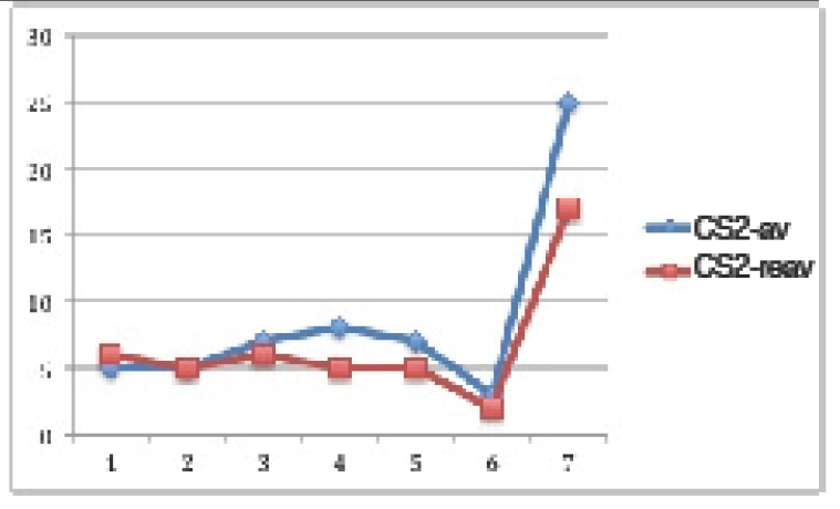
In normogonadic animals: Control group did not show SBP changes during the experiment. Nevertheless, there was a significant increment in the F group (p<0.01), in the OA group (p<0.0001) and in the FOA group (p<0.0001) compared to basal state. At the end of the experimental period, SBP was significantly higher in all treatment vs control group (p<0.0001). In addition, a significant difference was found between F vs OA and FOA groups (p<0.01), reaching maximal SBP levels in FOA, which was significantly higher than in the OA group (p<0.01) (Figure 1).

EDMJ 2019-111 - Soutelo J Argentina_F1

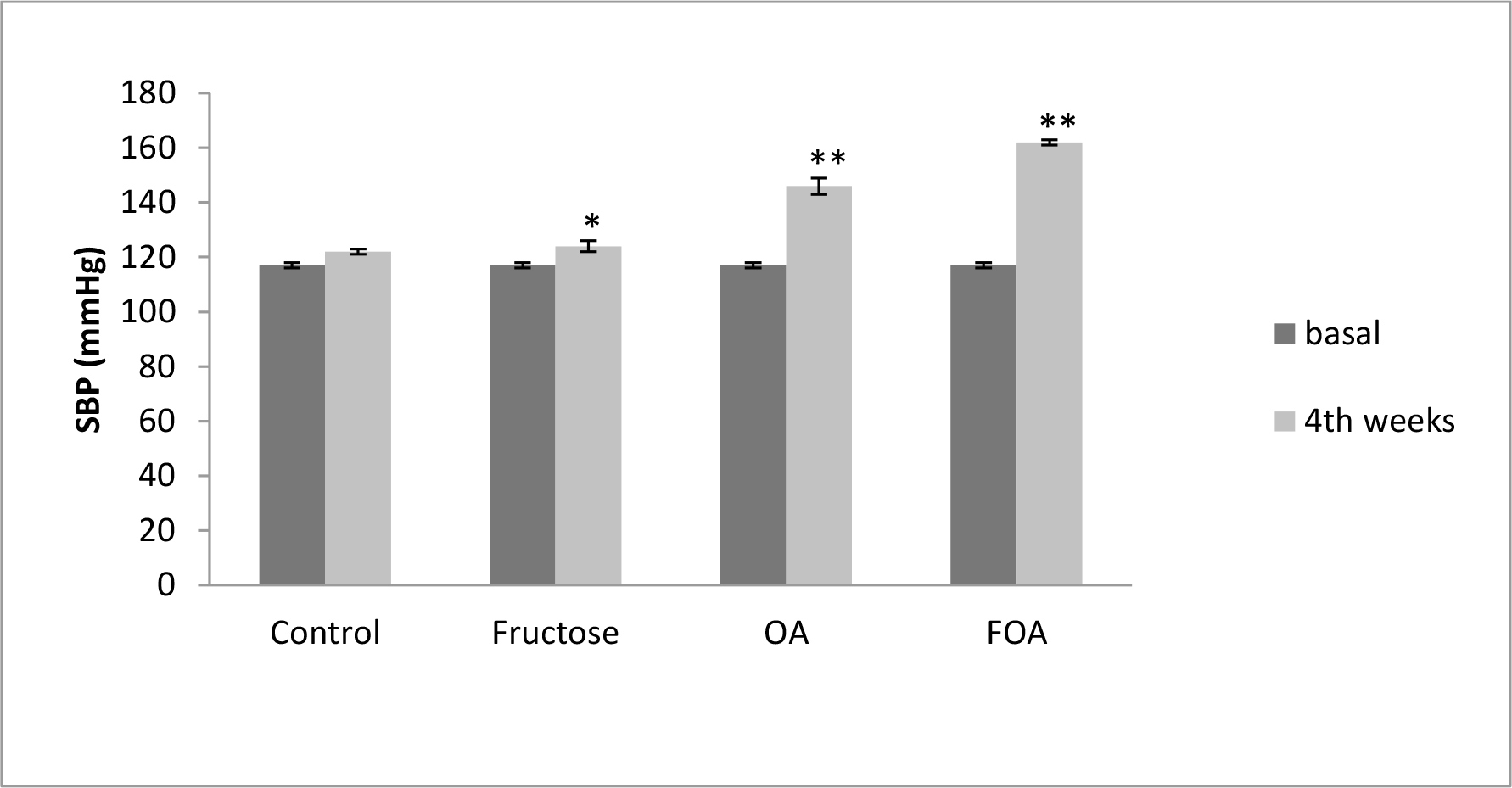
Figure 1. Blood Pressure in NGN groups, at basal and 4th weeks after beginning treatment.

Data are expressed as mean ± SEM. NGN (Normogonadic), OA (Oxonic acid); FOA (Fructose and oxonic acid)
* p<0.05 Fructose at 4th week vs Fructose basal.
** p<0.001 OA and FOA groups at 4th weeks vs respective basal groups.

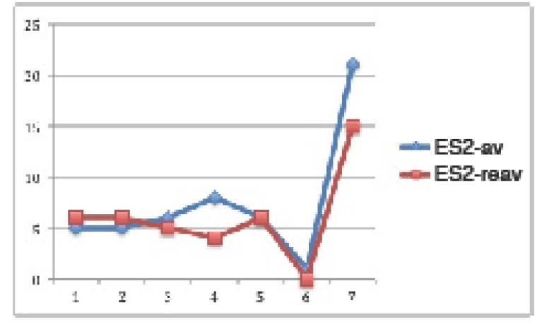
In hypogonadic animals: Contrary to normogonadic rats, the control group showed a significant (p<0.01) increment in SBP during the time of experiment. Also there was a significant (p<0.001) increment in the F, OA (p<0.0001) and FOA groups (p<0.0001). At the end of the experiment, SBP was significantly higher in all treatments vs. control group (p<0.001). In addition, a significant difference was found between F vs. OA and FOA groups (p<0.01), reaching maximal SBP levels in FOA, that were significantly higher than in the OA group (p<0.01). (Figure 2).

EDMJ 2019-111 - Soutelo J Argentina_F2

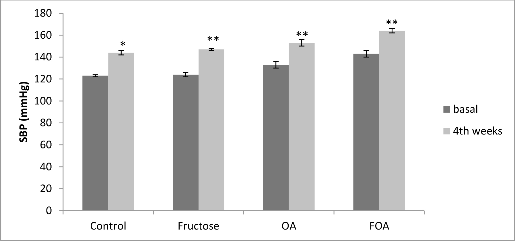
Figure 2. Blood Pressure in HGN groups, at basal and 4th weeks after beginning treatment.

Data are expressed as mean ± SEM. HGN (Hypogonadic), OA (Oxonic acid); FOA (Fructose and oxonic acid)
*p<0.001 Control at 4th week vs Control basal.
**p<0.0001 Fructose, OA and FOA groups at 4th week vs respective basal groups.

When comparing gonadal condition, HGN rats presented -in all groups and during the whole experimental time- higher SBP levels than NGN rats (p <0.0001), except at the end of experimental time in OA and FOA groups.

3.4 Biochemical Variables

As expected, testosterone levels decreased to a very low level in all hypogonadic animals compared to all non castrated rats (p<0.0001). Nevertheless, in both normogonadic and hypogonadic groups there were no plasmatic testosterone levels differences between different treatments. There was no difference in plasmatic creatinine levels when comparing treatment groups. Also no significant differences were observed in fasting glucose levels between normo and hypogonadic groups with different treatments.

Uric acid levels were significantly higher in normogonadic animals treated with OA (UA: 1.27 ± 0.13 mg/dl) and FOA (UA: 1.49 ± 0.1 mg/dl) when comparing them to the respective control group (UA: 0.97 ± 0.04 mg/dl) (p< 0.01). Also, uric acid levels were significantly higher in hypogonadic animals treated with FOA (UA: 1.29 ± 0.06 mg/dl) when comparing them to the respective control group (0.96 ± 0.67 mg/dl) (p< 0.01). Likewise, there were no significant differences when comparing NGN and HGN animals undergoing same beverage treatment.

Regarding the lipid profile, NGN animals showed no significant difference in Total Cholesterol (TC), triglycerides (TG), HDL-c levels, in no-HDL-c, in the TG/HDL index in the different experimental groups.

HGN animals in the fructose (F) group showed a significant increase (p<0.01) in TG and TC levels, accompanied by an increase of no HDL-c (p<0.05) with no changes in the HDL fraction. These changes translated into a significant increase (p<0.05) of the TG/HDL index. Conversely, the lipid profile showed no changes in OA and FOA groups when comparing them to the control group.

Hypogonadic animals showed -when faced to fructose administration- a significant increase in CT plasmatic levels (p<0.0001), TG (p<0.01), no HDL-c (p<0.0001) and a decrease of HDL (p<0.0001) accompanied by an increase of the TG/HDL index (p<0.004) as compared to the normogonadic animals. Likewise, hyperuricemia induction (OA) in HGN animals produced an HDL decrease (p<0, 001) with a rise of no HDL-c (p< 0, 001) accompanied by an increase of the TG/HDL index (p<0.02); and the combined treatment (FOA) in the same group showed a decrease of HDL (p<0.02) with an increase of no HDL (p<0.04) (Table 1)

Table 1. Lipid Profile. Comparative effect of the gonadal condition at different stages of treatments.

Cholesterol (mg/dl)

NGN

HGN

p (NGN vs HGN)

Control

53.71 ± 10.12

59.83 ± 2.56

NS

Fructose

56.00 ± 5.68

74.79 ± 3.21*

0.0001

OA

55.28 ± 5.85

60.22 ± 2.84

NS

FOA

63.00 ± 2.20

60.38 ± 4.11

NS

Triglycerides (mg/dl)

Control

64.14 ± 16.65

67.5 ± 6.85

NS

Fructose

69.28 ± 14.28

103.15 ± 9.58**

0.01

OA

48.00 ± 7.69

59.25 ± 5.66

NS

FOA

67.14 ± 15.84

68.03 ± 7.23

NS

HDL-c (mg/dl)

 Control

41.71 ± 4.39

27.75 ± 1.85

0.001

Fructose

44.71 ± 6.01

30.89 ± 2.06

0.0001

OA

44.28 ± 5.54

31.95 ± 1.57

0.001

FOA

44.00 ± 6.72

34.58 ± 2.21

0.02

TG/HDL

Control

1.05 ± 0.58

1.47 ± 0.31

NS

Fructose

1.52 ± 0.45

3.49 ± 0.54***

0.004

OA

1.10 ± 0.24

1.86 ± 0.16

0.02

FOA

1.58 ± 0.48

2.06 ± 0.31

NS

No HDL-c (mg/dl)

Control

22.00 ± 6.30

32.08 ± 0.95

0.01

Fructose

19.28 ± 4.59

43.9 ± 2.80

0.0001

OA

19.00 ± 2.45

28.26 ± 2.27

0.01

FOA

19.00 ± 4

25.20 ± 2.35

0.04

Data are expressed as mean ± SEM . NGN: normogonadic HNG: hypogonadic, OA: oxonic acid; FOA: Fructose and oxonic acid. NS: non significant.
*p< 0.01 HGN Control vs HGN Fructose group.
**p< 0.01 HGN Control vs HGN Fructose group
*** p< 0.05 HGN Control vs HGN Fructose group.
p< 0.05 HGN Control vs HGN Fructose group.

A weak positive correlation was found between the testosterone and HDL levels (r: 0.313, p<0.02) and a weak reverse relationship between the testosterone and no HDL levels (r -0.345, p<0.01) (Figure 3).

EDMJ 2019-111 - Soutelo J Argentina_F3

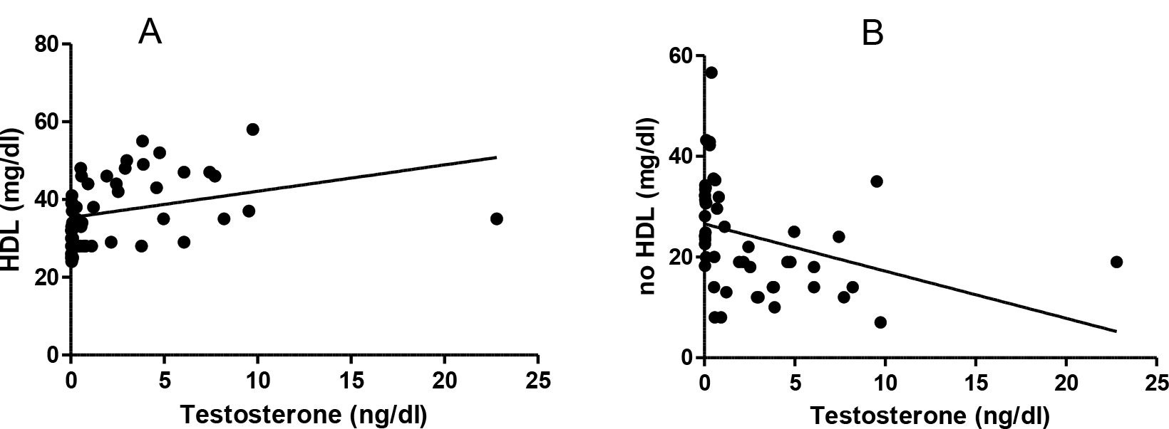
Figure 3. Testosterone and lipids correlation. A: Testosterone and HDL (r: 0.313 p< 0.02). B:  testosterone and no HDL levels (r -0.345, p<0.01).

3.5 Cardiovascular Histology

3.5.1 Morphometric determination of myocyte size

In the NGN group of animals there was significant difference between groups, the ones treated with FOA showed a greater volume (p<0.001), followed by the ones treated with OA (p<0.001); then animals treated with F (<0.05), while control animals had the lowest volume (Figure 4 A)

Same pattern with significant difference was observed in the HGN group. The FOA group showed a greater volume (p<0.001), followed by the ones treated with OA (p< 0.001); then animals treated with F (NS), while control animals had the lowest volume. (Figure 4B)

EDMJ 2019-111 - Soutelo J Argentina_F4

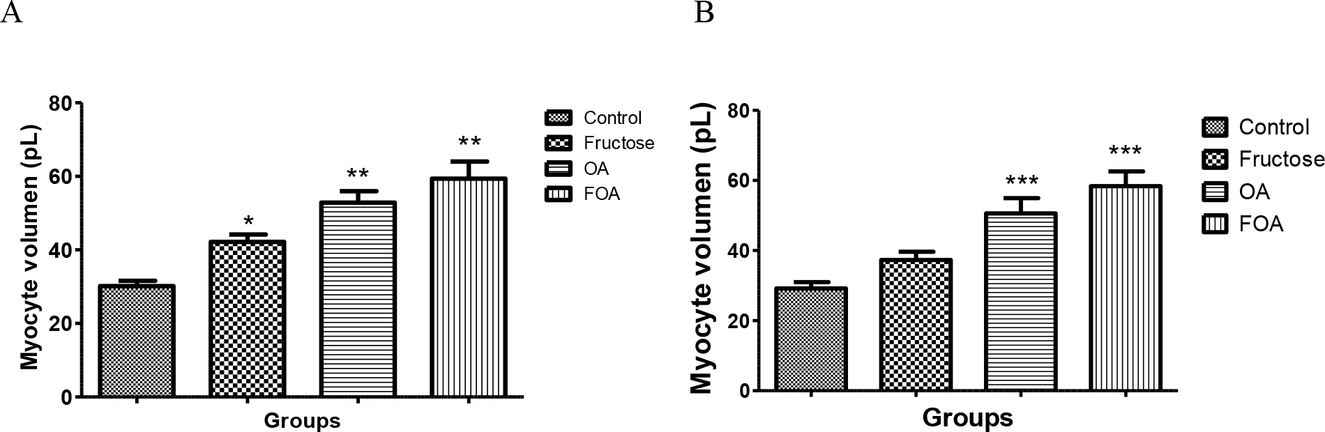
Figure 4. Myocyte volume of normogonadic (A) and hypogonadic rats (B).

Data are expressed as mean ± SEM. OA: oxonic acid, FOA: Fructose and oxonic acid.
*p < 0.05 Fructose vs Control NGN group
** p < 0.001 OA and FOA vs Control NGN group
*** p < 0.001 OA and FOA vs Control HGN group

No significant differences were found when analyzing different groups (NGN vs HGN) with same treatment.

3.5.2 Fibrosis

Animals in NGN groups treated with FOA showed a greater fibrosis percentage vs control (p<0, 009) also in F group (p<0.03 vs control). Animals in HGN groups treated with FOA and OA showed more fibrosis than control group (p<0.01) When comparing the different experimental groups (NGN vs HGN) under same treatment, we found a greater fibrosis in HGN animals treated with OA than in NGN animals under same treatment (p<0.04). Even though it was not significant, same pattern was observed in animals treated with FOA (Table 2).

Table 2. Percentage of miocardic fibrosis.

NGN

HGN

p

Control

2.69 ± 0.19*‡

2.56 ± 0.06**†

NS

Fructose

3.25 ± 0.14‡

3.26 ± 0.34

NS

OA

2.97 ± 0.17

4.33 ± 0.62†

0.04

FOA

3.43 ± 0.14*

5.96 ± 1.23**

0.06

Data are expressed as mean ± SEM. NGN: normogonadic,  HNG: hypogonadic, OA: oxonic acid; FOA: Fructose and oxonic acid.  NS: non significant
*p <0.009 control group NGN vs FOA group NGN
‡p <0.03 control group NGN vs F group NGN
**p< 0.01 control group HGN vs FOA group HGN. † p< 0.01 control group HGN vs OA group HGN

3.5.3 Intima media of aorta

The intima media was significantly thicker in FOA (p<0.001), OA (p<0.001) and F (p<0.001) groups when compared to control animals, with no evidence of differences between the treated groups (Figure 5)

EDMJ 2019-111 - Soutelo J Argentina_F5

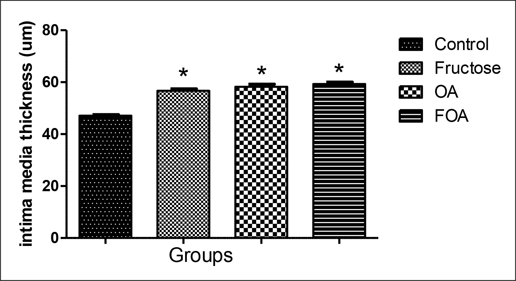
Figure 5. Intima media thickness in hypogonadic rats.

Data are expressed as mean ± SEM. OA: oxonic acid, FOA: Fructose and oxonic acid.
* p<0.001 Fructose, OA and FOA vs Control group.

4. Discussion

During our work we found that animals in both groups (NGN and HGN) treated with FOA showed a greater volume of myocyte, followed by OA groups while only normogonadic animals treated with F showed a greater volume when compared to control animals; this effect was not influenced by gonadal condition. Similar results were observed when systolic blood pressure was examined; probably the increase of same is -in part- the cause of the myocyte hypertrophy found.

Hyperuricemia leads -in its initial phase- to an endothelial dysfunction [25, 26], increase in the oxidative stress and activation of the rennin-angiotensin- aldosterone system [27] and in a second phase favors inflammatory changes [28]. Likewise, Saygin et al. showed recently that a high-fructose diet also produces endothelial damage [29]. Hypogonadic animals treated with F, OA and FOA showed an increase in the intima media when compared to the control group. So, we could observe that a high-fructose diet and hyperuricemia share mechanisms to favor the increase of intima media thickness, endothelial damage and arterial hypertension and in this way favoring arteriosclerosis [25, 26, 30] and cardiac hypertrophy. This is consistent with the observations made in our experiment where the FOA group showed the highest values of systolic blood pressure, greater thickness of the intima media and cardiac hypertrophy.

We also noticed that hypogonadic animals showed higher levels of blood pressure than the normogonadic ones. The blood pressure increase could be partly due to the weight increase observed in these animals, as well as insulin resistance and cytokine increase which favor vasoconstriction. Testosterone is a vasodilating agent; some experiments have theorized about an inhibition of the Voltage Gated Calcium Channels (VGCC) and/or activation of potassium channels in the vascular smooth muscle, and it could also be a result of an up regulation of endothelial nitric oxide synthase enzyme expression (eNOS) [31, 32].

Animals treated with FOA in both groups showed greater cardiac fibrosis when comparing with their respective controls: Likewise animals in the hypogonadic OA group showed a greater fibrosis compared with normogonadic OA, and the same tendency was observed in FOA groups. Chen et al. [33] showed that hyperuricemia increases cardiac fibrosis and Mellory et al. [34] found that the high-fructose diet also increases cardiac fibrosis. Unfortunately, there is a strong controversy regarding the role of testosterone over the cardiovascular effects [35] as to assert that the testosterone deficit causes cardiovascular damage and fibrosis.

Regarding the lipid profile, it is clear that hypogonadic animals showed a less favorable profile, where the fructose group is the one with higher levels of cholesterol, triglycerides, no-HDL and the TG / HDL ratio, a marker of insulin resistance [23].

High fructose diets lead to a hepatic insulin resistance with an increased influx of free fatty acids, synthesis and triglyceride storage, and VLDL synthesis excess. This overproduction of VLDL alters the lipoprotein lipase (LPL) function. Hypertriglyceridemia is normally associated with low HDL levels [36].

This occurs, and is partly due to an increment of the Cholesteryl ester transfer protein (CETP) activity which favors HDL decrease (36). In the resistance to insulin an alteration of hepatic and endothelial lipase activity has been observed; which increases the HDL catabolism [36].

So we can assert that high levels of triglycerides and a decrease in HDL levels are independents predictors of insulin resistance and cardiovascular disease [23].

Likewise, low testosterone levels are linked to a pro-atherogenic lipid profile. A positive correlation between HDL and testosterone levels was found in several works [37, 38]. Rancho Bernardo study also showed a reverse relation between testosterone and VLDL levels [39]. On the other hand, in all treatments we found a uric acid level increase in normogonadic animals compared to the hypogonadic ones. This could be the result of testosterone stimulant action on the urate transporter -1 (URAT-1) expression responsible for the reabsorption of urates at tubular level. Also the monocarboxylate transporter expression coupled with sodium type 1 and 2 which facilitates the presence of essential lactate for the urate/lactate transport by URAT-1 [40].

On the other hand we observed an inversely significant correlation between testosterone and body weight. Studies in humans [41–43] have shown that hypogonadic men have an increase in body weight (BMI) and in waist circumference. Although the mechanism has not been completely clarified, it was stated that adipocytes express androgen receptor [44] and that testosterone inhibits Lipoprotein Lipase (LPL) activity, responsible for the uptake of triglycerides by the fat cell, and so producing an inhibition of the triglyceride uptake and a decrease of visceral adipose tissue [45]. On the contrary, the lack of testosterone produces a higher triglyceride uptake with the subsequent increase of visceral fat. This increment favors a rise in the aromatase, increasing the estrogen synthesis. Likewise, it produces the resistance to insulin, which leads to an SHBG decrease, thus increasing the testosterone metabolism [46].

Undoubtedly animals treated with FOA, OA and F showed some morphologic and functional cardiovascular changes, shown by the increment in systolic arterial pressure and therefore a higher hypertrophy and fibrosis. These changes were affected by gonadal conditions. Likewise, hypogonadic animals showed greater weight, worse lipid profile (less HDL and more HDL) and a higher TG/HDL index as insulin resistance marker, which carries a greater atherosclerotic risk.

 Fructose is a simple sugar that is present in fruits and honey and is responsible for their sweet taste. Excessive fructose intake (>50 g/d) may be one of the underlying etiologies of metabolic syndrome and type 2 diabetes. [47] One of the more striking aspects of fructose is its ability to stimulate uric acid production. As ATP is consumed, AMP accumulates and stimulates AMP deaminase, resulting in uric acid production. Researchers have reported a dose dependent relationship between fructose ingestion and serum uric acid levels in both men and women, although in another study this relationship was confirmed only in men [45] It has been proved that fructose administration to normal rats for different time periods sequentially induces impaired glucose tolerance and type 2 diabetes [48–50]. Also rats have an active uricase, and these findings explain why higher concentrations of fructose are required to induce greater metabolic changes in rats, whereas humans, who lack uricase, appear to be much more sensitive to the effects of fructose. For this reason, we induce mild hyperuricemia with an inhibitor of uricase, oxonic acid. That in humans would be given by a serum value between 6 to 7.0mg/dl [51]. Likewise, we believe that the age of the rats used in this present experiment, did not influence the results, since the male Wistar rats acquire their reproductive capacity at 60 days. We wanted to simulate a state of hypogonadism similar to a man around the age of forty, when testosterone begins to decline [52], without becoming an elderly adult.

A high fructose diet as well as hyperuricemia conditions, favor the increment of blood pressure and cardiovascular damage, and these effects are more relevant in animals with both conditions simultaneously. Likewise, it was also shown that the lipid profile is linked to testosterone levels. The combination of these conditions might explain why western fast food diets associated with a testosterone decrease favor the presence of cardiovascular disease in older men. In any case, it is necessary to carry out more studies to understand the mechanisms involved in such changes.

In short, cardiovascular damage was worse in rats with hypogonadism with a condition induced by mild hyperuricemia exposed to a high fructose diet than the normal gonadal state with a similar condition.

Authors’ Contributions

Study design was conducted by Jimena Soutelo, who also performed experiments, analyzed data, and wrote the paper. Yanina Alejandra Samaniego and María Cecilia Fornari were responsible for sample collection. Carlos Reyes Toso was responsible for performed experiments and data analysis. Osvaldo Ponzo analyzed data and wrote the paper.

Acknowledgement

This work was supported by Grants from University of Buenos Aires (UBACYT) Project number 20020130100439BA. We thank Angela Ciocca Ortúzar for the manuscript translation and revision.

References

  1. World Health Organization (WHO) Global status report on noncommunicable diseases 2014. 2014: 1–302. Available at: http://www.who.int/nmh/publications/ncd-status-report-2014/en/
  2. Shores MN, Biggs ML, Arnold AM, Smith NL, Longstreth WT, et al. (2014) Testosterone, dihidrotestosterone, and incident cardiovascular disease and mortality in the cardiovascular health study. J Clin Endocrinol Metab 99: 2061–2068.
  3. Barrett-Connor E, Khaw KT (1988) Endogenous sex hormones and cardiovascular disease in men. A prospective population-based study. Circulation 78: 539–545.
  4. Contoreggi CS, Blackman MR, Andres R, Muller DC, Lakatta EG, et al. (1990) Plasma levels of estradiol, testosterone, and DHEAS do not predict risk of coronary artery disease in men. J Androl 11: 460–470. [crossref]
  5. Phillips GB, Yano K, Stemmermann GN (1988) Serum sex hormone levels and myocardial infarction in the Honolulu Heart Program. Pitfalls in prospective studies on sex hormones. J Clin Epidemiol 41: 1151–1156.
  6. Ohlsson C, Barrett-Connor E, Bhasin S (2011) High serum testosterone is associated with reduced risk of cardiovascular events in elderly men. The MrOS (Osteoporotic Fractures in Men) study in Sweden. J Am Coll Cardiol 58: 1674–1681.
  7. Malkin CJ, Pugh PJ, Morris PD, Asif S, Jones TH, et al. (2010) Low serum testosterone and increased mortality in men with coronary heart disease. Heart 96: 1821–1825.
  8. Wehr E, Pilz S, Boehm BO, März W, Grammer T, Obermayer-Pietsch B (2011) Low free testosterone is associated with heart failure mortality in older men referred for coronary angiography. Eur J Heart Fail 13: 482–488.
  9. Hyde Z, Norman PE, Flicker L, Hankey GJ, Almeida OP, et al. (2012) Low free testosterone predicts mortality from cardiovascular disease but not other causes: the Health in Men Study. J Clin Endocrinol Metab 97: 179–189. [crossref]
  10. Johnson RJ, Segal MS, Sautin Y, Nakagawa T, Feig DI, et al. (2007) Potential role of sugar (fructose) in the epidemic of hypertension, obesity and the metabolic syndrome, diabetes, kidney disease, and cardiovascular disease. Am J Clin Nutr 86: 899–906. [crossref]
  11. Oda M, Satta Y, Takenaka O (2002) Loss of urate oxidase activity in hominoids and its evolutionary implications. Mol Biol Evol 19, 640–653.
  12. Alderman MH, Cohen H, Madhavan S (1999) Serum uric acid and cardiovascular events in successfully treated hypertensive patients. Hypertension 34: 144–150.
  13. Fang J, Alderman MH (2000) Serum uric acid and cardiovascular mortality the NHANES I epidemiologic follow-up study, 1971–1992. National Health and Nutrition Examination Survey. JAMA 283: 2404–2410.
  14. Høieggen A, Alderman MH, Kjeldsen SE, Julius S, Devereux RB, et al. (2004) The impact of serum uric acid on cardiovascular outcomes in the LIFE study. Kidney Int 65: 1041–1049. [crossref]
  15. Wannamethee SG, Shaper AG, Whincup PH (1997) Serum urate and the risk of major coronary heart disease events. Heart 78: 147–153. [crossref]
  16. Culleton BF, Larson MG, Kannel WB, Levy D (1999) Serum uric acid and risk for cardiovascular disease and death: the Framingham Heart Study. Ann Intern Med 131: 7–13. [crossref]
  17. Wheeler JG, Juzwishin KD, Eiriksdottir G (2005) Serum uric acid and coronary heart disease in 9,458 incident cases and 155,084 controls: prospective study and meta-analysis. PLoS Med 2:76.
  18. Kim SY, Guevara JP, Kim KM, Choi HK, Heitjan DF, et al. (2010) Hyperuricemia and coronary heart disease: a systematic review and meta-analysis. Arthritis Care Res (Hoboken) 62: 170–180. [crossref]
  19. Ranjith N, Myeni NN, Sartorius B, Mayise C (2016) Association Between Hyperuricemia and Major Adverse Cardiac Events in Patients with Acute Myocardial Infarction. Metabolic syndrome and related disorders 15: 1–8.
  20. Tomiyama H, Higashi Y, Takase B, Node K, Sata M, Inoue T, et al. (2011) Relationships Among Hyperuricemia, Metabolic Syndrome, and Endothelial Function. American Journal of Hypertension 24: 770–774.
  21. Tapia E, Cristóbal M, García-Arroyo FE, Soto V, Monroy-Sánchez F, et al. (2013) Synergistic effect of uricase blockade plus physiological amounts of fructose-glucose on glomerular hypertension and oxidative stress in rats. Am J Physiol Renal Physiol 304: 727–736. [crossref]
  22. Cossio MA, Gómez R, Arruda M, Hochmuller R ( 2010) Valores de confiabilidad de indicadores somáticos en ratas machos wistar. Actualizacion en Nutricion 11: 296–302.
  23. McLaughlin T, Abbasi F, Cheal K, Chu J, Lamendola C, et al. (2003) Use of Metabolic Markers To Identify Overweight Individuals Who Are Insulin Resistant. Ann Intern Med 139: 802–809.
  24. Satoh H, Delbridge LM, Blatter LA, Bers DM (1996) Surface volume relationship in cardiac myocytes studied with confocal microscopy and membrane capacitance measurements: species-dependence and developmental effects. Biophys J 70: 1494–1504.
  25. Tian TT, Li H, Chen SJ, Wang Q, Tian QW, Zhang BB, et al. (2018) Serum uric acid as an independent risk factor for the presence and severity of early onset coronay artery disease: A case control study. Disease Markers Volume, Article ID 1236837, 8 pages.
  26. Lan M, Liu B, He Q (2018) Evaluation of the association between hiperuricemia and coronary artery disease. A strobe- compliant articule. Medicine 97: 44–50.
  27. Yang X, Gu J, Lv H, Li H, Cheng Y, et al. (2019) Uric acid induced inflammatory responses in endothelial cells via up-regulating (pro) rennin receptor. Biomedicine & Pharmacotherapy 109: 1163–1170.
  28. Kanbay M, Segal M, Afsar B, Kang DH, Rodriguez-Iturbe B, et al. (2013)The role of uric acid in the pathogenesis of human cardiovascular disease. Heart 99: 759–766.
  29. Saygin M, Asci H, Cankara FN, Bayram D, Yesilot S, et al. (2016) The impact of high fructose on cardiovascular system: Role of α-lipoic acid. Hum Exp Toxicol 35: 194–204. [crossref]
  30. Liu Z, Chen T, Niu H, Ren W, Li X, et al. (2016) The Establishment and characteristics of rat model of aterosclerosis induced by hiperuricemia. Stem Cells International  Article ID 1365257, 7 pages.
  31. Kelly DM, Jones TH (2013) Testosterone: a vascular hormone in health and disease. J Endocrinol 217: 47–71. [crossref]
  32. Chistiakov DA, Myasoedova VA, Melnichenko AA, Grechko AV, Orekhov AN, et al. (2018) Role of androgens in cardiovascular pathology. Vasc Health Risk Manag 14: 283–290. [crossref]
  33. Chen Ch,  Hsu YJ, Lee TM (2011) Impact of elevated uric acid on ventricular remodeling in infacted rats with experimental hiperuricemia. Am J Physiol Heart Circ Physiol 301: 107–117.
  34. Mellor KM, Bel JR, Young MJ, Ritchie RH, Delbrigde L (2011) Myocardial autophagy activation and suppressed survival signaling is associated with insulin resistance in fructose fed mice. Journal of Molecular and Cellular Cardiology 50: 1035–1043.
  35. Testes RC, Carneiros FS, Carvalho MHC, Reckelhoff JF (2016) Reactive oxygen species: Players in the cardiovascular effects of testosterone. Am J Physiol Regul Integr Comp Physiol 310: 1–14.
  36. Cornier MA, Dabelea D, Hernandez TL, Lindstrom RC, Steig AJ, et al. (2008) The metabolic syndrome. Endocr Rev 29: 777–822. [crossref]
  37. Haffner SM, Mykkänen L, Valdez RA, Katz MS (1993) Relationship of sex hormones to lipids and lipoproteins in nondiabetic men. J Clin Endocrinol Metab 77: 1610–1615. [crossref]
  38. Mäkinen JI, Perheentupa A, Irjala K, Pöllänen P, Mäkinen J, et al. (2008) Endogenous testosterone and serum lipids in middle-aged men. Atherosclerosis 197: 688–693. [crossref]
  39. Khaw KT, Barrett-Connor E  (1991) Endogenous sex hormones, high density lipoprotein cholesterol, and other lipoprotein fractions in men. Arterioscler. Thromb 11: 489–494.
  40. Hosoyamada M, Takiue Y, Shibasaki T, Saito H ( 2010) The effect of testosterone upon the uratere absorptive transport system in mouse kidney. Nuclesides, Nucletides and Nucleic Acids 29: 574–579.
  41. Vermeulen A, Goemaere S, Kaufman JM (1999) Testosterone, body composition and aging. J Endocrinol Invest 22: 110–116. [crossref]
  42. Tsai  EC, Boyko EJ, Leonetti DL, Fujimoto WY(2000) Low serum testosterone level as a predictor of increased visceral fat in Japanese-American men. International Journal of Obesity and Related Metabolic Disorders 24: 485–491.
  43. Corona G, Giagulli VA, Maseroli E, Vignozzi L, Aversa A, et al. (2016) Testosterone supplementation and body composition: results from a meta-analysis of observational studies. J Endocrinol Invest 39: 967–981.
  44. Bjorntorp P (1996) The regulation of adipose tissue distribution in humans. International Journal of Obesity and Related Metabolic Disorders 20, 291–302.
  45. Mårin P, Odén B, Björntorp P (1995) Assimilation and mobilization of triglycerides in subcutaneous abdominal and femoral adipose tissue in vivo in men: effects of androgens. J Clin Endocrinol Metab 80: 239–243. [crossref]
  46. Cohen PG (1999) The hypogonadal-obesity cycle: role of aromatase in modulating the testosterone-estradiol shunt–a major factor in the genesis of morbid obesity. Med Hypotheses 52: 49–51. [crossref]
  47. Johnson RJ, Perez-Pozo SE, Sautin YY (2009) “Hypothesis: could excessive fructose intake and uric acid cause type 2 diabetes?” Endocrine Reviews 30: 96–116.
  48. Thorburn AW, Storlien LH, Jenkins AB, Khouri S, Kraegen EW (1989) Fructose-induced in vivo insulin resistance and elevated plasma triglyceride levels in rats. Am J Clin Nutr 49: 1155–1163.
  49. Catena C, Giacchetti G, Novello M, Colussi G, Cavarape A et al. (2003) Cellular mechanisms of insulin resistance in rats with fructose-induced hypertension. Am J Hypertens 16: 973–978.
  50. Panchal SK, Brown L (2011) RodentModels forMetabolic Syndrome Research. Journal of Biomedicine and Biotechnology Volume 2011, Article ID 351982, 14 pages.
  51. Kuwabara M (2015) Hyperuricemia, Cardiovascular Disease, and Hypertension. Pulse 3: 242–252.
  52. Feldman HA, Longcope C, Derby CA, Johannes CB, Araujo AB, et al. (2002) Age Trends in the Level of Serum Testosterone and Other Hormones in Middle-Aged Men: Longitudinal Results from the Massachusetts Male Aging Study. J Clin Endocrinol Metab 87: 589–598.

Awareness of HPV Infection and Vaccination among Teens in Urban High School

DOI: 10.31038/AWHC.2019225

Abstract

To assess the level of awareness and knowledge of HPV infection and vaccination among 648 Teens from secondary schools and colleges in four metro cities of Port Harcourt Nigeria.

Materials and methods: This cross-sectional study was conducted by from 1st March to 31st August, 2018. Girls of 13–19 years, with an average of 16 years are targeted. A written questionnaire with two parts has been applied. A preliminary written questionnaire included questions of HPV infection and vaccination awareness.

Results: The study participants are poorly aware about HPV infection and vaccination but are intensely willing to know about it. 98% (n-632) are not aware of HPV infection, while, 98% (n–636) is not aware of the vaccination.

Conclusion: This study brings out the unawareness about HPV infection and vaccination in urban adolescent in four metro cities in Port Harcourt Nigeria. Adolescent understanding and being aware of the HPV virus is needed to have successful vaccination programs in Nigeria.

Keywords

Adolescents and HPV, Cancer cervix, HPV Awareness, HPV Vaccination

Background

Human Papillomavirus (HPV) is a common virus reported to be responsible to some type of cancers. HPV infection is the major risk factor for cervical cancer [1]. The prevalence rate of more than 85% global cervical cancer occurs in developing countries. This health burden is a public health issue that could be prevented by early vaccination of adolescents against HPV. A prophylactic vaccination against the HPV has the ability to reduce HPV infection and cervical cancer occurrence, thereby saving HPV many lives [2]. HPV vaccine approved by FDA has been in circulation since the year 2006 [3]. Notwithstanding, HPV vaccine implementation is still facing challenges in acceptance and lack of awareness despite its futuristic benefits. Awareness has been found to be low in some population and high in another [4, 5], but studies are yet to investigate the awareness of teens in Nigeria, If one exist, it does not measure the awareness of both the HPV infection and HPV vaccination of teenagers in high school. To enable practical programmed of HPV vaccination in the future, it will be required to assess the awareness and knowledge regarding HPV and vaccination among the teens population. If awareness could influence the acceptability of the uptake of the vaccination, it may then be wise to target public awareness as commonly done [6]. More research focus should be targeted on adolescent population as they constitute the interest group. This study therefor, explores the teens urban dwellers’ awareness and knowledge about HPV infection and it vaccination.

Methods

Design

A longitudinal cohort study design with a cross sectional quantitative analysis. The study assessed baseline awareness and knowledge of HPV infection and HPV vaccination among teens in a four selected High School in Port Harcourt, Nigeria. The study was conducted from 1st March to 31st August, 2018.

Study population and Recruitment

For the teens studied, the eligibility criteria included ages 13 – 19 years in High School. The method of selection and sampling units (high school) was through random. Four urban High Schools were purposively selected. Six hundred and forty eight (648) Teens were then randomly selected from the four high schools.

Data Collection

Each respondent completed a consent form and a questionnaire. Participation was voluntary and anonymous. Selected Teens were assembled in a room on the day of the survey. Each respondent filled the questionnaire under close supervision and the purpose and procedure of the survey explained. Data was collected, starting from the first school to the 4th school. The research questionnaire consisted of two parts; social demographic profile, and awareness questions form the second part. The demographic data included information regarding sexual activity. This was done in other to predict the sexual behavior risk factor to HPV infection.

Result and Statistical Analysis

In this study, simple descriptive statistics were used. The study question items were organized into categories: demographic 5 items, awareness based 8 items which include awareness of HPV infection and awareness of HPV vaccination. Questions were answered in “Yes” or ‘NO’ items. Those with answers “Yes” = 1 point and “No” = 0 point. All scores were summed up to calculate the overall awareness scores for HPV infection and HPV vaccination.

The study considered 648 Teens between the ages of 13 to 19 years, with 16 years as mean age. The rationale for the sample size and sampling is to see if there are any significant gaps between the level of awareness on HPV infection and HPV vaccination and between the girls and boys participants (Table 1). Of the study population (n-648) Teens, 41% (n- 264) were boys and 59% (n- 384) were girls. The preliminary questionnaire assessed the awareness. Awareness was low. Only 2% (n – 16) were aware of HPV infection, and 98% (n = 632) were not. Also 2% (n – 12) knew about HPV vaccination and 98% (n – 636) did not know. Out of 12 and 16 participants that had heard about HPV infection and HPV vaccination respectively, 1% (n-3) were boys and 3% (n –13) were girls. Majority of both boys and girls were not aware. Few of the participants that were aware heard about HPV from their parents and friends (Table 2).

Table 1. The demographic data of the study population.

Variable

Classification

Frequency

Percentage

Age

9 – 14

15 – 19

432

216

67%

33%

Sex

Boys

Girls

264

384

41%

59%

Have had sex

Yes

No

182

466

28%

72%

Age at first sex

13 years

14 years

15 years

16 years and above

7

22

48

105

4%

12%

26%

58%

Table 2. Awareness on HPV Infection and HPV Vaccination and between boys and girl.

Awareness

Classification

Frequency

Percentage

HPV Infection

No Awareness

Yes Awareness

632

16

98%

2%

HPV   Vaccination

No   Awareness

Yes  Awareness

636

12

98%

2%

 Boys

Yes  Awareness

No   Awareness

3

261

1%

99%

Girls

Yes  Awareness

No   Awareness

13

381

3%

97%

Discussion

The study found result not consistent with some previous studies on high school students’ knowledge and awareness of HPV infection and vaccination [7, 8]. It was anticipated that there would be unawareness across participants as there is no knowledge programmes in schools and colleges regarding HPV and its vaccination. Nigeria adolescences seem to be disadvantaged group both economically and by their lack of knowledge and health awareness like many other African countries. However the findings of this study may have limited generalizability to Nigerians with cultural diversity, different religions, and socioeconomic status that are distinctively different from western societies. Adequate knowledge and awareness of the clinical health importance of HPV infection and vaccination usually provide positive influence.

In addition, when comparing the social and demographic characteristics among the respondents from four schools based on their gender, the differences were rather small, majority of participants had little knowledge and awareness on HPV infection or HPV vaccination across the four schools in the study. Researchers from west found out that adolescents who have high knowledge levels about HPV and cervical cancer, their acceptance of vaccine is also high [9]. Ninety nine percent girls and 97% boys have not heard of HPV virus and but were willing to know more about the infection. The new option of HPV vaccine as a primary prevention in adolescents is a unique opportunity of this era which promises a significant reduction of cervical cancer in the coming decades [10]. More efforts are needed to provide adolescence with information which will help them to be excluded from among those thousands of women who die from this preventable condition.

Conclusion

 This study brings out the unawareness about HPV infection and vaccination in urban Teens in 4 High schools in Port Harcourt city, Nigeria. The study participants are poorly aware about HPV infection and vaccination but are intensely willing to know about them. In conclusion, findings of this study suggest that HPV infection and vaccination are not likely to encourage adolescent sexual activity. In addition, efforts should be made to increase knowledge-based programs in schools and colleges.

Acknowledgement

The authors gratefully acknowledged Dr. C. TobI-West, and Dr. EO. Oranu for their contributions. I also acknowledge all the high school students who participated.

Authors Contributions

FCD had primary responsibility for protocol development, data collection, analysis and writing of the paper. KND performed final data analysis, participated in the development of the protocol and analytical framework for the study and contributed to the writing of the study.

What this study Add

It raised alarm on the poor awareness on HPV infection and Vaccination. It also highlight that the relevant group for HPV vaccination are willing to receive the vaccine.

References

  1. Cohen J (2005) Public health. High hopes and dilemmas for a cervical cancer vaccine. Science 308: 618–621. [crossref]
  2. Smith JS, Lindsay L, Hoots B (2007) Human papillomavirus type distribution in invasive cervical cancer and high-grade cervical lesions: a meta-analysis update. Int J Cancer 121: 621–632
  3. Walsh CD, Gera A, Shah M (2008) Public knowledge and attitudes towards human papilloma virus (HPV) vaccination. BMC Public Health 8: 368.
  4. Fu LY, Bonhomme LA, Cooper SC, Joseph JG, Zimet GD (2014) Educational interventions to increase HPV vaccination acceptance: a systematic review. Vaccine 32: 1901–1920
  5. Small SL, Sampselle CM, Martyn KK (2013) Dempsey AF. Modifiable influences on female HPV vaccine uptake at the clinic encounter level: a literature review. J Am Assoc Nurs Pract [published online ahead of print August 22, 2013].
  6. Blasi PR, King D, Henrikson NB (2015) HPV Vaccine Public Awareness Campaigns: An Environmental Scan. Health Promot Pract 16: 897–905. [crossref]
  7. Jill B, Melissa KF, Michael JW Jr (2012) Adolescent understanding and acceptance of the HPV vaccination in an underserved population in New York City. J Oncol 2012, Article ID 904034.
  8. Ericka C (2001) Lambert-college students’ knowledge of human papillomavirus and effectiveness of a brief educational intervention. J Am Board Fam Pract 14: 178–183.
  9. Marlow LA, Waller J, Wardle J (2007) Public awareness that HPV is a risk factor for cervical cancer. Br J Cancer 97: 691–694. [crossref]
  10. Brabin L, Roberts SA, Henry CK (2007) A semi-qualitative study of attitudes to vaccinating adolescents against HPV without parental consent-BMC Public health 2007. http://www.biomedcentral.com/1471–2458/7/20.

Psychopedagogic intervention according to Piagetian Theory in three girls with Turner Syndrome: performance improvement revealed by Bender Test

DOI: 10.31038/AWHC.2019224

Abstract

Turner syndrome (TS) carriers seem to present cognitive compensation of profile of reduction in visuospatial skills during development. Thus, a psychopedagogical intervention was performed in order to optimize this mechanism.

Three pairs of age matched 45, X girls were evaluated at two moments (pre and posttest) for the Bender test (BT) and specific Piaget’s tasks or scales (PS). The experimental intervention through the environmental solicitation process was applied for one year to each pair girl with learning disabilities.

Experimental subjects (ES) revealed more ability to planning and organizing their graphic expression in the posttest, as well as presented a decrease in number of brain lesion indicators on their BT performance. ES also showed performance improvement in half of PS (e.g. conservation and measurement of volumes task).

Data suggest that the present intervention instruments may have contributed to the performance increment of the experimental TS girls.

Keywords

Turner syndrome cognition; cognitive compensation; psychopedagogic intervention; Piaget’s tasks or scales; Bender-Gestalt test.

Introduction

Turner syndrome is consequent to total or partial absence of one X-chromosome. Specific profile of reduction in visuospatial skills besides other cognitive or neuropsychological (arithmetic and executive function) and neurophysiological characteristics among subjects with Turner syndrome (TS) has been widely described [1–8]. TS girls seem to have cognitive compensation of that visual deficit throughout their growing-up [7, 9]. With the hypothesis of a gradual increasing to this mechanism, a Piagetian psychopedagogic intervention was planned in order to optimize the heteromodal connections of brain areas, associated to the development of neuropsychological compensation.

Piagetian scales and experimental intervention through the environmental solicitation process [10] “make use of the clinical method [11], which guarantees an affective-emotional optimization in order to promote the best child intellectual performance and to more accurately evaluate their individual cognitive potential”[7].

In addition, clinical method seems to be particularly appropriate for a evaluation or/and intervention with Turner syndrome carriers as far as their verbal performance is within (or above) most population average [12–15], since this method facilitates the establishment of relations and the argumentation in favor of the subject’s own ideas [11, 16]. ZAIA, [17] still expounds that “the critical clinical method is to follow the unfolding of the child’s thinking, adapting questions to the actions and speeches of the child, enabling the free and personal expression of the child’s ideas. This process is still characterized by the adult’s effort to use the child’s language, not to suggest anything, not to give clues to the answers and to understand the child’s point of view without deforming it.”

Materials and Methods

This investigation was approved by the Ethics Committee of Federal University of São Paulo- Paulista School of Medicine (UNIFESP-EPM), protocol number 1040/08. Only patients whose legal guardians signed an informed consent were included in this study.

Three pairs of age matched 45, X girls were evaluated at two moments (evaluation and revaluation; or pretest and posttest) for the Bender test (BT) and specific Piaget’s tasks or Piagetian scales (the latter apparently have been applied to TS carriers only twice before [4, 7]).

In between, the experimental intervention through the environmental solicitation process (developed by Mantovani de Assis [10], according to Piaget’s theory; and adapted to psychopedagogic intervention by Zaia [18]) was applied to experimental subjects in order to propitiate thinking development. The experimental intervention, lasting one year, was performed to each pair girl that presented learning disabilities.

The BT was administered according to Clawson [19] (Figure 1). The examiner registered each subject’s visual motor performance, which was further analyzed with Koppitz method [20] by psychologists specialized in BT.

AWHC 2019-110 - Fatima Ricardi Brazil_F1

Figure 1. Stimulus-models from Bender visual motor gestalt test used on the present investigation according to Clawson (1980).

Piagetian scales (PS) consisted of the construction of reflective abstraction (Correlate Formation (FC); and Relationships between Surfaces and Perimeters of Rectangles (SP) [21]); representation space (projective straight line construction (PSL); and viewpoints or perspectives coordination (“three mountains”; 3M) [22, 23]; and conservation of physical quantities (Conservation and Measurement of Volumes; CMV [24]) tasks. In addition, the Discover the Animal Game (DA) [25] was employed to evaluate logical structures and concept construction.

Intervention process comprised a series of appropriate psychopedagogic instruments, selected in order to develop those aspects which presented development delay during evaluation through Piagetian tasks: Discover the Animal Game (classification) [25], Memory Game (memory) [26], activity of making rolls into several formats using the same amount of modeling clay, for the construction of conservation of continuous quantities [27] and wooden building blocks (Playing Engineer [28]) for the conservation of discrete quantities. For the construction of spatial relations, Headsnake [29] and Magic Bag [29] games were picked out as well as activities like puzzles [30], figures assembly [31], free drawing and geometrical figures fitting. Causal relationships establishment was stimulated by the Semblance [29], Total Balance [29] and Pick-up-Sticks [32] games besides the fluctuation of body’s activity [27]. Furthermore, narrative construction [33] and activities with numbers (Semblance game [29], e.g.) were applied to facilitate the construction of school skills. Finally, in order to provide own body knowledge and mastery, the following activities were proposed: body scheme, making the child’s body outline with wax pencil followed by requesting her to represent different body parts of her own; songs with gestures (e.g.: “Hokey- Pokey”); clay doll followed by its drawing. Table 1 presents the games and activities used in the intervention for each experimental subject beyond the total number of intervention sessions per subject.

Table 1. Games and activities employed during psychopedagogical intervention (environmental solicitation) applied to each experimental subject (ES) and their respective purposes; and total number of sessions per subjects.

Games and activities to propitiate

Subjects

Logical relationships development

ES1

ES2

ES3

Classification: Discover the animal Game

X

X

X

Continuous quantities conservation: making rolls

X

X

X

Memory: Memory Game

X

X

X

Real construction

Space:

Headsnake Game

X

Puzzles

X

X

X

Magic Bag Game

X

X

X

Free drawing

X

X

X

Playing Engineer (wooden building blocks)

X

X

X

Figures assembly

X

X

X

Geometrical figures fitting

X

Causality:

Sambalance Game

X

X

X

Activities with objects that float or sink

X

X

Total Balance Game

X

X

X

Pick-up sticks Game

X

X

Oral and written language construction

Narrative construction

X

X

X

Dictation

X

Number recognition (and arithmetic operations)

Sambalance game

X

X

X

Calculation (regarding the pick up sticks game play)

X

Psychomotor activities: body image construction, rhythm and movement control

Body outline made with wax pencil complemented with different body parts representation

X

X

“Hokey Pokey” (music with gestures)

X

Modeling-clay (e.g.: clay doll)

X

X

X

Doll drawing

X

Songs

X

X

X

TOTAL NUMBER OF INTERVENTION SESSIONS

36

36

27

It is noteworthy that both the application of Piagetian tasks and the process of psychopedagogic intervention used the critical Piagetian clinical method [11, 16–17], which fundamental aspects were previously reported in this paper.

Results

Bender test

Data referring to performance by experimental and control subjects on Bender test (Table 2; Figures 2 to 7) exhibit that from seven years old on impulsivity diminished. On the other hand, time required to perform BT increased with age (Table 2). Furthermore, both experimental TS girls younger than ten during posttest (ES1 and ES2) improved the use and planning of spaces of gestalts.

AWHC 2019-110 - Fatima Ricardi Brazil_F2

Figure 2. Evaluation of ES2, at five yr and 2nd examination at six yr.

AWHC 2019-110 - Fatima Ricardi Brazil_F3

Figure 3. Evaluation of CS2, at five yr and 2nd examination at six yr.

AWHC 2019-110 - Fatima Ricardi Brazil_F4

Figure 4. Evaluation of ES1, at six yr, and 2nd evaluation at eight yr.

AWHC 2019-110 - Fatima Ricardi Brazil_F5

Figure 5. Evaluation of CS2, at seven yr, and 2nd evaluation at eight yr.

AWHC 2019-110 - Fatima Ricardi Brazil_F6

Figure 6. Evaluation of ES3, at nine yr. and 2nd evaluation at 10 yr.

AWHC 2019-110 - Fatima Ricardi Brazil_F7

Figure 7. Evaluation of CS3, at nine yr, and new assessment at 10 yr.

Legend
1- Age; 2-Time; 3-Form distortion; 4-Rotation: 5-Integration; 6-Perseveration: 7-Score

Table 2. Performances of experimental (ES) and control (CS) subjects on Bender test and respective ages (A) and significative (+) or not significative (-) number of brain lesion (BL) indicators.

Subjects

A (y;m)

Step

Time

F

R

I

Pe

Score

Visual motor perception level

BL

BL#

BL##

BL (meaning)

BL (difference) (R-E)

ES1

(06;10)

Evaluation

11’

7

7

5

0

19

Below 5 years

21

+

+

anxiety and space disorganization

-6

(07;11)

Revaluation

18’

4

5

5

2

16

Below 5 years

15

+

+

improved in space planning and decreased in impulsivity

CS1

(07;01)

Evaluation

12’

4

6

4

3

17

Below 5 years

17

+

+

CS1 erased during performance and exhibited disorganization and lack of space planning

-4

(08;02)

Revaluation

11’

5

5

3

0

13

5 years and 5 years and 5 months

13

+

+

disorganization and lack of space planning

ES2

(05;00)

Evaluation

05’

6

8

6

1

21

Below 5 years

13

+

+

impulsivity and space disorganization

-5

(06;02)- (06;03)

Revaluation

06’

5

4

6

0

15

Below 5 years

08

+

ES2 retouches copies during performance and shows impulsivity and disorganization

CS2

(05;03)

Evaluation

05’

7

8

7

3

25

Below 5 years

25

+

+

CS2 retouches a lot during performance besides exhibiting space disorganization

-5

(06;02)

Revaluation

05’

6

5

5

2

18

Below 5 years

20

+

+

CS2 showed improvement in perception although space kept disorganized

ES3

(09;00)

Evaluation

03’

6

8

5

0

19

Below 5 years

20

+

+

impulsivity and lack of space planning

-16

(10;04)

Revaluation

06’

5

4

2

0

11

5½ and 5 years and 11 months

04

Anxiety. Nevertheless, ES3 exhibited an outstanding improvement in space planning

CS3

(09;02)

Evaluation

07’

2

4

2

0

08

6 and 6 years and 5 months

05

CS3 retouches during performance

2

(10;05)

Revaluation

13’

3

2

2

0

07

6½ and 6 years e 11 months

07

CS3 erased a lot and retouched copies. Also still showed good usage of space. However, exhibited anxiety to improve gestalt

Age = (years; months); F = form distortion; R = rotation; I = integration; Pe = perseveration; #UnG (Guarulhos University) standard: n = 8; ##APEP (Psychology and Psychotherapy Studies Association) standard: n = 10; E = evaluation; R = revaluation.

Table 2 also shows that most (5/6) of all subjects presented decrease in number of brain lesion indicators from evaluation to revaluation. It is noteworthy that such difference was greater for two experimental subjects (ES1 and ES3).

On the contrary, ES2 did not present a BL decrease greater than her respective control (CS2). Although ES2 and CS2 showed the same BL decrease, the former diminishment was from a significative to a non significative value, according to one of the two considered standards (Table 2).

Moreover, nearly all propositii (5/6) showed improvement on all scores of their performances on Bender test (evaluation and revaluation compared; table 2), either experimental (3/3) or control (2/3) ones.

Piagetian tasks or Piagetian Scales

Data referring to performance by experimental and control subjects on conservation and measurement of volumes and other Piagetian tasks during evaluation and revaluation are given in Table 3.

Table 3. Performances of experimental (ES) and control (CS) subjects classified by Piagetian Scales (PS) and respective ages (A) and final diagnoses (FD)

AWHC 2019-110 - Fatima Ricardi Brazil_F9

PS tasks: CMV = conservation and measurement of volumes; 3M = viewpoints or perspectives coordination (“three mountains”); CF = correlate formation; SP = relationships between surfaces and perimeters of rectangles; PSL = projective straight line construction. Game: DA = discover the animal. For abbreviations of diagnosis, see legend to Table 4.

Table 4 exhibits the reference values for the final operatory diagnosis corresponding to the sum of points of individual performance on each Piagetian task.

Table 4. Reference values for the final operatory diagnosis corresponding to the sum of points of individual performance on each Piagetian task.

Total points

Final diagnosis

6,0

PO1

6,5 – 11,5

tPO1/2

12

PO2

12,5 – 17,5

tPO/CO

18,0

CO1

18,5 – 22,5

tCO1/2

23

CO2

23,5 – 24,5

tCO/FO

25 – 30

FO1

PO = pre-operatory period; PO1 = the beginning stage of the pre-operatory period; tPO1/2 = transition between Pre-Operatory Stage 1 and the Pre-Operatory Equilibrium Level; PO2 = Pre-Operatory Equilibrium Level; tPO/CO = transition between pre-operatory and concrete operatory periods; CO = concrete operatory period; CO1 = the beginning stage of the concrete operatory period; tCO1/2 = transition between the beginning stage and the stage in equilibrium of the concrete operatory period; CO2 = concrete operatory equilibrium level; tCO/FO = transition between the concrete operatory and the formal operatory periods; FO1 = the beginning stage of the formal period; FO = formal operatory period.

In addition, Figure 8 presents the comparison between experimental (ES) and control (CS) subjects performances classified by Piagetian scales (PS) during evaluation and revaluation. Table 3 and Figure 8 analyses enable to compare the progress achieved by the experimental and control subjects in the different Piagetian tasks.

AWHC 2019-110 - Fatima Ricardi Brazil_F8

Figure 8. Comparison between experimental (ES) and control (CS) subjects performances classified by Piagetian scales (PS) during evaluation and revaluation. PS tasks: CMV = conservation and measurement of volumes; 3M = viewpoints or perspectives coordination (“three mountains”); CF = correlate formation; SP = relationships between surfaces and perimeters of rectangles; PSL = projective straight line construction. Game: DA = discover the animal.

The great majority (5/6) achieved progress in Conservation and Measurement of Volumes task (CMV). In groups comparison, all experimental (three) and most (2/3) control subjects presented better performance in CMV during posttest than in the pretest (Table 3)

Thus, in CMV task and among controls, one subject (CS3) did not achieve progress, whereas the other two presented one (CS1) and two (CS2) levels of progress. In addition, among experimental subjects, two (ES2 and ES3) achieved one level while ES1 achieved two levels of progress (Figure 8). However, in Discover the Animal game 4/6 of the subjects presented progress on their performances, that means every control and one experimental subject (ES2; Figure 8).

Data referring to performance by control and experimental subjects in representation space tasks revealed that two of each group (4/6) achieved progress in the “Three Mountains” (CS2 and CS3; ES2 and ES3) and in the Projective Straight Line (CS2 and CS3; ES1 and ES3; Figure 8) tasks. Also considering the progress amplitude, only ES2 presented two levels whereas the other experimental and both control subjects showed one level of progress in 3M, in pretest and posttest comparison.

Concerning reflective abstraction, half of the subjects (3/6) achieved progress on their performance in Correlate Formation task that means two controls (CS1 and CS2) and one from the experimental group (ES3). The progress amplitude corroborates this observation, as far as one of the controls (CS2) presented three levels while CS1 and the experimental subject showed only one level of progress (Figure 8).

Nevertheless, still considering abstraction, despite only one subject of each group (CS1 and ES3) have achieved progress on their performances in the Relationships between Surfaces and Perimeters of Rectangles task, the control showed one level whereas the experimental subject, two levels of progress.

Discussion

It is noteworthy that Turner syndrome has a cognitive impact besides a physical one [8, 34–36]. Thus TS girls and teenagers should be provided a demanding formal assessment, orientation and/or intervention [7, 8, 34, 37].

In the present investigation, there was a difference on BT performance in favor of the experimental subjects (ES), who presented more ability to planning and organizing their graphic expression.

Increase in time required to perform BT with age was probably due to older subjects effort in order to more carefully accomplish gestalts.

Furthermore, improvement in use and planning of spaces of gestalts of both experimental TS girls younger than ten during posttest (ES1 and ES2) suggests those subjects took more advantage of intervention procedures than the eldest one (ES3). Also, it is likely that neurological functions in development have been benefited by intervention process stimuli. This assumption is based on number of brain lesion indicators (BL) decrease revealed by BT.

Unlike both other experimental subjects, ES2 did not present a BL decrease greater than her respective control (CS2) inasmuch as the observed hindrance to cooperate in the beginning of the intervention process, specifically refusing several times to accomplish or to repeat the suggested activity. The latter behavior could be due to pre-operativeness characteristics ES2 presented at that time.

Improvement of nearly all propositii on all scores of their performances on Bender test may be assumed mostly to their natural development, in controls case. On the other hand, it is reasonable to consider the benefit of intervention that favours the subject in order to explain the considerable decrease ES3 presented for brain lesion indicators on her BT performance.

However, a new investigation with a greater number of subjects and its replication by other researchers would be necessary to corroborate such supposition.

The present data may reinforce how important psychopedagogic intervention process can be to Turner syndrome carriers. In addition, the earlier such intervention occurs the more convenient it will be, as well as should the family be advised with this purpose.

Besides, on three Piagetian tasks (CMV, 3M and SP) a greater progress was observed in most experimental subjects.

This mentioned difference, if explained by the influence of psychopedagogic intervention, shows how the instruments employed along the latter might influence conservation construction on TS girls.

This may be confirmed by the amplitude (namely the relationship between the beginning performance level at the pretest and that achieved at the posttest) of progresses achieved by experimental and control subjects performances analysis.

This relationship suggests the intervention process might have influenced on the conservation construction.

Moreover, on projective straight line task subjects performances were similar in both groups.

On the other hand, controls achieved higher progress in CF and in Discover the Animal game as in number of subjects as in the amplitude of progresses.

Therefore, this led us to consider that concept construction and classification were not influenced by psychopedagogic intervention instruments. This finding may be confirmed by the progresses amplitude as far as the only experimental subjects and all the controls achieved one level of progress each.

So, considering the number of subjects which achieved progresses in the space construction, we do not find there was influence from the intervention process, as far as it was the same in both experimental and control groups. However, if we also consider the progress amplitude, only ES2 presented two levels whereas the other experimental and both control subjects showed one level of progress in 3M, in pretest and posttest comparison. In conclusion, we suppose there was a slight influence of psychpedagogic intervention, in this case. Finally, it is noteworthy that a more detailed analysis of intervention through the environmental solicitation process and its influence on experimental subjects performances will be addressed in an upcoming publication.

Conclusion

According to Bender [38], the gestalt visual-motor functions basically may be associated with language aptitude, time and space organization, and to manual motor abilities. One concluded there was an improvement on the quality of gestalt functions, observed in the paper organization and drawings planning, and that psychopedagogical activities have probably influenced on the Bender test organization and planning, so presumably contributing to ameliorate cognitive performance of three girls with Turner syndrome. In conclusion, greater performances progresses observed among experimental subjects on Bender test, when compared to those presented by controls, suggest the psychopedagogic intervention accomplished during the present study may have contributed to optimize the cognitive compensation of the experimental subjects visual motor deficit.

Nevertheless, the environmental solicitation process should be applied to a greater number of TS girls and preferably for a longer period of time in order to confirm the conclusion above.

References

  1. Shaffer JW (1962) A specific cognitive deficit observed in gonadal aplasia (Turner’s syndrome). J Clin Psychol 18: 403–406. [crossref]
  2. Garron DC (1977) Intellegence among persons with Turner’s syndrome. Behav Genet 7: 105–127. [crossref]
  3. Money J (1968) Cognitive deficits in Turner’s syndrome. In: Vandenberg SG. (Ed.) Progress in human behavior genetics. J. Hopkins, Baltimore, USA.
  4. Ricardi FCF (1996) Cognição na síndrome de Turner: investigação de 28 sujeitos com o Teste de Bender e as Escalas de Piaget. Masters of Science thesis, São Paulo: USP.
  5. Ross J, Stefanatos GA, Kushner H, Zinn A, Bondy C, Roeltgen D (2002) Persistent cognitive deficits in adult women with Turner syndrome. Neurology 58: 218–225. [crossref]
  6. Ross J, Roeltgen D, Zinn A (2006) Cognition and the sex chromosomes: studies in Turner syndrome. Horm Res 65: 47–56. [crossref]
  7. Ricardi FCF, Zaia LL, Pellegrino-Rosa I, Rosa JT, Mantovani de Assis OZ, et al. (2010) Psychogenetics of Turner syndrome: an investigation of 28 subjects and respective controls using the Bender test and Piagetian scales. Genet Mol Res 9: 1701–1725. [crossref]
  8. Hong DS, Reiss AL (2012) Cognition and behavior in Turner syndrome: a brief review. Pediatr Endocrinol Rev 9: 710–712. [crossref]
  9. Ricardi FCF, Pellegrino-Rosa I, Rosa JT, Saldanha PH (2002) Síndrome de Ullrich-Turner: investigação de 21 sujeitos com o teste de Bender. Águas de Lindóia: Sociedade Brasileira de Genética (Ed.), Resumos do 48. Congresso Nacional de Genética, CD-ROM.
  10. Mantovani de Assis OZ (1976) A solicitação do meio e a construção das estruturas lógicas elementares na criança. Ph.D. thesis. Campinas: Unicamp.
  11. Vinh-Bang (1970) El método clinico y la investigación en psicologia del niño. In: Ajuriaguerra J et al. (Eds). Psicologia y epistemologia genéticas: temas piagetianos. Buenos Aires: Proteo.
  12. Shankar RK, Backeljauw PF2 (2018) Current best practice in the management of Turner syndrome. Ther Adv Endocrinol Metab 9: 33–40. [crossref]
  13. Temple CM, Shepard EE (2012) Exceptional lexical skills but executive language deficits in school starters and young adults with Turner syndrome: implications for X chromosome effects on brain function. Brain Lang 120: 345–359. [crossref]
  14. Hong D, Scaletta Kent J, Kesler S (2009) Cognitive profile of Turner syndrome. Dev Disabil Res Rev 15: 270–278. [crossref]
  15. Ross JL, Stefanatos GA, Kushner H, Zinn A, Bondy C, Roeltgen D (2002) Persistent cognitive deficits in adult women with Turner syndrome. Neurology 58: 218–225. [crossref]
  16. Piaget J (1975) Os problemas e os métodos. In: A Representação do Mundo na Criança. Rio de Janeiro: Brazil.
  17. Zaia LL (2015) O atendimento psicopedagógico pelo processo de solicitação do meio. Revista Educação 9: 117–127.
  18. Zaia LL (1996) A solicitação do meio e a construção das estruturas operatórias em crianças com dificuldades de aprendizagem. Ph.D. thesis, Campinas: Unicamp, Brazil.
  19. Clawson A (1980) Bender infantil: manual de diagnóstico clínico. Porto Alegre: ArtMed, Brazil.
  20. Koppitz EM (1966) The Bender gestalt test for young children. New York: Grune & Stratton, USA.
  21. Piaget J (1995) Abstração reflexionante: relações lógico-aritméticas e ordem das relações espaciais. Porto Alegre: ArtMed, Brazil.
  22. Piaget J, Inhelder B (1993) A representação do espaço na criança. Porto Alegre: Artmed, Brazil.
  23. Kobayashi MCM (1998) A construção das relações espaço-geométricas em crianças de educação infantil: um estudo de epistemologia genética. Masters of Science thesis. Marília: Unesp, Brazil.
  24. Piaget J, Aebli MH, Pitsou MF (1948) La conservation et la mesure des volumes. In: Piaget J, Inhelder B, Szeminska A. La géométrie spontanée de lénfant. Paris: Presses Universitaires de France, France: 448–483.
  25. Piaget J, Sakellaropoulo M, Henriques-Christophides A (1996) Em direção à circularidade dialética mais geral das conexões lógicas: a determinação de alguns animais ou objetos. In: Piaget J. As formas elementares da dialética. São Paulo: Casa do Psicólogo, Brazil.
  26. Jogo da Memória- Brinquedos (Memory Game- Toys) (24 peças em madeira). Xalingo brinquedos. Fabricado por: Xalingo S/A Indústria e Comércio. CNPJ: 95.425.534/0001-76. Santa Cruz do Sul, Brazil.
  27. Mantovani de Assis OZ, Assis MC (Orgs.) (2004) PROEPRE: prática pedagógica. Campinas: Graf. FE; LPG, 3.
  28. Brincando de Engenheiro (Playing Engineer) (42 peças). Xalingo brinquedos. Fabricado por: Xalingo S/A Indústria e Comércio. Santa Cruz do Sul, Brazil.
  29. Zaia LL (2008) A construção do Real na criança: a função dos jogos e das brincadeiras. Schème 1: 74–94.
  30. Quebra-cabeça: The Flintstones (30 peças em madeira- 22X18 cm). Start. Fabricado por: L. Moller. São Paulo, Brazil.
  31. Monta-figuras (Mounting Pictures): Turma da Mônica (24 peças em madeira). Xalingo brinquedos. Fabricado por: Xalingo S/A Indústria e Comércio, Santa Cruz do Sul, Brazil.
  32. Pega-Varetas (28 varetas). Fabricado por: Algazarra Ind. e Com. de Brinquedos Ltda. São Paulo, Brazil.
  33. Sauer MIM (2000) A construção da narrativa infantil e suas relações com a construção do espaço. Masters of Science thesis, Campinas: UNICAMP, Brazil: 73–107.
  34. Erhan H, Belotserkovsky J (2014) Neuropsychological impact of Turner syndrome: importance of parent education, early detection and intervention: a case study. Arch Clin Neuropsychol 29: 544–545.
  35. Saad K, Abdelrahman AA, Abdel-Raheem YF, Othman ER, Badry R, Othman HA, Sobhy KM (2014) Turner syndrome: review of clinical, neuropsychiatric and EEG status: an experience of tertiary center. Acta Neurol Belg 114: 1–9. [crossref]
  36. Kesler SR, Sheau K, Koovakkattu D, Reiss AL (2011) Changes in frontal-parietal activation and math skills performance following adaptive number sense training: preliminary results from a pilot study. Neuropsychol Rehabil 21: 433–454. [crossref]
  37. Mueller SC (2013) Magnetic resonance imaging in paediatric psychoneuro-endocrinology: a new frontier for understanding the impact of hormones on emotion and cognition. J Neuroendocrinol 25: 762–770. [crossref]
  38. Bender L (1938) A visual motor Gestalt test and its clinical use. New York: American Orthopsychiatric Association, Res. Monograph, USA: 103.

Caring for Confused Community Dwelling Seniors: Contributing and Obstructing Factors in Daily Care for Healthcare Assistants in District Nursing

DOI: 10.31038/ASMHS.2019321

Abstract

Older people with confused behavior, have behavioral problems due to dementia, mental problems or social problems. For the Healthcare Assistant (HA) in district nursing, it is a daily challenge to care for older people with confused behavior. Aim of this research is to achieve an insight in the strategies the HA uses to deal with the daily care for older people with confused behavior. It is also the aim to have insight in factors which contribute to the daily care in a positive or negative way. Method: a qualitative explorative research. 17 HA’s in district nursing participated in semi-structured interviews. All respondents had experience with caring for older people with confused behavior. The most important influencing factors are the experienced relationship between HA and the client and the experienced support by the team. Particularly behavioral problems due to mental problems can impede a relationship with the client. Further research is recommended to study the level of knowledge and competences of all levels of employees in district nursing.

Keywords

Confused persons, vulnerable elderly, older adults living at home, mental disorder, home care staff, district nursing

Short Commentary

The number of seniors confronted by chronic illness and dementia is rising, due to the fact that people are getting older and stay at home longer. Because of rising healthcare costs, people are to assumed to stay at home as long as possible [1]. Though, age-related physical and cognitive decline can make aging at home challenging [2]. Psychiatric problems among community dwelling seniors is frequently recognized by healthcare assistants (HA’s) in district nursing [2]. One of the consequences of these problems is an increase of the so-called confused community dwelling seniors. Seniors with confused behaviour show problems like agitation, aggression or apathy. For HA’s it is a daily challenge to manage the care for confused community dwelling seniors. It is important that HA’s have sufficient knowledge of confused behaviour in seniors in order to recognize and manage psychiatric problems [2]. However, little is known about what HA’s experience themselves as contributing or obstructing factors to daily care and how to respond in a right way to confused behaviour as well as to provide good care. We conducted an explorative research among seventeen HA’s in the Netherlands to gain insight in these contributing and obstructing factors. The qualified HA’s, with a variety in age and work experience, provide basic personal care under supervision of a district nurse.

The professionals were asked, in an individual interview, to share their experiences with confused community dwelling seniors and how they manage daily care. The Cohen-Mansfield mapping of problem behaviour, an instrument for assessing agitation, was used to stimulate participants to give more detailed information and examples of problem behaviour they experienced [3]. Most of the participants experience an increase of confused community dwelling seniors, which was in line with research conducted by Grundberg and colleagues concerning the role of home care assistants recognizing mental health problems in community dwelling seniors [4]. HA’s reported daily difficulties and challenges in caring for confused community dwelling seniors. The HA’s were confronted with verbal aggressive behaviour like threats, berate or curse by the seniors. They were also confronted with physically non-aggressive behaviour like compulsive or risky behaviours of the seniors. According to the HA’s, there is no ‘standard recipe’ to manage problem behaviour: the way the HA’s approach confused seniors depends on factors related to the seniors as well as characteristics of the HA. These factors are for example age and work experience of the professional as well as the type and cause of behaviour problem of the senior. The presence or absence of a relation of trust, information and support are the main contributing or obstructing factors.

Despite the fact that a relation of trust is seen as essential for providing good care, it seems very difficult to achieve such a relation with confused community dwelling seniors. According to the HA’s, a relation of trust can be reached when they have the opportunity to get to know the client. HA’s experience more difficulties to become familiar with the senior and building up a relation of trust in case a diagnose is missing and psychiatric problems are suspected. A study of Gleason and Coyle regarding the experiences of home care workers providing homecare to clients diagnosed with mental and behavioural problems, also mentioned the struggles in building a relation of trust in such cases [5]. In addition, in cases where psychiatric problems are suspected, the HA’s in our study seemed to have less tendency to build this relation of trust. Several factors might explain this low tendency: having a primary focus on personal care and not on a relation of trust, uncertainty of the HA or not having the required knowledge on how to respond to clients with (suspected) psychiatric diagnoses. These factors are also identified in a study on how nursing staff in a hospital manage patients with psychiatric problems [6].  Also a lack of collaboration with professionals in social care can obstruct a relation of trust [7].

The second factor, as mentioned by the HA’s, is the presence or absence of information on the underlying cause of the behaviour problem, for example dementia or loneliness of the senior. Olivera and colleagues studied the factors contributing to the development of psychiatric problems in community dwelling seniors [8]. When professionals in home care are aware of these factors, problems could be recognized at an early stage. In our study, HA’s stated that when information about the cause and reason of the behaviour is available, this increases their ability to recognize the problems. In such cases, HA’s experience a better management of problem behaviour and they feel like they succeed (more) in providing daily care.

At last, the HA’s perceive the available support of the district nursing team as a contributing or obstructing factor in the daily care for confused community dwelling seniors. Important is sharing knowledge and experiences within the team and with other professionals in healthcare and social care The importance of exchanging knowledge and experiences is confirmed in a study by Gleason and Coyle [5]. When confronted with problem behaviour, (emotional) support of other experts seems very important. Of course, attention is also still needed by strengthen (theoretical) knowledge about psychiatric problems and how to recognize such problems.

Stay longer at home by seniors, including confused community dwelling seniors might be desired and needed because of rising health costs. Though professionals both health and social care need help and more attention to overcome contributing and obstructing factors. Specifically providing information and (emotional) support, can support professionals in district nursing and can increase their feeling of being capable of building up a relation of trust with confused community dwelling seniors. Subsequently, this relation of trust can contribute to the provision of optimal care to the client.  Development of further collaboration of health care and social care professionals can contribute to more optimal and integrated care for both personal and mental health care [1, 7].

Note: Accepted for publication in the Dutch Journal of Gerontology and Geriatrics in Dutch (Tijdschrift voor Gerontologie en Geriatrie).

References

  1. Josefsson K, Meranius, MS (2018) Complexity in daily living of older adults with multimorbidity: health, social and informal care utilization and costs. J Gerontol Geriatr Med 4:017.
  2. Haddad M, Plummer S, Taverner A, Gray R, Lee S, Payne F, Knight D (2005) District nurses’ involvement and attitudes to mental health problems: a three-area cross-sectional study. J Clin Nursing 14: 976–985. [Crossref]
  3. Cohen-Mansfield J, Marx MS, Rosenthal AS (1989) A description of agitation in a nursing home. J Gerontology 44: 77–84. [Crossref]
  4. Grundberg A, Hansson A, Religa D, Hilleras P (2016) Home care assistants’ perspectives on detecting mental health problems and promoting mental health among community-dwelling seniors with multimorbidity. J Multidisciplinary Healthcare 9: 83–95. [Crossref]
  5. Gleason HP, Coyle CE (2016) Mental and behavioral conditions among older adults: implications for the home care workforce. Aging Mental Health 20: 848–855. [Crossref]
  6. Lethoba KG, Netswera FG, Rankhumise E (2006) How professional nurses in a general hospital setting perceive mental ill patients. Curationis, 29: 4–11. [Crossref]
  7. Rämgård M, Blomqvist K, Petersson P (2015) Developing health and social care planning in collaboration. J Interprofessional Care, 29: 354–358. [Crossref]
  8. Olivera J, Benabarre S, Lorente T, Rodrigues M, Barros A, Quintana C, Pelegrina V, Aldea C (2011) Detecting psychogeriatric problems in primary care: factors related to psychiatric symptoms in older community patients. J Mental Health in Family Medicine 8: 11–19. [Crossref]

Synovial Chondromatosis with Os Trigonum

DOI: 10.31038/IJOT.2019221

Case

A 25-year old male physical therapist had severe right ankle pain immediately following a workout with lunges, drop jumps, and single leg hops. The patient reported 10/10 anterior and posterior ankle pain and was unable to run or fully weight bear immediately or for the next 24-hours.  The patient had a long history of multiple inversion sprains resulting in ankle pain and edema throughout his prior high school/college football career.

Motion loss of 10 degrees in dorsiflexion/plantarfexion resulting in a reduced stance, stride, and push-off (antalgic gait) was evident. Palpation tenderness was noted over the anterior talus and distal tibia. The patient referred himself to an orthopedic physician two days following the injury due to the palpatory tenderness over the anterior distal tibia and pain with weight bearing (Ottawa Ankle Rules) [1].

No evidence of a fracture was noted on the anterior-posterior or mortise radiographic views of the ankle (Figure 1). Synovial chondromatosis [2], or “popcorn” was present on both anterior-posterior and lateral views.  Os trigonum [3] (an accessory ossicle) was also present between the posterior talus and calcaneus (Figure 2).   MRIs confirmed radiographic findings. The patient was referred to an orthopedic ankle surgeon for an arthroscopic resection of the os trigonum and synovial osteochondromatosis 3-weeks post-injury date. The patient wore a CAM boot for 2-weeks at WBAT status and resumed full weight bearing ambulation at 3-weeks post-op without residual pain or dysfunction. He resumed running at 2-months post-surgery without pain.

IJOT 19 - 113_Marsha Rutland_F1

Figure 1. Anterior- posterior and Mortise view radiographs of the right ankle. Some small chondromatosis is seen in mid area of distal tibia and talus.

IJOT 19 - 113_Marsha Rutland_F2

Figure 2. Lateral view radiograph of right ankle, demonstrating significant synovial chondromatosis in the anterior aspect of the tibia and talus. Os trigonum is also present between the posterior talus and calcaneous.

References

  1. Stiell IG, McKnight RD, Greenberg GH, et al (1994) Implementation of the Ottawa ankle rules. J Am Med Assoc 271:827-832.  [Crossref]
  2. Sedeek SM, Choudry Q, Garg S (2015) Synovial chondromatosis of the ankle joint: clinical, radiological, and intraoperative findings. Case Rep Orthop. 2015.
  3. D’Hooge P, Alkhelaifi K, Almusa E Tabben M, Wilson MG, Kaux JG (2018) Chronic lateral ankle instability increases the likelihood for surgery in athletes with os trigonum syndrome. Knee Surg Sports Traumatol Arthrosc 2018.

Successful Delivery at 37 Weeks via C-Section and Simultaneous Total Hysterectomy, After IVF Oocytes Donor Transfer in an Ovarian Cancer Patient

DOI: 10.31038/IGOJ.2019223

Abstract

A salpingo-oophorectomy due to an ovarian malignancy could be accompanied with great stress and a number of risks for childless but, still fertile women. This situation poses a great challenge for an obstetrical point of view. A successful twin pregnancy at 37 weeks via c-section with simultaneous total hysterectomy is reported to a woman who was diagnosed with ovarian cancer (sex cord granulose tumor) and treated with right salpingo-oophorectomy 3 years prior to a successful fourth trial of IVF implantation. This woman underwent a c-section at 37 weeks, giving birth to twin females neonates with a simultaneous total hysterectomy. Antenatally, no complications were reported, rather than an admission due to premature contractions at 26+3, which resolved with adequate medication 5 days later, when the patient was fit for discharge and appointed for the operation. Her post-operative course was uneventful.

Introduction

Infertility causes a great deal of stress to a large number of women all over the world. Imaging complicates an already complex situation with the presence of ovarian cancer. Time limits become narrower and the obstetric team needs to evaluate and assess the risk of cancer versus the risk of failure in conception. Ovarian sex cord-stromal tumors (SCSTs) are extremely rare with a favorable long-term prognosis [1]. These types of ovarian cancer results to 7% of the all ovarian tumors [2]. For the reasons above, fertility sparing technique should be encouraged. Since the majority of women affected are young and at a reproductive age the safety of fertility-sparing operations must be assessed [3]. More specifically, GCTs illustrate a 5-year-survival prognosis of 92% to 100% in cases of surgical staging [4, 5].

History

A 40-yer-old gravida 1 para 0 with a history of thrombophilia (antithrombin III deficiency) underwent a laparoscopy due to a cyst foun via ultrasound suring her preIVF assessment in late 2014. Due to the size of the cyst, the specimen had to be resected in order to be removed through the trocar openings. The pathologist review revealed a sex cord ovarian tumor-granulosa cell tomor (GCT) stage IC. The stage of the tumor was possibly overestimated since the resected specimen could not provide to the pathologist clear limits for staging. After 2 months, the patient underwent an open surgery removal of right adnexa, appendicectomy and omentomectomy. No chemotherapy was proposed. In addition, she received IVF with 2 cycles of citric clomifaine ovarian hyperstimulation without any result since she had an incident of miscarriage at 8 weeks. Another 2 courses of treatment with donor ovarian foccicles were perfomed. The first led to a miscarriage at 5 weeks but the latest led to success with 2 MCDA twin female embryos. During her pregnancy no pathology was noted. At 26+3 weeks she was admitted in the hospital due to premature contractions for which she received 2 courses of Atosiban till 27+4 weeks. At 27+5 she was discharged by the hospital and she was appointed for a ceasearian section at 37 weeks with a simultaneous hysterectomy and left salpingo-ophorectomy. The perinatal outcome was successful resulting to twin healthy females neonates with an Apgar score 10 out of 10 within the first minute of their birth and adequate weight. They required no incubators.

Literature Review

Fertility-sparing operation has been proposed in order to achieve a viable pregnancy for women who are younger and usually present with early stages of cancer [6]. Nevertheless, the risk of recurrence is high, almost 45% in cases of cystectomy which means that fertility methods must work in a timely manner [6, 7]. In this case no chemotherapy was proposed, since international literature suggests platinum-based chemotherapy in cases of advanced ovarian cancer or recurrent disease [8].

Due to the rarity of this GCTs, no large scale randomized trials have been conducted, leaving a vague approach in cases, where fertility must be preserved [9, 10]. However, it seems that women at stage I have a very low risk of further recurrence, when adequate surgical staging has been performed [3]. Further investigation in literature reveals a unexpected increased female neonates ratio as well as a full term births for women in pregnancies complicated with SCTs [11]. Usually after delivery, a second operation must be perform [12, 13]. Total hysterectomy with bilateral salpingo-oophorectomy must be performed ± chemotherapy [14].

Conclusion

The evolution of obstetrics and gynecology even in severe cases such as ovarian cancer, gives women, who wish to maintain their fertility, many options. In this case, this patient not only was treated for ovarian cancer, patient’s fertility was preserved for further pregnancy. IVF treatment was successful and the obstetrics team achieved an appointed birth via c-section at 37 weeks despite all odds.

Discussion

For complicated cases as the one presented above, a multidisciplinary team must be sought [15–17]. The need for a collaboration of an obstetrician, a pediatrician, an oncologist and a pathologist is self-explanatory [18]. Platinum –based chemotherapy should be sought since the stage of the ovarian tumor is higher than I. (see figure 1 below) [19].

IGOJ - Orestis Tsonis - F1

Authors have no conflict of interest.

References

  1. Serov SF, Scully RE, Sobin LH (1973) Histological typing of ovarian tumours: Citeseer.
  2. Koonings PP, Campbell K, Mishell JD, Grimes DA (1989) Relative frequency of primary ovarian neoplasms: a 10-year review. Obstetrics and Gynecology 74: 921–926.
  3. Evans AT 3rd, Gaffey TA, Malkasian GD Jr, Annegers JF (1980) Clinicopathologic review of 118 granulosa and 82 theca cell tumors. Obstet Gynecol 55: 231–238. [crossref]
  4. Malmström H, Högberg T, Risberg B, Simonsen E (1994) Granulosa cell tumors of the ovary: prognostic factors and outcome. Gynecologic oncology 52: 50–55.
  5. Zhang M, Cheung MK, Shin JY (2007) Prognostic factors responsible for survival in sex cord stromal tumors of the ovary-an analysis of 376 women. Gynecologic oncology 104: 396–400.
  6. Cadron I, Leunen K, Van Gorp T, Amant F, Neven P, et al. (2007) Management of borderline ovarian neoplasms. Journal of clinical oncology 25: 2928–2937.
  7. Marret H, Lhommé C, Lecuru F (2010) Guidelines for the management of ovarian cancer during pregnancy. European Journal of Obstetrics & Gynecology and Reproductive Biology 149: 18–21.
  8. Colombo N, Parma G, Zanagnolo V, Insinga A (2007) Management of ovarian stromal cell tumors. J Clin Oncol 25: 2944–2951. [crossref]
  9. Gershenson DM (2005) Fertility-sparing surgery for malignancies in women. J Natl Cancer Inst Monogr 43–47. [crossref]
  10. Morice P, Denschlag D, Rodolakis A (2011) Recommendations of the Fertility Task Force of the European Society of Gynecologic Oncology about the conservative management of ovarian malignant tumors. International Journal of Gynecological Cancer 21: 951–963.
  11. Blake EA, Carter CM, Kashani BN (2014) Feto-maternal outcomes of pregnancy complicated by ovarian sex-cord stromal tumor: a systematic review of literature. European Journal of Obstetrics & Gynecology and Reproductive Biology 175: 1–7.
  12. Young RH, Dudley AG, Scully RE (1984) Granulosa cell, Sertoli-Leydig cell, and unclassified sex cord-stromal tumors associated with pregnancy: a clinicopathological analysis of thirty-six cases. Gynecologic oncology 18: 181–205.
  13. Behtash N, Zarchi MK, Gilani MM, Ghaemmaghami F, Mousavi A, et al. (2008) Ovarian carcinoma associated with pregnancy: a clinicopathologic analysis of 23 cases and review of the literature. BMC pregnancy and childbirth 8: 3.
  14. Zhao X, Huang H, Lian L, Lang J (2006) Ovarian cancer in pregnancy: a clinicopathologic analysis of 22 cases and review of the literature. International Journal of Gynecological Cancer 16: 8–15.
  15. Bernhard LM, Klebba PK, Gray DL, Mutch DG (1999) Predictors of persistence of adnexal masses in pregnancy. Obstet Gynecol 93: 585–589. [crossref]
  16. Wong H, Low J, Chua Y, Busmanis I, Tay E, et al. (2007) Ovarian tumors of borderline malignancy: a review of 247 patients from 1991 to 2004. International Journal of Gynecological Cancer 17: 342–349.
  17. Amant F, Van Calsteren K, Halaska MJ (2011) Gynecologic cancers in pregnancy: guidelines of an international consensus meeting. Rare and Uncommon Gynecological Cancers: Springer 209–227.
  18. Sherard GB 3rd, Hodson CA, Williams HJ, Semer DA, Hadi HA, et al. (2003) Adnexal masses and pregnancy: a 12-year experience. Am J Obstet Gynecol 189: 358–362. [crossref]
  19. Schneider DT, Calaminus G, Wessalowski R, Pathmanathan R, Selle B, et al. (2003) Ovarian sex cord-stromal tumors in children and adolescents. J Clin Oncol 21: 2357–2363. [crossref]

Oral Drug Compounding in Pediatric Patients: a Japanese Perspective

DOI: 10.31038/JPPR.2019231

Body Text

Many medicines used for pediatric patients are not available in pharmaceutical forms adapted to their needs. Actually, most oral medications are developed for adults as tablets and capsules. These forms are lack of dosing flexibility and it does not meet dosage requirements for pediatric patients from neonates to adolescents [1]. According to the updated review that including new pediatric formulations marketed in the United States (US), the country of European Union (EU), and Japan spanning the years 2007 to 2018, 16 kinds of pediatric oral formulations of which 7 drugs are ready-to-use and manipulation is required in 9 drugs, and 51 total new pediatric oral formulations of which 21 drugs are ready-to-use and manipulation is required in 30 drugs [2]. Furthermore in Nigeria, that is one of the low-middle income countries, 121 of 143 oral essential medicines (85%) were not available as flexible solid oral dosage forms and manipulation is forced [3].  When this manipulation is forced, the adult dosage forms are manipulated by either a health care provider, such as pharmacist, or by the parents and caregivers (e.g., crashing or grinding a tablet, capsule opening and sprinkling it into some foods or drink). These processes are called compounding and are commonplace for those medicines that lack pediatric formulations. Compounding procedures are regulated by provincial pharmacy standards, based on guidelines published by the National Pharmacy Regulatory Authorities (NPRA) in Canada or the US Pharmacopeia (USP). However, compounded medicines are not approved by rigorous process in each country such as Food and Drug Administration (FDA), European Medical Agency (EMA), Health Canada, Pharmaceuticals and Medical Devices Agency (PMDA) and the other region’s regulatory authorities.

The compounded medication’s characteristics and its physical property and specifications are not always known, and its compounding procedure is not well established or controlled and validated before their use in children. This is particularly true with reference to: stability, potency, content uniformity, purity or bioavailability, and so on. First and most important, the administration of the appropriate dose cannot be guaranteed. Moreover, most of the compounded medicines have an unpleasant and bad taste, which leads to adherence challenges. Even if every compounding process is taken to ensure, errors have a potential to do occur. Some compounding is needed in oncologic drugs with the concerns and obvious health risks to health providers and/or caregivers. It has the potential to expose the entire family to these toxic chemical agents.  Furthermore, the vehicle (e.g., juice, milk or yogurt) used to dissolve drugs and administer these compounded medicines, or to mask the bad taste of compounded drugs, has a potential to influence drug absorption. Especially, physicians and pharmacist need to be aware of the consequences of compounded medicines for drugs with a narrow therapeutic and safety drug index. Compounding at home also increase the variability in the product by inaccurate measurement, issues with stability or errors in instruction for manipulation [4]. Caregivers often mistake the procedure of the extemporaneous preparation [5]. A lack of bioequivalence study of compounded drugs is also concerned [6]. It is important as the formulation can lead the difference between successful treatment and therapeutic failure.

According to our survey results of 328 hospitals that have a pediatric department in Japan, that account for approximately a half of pediatric department, a total of 320 compounded drugs were identified and most of them were administered as a powder formulation. Top five percentile of compounded drugs were briefly indicated in Table 1. In Canada, the Goodman Pediatric Formulation Center which is a not-for-profit organization that is working as a facilitator between industry, regulatory and reimbursement agencies to bring commercialized pediatric formulations into Canada, also conducted a survey with hospital pharmacists from a dozen pediatric Canadian institutions in 2017. A total of 12 drugs were identified as a priority by at least one third of investigated institutions. Noteworthy, 11 of 16 drugs were same with the result of our survey, suggesting that a lack of appropriate formulation for pediatric patient is common issue in the world and global drug development may be an effective solution (Table 1, the 5th column).

Table 1. The current state of compounding in 328 hospitals that have pediatric departments in Japan and availability in other regulatory authorities.

Active ingredients name

Frequency of compounding No. (%)

Pre-compounded dosage form (strength)

Compounded Dosage Form

Common desired flexible dosage forms among Japan and Canadaa

Already approved flexible dosage forms in FDA, MHRA and EMAb

Dantrolene sodium hydrate

67 (20.6%)

Capsule (25mg)

Powder

N/A

Ramelteon

65 (20.0%)

Tablet (8mg)

Powder

N/A

Baclofen

58 (17.9%)

Tablet (10mg)

Powder

Oral liquid 5mg/5ml

Hydrocortisone

56 (17.2%)

Tablet (10mg)

Powder

N/A

Dexamethasone

49 (15.1%)

Tablet (0.5mg)

Powder

Elixir 0.1mg/mL

Prednisolone

49 (15.1%)

Tablet (5mg)

Powder

Syrup 3mg/mL

Enalapril maleate

45 (13.9%)

Powder (5mg)

Powder (dilution)

Oral solution 1mg/mL

Tadalafil

42 (12.9%)

Tablet (20mg)

Powder

N/A

Carvedilol

41 (12.6%)

Tablet (2.5mg)

Powder

N/A

Sildenafil citrate

30 (9.2%)

Tablet (20mg)

Powder

Powder for oral suspension 10mg/ml

Clonidine hydrochloride

30 (9.2%)

Tablet (75µg)

Powder

N/A

Levothyroxine sodium

30 (9.2%)

Tablet (50µg)

Powder

Oral solution 100µg/5ml

Diazoxide

26 (8.0%)

Tablet (25mg)

Powder

Oral suspension 50mg/mL

Aspirin

23 (7.1%)

Powder (1g/g)

Powder (dilution)

N/A

Propranolol hydrochloride

22 (6.8%)

Tablet (10mg)

Powder

Oral solution 8mg/mL

Methotrexate

22 (6.8%)

Tablet (2.5mg)

Powder

Oral solution 2mg/ml

a. The information about compounding in Canadian hospital was provided by the Goodman Pediatric Formulations Centre. Check marks (✔) indicate the common drugs that are desired from both Canada and Japan.
b. Approved drug information was searched using the websites provided from Food and Drug Administration (FDA) in the United States, Medicines and Healthcare products Regulatory Agency (MHRA) in the United Kingdom, and European Medical Agency (EMA)
Compounded drugs in the top 5 percentile was indicated. No., number; N/A, not available.

If the global product development is proceeding, which formulation is acceptable?

In many cases, commercialized pediatric formulations are available in other jurisdictions, such as in the US and in Europe and these are often developed as liquids or suspensions (Table 1, right column). Oral liquid medicines have some disadvantages over solid medicines. The major barrier in development of oral liquid formulations is taste-masking of drugs as almost all of health provider for pediatric patients in the US reported that a taste and palatability were the greatest barriers to appropriate medication [7]. The excipients used in the development of a product need to be safe and acceptable for use in children. Excipients are typically used to optimize the formulation of the medicine to improve palatability, shelf-life and/or manufacturing processes [8]. Another problem is that liquid medicines are less chemically stable than solid medicines and require refrigeration in hot climates to guarantee their quality and efficacy. When a company manufactures develop a product for different regions, it may be necessary to adapt tastes and flavors in order to different regional preferences. Having knowledge about caregivers’ perceptions would also be needed. These issues become a bottleneck restricting to facilitating the age-appropriate drug development. In 2008, the challenges of ensuring access to appropriate drug formulations for pediatric patients led the World Health Organization (WHO) to propose flexible solid oral dosage forms as the preferred formulations for them [9]. The use of oral solid dosage forms such as dispersible tablets, powders, granules, films or sprinkles for reconstitution have a potential to be an excellent substitute for liquid formulations, because the solid product has typically better stability compared with a liquids. However, the instructions for reconstitution can be complicated for untrained or uneducated individuals, yet it is important that the final product contains the correct dosage for the patients. If these forms are administered in the absence of water they are only applicable to infants who are accepting solid such as a baby food. The risk of any error also remains.

From these compelling issues, the Academy of Pharmaceutical Science and Technology in Japan and its subcommittees named ‘the individualized medicine focus group’ and ‘the clinical formulation focus group’, decided to prepare countermeasures in medically ensured compounding procedures. We aimed to collect accurate information about the present status of compounding and unclose what information is needed to the medically ensured compounding procedure.

To facilitate the drug development globally, no regulatory and financial drivers to develop age-appropriate medicines for pediatric patient become a heavy drag, especially about off-patent drugs. In the European countries, there is a significant number of existing drugs where age-appropriate formulations are needed [10, 11]. Almost all of these drugs are generic drugs developed in the remote past for which there are no incentives or any intellectual property protection, making these drugs less interesting to invest time and cost. Requesting an age-appropriate drug development to industrial companies is one of the best solutions, however, ensuring the quality of compounded formulation by the health professionals is required as an urgent issue. While the future of ideal pediatric oral formulations may increasingly be with taste-masked, preservative-free, and user-friendly formulations including multi-particulate solid dosage forms such as mini-tablets, orally disintegrating tablets, and granules, to assure the quality of compounded drug that is not on the radar of manufacturer is needed.

Acknowledgement

This work was supported by a Research Program from the Japanese Agency for Medical Research and Development under Grant Number JP19mk0101134, awarded to H.N. We are grateful to collaborated 328 hospital pharmacists who supported us to perform our survey.

References

  1. Venables R, Marriott J, Stirling H (2012) FIND OUT: key problems with children’s medicines formulations … It’s a taste issue! Int J Pharm Pract 20: 23.
  2. Strickley RG (2019) Pediatric oral formulations: an updated review of commercially available pediatric oral formulations since 2007. J Pharm Sci 108: 1335–1365.
  3. Orubu ES, Tuleu C (2017) Medicines for children: flexible solid oral formulations. Bull World Health Organ 95: 238–240.
  4. Richey RH, Shah UU, Peak M, Craig JV, Ford JL, et al. (2013) Manipulation of drugs to achieve the required dose is intrinisic to paediatric practice but is not supported by guidelines or evidence. BMC Pediatr 13: 1–8.
  5. Tomlin S, Cokerill H, Costello I, Griffith R, Hicks R, et al. (2009) Making medicines safer for children – guidance on the use of unlicensed medicines in paediatric patients. Guidelines 1–12.
  6. Batchelor HK, Marriott JF (2015) Formulations for children: problems and solutions. Br J Clin Pharmacol 79: 405–418.
  7. Milne CP, Bruss JB. (2008) The economics of pediatric formulation development for off-patent drugs. Clin Ther 30: 2133–2145.
  8. Fabiano V, Mameli C, Zuccotti GV. (2011) Paediatric pharmacology: remember the excipients. Pharmacol Res 63: 362–365.
  9. WHO. Annex 5. (2012) Development of paediatric medicines: points to consider in formulation. World Health Organ Tech Rep Ser 970: 197–225.
  10. EMA. (2013) Revised provisional priority list for studies into off-patent paediatric medicinal products. EMA/98717/2012: 1–12.
  11. WHO. (2015) WHO Model List of Essential Medicines for Children. 5th Edition: 1–42.