Archives

Egg and Sperm Donors: Parental Identity Formation

DOI: 10.31038/IGOJ.2019221


Infertility is a medical condition that affects many aspects of human life. It does affect one’s relationship with self and consequently will impact one’s self- Identity as a mother or father. Individuals who resort to Assisted Reproductive Technology methods will recognize the emotional impacts, physical, social and as well as economic hardship on their families.

How individual deal with these unexpected emotions, depends on one’s personality style, coping mechanism and external and familial support systems.

Often facing infertility and having to deal with its traumatic impact leads to denial and projection. The traumatic loss of one’s ideal self makes it a challenge for the individual who has to deal with infertility. One cannot rely upon secondary process thinking. The unconscious wishes will always be carrying reason along in the course of the decision making process and what goes into choosing egg or sperm donor.

In considering the psychological implications of ART, here we first discuss the various reproductive technologies available to couples. There are many resources that patients can access to learn about infertility, reproductive facts and Assisted Reproductive Technology. They can also access mental health professionals to help them with their unresolved emotions regarding their infertility, use of egg and sperm donor or surrogate mother.

In the United States, the American Society of Reproductive Medicine (ASRM) has been instrumental in providing specific educational resources to guide people who are facing infertility. American Fertility Association (AFA) is an organization created to educate the public about reproductive disease and support families during struggles with infertility and adoption, (http://www.theAFA.org)

In the United Kingdom since 2005, the child who is the product of egg donation can now find out non-identifying information about its donor at sixteen, and more detailed information, including name and address, when it reaches eighteen. Although the numbers of egg donors didn’t collapse after this, as feared, fewer new donors registered and there has been a shortage as demand has increased – around 1,300 women every year in the UK are treated with donated eggs – with waiting lists of around a year at some clinics, which has resulted in many women and couples seeking treatment abroad. In the UK, the number of women treated using donor eggs has hovered around 1,300 every year since 2007 [1]. (The Guardian, Dec 12, 2012).

The new trend is to air for openness and transparency when individuals resort to egg donors and sperm donors, much similar to what adoption is now a days. More parents do plan to tell such children how they were conceived. There are counseling centers that help couples on how best to do it. Anonymity and secrecy is no longer in fashion. There are still some cases that do not dare to break the secrecy, since struggle with stigmatization, fear of forming weaker attachment bonding and inner conflicts.

Many gay and lesbian couples pioneered openness about using donors; many form relationship with their child’s biological parent.

Today, one in every 100 babies in the U.S is created through some form of in vitro fertilization, or IVF, even many heterosexual couples like to tell their children this modern-day version of the facts of life. (npr report) http://www.npr.org/2011/09/17/140476716/a-new-openness-for-donor-kids-about-their-biology When and how to tell a child about biological genes, depend on the child’s developmental and cognitive capacity to understand the meaning of the information they receive. Dishonesty affects the child’s sense of trust toward their parents. It potentially creates disequilibrium in their psychic function and object relation.

Egg Donors

For woman with premature ovulation failure woman’s ovaries, advanced age, lower ovarian reserve, autoimmune disease, failed IVF, or concerns about genetically transmitted disease, donor egg IVF may be considered. When in-vitro fertilization treatments are not effective, donor eggs can allow an infertile woman to carry a pregnancy to term and give birth. In men with deformed or absent sperms, the use of a technique called, ICSI (Intra Cytoplasmic Inoculation) could potentially present risks to the future child.

Although egg donation is scientifically analogous to artificial insemination with donor sperm, it raises questions concerning the medical risks and potential psychological impact on the mother, couple, and future child. The risks include expense to the woman donating the egg, the risk of carrying multiple babies to term, as well as ethical and legal issues involved in egg donation that have yet to be resolved. There are many psychological, social, and economic issues associated with the use of ART.

During the course of infertility work up, the attention is often focused on the external medical team than an individual‘s mind and unconscious fantasy life. The new infertility techniques help many infertile women to get pregnant, but the interaction between fantasy and reality stimulated by the adult wish for a baby (Pines, 1972) is complicated both before conception and during the pregnancy and the concerns transference feelings toward the donor egg or donor sperm.

After consultation with the reproductive physician, the couple can meet the egg donor though the donor egg coordinator in conjunction with the physician. Psychological issues often appear during the selection process. Some of these cases need help with the entire Egg IVF treatment, donor selection, cycle coordination, egg retrieval, and transfer of embryos.

Unlike their European colleagues, reproductive specialists in the U.S. tend to transfer multiple embryos at a time, resulting in an increased risk of multiple birth pregnancies. Although patients have opportunities to explore the concrete details of IVF available in clinician offices or on websites, they often do not explore their own internal fantasy construct. One example of denial of what reality may present is dealing with multiple babies. There is potential for both pleasure and peril in carrying multiple babies to term and parenting multiples; however pre-warning educative stance could be the most crucial task of reproductive physician and his team.

Sperm Donor

Some men must confront their own infertility issue once the actual infertility like their female counterparts.

In approximately 40% of infertile couples, the male partner is either the sole or a contributing cause of infertility. There may be abnormality related to the volume or amount, motility, and morphology of the sperm. A male fertility work up can involve genetic testing and other hormonal testing. In some cases, no obvious cause of poor sperm quality can be found.

Treatment for male factor infertility may include antibiotic therapy for infection, surgical correction of varicocele (dilated or varicose veins in the scrotum) or duct obstruction, or medications to improve sperm production. In some men, surgery to obtain sperm from the testis can be performed. In some cases, no obvious cause of poor sperm quality can be found. Intrauterine insemination (IUI) or IVF may then be recommended.

When the male infertility is the cause of the problem, individual has the option of choosing a sperm donor. Direct injection of a single sperm into an egg called ICSI, (intra-cytoplasmic sperm injection) may be recommended as a part of the IVF process. In men with deformed or absent sperms, the use of this technique could carry some risks. The physician may recommend using a sperm donor. Insemination with donor sperm may also be considered if IUI is not successful or if the couple does not choose to undergo IVF. Psychologically, men are not immune to devastating feelings of despair when you discover they are infertile.

Men might feel infertility as loss of success—that is as failure or life crisis. As one of my male patients described his feeling of failure, he felt his life came to disequilibrium. He felt a chronic sense of fear and anxiety that felt unending. The recovery from the loss may never reach to a full resolution. The mourning period can linger on for a long time and becomes an unfinished mourning in some cases. A good number of cases are unable to ask for help to process the intense feeling of disappointment to recover from the loss. The feeling of “Why me”, in particular can erodes one’s sense of self-confidence and self-doubt sets in.

Cases

Here we describe three cases in psychodynamic psychotherapy that illustrate the complex psychological ramifications of fertility and the impact of ART.

Nelly

Nelly is a 32-year-old married woman, who came to psychotherapy treatment for depression. She was unhappy in her life, suffered from poor self-esteem and harbored ambivalent feelings towards getting pregnant.

There was no history of childhood disturbances and she was used to being an only child. She had close healthy attachment to her mother. Her developmental milestones were reportedly normal. She had no memory of any other caregivers besides her mother.

Unconsciously she identified with her mother who denigrated her stepfather. Her mother was not a nurturing woman and was often angry with Nelly when she was a young child. In contrast her stepfather was a caring man who showed affection toward her. As a child, she was under the impression that she was conceived out of wedlock. After Nelly turned sixteen, her mother told her that she was conceived with the help of a sperm donor. Her mother refused to tell her whether she had used a sperm bank or the sperm from a friend or acquaintance. Her world suddenly became upside down—not knowing anything about her origin. Her sense of identity was shaken up. Initially shocked by this tightly kept secret. Nelly became depressed when she could not find out the identity of her biological father. She was angry with her mother who concealed the truth from her. She developed an obsession with the thought of wanting to find her biological father.

She needed to know where her chin or nose came from and also why she developed an interest in international relation and cross-cultural psychology. She finally was able to find her father through Internet search after a long detective work. She wanted to solve the puzzle of “Who am I?”

Nelly hoped to forgive her mother for having her out of wedlock, and that someday her mother would tell her about the man whom she got pregnant. She started to understand her mother anxiety about her biological clock and uncertainty about her sense of femininity.

Her mother explained that she was not sure she could marry at age 34 and the clock was ticking away. She did not think she could attract a man to marry her. She decided to use anonymous sperm donor. She told her that her doctor helped her with an anonymous sperm donor. She wanted it to be something like an immaculate conception. Nelly’s mother for reasons of her own did not tell her how she was conceived or where she found the sperm donor. She did not think that far ahead about Nelly’s future inquiry into her root. All Nelly knew was that the man she called Dad was not her biological father. None of her friends knew that Nelly had stepfather.

Once Nelly became aware of the intensity of her chronic anger toward her mother, she decided to get psychotherapeutic help. She felt frozen in her life and could not move forward.

She was in tear one day and said she cannot free herself from her rage. “I don’t understand why my mother did not think about what would happen to me when I had to live in her secret world of lies and deceit! Did she want empathy from me that she was not marriageable and she had to resort to lying to everyone especially to me? I am a person too”.

After she discovered the biological father, she became angry with him as well. One day she related,  “Now I know who was the donor I am angry with him too. It is like staying anonymous and absolved from his responsibility finished his job! I feel miserable. My mother brought me into this world just to please herself by affirming her feminine and fertile self”.

In therapy, she worked on her revived deep sense of shame and narcissistic disappointment. She was ashamed of herself and her mother who lacked self-confidence. She had anxious fantasies that she was joined with her mother eternally and could not find her own autonomous self. She worried her rage would destroy her connection with her mother. Her fear of losing her god-like father of her childhood image of her stepfather mixed with a newly emerging image of this other “father”, these fears turned into an anxiety about her identity diffusion. She felt regret for getting herself into “this mess” without knowing how to get out of it. She wondered about repeating what her mother had done. She fantasized about staying and childless to get revenge from her mother. She did not want to let technology decide her conception. She struggled with her husband’s wish for wanting to have children.

She became aware of her ambivalent feeling about “motherhood” and was willing to work through her childhood wish to conceive and bear a child. She learned to value her academic achievement, which was so much greater than her mother’s without having to feel guilty of surpassing her.

Todd

Todd and Sherry, as a couple, parent of a four-year-old son came to see me because Todd had difficulty relating to their son. He was conceived through using sperm from a sperm donor who was a friend of family and Sherry’s eggs.

Sherry was 35 when after many years of trial to get pregnant; the male infertility was identified as the main factor in failing to conceive. Todd recalled the sense of failure was made infertility so difficult to deal with. It felt like a crisis for him and he went through cycles of hope, fear and despair. It felt for like an emotional roller coaster each time they started a new round of ICSI.  After multiple trials, they came to accept the fact that they had to use a donor sperm. He wanted to use his close friend’s sperm and his wife agreed to that. When Sherry became pregnant, they were able to go through feeling of anticipation and excitement together as a couple. The pregnancy was uneventful their healthy baby boy was born. He was a beautiful boy who did not resemble Todd.

He found himself feeling surprised having noticed some strange feeling toward his infant son and his friend with whom he had a close friendship and working relationship. In retrospect, he started to have doubting feeling about his decision using his friend’s sperm rather going to a sperm bank. It felt right to ask his good friend at that time, but after Sherry gave birth to their son, he noticed he was having many doubts about his ability to be a good father. How could he have ignored his own longing for biological relationship to his child? After all many people went to “Cryobank” and arranged to get donor sperm. He felt his decision asking his friend was a reasonable one at the time. He did assume the biological root did not matter and what mattered was “the end product”. He secretly blamed his wife for rushing to get pregnant. Now that their son is four years old, he looks so much like his friend Peter. He avoids getting together with Peter because he worries his son would show more affinity to Peter as well as Peter towards him.

He wondered if he could ever have the stamina keeping the secret from his son. He worried if he discloses to his son about his genetic root, he would turn away from him. Did he strip his son from the right to know who both of his parents came from? How can he and his wife explain all of these to him?

He argued with himself that his son has the right to know that Todd is not his biological father. Furthermore, he feared his friend Peter would someday reclaim his right as biological father. He kept wondering what would happen to their marriage when all three of them would attend gatherings especially when they would also take their son with them. What if Sherry developed closer connection to Peter and consequently ends their marriage.

The new dilemma he was facing caused the two of them a lot of tension. Sherry communicated to him it was his entire fault that they had to resort to his friend for help. She reminded him that she preferred an anonymous donor. They did yell at each other and it seemed like they could not agree about anything especially when it came down to decisions about the kind of day care their son was to attend or issues around discipline rules. He recalled how he felt mixed, when there was an expectation to be a hand holder when Sherry was going through pregnancy.

He did not have time to think through his decision and was expected to offer Sherry emotional support. He did not know he had come to terms with his own infertility let alone to play a new role supporting his wife. He did not know how he could offer support especially when another man’s sperm was involved in this process. The sense of profound failure as a male was a prevailing feeling. He was there for her when she went through wrenching experience of the harvesting of the ova, but now he felt as if his son was a stranger.  Now only in retrospect, he was able to think more deeply and wondered what effect his consenting to use donor sperm would have had on his sense of father identity and paternity right of their son?

He tormented himself with another question; his fear of seemingly weakened bond with his son. He wished he could have foreseen and prevented psychological complications of his rushed decision.

In his therapy Todd was able to discuss all of his many fears and self-doubt as non-biological father as well as his role as husband to Sherry. He felt he had to prepare himself to be less emotionally reactive or erratic when he encountered his friend. After all, he could not be ungrateful to his friend, since he was an honorable, altruist man who helped to create their son.

He just could not believe that his feelings were going to be so out of control after the baby was born. He agreed to freeze two of Sherry’s embryos for the future use. He resented the thought of having more kids, fearing having to endure the same familiar range of intense emotions. The emergence of a powerful destructive fantasy was explored during this period of his work with me. How could he deny Sherry for wanting to have more babies?

His individual therapy helped him to work through his destructive feelings, mange his intense negative affect, develop a better tolerance, and not acting out on them.  After much individual work, He became interested to start couple therapy. He felt he was in a much better place since he developed a more consolidated sense of himself. He wanted to work on stability of his marital relationship.

From a post therapy contact by phone, Todd appeared that his relationship with his son improved and overall he was able to enjoy a healthy relationship with him. He learned the importance of openness and wanted to tell his son about the sperm donor when he was at a right age to comprehend it’s meaning. His son temperamentally resembled him but not physically.

Emily

Emily a married professional of 45 came to therapy because of conflicts about her desire to become a mother and martial problem. She was doubtful about their capacity to become parents.

She postponed her decision to have a child for many years and suddenly realized time was running fast. Emily was the second child from a family of three children. Her sister was five years older than her and was very popular girl. She could never be like her sister and she felt her mother preferred her sister because they both were brunet and had more in common. Emily was blond and petit. The youngest was a girl who was born eight years after her birth. Emily grew up with a pervasive feeling that she was damaged, a feeling that was reinforced by her over anxious mother throughout her childhood. She was maternal to her younger sister and fiercely competitive to her older sister. Her father drank a lot and used fowl language when she was drunk. She learned to avoid him and his abusiveness.

Emily was an obedient girl who thought she was not intelligent. Her mother constantly compared her to her older sister and her father jokingly called her dumb little blond. She was filled with anxiety and self-doubt. Her family was struggling financially, and she felt inferior to her class -mates who were living in comfort.

In her early adolescence, she befriended a girl from a high socioeconomic class. She could ride in her friend’s car going to school while enjoying the association to her friend’s especial privilege of being driven by her chuffer. She felt envious of her friend and secretly wished she could be her. Her friend’s grandfather, a financially prominent man lived with his Emily’s friend’s immediate family. He touched her chest several times inappropriately during her pubescent years. She recalled those early experiences exciting and at the same time was fraught with shame and guilt. Her friend’s move to another school put an end to it. She liked the attention the old man was giving her by having her sit on his lap. She never told anyone about this.

Emily finished her high school years and was able to enter a prestigious collage. She met her husband in collage and after short few months, they decided to get married. The couple waited for few years before they decided to give it a try to conceive.

Their trials for conception failed for few years and finally the gynecological work up revealed multiple calcified fibromyoma in her uterus.  Her mother also suffered from the same problem. However her mother had her three children before her fibromyoma became problematic.  Additionally Emily’s contributing psychological component complicated further to her infertility problem. Her difficulty conceiving made her feel as though she was “damaged goods”. She could not become a mother unlike her mother and sister. She was bitter and could not accept that her uterine abnormality was an important reason for not conceiving. She felt it was not fair that her sister had children and she could not get pregnant. She was a “virtuous good girl” and her mother relied on her when her family needed help. She felt she was denied something very important, meaning getting pregnant like anyone else. “It was my birth right, it was not fair!”, one day she uttered, and she wanted me to agree with her.

On the other hand, after she found out she could not conceive, she thought “there was a big sign on me like in Scarlet letter that I was Infertile”. The fertility clinic told her she needed to talk to a psychiatrist because her infertility was “psychogenic”. I was going to a full circle. I thought I was grieving. I was so disappointed each time I tried. My husband was not grieving. But, to be fair to him he actually was very supportive of me and left me free to make a decision how to go forward. He even was willing to be childless if I wanted to.

In one of her analytical hours, she said “I must have made myself infertile by being neurotic. See, I am not smart to catch on to things quickly. See, pregnancy may never happen for me. May be I wanted too much like I wanted my friend’s wealth and prestige. I wanted to wear expensive clothes, drive expensive car and marry a rich man. It may want everything or wanting too much, being greedy has to do with not getting what I really want, a family is what I have always dreamt of.” She had tears in her eyes. She was in despair and full of doubt. She continued, “I did not even ask why I needed to talk to a psychiatrist. I followed their advice. Looking back I could tell why I needed to see someone and now I am here to see you in order to understand my ambivalence feeling about motherhood.”

She felt she needed me to help her learning to accept her fate, or maybe she would regain her stamina to pursue other options such as using egg donor or resigning to the prospect of being childless.

She had contemplated using a surrogate mother but wanted to give it a shot to egg donor option and her newly reconstructed uterus a good try (She had gone through extensive myomectomy). Her two IVFs and GIFT (Gamete in the Fallopian tube) procedures were unsuccessful. These failures caused her a great anguish, hopelessness and self -doubt.

She realized how angry she was with her mother who did not empower her and instead made her a damaged woman. She also felt that I was an impotent analyst and that her problem was far more complicated, beyond the scope of my expertise to be able to solve it. She was unsure about the help I could offer her. In the transference I turned in to an “Infertile analyst”. She agreed with my interpretation of how she wished I could have had a magic formula to help her damaged uterus, damaged mind and damaged body. She felt I was letting her suffer in her despair and was being indifferent to her pain. I emerged as a controlling pre-oedipal mother who expected a total obedience and submission.

The decision about using an egg donor and who to ask became an obsession that agonized her for many months. She thought maybe she could ask her younger sister to become her egg donor. She asked herself if her sister could have a second thought about going through the medical procedure. She wanted her child to have her family genes and not to go to a stranger for eggs.

After several month of deliberation, she got her courage up to ask her sister Ann to see if she was willing to be her donor.  Ann was a mother of two and was happily married. She was eager to help her older sister whom she was very fond of. Emily was static when she heard her sister accepted to be her donor egg. We tried to explore the meaning of using the eggs from her sister.

Her desire to use her sister eggs was so strong that she was not interested to explore the meaning of her decision. Her conscious desire was to have her family gene pull with her husband’s sperm than using some stranger whom she did not know. She was set to have her own child and if her sister was willing to give her eggs, it meant that those were like her own eggs. It was like a dress they would share both. That is how she put it with a glee on her face. Shortly after this decision, she started to have fears about her sister’s children who were her child’s half siblings. How could she bear the thought of keeping it secret or have it out in open?

One happy thought countering her fear was “why not to become a big happy family?” She would carry the baby to term and everyone would understand and support her. She also thought she was being greedy to ask for her sister’s eggs, which were not hers. To her it was like stealing! This self-accusation led to an association to an earlier memory about her mother. She recalled how her mother declared one day that how could she feed her children when there was hardly any food at home. Her father, an alcoholic was not helping the family when he spent money on his alcohol. Food was scares and the family had to deal with extreme deprivation.

Together we had done good deal of analytical work to help her with feelings of envy, greediness and stealing charge. She worked hard to overcome her doubt and move forward with her decision to enter into the world of motherhood.

After this unsettling emotional period was over, a much hopeful anticipation to “become a mother” prospect, made Emily overcome her disabling doubts. Her pregnancy was uneventful and she gave birth to a healthy baby girl. She brought her baby infant to one of her sessions. The baby had resemblance to her. She wanted to hear my reassurance   that indeed her daughter looked like her. It was very important to her to hear it from me.

Two years later, after a period of hiatus in her treatment, she phoned to see me for a follow up visit. She was caught by surprise when she began feeling confused about her reaction toward her sister in a holiday family gathering. She felt her daughter seemed to go to her aunt (her biological mother) and she imagined her sister also made it obvious through her non-verbal interaction. She told me that she had never thought about the future encounters and her unexpected emotional reaction. She knew her sister had not revealed to her own two children about having volunteered to be an egg donor to her older sister. Emily was not prepared to talk about it with her daughter and was not sure that she would have wanted to disclose to her.

There was more work for us to do in therapy, exploring the meaning of her motivation giving promise to herself “the promise of anonymity”. She felt competitive toward her sister and felt inferior to her. She struggled with the old feeling about her body that betrayed her.

Discussion

For men like Todd, infertility does not directly affect their traditional and gender role, although it can be profoundly disappointing and be experienced as performance failure and a failure of masculinity, as well. In some cultures, masculinity is directly linked to being able to produce children and as a woman’s value to the man is diminished after her child bearing ends at menopause. There is no parallel to the visible personal bodily changes that the woman experiences in pregnancy.

Both men and women go through the physical and emotional turmoil of infertility. Infertility is both unique and complex. Although children are born through ART, the emotional impact of such reproductive methods is in need of further studies.  The experience of going through fertility work up and ultimately having to go through many medical and psychological phases leading to a successful outcome is very unique for each individual. It can be a lonely and private experience for both men and women. It does affect their sense of which they are and who they imagined in their youth wanted to be. The fundamental value of capacity for creation has been so deeply rooted in sense of bodily self that individual belief systems about her/his sense of identity become challenged. Patient will struggle consciously and unconsciously with the meaning of borrowing eggs or sperms from other living beings.

The intense emotions erupt without knowing how to manage these strong emotions while they go through reproductive procedures.

Analysts and psychotherapists roles offering a holding environment at such trying times are each of these cases presents a complex of social, familial and cultural, unconscious and conscious elements which intern influence the ways in which navigate through the turmoil of their development challenges and unexpected losses. The earlier unconscious conflicts reappear and affect their sense of identity and their couple relationship.

References

  1. The Guardian, UK, Wednesday 12 December 2012
  2. Robert Nachtigall, Elizabeth Mehren (1991) Overcoming Infertility a practical strategy for navigating the emotional, medical, & financial minefields of trying to have a baby. Doubleday Book

Ethics of Belief and Ethics of Ambiguity: Demystifying the Ethics of the USPHS Syphilis Study at Tuskegee [1]

DOI: 10.31038/AWHC.2019222

Abstract

One of the lead physicians defending the “United States Public Health Service Study at Tuskegee of Untreated Syphilis in the Negro Male” was Dr. John C. Cutler. His defense of the study was (roughly) because the benefits to society in general, and the black community in particular, far outweighed any corresponding risks or harms. His moral judgment controverted the large number of thoughtful essayists decrying the Study as a straightforwardly and racist dehumanizing of men, women, and families. In this essay, I seek to show two things. First, I seek to show how an action or event done at a particular time may lack the objectivity that comes with distanciation, thus a (malicious) systemic distortion may be ambiguously reducible to a historical error, especially if there is no critical theory borne from critical reflection to hold the action or event morally accountable. Second, I show how two things should be apparent with disambiguation of the historical narrative and with critical reflection: 1) an ethic of ambiguity (and the integrity thereof) should demand that moral propositions be critically assessed at the time of an action or event from more than one angle to ensure the protection of all humans, especially historically marginalized individuals and groups; 2) an ethic of belief, given its Intentionality and intentions, should understand its inherent vulnerabilities and accentuate the import of evidence in moral epistemology.

Keywords

Ethics of Belief, Ethics of Ambiguity, Intentionality, Moral Epistemology, Syphilis Study, Tuskegee, USPHS

A young boy and his mother stood viewing a museum art piece depicting a man slaying a lion. Looking incredulous, the boy argued, “Mama, everyone knows that a man can’t beat a lion.” The mother prudentially responded, “Yes, but remember son, it was a man who painted the picture.” – anonymous source

there can be mutual reinforcement between an explanation and what it explains. Not only does a supposed truth gain credibility if we can think of something that would explain it, but also conversely: an explanation gains credibility if it accounts for something we supposed to be true. – Quine and Ullian The Web of Belief.

Introduction

There is a uniquely discursive and unexpected feature of the notorious United States Public Health Service Study on the effect of Untreated Syphilis on the Negro Male at Tuskegee (the Study), one that points to either a weakness in the structure of ethical arguments or the propensity of agents to put forth invalid ethical arguments due to an accentuation of fallacious propositions, maniacal beliefs, or justifiable ambiguities. This paper shows that an in-principle attempt to justify the treatment (or non-treatment, as it were) of 623 Negro men (and their families) survives no credible moral epistemology or justification, cannot rely on an ethic of ambiguity to demonstrate its veracity, and cannot avoid its inherent racism. We are almost five decades since the Study, and almost nine decades since its inception. Given this near half-century of critical reflection, analysis, and moral inferences, I look at the force of predominant ethical theories to see whether an ethic of ambiguity is a sustainable defense for the Study’s protagonists.

In this essay, I use Dr. John Cutler as a symbol of the primary protagonists. Born in 1915, Dr. John Cutler, finished Western Reserve University Medical School in 1941 at the age of 26, and one year later, ten years after the Study began, started his career at the United States Public Health Service—the government organization that authorized the Study. His participation in the Study as one of the primary doctors, his furtherance of a similar study in Guatemala, and his unswerving commitment and defense of the Study decades after its public revelation, are all indicative of why he surmounts several others as the Study’s protagonist. There’s a prima facie sense in which his words can be perceived as genuine empathy: “The Tuskegee Study has been grossly misunderstood and misrepresented this way. And the fact was it was (our) concern for the black community, trying to set the stage for the best public health approach possible and the best therapy, that led to the Study being carried out” [2]. Defensive terms like “grossly misunderstood,” “misrepresented,” “concern for,” “best…approach,” and “best therapy” suggest the deep lack of empathy and care that the United States Public Health Service visited upon the families of Macon County, Alabama between 1932 and 1972 [3]. If Dr. Cutler is correct, that ambivalence surrounding the Study was only a gross “misunderstanding” and that there was nothing duplicitous about it, then the last half-century of critical analysis amounts to nothing more than the subjective misinterpretation that is (potentially) commonplace to all historical events. The “historical events” to which I refer are those events that come into being—that occur—due to actions caused by Intentional mental states (beliefs) and intentional (deliberate) actions. In this regard, I am not referring to anything environmental; [4] rather; I am referring to a particular narrative that was imposed upon one group of humans by another group of humans. Historical events of this ilk, such that they are the consequence of human actions, are themselves narratives—they occurred because of (1) particular Intentional mental states, attitudes, beliefs, etc. (2) and intentional, planned out/premeditated actions. Thus, the historical event of my present concern is the USPHS Syphilis study at Tuskegee

Undergirding many mental states (beliefs, desires, etc.) is the property of Intentionality. [5] All beliefs are mental states having certain properties: 1) beliefs (because they have Intentionality) have aboutness or directedness. “If I have a belief, it must be a belief that such and such is the case” [6]. Not all mental states have aboutness. I may have an attack of nervousness, for example, that is not about anything in particular. Beliefs, on the other hand, is not of that kind. It is a mental state that is about something. This notion that my beliefs have aboutness (or directedness), i.e., that my beliefs are always traveling towards something is philosophical jargon for inferring that my beliefs have Intentionality. Also, beliefs have 2) intentions. In this regard, I am referring to intentional in the classical, non-philosophical sense of having goals, objectives or purposes. Intentions of this sort cannot exist independent of background mental states. To speak of Intentionality, then, is to speak in a discursive and reflective way on a necessary property of belief. One can’t speak of the ethics of belief without acknowledging the belief’s Intentionality, i.e., “what is it about?” If I say I have a certain belief, the very next question would (or should) be “what is your belief about?”

The Study didn’t just happen, like say, an accident of history, void of an Intentional human narrative. The Study was a non-natural act, the result of intentional mental state, attitudes, and belief. Such states need a (mind-to-world) direction of fit. This direction of fit disqualifies interpretations that are inconsistent with an agent’s belief. On this account, there are two kinds of beliefs worthy of mention: de dicto and de re. A de dicto belief is essentially a “belief,” i.e., it is a mental state with content and Intentionality. I am not going into the richness of this discussion, which would take me too far afield of my present concern, except to say, very roughly, that a de dicto belief is an a priori belief, the kind of belief that can be had (arguably) by a brain in a vat, independent of experience. Thus, there is a redundancy in uttering a term like “de dicto belief” [6].

On the other hand, a de re belief is far more fundamental to my present concern. A de re belief is a belief that denotes something existential—or external. One can say that a de dicto and a de re belief have the same starting point. They have the same initial properties for sure. However, de re beliefs interacts with the actual, real world. Commonly, these distinctions are of no significant consequence beyond the philosophy of mind and the philosophy of language but given my showing of how contextual beliefs can function to marginalize fellow humans, this distinction helps in clarifying the ethics of belief. Thus, we will start with a clarification of belief, namely, what it means to have an ethical belief and how an unethical belief can yield ambiguous concern and render harm to others and self. Concerning the Study, unethical beliefs began as de dicto mental states and ended with the (de re) marginalization of 623 black men in Macon County, Alabama, then extended to their families.

The Ethics of Belief

Historical events, caused by intentional actions, happened because human minds made them happen. Beliefs about the historical narrative, if they are untrue, don’t count for anything ethical. If the trajectory of their beliefs is to do good, then the truth value of their beneficent proposition is ethical. If, on the other hand, the trajectory of their belief is to harm, then their actions are maleficent and unethical.  I. L. Humberstone surmises, “Beliefs aim at being true, and their being true is their fitting the world; falsity is a decisive failing in a belief, and false beliefs should be discarded; beliefs should be changed to fit with the world, not vice versa” [7]. Facts about the world (e.g., the grass is green, snow is white, Nigeria is in Africa, and water is wet) should always be prioritized over individual opinions about the world, both epistemically and ethically. If the beliefs we have aren’t fortified by facts (i.e., don’t correspond to facts about the world) and good evidence (or valid arguments in defense of facts and evidence) we should set them aside. First person authority of one’s beliefs—i.e., the fact that I have access to my beliefs in a way that no one else is able—tends to render a superiority (or perhaps even an arrogance) that is not necessarily justifiable [8]. It is a special positioning, with special access, but it is not sacrosanct or devoid of error. A person, for good or for ill, may feel fully justified in believing her beliefs—and that makes perfect sense (unless one knows oneself to be delusional)—but if that belief is taken to be perfect, free from error, hubris may prevail. Donald Davidson reminds us that “Error is possible” [8]. Error, mistakes and miscalculations are always possible, “Though there is first person authority with respect to beliefs and other propositional attitudes, an error is possible; this follows from the fact that the attitudes are dispositions that manifest themselves in various ways, and over a span of time” [8]. Thus, beliefs (both de dicto and de re) are vulnerable to error and doubt, “For first-person attributions are not based on better evidence but often on no evidence at all” [8]. Fashioning one’s actions based on uncritical beliefs, therefore, is hubris. It is the beginning of personal folly, bigotry, and unethical behavior. It seems rather difficult for the protagonists in the Study to discount the word-evidence of the credible, namely, one of the primary participants in the Study and the one who brought the lawsuit, Charlie Pollard when he said, “And when this first started up, I didn’t know nothing—just a country boy, as they say. And when they got down here in Alabama, they found what they wanted—they just went to doctoring on us. And said they gon treat us. They just said, ‘bad blood’” [2]. It is clear that Pollard’s words, once juxtaposed with the goal and title of the Study, “the Effects of Syphilis in the Untreated Negro male” (italics mine), successfully refute Dr. Cutler and the other protagonists’ intentions. So long as their de dicto beliefs were only beliefs, there were no objective ethical issues. The moment their beliefs became de re (actual), i.e., the United States Public Health Service came to Macon County, Alabama and objectively fulfilling their beliefs, they were officially involved in unethical behavior. It’s time now to distinguish the two types of beliefs more clearly.

A de dicto belief is not necessarily of moral consequence. Indeed, I cannot be held morally responsible (or reprehensible) for thinking an act that I don’t commit. My subjective beliefs are of no moral consequence unless they are de re beliefs—i.e., my beliefs are of no consequence unless they are beliefs that are imposed upon real external objects. The external objects of our present concern are 623 black men and their families. If it was the case that Cutler and other protagonists of the Study opined privately—like a brain in a vat—nothing ethically hung on their beliefs (or the content thereof). But the moment their beliefs were imposed upon the men (and ostensibly, their spouses and children), causing unjustifiable harm, their beliefs in correspondence with their actions became unethical [9]. Beliefs remaining in one’s head may be metaethical moorings—analogical attempts at resolving puzzles, for instance. On a regular basis, philosophers ponder counter-intuitions, counterfactuals or expedient analogies, everything from pedophilia to murder to genocide are considered. None of these ponderings (or moorings) are considered unethical as such. They are just thoughts, even though some of these thoughts may rise to the level of beliefs. But if their beliefs move from private to public, impacting real human (or non-human) animals in the real world, their beliefs, and their corresponding actions are susceptible to (normative) moral judgement. Let’s say, for example, that a certain man (Paul) dislikes his father-in-law (Sam), which is not abnormal. Paul so dislikes Sam that he regularly ponders murdering him. He envisages a myriad of scenarios about how to kill Sam, and he even practices shooting at an effigy of Sam pinned to a tree stump in the woods. However, because Paul’s ponderings are de dicto—words and thoughts only—he committed no ethical wrong. [10] If he murders his father-in-law through premeditation, he has committed a moral and a legal de re wrong. But is there a possibility of ambiguity here? Perhaps. Imagine that Paul is so angry that with Sam that he goes to the woods and continues his vitriolic target practice with Sam’s effigy attached to a tree stump. As Paul aims at the effigy, unbeknownst to him Sam is taking a walk in the woods and crosses Paul’s line of fire precisely as Paul pulls the trigger. Sam is killed immediately. This is a homicide, but is it murder? Certainly, there is a great likelihood that Paul may be convicted, especially after the jury hears how much he despised Sam, wanted him dead, and often premeditated his murder even to the point of practicing it.

The truth of the matter is that this is a case of accidental death because there is no causal relationship between Paul’s de dicto beliefs and Sam’s death. The de re direction of fit is absent in Paul’s mind. In short, de re beliefs are subject to ethical judgement due to their interaction with the real world in ways that de dicto beliefs do not. Cutler’s (et al.) beliefs moved from de dicto to de re, and, as such, their uncritical analysis of their actions make them susceptible to the prevailing evidence against the Study and made them susceptible to moral judgement. Paul can legitimately plead ignorance of his father-in-law’s presence when the homicide/death took place. Cutler, however, fully believes that his beliefs were justified, and his actions were of no negative consequence. But he is wrong—again. He was wrong with his participation in the Study and he is wrong in the way he frames and delivers his reflections. The evidence is against him. Let me say why.

W. K. Clifford poignantly claimed in the 19th century,

He had no right to believe on such evidence as was before him. He had acquired his beliefs not by honestly earning it in patient investigation, but by stifling his doubts. And although in the end he may have felt so sure about it that he could not think otherwise, yet inasmuch as he had knowingly and willingly worked himself in that frame of mind, he must be held responsible for it [11].

The most compelling function of the ethics of belief is an epistemic one: one is never justified in making assertions in the absence of evidence. Evidence is sacrosanct. Also, justification for believing is intimately connected to evidence. Certainly “whether or to what degree a person is justified in believing something may vary with time” [12]. The protagonists, when faced with mounting evidence, over 40 years, chose to believe corrupted beliefs and false truths rather than good evidence. Once again Clifford makes excellent ethical sense, “However convinced you were of the justice of your cause and the truth of your convictions, you ought not to have made a public attack upon any man’s character until you had examined the evidence on both sides with the utmost patience and care” [12].  There was never intent to treat these men. When penicillin was discovered as a treatment for syphilis they were never administered this potentially curative antidote. And as an aside, in 1932, the same year the Study began, notorious gangster Al Capone was incarcerated in the Atlanta U.S. Penitentiary with syphilis and gonorrhea. When penicillin was found to be curative for syphilis, the ruthless, alleged murderous gangster and convicted income tax evader, was offered the medication though he refused it thinking it was poisonous [13]. Then hard-working black men in Macon County, some of whom were share-croppers, were given less consideration than an acrimonious gangster. Speaking with respect to his honest feelings about the Study, Pollard quietly said, “It did make me, you know, I might have said some curse words-when I was by myself-but they ought to have been ashamed of themselves. I wouldn’t have done them like that” [1, 46:00]. Experimenting on humans without letting them know, and without their consent, is unethical. The men thought they were patients. The protagonists knew they were objectifying these human subjects. If the Study had any integrity they would have been told them the truth; they would have asked for their consent. Whether we like it or not, beliefs, if they are to be regarded as ethical, have social constraints. Clifford offers the following refrain, “And no one man’s belief is, in any case, a private matter which concerns him alone. Our lives are guided by that general conception of the course of things which were created by society for social purposes” [11]. Social justice should never be a privilege—it is an ethical responsibility that begins with doing the good.

The Ethics of Ambiguity

Beliefs are said to be ambiguous or false if they are derived from an interpretation of a historical event in which the mind-to-world direction of fit is somehow fuzzy or unclear. Ambiguity takes place in the mind. It is a confusion of beliefs—justifiably or without justification—based upon evidence and the interpretation of evidence derived from a historical event. As a result, the ethics of ambiguity boils down to what a person believes to be ethical truths based upon his or her interpretation (assuming there are no guiding moral absolutes). If there are no moral absolutes or if there is no objective moral truth, any person seems entirely justified in his or her beliefs, even if said beliefs lead to moral relativism, moral skepticism, or even more extremely, moral nihilism—and ethical ambiguity has room to thrive. Facing this quirk in the complexity of moral philosophy, Fyodor Dostoyevsky’s (The Brothers Karamazov) way of inveighing against such views is to couch his own beliefs as a religious ethic, “Without God everything is permitted.” This relativistic view, which is also found in the Hebrew Bible, “In those days there was no king in Israel; every man did what was right in his own eyes” (Judges 17:6), was refuted by Plato in the Euthyphro over two millennia ago. On Plato’s account, the rightness or wrongness of an action is not contingent upon a Divine Commander who makes an action right because it is “loved by the Gods.” An action tends to be right or wrong for other (more human) reasons (e.g., reason, consequences, sentiments, virtues or caring relations). Nevertheless, there is something that the relativists and the Universalists have in common: they are both necessarily conjoined by the fact that there is no way to construct an argument independent of beliefs. Belief (or having a belief) is a necessary (but not sufficient) condition for having knowledge. If I say I know any particular thing I must also believe it. It makes no sense to say “I know that my mother’s name is Alice, but I don’t believe it.” With respect to moral epistemology (i.e., moral knowledge), the same rule applies. I can’t say, for example, “I know that murder is wrong, but I don’t believe it is wrong.” The challenge question in making such a moral inference, particularly with respect to analytic philosophy, is do I have a supporting argument in defense of my belief and my moral knowledge? [14] So, a legitimate question for John Cutler and others of his ilk could be, “How did you understand the nature of your beliefs and the nature of your ethics during and after the Study? Also, in the face of moral outrage, lawsuits, personal objections and a demand for social justice, why did you hold fast to your beliefs?”

Something is said to be ambiguous if its’ significance is such that more than one interpretation can be offered. Immediately it is clear that ambiguity has something to do with mind and language—mind in the sense that it is here that interpretations are done, and language in the sense that this is the medium through which interpretations are played out both in the mind and in conveyance to others. These terminologies are governed through both cognitive meanings and emotive meanings. From a logical point of view, and in order to get to the particular ends of which I see in this essay, I am more concerned with the cognitive meaning than the emotive one. Cognitive meanings demand evidence to determine the truth-value of particular statements/propositions, but emotive meanings tend to allow emotions and feelings that are vulnerable to biases in a way that facts aren’t. An act or an event is never ambiguous—They are facts. They occurred. The motivations that brought about a particular event may need unpacking, but the act or the event can be independently verified. Acts or events take place in the presence of certain irreversible contingent properties; therefore, “with regard to the past, no further action is possible” [15]. Interpretations of the actions or events are certainly all we have—interpretations. Interpretations such can be correct or incorrect, wrong or right—They can be ambiguous based upon several human factors. But the action itself, or the event, is a fact. It occurred. It represents a historical reality—it is not ambiguous—our interpretations are.

Thus, since an action or event lacks the property of ambiguity, and are, as such, factual or evidentiary, the true focus of our investigation lies in the nature of interpretation—for it is here that the ethics of ambiguity truly finds itself. The nature of interpretation must begin, in logical form, with the truth value of propositions. Such that a proposition is true, it has sustainability in valid and sound logical arguments. There is no ambiguity here. Susan Haack pointedly articulates what propositions can and can’t be if they are to have unequivocal truth-value as premises in a valid argument: “For it is valid if it couldn’t have, not just doesn’t have, true premises and false conclusions” [12]. So the proposition, “The men in the Syphilis study were inhumanely violated,” is true if and only if this proposition can be shown to be true. It is false otherwise—so says the Law of Non-Contradiction. And if this conclusion is true, and if the major and minor premises are constructed with truthful propositions, then the logical dictum—there is no conclusion that is false if all of the premises are true—remains. Every argument such is valid and sound, having true premises (none of which are false) and a true conclusion. This is quite straightforward and an irrefutable philosophical yardstick, especially in analytic philosophy, from Aristotle to today. The strength of any successful refutation would have to show a compromise (or weakness) in any of the propositions (either in the premises or the conclusion) such that either one is less formidable making the premises indeterminable, the argument (potentially) unsound, and is thus ambiguous.

Propositions, then, must be of such disambiguation that they are inaccessible to falsification or pliability. To this end, Scott Soames says the following about propositions, “The key constraint shaping the account is that real propositions are not things we, or other cognitive agents, interpret; they are not instruments we or they use to carry information; they are not entities we or they endow with Intentionality. Rather, propositions are inherently representational entities that are capable of being true or false, independent of any actual use to which we or other agents put them” [16]. The key to Soames benchmark definition is “real propositions”—i.e., as opposed to, say, unreal, artificial, fake, or illusory propositions. The latter has cognitive meaning buttressed by unequivocal evidence, the latter is suspect—suspicious even—a fantasy of sort that seems to render an historical event ambiguous, if it were able—more open to (subjective) interpretation. But again, with respect to the ethics of ambiguity, de Beauvoir says, “With respect to the past, no further action is possible. There have been war, plague, scandal, and treason, and there is no way of our preventing their having taken place; the executioner became and executioner and the victim underwent his fate as a victim without us; all that we can do is to reveal it, to integrate it in the human heritage…” [16]. The only way, it seems to me, to reveal the facts of human history, especially those facts that rise to heinous acts against others, given human tendency toward tribal revisionism, is to disambiguate the propositions of those promoting dehumanization as a matter of course,

I need to be clear: Cutler did not commence the Study, neither is he its only defender, he is more of a metaphor for its advocates. As historian Susan Reverby avers, there were some, even blacks, who saw “nothing wrong with what was done” [17]. But the preponderance of evidence suggests a malicious complicity. The researcher/playwright David Feldshuh of Miss. Ever’s Boys, the fictionalized account of the Study, usefully argues, “Here’s this reprehensible study that very caring people partook in that made me wonder what guidelines would you follow…If our intention are good, how are we to be certain that we’re not engaged in something that will in the future prove to be morally reprehensible or at least morally questionable” [17].  Ambivalence of this sort often hurries to a kind of consequentialism—a utilitarian defense that would concede moral rightness if a reprehensible (or sacrificial) act is such that it produces a greater good, yay, a greater happiness overall. On this account, there is no moral uncertainty; there is no ethical ambiguity. An action is right—not intrinsically, but consequentially—if it tends to promote a better state of affairs overall. Cutler, then, is not at all ambivalent about the Syphilis Study. He quite clearly states, “We were dealing with a very important study that was going to have the long term results of which was to actually improve the quality of care for the black community—so that these individuals were actually contributing to the work towards the improvement of health toward the black community, rather than simply serving as merely guinea pigs for the Study. And, of course, I was bitterly opposed to cutting off the Study for obvious reasons” (italics mine) [2].

The amount of rational, learned professionals, mostly employed by the Centers for Disease Control (CDC) or other parts of the USPHS, who were deeply lacking in human empathy, is so astounding that one may ask why choose Cutler as the metaphor for the Study? His words, as I’ve already shown, in the wake of an escalating moral epistemology decrying the Study, seems heartless—“It was important that they were supposedly untreated, and it would be undesirable to go ahead and use large amounts of penicillin to treat the disease, because you’d interfere with the study.” His willingness to make these claims on camera for the Nova documentary Deadly Deception is instructive, and his responses to the views of others in the USPHS is confirming. Sidney Olansky, Chief of the Division of Venereal Disease Research Laboratory, wrote to Cutler, “Dear John…We agree wholeheartedly with your premises for the validity of the study, your arguments for the importance of this follow-up, and your recommendations for the clinical examination” [17].

Cutler’s argument that it would be “undesirable” to use “large amounts of penicillin to treat the disease” is in itself a violation of the Hippocratic Oath as a physician he vowed to uphold, particularly the stanza, “I will abstain from all intentional wrong-doing and harm, especially from abusing the bodies of man or woman, bond or free.” Do no harm is the common phrasing. But in the minds of Cutler et al, were they committing harm? Not according to their interpretation of their actions or the event. On their account, “the validity of the study” (i.e., the argument that the Study should be prioritized over all other interpretations) or the economy of the Study was sacrosanct—it was an investment in something noble, regardless of other interpretations. Olansky continues in 1951, one-year shy of two decades into the Study, “We have an investment of almost 20 years of Division interest, funds and personnel; a responsibility to the survivors both their care and really to prove their willingness to serve, even at the risk of shortening life, as experimental subjects. And finally, a responsibility to add what further we can to the natural history of Syphilis” [17].

The protagonists of the Study were very clear: the Study was a noble act; there was no ambiguity with respect to their interpretation, even when it meant the direct infliction of pain for no curative end, which is entirely in view with Olansky’s words to Cutler, “Careful studies of spinal fluid and neuromuscular system are advised.” The trauma associated with this kind of invasive medical procedure was not only extremely painful, but it was also dangerous during its early 20th Century usage, which was only a few decades in use before the Study’s conception. Charlie Pollard’s testimony clearly and heroically states, even during the associated agony of the spinal tap, “It was pretty bad—that spinal tap—course, I did along pretty well with it, but uh…I stayed in the bed a week or two” [1]. These men were told that the spinal tap was a “special free treatment.” It is clear that the men saw themselves as patients receiving special care from the United States Public Health Service and the scientist who conducted Syphilis Study at Tuskegee saw the men as “experimental subjects” (and falsely) with a “willingness to serve.” Any willingness they may have had was based upon deception.

The key words in Olansky’s letter to Cutler, which served to affirm and congratulate him, were all given in the best interest, not for the men, but of the Study. Terms like “investment,” “Division interest, funds and personnel,” as well as the paternalistic ambiance of their tone, “their willingness to serve,” “at the risk of shortening [their] life,” and “experimental subjects” are omniscient language in which they offered no unadulterated evidence. Their “willingness to serve” begs the question; did they know they were serving some grand humanitarian end? And if they did know, did they know they were “experimental subjects” and that their lives (and their families’ lives) were at risk? The omniscient, omnipresent and omnipotent languages of these doctors bespeak their God-complex in the lives of an extremely vulnerable population of Negro men in Macon County, Alabama. One year after Olansky’s letter, Eleanor N. Walker, writing to Cutler, continues to encourage him to keep the course: “It seems to me that after 20 years we have too much at stake in this study to let it slide now for the sake of a few dollars.” And, as for those “lost” men who were able to slip through the idiomatic cracks, “In the case of those residing in Alabama it should be simple enough to have them located and examined.” Once again it is clear: the administrators of the Syphilis Study wanted nothing to do with its ceasing, even after two decades of watching misery and death. Their empathy for this population of black had either eroded or never existed. And their urgency didn’t recede—it abounded as we see in Walker’s search for options to ensure the Study’s continuance, “Would it be possible to think about tying in more closely with the [Tuskegee] Institute staff in some way. Certainly, something should be done right now to put the study on a firmer foundation if it is determined it should be continued.” Apparently, the firmer foundation was solidified since the Study continued for another two decades.

The Ethics of the Study

With the help of Simone de Bouveur et al., I have tried to show how that the Syphilis Study wasn’t at all ambiguous—it was a straightforward historical event. Any potential ambiguity would have to hang on a misinterpretation of the Study—a misinterpretation that I take to be wholly unjustified given the level of deception and harm. If the doctors in the Study desired moral salvation, it would have had to be because they thought they weren’t doing something morally prohibited. In their minds, their interpretation of the Study was the correct one. There was no moral or ethical ambiguity in their imagination. I believe that Cutler’s beliefs were morally groundless and unjustifiable, but what of his/their actions? Cutler says, “The fact was it was a concern for the black community.” The morality of the Study rests on the truth-value of this proposition. I think I’ve shown this proposition to be false. And I’ve also showed how Clifford’s scourge against false beliefs, especially when used as propositions in the construction of an argument, is also embraced by Bertrand Russell, “It is undesirable to believe a proposition when there is no ground whatever for supposing it true.”

The USPHS Syphilis Study at Tuskegee was an experiment with (objectified) human subjects, but for these black men in rural Alabama it was an unconscionable disrespect of their freedom and bodies by none other than their government—the United States of America, the “land of the free.” Their basic human liberty was denied and for four decades empathy was an irrelevance. This “study” was anti-black racism. There can be no gainsaying the fact that the USPHS Study on Untreated Syphilis in the Negro Male at Tuskegee was an intentionally racist study; it wasn’t simply a human rights violation. It was an antiblack racist act. None of the men in the Study were white. It was titled as a study on Negro males. An argument could be made that the Study would not have been done with black slave men, because during slavery commodified black bodies had value. But free black men of the Deep South were disposable (this is clear from the post-slavery lynchings), thus the nomenclature of “anti-black racism” of which I give Lewis Gordon’s reflection a generous quote.

In this explicit foregrounding of the self, Gordon’s focus is on the ontology (not the psychology) of everyday black and white egos, the interactive dynamics between these ontologies, and their relations to the origins and maintenance of antiblack racism. The interactive dynamics between these ontologies have trapped black and white ego formation in classic imperial battles for ontological space. By ontological space, I mean space to be, to posit oneself and realize that self-positing. The imperial nature of this battle derives from the fact that Europeans and Euro-Americans have defined the ontological space of white ego genesis in a way that requires the evading of the humanity of Africans. This evasion is effected through the racial redefining of Africans as blacks, Negroes, or more pejoratively as “niggers.” The result is an imperial ontology that restricts the space of black ego genesis and appropriates its ego-formative resources in the interest of white self-formation. (italics mine) [18].

Black men in 1932 Alabama lacked the power and the resources to defend themselves against the imperial threat of a United States government sanctioned, white supremacist ideology. They had nothing with which they could resist the invasion of whiteness—they knew that many of their family members were sick with “bad blood” so they trusted their bodies (and their ontology) to those whose interest was self-centered. The men deserved an ethic of empathy and care. Instead, they were the recipient of ethical egoism—the USPHS did what was right in their own rational self-interest, regardless of the collateral damage in the form of devalued black bodies (and later, Guatemalan brown bodies).

The underlying conceptual scheme facilitating the Study was a white supremacist, anti-black racism that envisioned black lives and black bodies as non-free property, void of any significance or import that whites were duty-bound to respect. Truly antiblack racism envisaged black bodies as non-free regardless of time or space. Consider for a moment the case of Dred Scott, the black slave who accompanied his master, Army officer Dr. John Emerson, to various northern Free states, where it was decided in 1852 by Chief Justice Roger Taney that

…a negro, whose ancestors were imported into the [United States of America], and sold as slaves…had for more than a century before been regarded as beings of an inferior order, and altogether unfit to associate with the white race, either in social or political relations; and so far inferior, that they had no rights which the white man was bound to respect; and that the negro might justly and lawfully be reduced to slavery for his benefit. He was bought and sold, and treated as an ordinary article of merchandise and traffic, whenever a profit could be made by it. This opinion was at that time fixed and universal in the civilized portion of the white race. It was regarded as an axiom in morals as well as in politics, which no one thought of disputing, or supposed to be open to dispute; and men in every grade and position in society daily and habitually acted upon it in their private pursuits, as well as in matters of public concern, without doubting for a moment the correctness of this opinion [19].

The Dred Scott decision, among other things, set the stage for the white supremacist arrogance that would eventually enable hideous actions like the Syphilis Study. Chief Justice Taney’s conceptual scheme was not based upon a parochial decision—it was the overarching decision with only two members of the Court dissenting. The Civil War may have put an end to physical (body) enslavement, but the white supremacist conceptual scheme promoting black inferiority through a pervasive anti-black racism was institutionalized in many white minds through their beliefs, creating the kind of patriarchal white supremacist culture that would make the Syphilis Study a possibility among enlightened, rational people who lived in the United States. The correlation of Scott and the men of the Study accentuates the absence of two values in the overarching conceptual scheme: freedom and empathy.

Conclusion

In conclusion, de Beauvoir, writing on what it means to be free, says, “But if man is free to define for himself the conditions of a life which is valid in his own eyes, can he not choose whatever he likes and act however he likes?” [15]. I am not binding myself to the French existentialism Beauvoir is defending here, but as a definition of freedom, I find much purchase. In other words, 1932 Macon County, Alabama had an unusually high rate of syphilis, roughly one-third of the men tested had the sexually transmitted disease. This “fact” caught the attention of the USPHS, whereupon they commenced a study—and experiment—of what would happen if these black men were untreated. To our present knowledge, the USPHS didn’t give the men the syphilis virus, neither through injection nor any other form (though we can’t discount this possibility that they may have, given the nefarious action was done a few years later in Guatemala by some of the same scientists, including Dr. Cutler), but what they did was of such deceitfulness that it interrupted history. The force of history is such that, in the words of theologian David Tracy, [20] “it is not only contingent; history is interruptive” [19]. The historical interruption seems clear enough:

The men thought that they were patients of a joint federal and local medical and nursing program at the Tuskegee Institute and the Macon County health department for their “bad blood,” a local idiom that encompassed syphilis as well as anemias. They did not consider themselves subjects since they did not know the study existed. The PHS followed the men for forty years (from 1932 to 1972), actively keeping them from many forms of treatment (including penicillin when it became available in the 1940s), never giving them a clear diagnosis, but providing them with the watchful eyes of a nurse as well as exams (including a diagnostic spinal tap), placebos, tonics, aspirins and free lunches. Burial insurance became an additional inducement for their participation. In exchange, the men or their families agreed to allow for autopsies without knowing that the researchers needed to confirm the ravages of syphilis on the men’s organs and tissues. [17].

Among the many pieces of evidence of what seems somewhat sadistic, or apathetic is the organizational structure derived from the conceptual scheme. There is no happenstance in this socio-scientific-medical construction. Instead, we see the properties of what injustice demands: an abuse of power by those who were in structurally powerful positions to halt the freedom of the powerless. The ethical violations, the human rights violations and the racism that hovered over this successful effort to constrain the vulnerable is clarified further by Tracy’s critique, “To be an American is to live with pride by participating in a noble experiment of freedom and plurality. But to be a white American is also to belong to a history that encompasses the near destruction of one people (the North American Indian, the true native Americans) and the enslavement of another people (the blacks)” [19].

In contrast to Dred Scott, a slave, the syphilitic (as well as the controlled group) men of Macon County were not slaves, but they were deceived just the same. They thought the actions of the doctors were noble. Herman Shaw, one of the men in the Study, said, “The way I heard about it was through a rumor that the people, and this came out of Macon County, said that you can get free medicine for yourself, and things of that kind. And therefore, I went. On that Saturday afternoon when we went over there, they said we would get free medicine, that wouldn’t cost us anything and the doctor… We will get free doctoring” [2]. They went to become patients of medical doctors. They were made interruptions of history as objectified experimental subjects. There is no way that deception of this kind can be morally prudential.

References

  1. A non-digitalized version of this article was previously printed without open access as a commentary in Journal of Healthcare, Science and Humanities. Vol. VIII, Number 1, Spring 2018.
  2. Deadly Deception: https://www.youtube.com/watch?v=qNa8CnC4sSU Deadly Deception 3:31
  3. Guatemala Syphilis Experiment https://www.britannica.com/event/Guatemala-syphilis-experiment; Recent questions regarding the existence of the specimens gives good reason to doubt the Study’s 1972 termination. See,
    1. Roy, Benjamin (1987) The Tuskegee Syphilis Experiment: Biotechnology and the Administrative State. Journal of the National Medical Association 87: 1;
    2. Spector-Bagdady, Kayte and Paul A. Lombrardo (2018) From in vivo to in vitro: How the Guatemala STD Experiments Transformed Bodies into Biopecimens” The Milbank Quarterly 00:1–28.
  4. Such as hurricanes. In 2017 Hurricane Irma and Maria devastated the Caribbean, but there were no underlying mental states. Though we tend to use generic language like “acts of God” to contextualize a certain kind of phenomenon, hurricanes and other kinds of environmental events are natural historical events having no import for my present concerns.
  5. The “I” is capitalized to differentiate between “Intentionality” as a function of mind in which mental states are very often about something and “intentions” or to “intend.”
  6. Searle, John. R. (1983). Intentionality: An Essay in the Philosophy of Mind. Cambridge, New York, Oakleigh, Melbourne: Cambridge University Press.
  7. Humberstone L (1992) Mind, New Series. Cambridge University Press 101: 59–83..
  8. Davidson, Donald (2001). New York: Oxford Press.
  9. Slote, Michael (2001) Morals from Motives. New York: Oxford University Press, Inc.
  10. There are some ethical theories, like Christian ethics, for example, “For as he thinks in his heart, so is he” (Proverbs 23:7) and other words spoken by Jesus on his Sermon on the Mount, that take motives and other de dicto beliefs to be on the same level of committing the action.  But this doesn’t work rationally. It is hard to envision an argument that would put thinking about pedophilia as being equivalent to actual pedophilia. Doing an act requires far more commitment than thinking an act. Either way, Christian ethics would also decry the Study even to a greater extent than other non-cognitivist and cognitivist approaches.
  11. Clifford, William Kingdon (2008) The Ethics of Belief, A. J. Burger.
  12. Haack, Susan (1995). Evidence and Inquiry Malden: 1995.
  13. Al Capone dies in Florida villa”. Chicago Sunday Tribune. Associated Press. January 26, 1947.
  14. As well as, to what degree am I justified in believing my beliefs? It does make good sense to believe my beliefs—even if they are ambiguous, but I am justified to a lesser degree—because if I don’t believe my beliefs, I would believe nothing at all, unless I find that my beliefs are delusional or fail to correspond to the “experts” or facts about the real world.
  15. de Beauvoir, Simone. (1948). The Ethics of Ambiguity. New York: Philsophical Library.
  16. Soames, Scott. (2015). Rethinking Language, Mind, and Meaning. Princeton, Oxford: Princeton University Press.
  17. Reverby, Susan M (2000) Tuskegee’s Truths: Rethinking the Tuskegee Syphilis Study.Chapel Hill, London: University of North Carolina Press.
  18. Gordon, Lewis R. (1995). Bad Faith and Antiblack Racism. Amherst: Humanuty Books.
  19. Scott v. Sanford, 60 U.S. (19 How.) 393 (1857).
  20. Tracy, David (1987) Plurality and Ambiguity. Chicago: University of Chicago Press.

The Pregnancy Ecological Momentary Assessment Pilot Study: Applications of Mobile Technology

DOI: 10.31038/IGOJ.2019215

Abstract

The Pregnancy Ecological Assessment (PregEMA) Pilot Study was designed to apply EMA approaches (i.e., capturing experiences in real time in the natural environment) among a population of pregnant women.  A total of 28 women completed EMA surveys on their smartphone over a 4 week period during their second and third trimester.  We assessed mood, eating, physical activity, stress, location and context at the beginning of the day, end of day, and randomly throughout the day and week.  The majority of participants completed EMA surveys (average 76% completion rate) with limited missing surveys. Key findings from the EMA data were that on average, women indicated having trouble sleep 71% of the time, were taking prenatal vitamins 73% of the time, were physically active 55% of the time, and spent 5.7 hours per day sitting. The innovative approaches employed in PregEMA allowed us to understand women’s experiences in real-time and in women’s natural environments.  Future work could consider how these methods may be integrated into larger population studies, trials and clinical or community-based interventions.

Keywords

Pregnancy; Ecological Momentary Assessment (EMA); mobile technology

Background

Ecological momentary assessment (EMA), the repeat assessment of experiences, behaviors, and mood in real time within one’s own environment has been applied in multiple studies, particularly in areas related to health [1].  EMA has been applied in numerous contexts including but not limited to studies related smoking cessation[2, 3], eating patterns[4], and cardiovascular activity[5]. EMA has been useful in addressing limitations in data collection methods that rely on recalling previous experiences over long periods of time as well as capturing dynamic processes that may fluctuate or change over short periods of time. EMA has rarely been applied in studies of pregnant populations; however some prior examples include the use of technologies such as personal digital assistants (PDA) to assess the psychological state of pregnant women to predict the length of human gestation [6, 7] and repeat collection of saliva samples and psychological distress questionnaires administered at short intervals to assess psychological distress and pregnancy outcomes [8].

Previous studies have used mHealth (mobile health) approaches among pregnant populations. However, there are no known published studies to date that specifically use mobile technology or smartphones coupled with EMA data collection to assess the health and experiences of pregnant women in real time. The application of EMA through current technologies such as smartphones can be useful for data collection among pregnant women given the dynamic, complex nature of pregnancy [9].

As a result, we developed the Pregnancy Ecological Momentary Assessment (EMA) Pilot Study (PregEMA), which was designed to examine the central question of whether EMA data collection through smartphone technology could be applied among a pregnant population to understand their contexts, behaviors/lifestyle factors, and psychosocial well-being. As a secondary aim, we explored the possibility of collecting location data in real-time, an extension of EMA known as geographical momentary assessment (GMA), in order to assess context/environments [10]. Assessment of location and environment in real time provides an avenue to capture women’s multiple environmental exposures outside of their residential neighborhood as typically measured in prior studies. Today, the smartphone allows for instant access to participants for real time data collection and transfer of information, and the GPS built into smart phones permits collection of information on subject location. Data from Pew show that smartphone use is common enough to support applications and are feasible among the proposed study population with a high percent of smartphone ownership[11]. These approaches can remotely capture women’s daily experiences and provide insight into effective strategies and interventions for addressing health during pregnancy. This paper presents the methods and design of PregEMA and some descriptive data of the study population.

Methods

Recruitment and Sample

PregEMA is a pilot ancillary study to the GDM2 Trial, a randomized controlled trial designed to assess two screening approaches for diabetes in pregnant women (i.e., gestational diabetes)[12, 13].  Participants recruited for GDM2 are a diverse sample of pregnant woman between the ages of 18–45 with singleton pregnancies between 18–24 weeks of gestation.  Exclusion criteria are preexisting type 1 or 2 diabetes, hypertension requiring medication, inability to complete or already completed glucose testing, advanced HIV or liver disease, and recent steroid use. The study takes place at a hospital in Pittsburgh, PA. Women are recruited directly through the hospital clinic, flyers, electronic announcements, social media advertisements,  and through other physicians’ offices.  Study recruitment for GDM2 began in July 2015.  Recruitment for PregEMA began October 2015 and ended in Jan 2016.  Women from GDM2 were approached during their first or second study visit (between 24 and 28 weeks gestation) to gauge their interest in participating in PregEMA. After their GDM2 visit, in which they were either completing their initial glucose tolerance test (visit 1) or randomized to take the follow-up glucose tolerance test (visit 2), women were asked if they were interested in hearing about PregEMA; and if so, the research coordinator would describe the study, review the eligibility criteria, and review informed consent.  Women also received the study coordinator’s contact information if any technical issues or other problems were to arise. Eligibility criteria for PregEMA included having a smartphone and willingness to use their own smartphone for completing EMA surveys. We approached 37 women from GDM2, and 3 did not have a smart phone and 2 were not interested. A total of 32 women completed consent forms while 31 initiated the web-based application. Our final analytic sample included 28 women who completed at least 16 surveys over the 4-week study period. The Institutional Review Board for the University of Pittsburgh approved this research study.

Study Design and EMA Data Collection

PregEMA is a prospective study where women were followed over a 4-week period. Over the 4-week period, women received prompts through the smart phone via text, linking them to time-specific surveys through an online system.  Participants would receive prompts to complete surveys at the beginning of the day (BOD), the end of day (EOD), and at a random day and time. Participants received prompts to complete the BOD and EOD surveys 3 times a week (various days) as well as a random prompt twice per week. Participants set the times for which they would like to receive their BOD and EOD, with the times being at least twelve hours apart.  Participants are given 30 minutes to complete the BOD and EOD surveys with reminder text messages sent every 10 minutes. They were given 60 minutes for the random survey with reminder text messages sent every 20 minutes. After that time period, the survey would cancel and would be considered missing.  Figure 1 includes example screen shot of the EMA prompts as they appear on the smartphone.

After the initial protocol was set up on the participant’s phone through the online system, their information was stored in their browser’s cookies.  The cookies were then sent to a secondary server, which was separate from the primary server used for the back-end program designed to send the survey prompts. Information was communicated back to this secondary server, which included a program designed to check when surveys were available.  Once the participant completed and submitted their survey, that information was also sent back to the secondary server, where the data was loaded into a database. Each time the participant completed a survey, their GPS coordinates (longitude and latitude) were also recorded. Participants had the option of turning on/off their location services. After the 4-week period, women were invited to complete a 20–30 minute exit interview over the phone for feedback about the protocol and overall study. Women were compensated $35 if they completed 3 surveys per week over the 4-week study period and an exit interview. Women did not receive direct payment for their phone or data charges.

IGOJ 2019-104 - Dara Mendez USA_F1

Figure 1. Screenshot of EMA Prompts on Smartphone

Measures

PregEMA included a variety of measures for the BOD, EOD, and random surveys. Many of the measures had parallel items from existing tools and scales. For example, the Cohen Perceived Stress Scale is a validated scale with multiple measures [14]. Although not developed for the EMA context, we adapted measures from this scale and others for this study. Some other measures were derived from other EMA studies [15].

BOD Survey. The BOD survey was designed to assess women’s mood and experiences in the morning and what may have occurred overnight. We collected measures on sleep, eating patterns, and mood. Example questions were “Did you have trouble falling asleep” with a yes or no response; “Rate how well you slept last night” with a response of 0 = poor to 4 = excellent; and “How are you feeling” with various options that they could check all that apply such as Happy, Angry, Stressed and Content.

EOD Survey. The EOD survey was designed to assess women’s experiences during the day and was only collected once per day, not throughout the day. Once again, we asked questions about rest, sleeping patterns, physical activity, and eating patterns. Exampled questions included “Were you able to eat today” with the option of yes or no. There were several follow up questions such as “How many meals and snacks did you eat today?”; “Did you eat anything before 12 noon”; “Did you change what you ate today because of weight gain?” Additionally, we asked that if participants purchased foods for the day, that they take a picture of the receipt. They had an opportunity during the EOD day prompt to upload pictures of receipts of foods they purchased. This could have been food purchased at any convenience or grocery store, including anything eaten a restaurant.

Random Survey. The random survey questions were sent at a random time during the waking hours (between times of the BOD and EOD alarm prompts) and any day of the week. They received a maximum of two random surveys per week. The random survey included items of discrimination, stress, and experiences in healthcare settings [16]. Example questions were “In the past week, have you seen a doctor, nurse or other healthcare worker for this pregnancy?” with follow-up questions such as “How satisfied were you with the care you received” on a scale of 1–5; “In the past week, were you treated unfairly because of your race or ethnic group?” If they answered yes, the follow-up question was “Where?” with options such as at the doctor/healthcare clinic, at a restaurant, at work.

Direct questions about context were sent with each EMA prompt: “Where are you?” (e.g., home, work, outside). “Who are you with?” (e.g., alone, with partner/spouse, other family member, other). GPS (longitude, latitude) capabilities are embedded in the current technology for smartphones allowing us to collect location as participants complete the various EMA prompts.

Analyses

The primary analyses are descriptive and show trends and prevalence of the core survey questions as well as location. Location information (longitude/latitude) were mapped using ArcGIS version 10.5 (ESRI; Redlands, CA). Since participants were completing the same survey questions multiple times over the 4 week period, univariate analyses were conducted by taking an unweighted average within subject and then averaging responses across subjects. Descriptive analyses were completed using SAS version 9.4 (Cary, NC).

Results

Table 1 includes characteristics of the population of women who completed a minimum number of EMA surveys. The characteristics of women in the pilot PregEMA were similar to the overall characteristics of the source population from the GDM2 Study.

A total of 880 surveys were sent to the 28 participants.  In total, 665 surveys were attempted/completed (BOD=278 out of 388, EOD=292 out of 372, RAND=95 out of 120) for completion rates of 72%, 78%, and 79%, respectively, and an overall completion rate of 76%. The total number of responses varied from a low of 12 to a high of 28 with a mean of 23 and a median of 25 data collection points.On average participants completed 9.7 BOD and 10.4 EOD surveys over the month period.  Subjects had good compliance with random surveys. Only two participants completed 1 or 2 random surveys while the remainder completed 3 or 4.  Within each time point, participant’s tended to have answered all the survey questions.  An examination of the main questions of each time point (excluding the food receipt upload option) shows high response rates within each BOD, RAND, and EOD time point with the average number of answered questions as follows: BOD 8 + 0.8 (out of 9 questions), RAND 8 + 0.5 (out of 9 questions), and EOD 9 + 0.9 (out of 11 questions). The most commonly missed BOD question was BOD6 which asks subjects to rate their level of hunger.  The most commonly missed RAND question was RAND3 which asks subjects about unfair treated today because of her race or ethnic group.  In part this was driven by one subject who never answered this question.  There was not a pattern of missing questions within the EOD surveys. Table 2 includes each type of survey completed per participant.

Table 1. Characteristics of the Population (N = 28)a

Estimate

Age, M (Range)

26 [19–37]

Pre-Pregnancy BMI, M (Range)

29 [22–41]

Weeks Gestation, M (Range)

27 [20–34]

Race, %

   Non-Hispanic Black

48%

   Non-Hispanic White

43%

   Mixed Race

9%

Income (yearly), %

   <$20K

57%

   $80K+

13%

Education, %

   HS/GED

35%

   Some college

22%

   College degree

22%

a. Height and weight (to calculate BMI) were all taken at participant’s 1st visit, while gestation was collected at the start of the surveys. There were several income and education categories, but only a few are presented. Yearly income was not adjusted for family size.

Table 2. Surveys Completed Per Participant (N = 28)

Time Category

Minimum count per person

Maximum count per person

M (SD)

Across all time categories

12

30

23 (3.9)

BOD

4

16

9 (2.5)

EOD

5

13

10 (0.6)

R

1

4

3 (0.7)

Participants in the study had the option to allow location data to be included with their survey responses. The majority of surveys included location data (80.3%).  Missing location data were subject dependent rather than time point dependent.  Missing location information for the three time periods (BOD, RAND, and EOD) occurred on 20.7%, 20%, and 18.6% of surveys respectively.  However, 65.1% of the missing location data occurred in just 4 participants.  No information is known about device or cellphone provider so it could not be determined if one of these factors contributed to missing location data. Participants were not restricted to any particular area during study participation, but the subject’s location data was predominantly within Allegheny County with a few locations outside of Allegheny County in PA, OH, and FL.  The location data revealed that very low location variability was collected, both overall and between time points (BOD, RAND, and EOD).    Three extreme location outliners, 2 in Florida and 1 in Ohio, were removed before examining the data.  Minus these locations that were far out of the subject’s normal pattern, the maximum range between survey points within subject was between 0.0171 and 96.7 km with an average (standard deviation) of 24.0 ± 24.7 km.  A given subject’s location data is predominantly found within 10 km of other location data from that same subject.

Tables 3–5 include descriptive analyses of the overall population by taking the within person unweighted average and then averaging across all participants for the study time frame (i.e., 4 weeks). In table 3, the beginning of day responses indicate that a majority of participants had trouble falling asleep but on average slept 7 hours each night. The rating of their sleep quality was average although a majority indicated being tired when asked about mood/feelings. In table 4 at the end of the day, on average, women at almost 4 meals or snacks per day, with a majority eating a meal/snack prior to noon. Not many participants indicated changing what they ate due to concerns with weight gain. About half of the participants indicated being physically active on average while 65% of the population spent 5 hours or more sitting in a given day.  As indicated in Table 5, which were random prompts, women were around 28 weeks pregnant when they participated in the study. About 41% saw a healthcare provider within the past week; and out of those, a great majority were satisfied with their care.  No one in the population indicated being treated unfairly due to their race (racial discrimination). Two core question related to perceived stress were queried and indicated that women have a sense of control over important aspects of their life and the ability to handle important life issues.

Table 3. Beginning of Day Responses

Survey Question

Estimate

Did you have trouble falling asleep last night? (yes)

71.9%

How many hours of sleep did you get?

7.0

Rate how well you slept last night (0=poor, 4=excellent)

                         0

7%

                         1

19%

                         2

34%

                         3

31%

                         4

9%

Do you feel nauseous or ill? (yes)

16%

Please rate your level of hunger (0=not hungry, 4=very hungry)

                        0

16%

                        1

30%

                        2

27%

                        3

15%

                        4

12%

Have you had anything to eat or drink this morning? (yes)

62%

Did you eat overnight or late at night after dinner? (yes)

31%

How are you feeling?  (check any that apply)

Selected items

Tired: 88%

Happy: 29%

Content: 40%

Table 4. End of Day Responses

Survey Question

Estimate

Were you able to eat today? (yes) a 

99%

(if yes) How many meals and snacks did you eat today?

3.8

(if yes) Did you eat anything before 12 noon? (yes)

62%

(if yes) Did any pain, fatigue, or other symptoms of pregnancy affect how you ate today?

16

(if yes) Did you change what you ate today because of concern about weight gain? b

7%

Did you take a pre-natal vitamin today? (yes)

73%

Were you physically active today? (yes)

55%

How many hours did you spend sitting today? c

5.7

Did you sleep or rest during the day today? (yes)

32%

(if yes) How many hours?

2.6

a. Only two records where a subject reported not eating for the day
b. Only 4 participants changed eating due to concerns about weight gain
c. 65% spent 5 or more hours sitting

Table 5. Random Survey Responses

Survey Question

Estimate

How many weeks pregnant are you?

28.1

In the past week, have you seen a doctor, nurse or other healthcare worker for this pregnancy? (yes)

41%

(If yes) How satisfied were you with the care you received (on a scale of 1–5, with 5 being most satisfied and 1 being least satisfied)?

                  1

3%

                  2

11%

                  3

13%

                  4

15%

                  5

58%

(If yes) Please select the topics you discussed (check all that apply)

Top topics:

Weight gain: 73%

Gestational diabetes: 37%

Medicines: 37%

Preterm labor signs: 34%

Breastfeeding: 24%

In the past week, how often have you felt like you were unable to control the important things in your life? 0=never; 1=almost never; 2=sometimes; 3=fairly often; 4=very often

                          0

41%

                          1

27%

                          2

18%

                          3

12%

                          4

2%

In the past week, how often have you felt confident about your ability to handle your personal problems? 0=never; 1=almost never; 2=sometimes; 3=fairly often; 4=very often

                          0

9%

                          1

3%

                          2

20%

                          3

27%

                          4

41%

Based on some results from the exit interview, we identified several ways in which we could tailor a future protocol and learned of participants’ perceptions of using mobile technology in order to assess pregnancy health and well-being in real time. A few key themes emerged: participants discussed dividing questions about food intake by meals and snacks rather than one question on meals; tailoring questions related to physical activity  and amount of sitting each day; eliminating the feature to upload pictures of participants food and restaurant receipts as many participants did not collect paper receipts. We also asked women during the exit interview about the potential burden of receiving EMA prompts at least 4 times per week, and no participant cited this as a major burden or deterrent from participating. The majority of participants also said they would have been willing to completed surveys for longer than four weeks. Further analyses from these exit interviews will be useful in continuing to tailor protocols for EMA data collection among pregnant women.

Discussion

PreEMA is an approach to advancing EMA data collection during pregnancy with the goal of understanding some of the dynamic processes women undergo extending beyond the physiological changes typically assessed in biomedical research. The innovative approaches employed in PregEMA allows us to understand women’s pregnancies in real-time and in women’s natural environments.  This is the first study of our knowledge that incorporates real-time measurements of behavioral, psychosocial, and environmental factors and processes during pregnancy and over a 4-week period.

The measures collected in this study allows us to apply multi-level frameworks to complex processes by integrating individual and environmental level data. The psychosocial measures of mood, stress, and discrimination have been collected in previous EMA studies, including assessment of diurnal patterns of salivary alpha amylase in pregnant women via PDA prompts for women to collect their own saliva samples while also completing mood assessments using paper and pencil methods [17]. However, no studies of our knowledge collect EMA data of stress as well as mood and experiences of discrimination via mobile technology and beyond the short 1 week intervals that are typical of EMA studies. Additionally, the collection of women’s location via the GPS capabilities embedded in the smartphone allowed for the implementation of geographical momentary assessment to understand location in tandem with participants’ reports of their environments. These location measurements present an opportunity to uncover patterns of mobility among pregnant women over an extended period of time and implementation of geospatial analyses that can aid in identifying clustering and patterning of health risks and behaviors in space and time.

There are some limitations in EMA data collection that are not characteristic just to this study. Although EMA methods allow for data collection in real time, repeat, intensive measurement can be burdensome over long periods of time. EMA helps to minimize recall bias, particularly among items that are dynamic and change within shorter intervals. However, the items from longer, validated scales have not been necessarily validated for EMA data collection nor designed to capture micro-processes. The technology developed for this pilot study is similar to other applications outside of EMA data collection. However, there is a challenge of developing an application that may not be transferable across operating systems. In order to address this, our pilot included a web-based application that required the participant to be connected to Wi-Fi either directly or through their data plan. This sometimes posed as a challenge for participants who did not always  have a data plan or who could not connect to Wi-Fi. A future protocol should include a phone-based and web-based component that allows the participant to complete EMA prompts when they are not connected to Wi-Fi or a data plan but then automatically uploads data whenever connected.

This pilot has also informed future research for a larger EMA cohort as well as continuation of EMA data collection to understand real-time micro-processes during the postpartum period, a time when mothers are going through tremendous changes, including taking on new maternal roles. The results of this pilot will aid in providing new research questions and hypotheses related to the multiple pathways between context and health among childbearing women to develop future interventions, programs and policies. Furthermore, the use of mobile technology to assess women’s health in real-time during pregnancy can directly inform a future intervention designed to address the multiple demands and challenges women face during pregnancy and beyond. Future work using mobile technology should apply person-centered design coupled with supportive services in real time and when necessary (just-in-time interventions) as well as support current modes of clinical and social service care for women and their children.

Acknowledgement

We would like to thank the women who agreed to participate in the pilot study. We would also like to thank the staff and co-investigators of the GDM2 Trial, including Dr. Esa Davis (PI). We would like to thank the PregEMA pilot study staff and students; including the recruitment and retention team, Jessica Duell and Rashaa Brown; and the technology team Er Wang and Dr. Hassan Karimi. We would also like to thank Dr. Lora Burke for her guidance in developing the protocols for the EMA mobile application and using her EMPOWER study as a prototype. Finally, this pilot study was made possible in part from funding from the Academy Health/Aetna Foundation Scholars-in-Residence Fellowship. The PI, Dr. Dara Mendez was funded by a Diversity Supplement NIH/NHLBI R01HL107370-S1 (Mendez/Burke) while she was conducting this pilot study. This pilot study led to the development of the NIH/NHLBI funded Postpartum Mobile Mothers Study (PMOMS).

References

  1. Shiffman S, Stone AA, Hufford MR (2008) Ecological momentary assessment. Annu Rev Clin Psychol 4: 1–32. [crossref]
  2. Shiffman S, Gwaltney CJ, Balabanis MH, Liu KS, Paty JA, et al. (2002) Immediate antecedents of cigarette smoking: an analysis from ecological momentary assessment. J Abnorm Psychol 111: 531–545. [crossref]
  3. Hufford MR, Shields AL, Shiffman S, Paty J, Balabanis M (2002) Reactivity to ecological momentary assessment: an example using undergraduate problem drinkers. Psychol Addict Behav 16: 205–211. [crossref]
  4. Engel SG, Kahler KA, Lystad CM, Crosby RD, Simonich HK, et al. (2009) Eating behavior in obese BED, obese non-BED, and non-obese control participants: a naturalistic study. Behav Res Ther 47: 897–900. [crossref]
  5. Kamarck TW, Shiffman SM, Smithline L, Goodie JL, Paty JA, et al. (1998) Effects of task strain, social conflict, and emotional activation on ambulatory cardiovascular activity: daily life consequences of recurring stress in a multiethnic adult sample. Health Psychol 17: 17–29. [crossref]
  6. Spicer J, Werner E, Zhao Y, Choi CW, Lopez-Pintado S, et al. (2013) Ambulatory assessments of psychological and peripheral stress-markers predict birth outcomes in teen pregnancy. J Psychosom Res 75: 305–313. [crossref]
  7. Entringer, S, Buss C, Andersen J, Chicz-DeMet A, Wadhwa PD (2011) Ecological Momentary Assessment of Maternal Cortisol Profiles Over a Multiple-Day Period Predicts the Length of Human Gestation. Psychosomatic Medicine 73: 469–474. [crossref]
  8. Giesbrecht GF, Poole JC, Letourneau N, Campbell T, Kaplan BJ, et al. (2013) The Buffering Effect of Social Support on Hypothalamic-Pituitary-Adrenal Axis Function During Pregnancy. Psychosomatic Medicine 75: 856–862. [crossref]
  9. Newham JJ, Martin CR (2013) Measuring fluctuations in maternal well-being and mood across pregnancy. Journal of Reproductive and Infant Psychology 31: 531–540.
  10. Epstein DH, Tyburski M, Craig IM, Phillips KA, Jobes ML, et al., (2014) Real-time tracking of neighborhood surroundings and mood in urban drug misusers: application of a new method to study behavior in its geographical context. Drug Alcohol Depend 134: 22–29. [crossref]
  11. Pew (2013) Internet and American Life Project.
  12. Abebe KZ, Scifres C, Simhan HN, Day N, Catalano P, et al. (2017) Comparison of Two Screening Strategies for Gestational Diabetes (GDM(2)) Trial: Design and rationale. Contemp Clin Trials 62: 43–49. [crossref]
  13. Scifres, C.M., et al., (2015) Gestational diabetes diagnostic methods (GD2M) pilot randomized trial. Matern Child Health J 19: 1472–1480. [crossref]
  14. Cohen S, Kamarck T, Mermelstein R (1983) A global measure of perceived stress. J Health Soc Behav 24: 385–396. [crossref]
  15. Burke LE, Shiffman S, Music E, Styn MA, Kriska A, et al. (2017) Ecological Momentary Assessment in Behavioral Research: Addressing Technological and Human Participant Challenges. J Med Internet Res 19: 77. [crossref]
  16. Williams DR, Yan Yu, Jackson JS, Anderson NB (1997) Racial Differences in Physical and Mental Health: Socio-economic Status, Stress and Discrimination. J Health Psychol 2: 335–351. [crossref]
  17. Giesbrecht GF, Campbell T, Letourneau N, Kooistra L, Kaplan B, et al. (2012) Psychological distress and salivary cortisol covary within persons during pregnancy. Psychoneuroendocrinology 37: 270–279. [crossref]

Gene Machine: The Race to Decipher the Secrets of the Ribosome by Venki Ramakrishnan, Basic Books, 2018

DOI: 10.31038/JMG.2019212

Book Review

When I was a Yale Graduate student in the Department of Molecular Biophysics and Biochemistry, from 1972 to 1978, there were several notable scientists in the department including Joan Steitz, her husband Tom Steitz, Don Engelman, Peter Moore, and Sidney Altman. In 1978, just as I received my Ph.D. from Yale, a new postdoctoral student named Venki Ramakrishnan was starting work in Peter Moore’s lab. Joan Steitz was studying ribosomal RNA binding sequences. Tom Steitz was using X-ray crystallography to study the three dimensional structure of large proteins like hexokinase. Don Engelman and Peter Moore were using neutron diffraction to study distances between deuterated pairs of ribosomal proteins. Sidney Altman was investigating the chemical properties of tRNAs. Venki Ramakrishnan’s work with Peter Moore on the ribosome would, three decades later, lead to a path of Ramakrishnan winning the Nobel Prize in Chemistry, along with Tom Steitz and Ada Yonath.

Finding Ramakrishnan’s book for sale at Barnes & Noble, and knowing some of the principals involved in this story, I had to read it. It is a beautifully written, riveting book. In some ways, Gene Machine bears similarities to another seminal scientific memoir The Double Helix written by James Watson, describing his race with his partner Francis Crick to decipher the secrets of DNA. In other ways, Ramakrhishnan’s narrative is different.

As related by the author in his book, Ramakrishnan received his undergraduate degree from Baroda College in India and his Ph.D. from the University of Ohio. After his post doc in Moore’s lab, Ramakrishnan landed his first independent job as a biophysicist at Brookhaven National Laboratory in 1983. In 1988, to learn X-ray crystallography, he took a two-week course in the subject at Cold spring Harbor Laboratory. In 1989, to learn more about crystallography, Ramakrishnan took a sabbatical year at, and several years later, took a staff position in the famous MRC Laboratory of Molecular Biology (LMB), where Watson and Crick had deduced the structure of DNA after Watson had looked at her Rosalind Franklin’s X-ray photographs of DNA, and for which Watson and Crick won a Nobel Prize shared with Franklin’s supervisor Maurice Wilkins in 1962. Sadly, Franklin died before that Nobel Prize was given.

In the early 1980s, Sidney Altman and Tom Cech discovered that RNA can catalyze chemical reactions much like protein enzymes, for which they won a Nobel Prize. Such RNA catalysts are called ribozymes and led to the speculation that life on earth originated in a RNA World where the first catalysts were ribozymes instead of proteinacious enzymes

Also, in the 1980s, Harry Noller at the University of California, Santa Cruz, was noticing that the ability of the ribosome to translate the genetic code written in messenger RNA into proteins seemed to be largely resistant to enzymes that cut up the ribosomal proteins, implying that ribosomal synthesis of protein from amino acids was catalyzed by ribozymes within the ribosome rather than ribosomal proteins. It was also around this time that Ada Yonath, of the Weizmann Institute in Israel, demonstrated that it was possible to obtain crystals of the ribosome which produced X-ray diffraction patterns. From these it would be theoretically be possible to determine the precise three dimensional map of the structure of the ribosome to atomic scale precision.

Finally, in this period, Tom Steitz and Peter Moore teamed up to begin studying the three dimensional structure of the ribosome by X-ray crystallography.

Thus, at the end of the millennium, the stage was set for race to discover the complete three dimensional structure of the ribosome, the organelle inside the biological cell that translates the genetic code into proteins that form the structures and enzymes of each cell. As Ramakrishnan’s scintillating narrative shows, this race was not going to be easy. In fact, all of the groups of researchers studying the structure and chemistry of the ribosome employed many grad students, post docs, and collaborators with enormous talents and skills in biomolecular analysis. The human character and brilliant contributions of these diverse molecular biologists are told with a considerable charm by Ramakrishnan in his book. Ramakrishnan is excellent at giving credit where credit is due, something which does not always happen in the modern competitive scientific world. It is very nice to see it here.

Not that there was any lack of tension in the race to the ribosome structure. Each of these researchers was working full speed in the hope of scooping the opposition. In the way that things worked out, the Steitz & Moore group concentrated on deciphering the structure of the 50S subunit of the ribosome, Ramakrishnan’s group concentrated on the structure of the 30S subunit, while Noller’s group and Yonath’s groups concentrated, by differing methods, on the whole 70S ribosome. All of these people who worked on the ribosome were friends and knew each other and met regularly to talk and compare notes at various conferences. Throughout his book, Ramakrishnan describes the honorable illuminating competition that goes on in science everyday and that is the glory of this subject. By the end of the millennium the thrilling race told by Ramakrishnan was essentially over, and the different research groups displayed their results in a really beautiful Volume LXVI of the Cold Spring Harbor Symposium in 2001.

The ribosome is one of the most complicated cell organelles ever studied. It is a monstrously complex concatenation of RNA and protein, having a diameter of approximately 200 Angstroms and a total molecular weight of about 3 million Daltons. It is comprised of 80 ribosomal proteins, making up 35% of its weight and 4 large ribosomal RNAs (rRNAs), making up 65% of its weight. A picture of the ribosome at atomic scale, shown on the cover of Ramakrishnan’s book, looks like an extremely complicated tangle of spaghetti. Most of the proteins are on the surface of the ribosome. Most of the interior is comprised of RNA. Thus, the synthesis of most proteins that goes on in cells is catalyzed by ribozymes, not enzymes. In this regard, the RNA World currently lives on in every biological cell on Earth.

As is often the case, the Nobel Committee in Sweden took several more years, until 2009 to award the Nobel Prize in Chemistry to Tom Steitz, Venki Ramakrishnan, and Ada Yonath. The rules of the Nobel Foundation limit the prize to three recipients. If there were no such limitation, then Peter Moore and Harry Noller would undoubtedly also have also shared in the Nobel Prize. While appreciating the great honor given to him by his peers, Ramakrishnan takes distinctly philosophical point of view by pointing out that winning a Nobel Prize in science is largely a matter of luck and of being in the right place at the right time. He also points out that many prestigious science prizes now bestow much more money than the Nobel Prize. Still, I have to give Ramakrishnan much credit for writing a book that is as riveting as The Double Helix but told with a natural grace and honesty that is lacking in Watson’s triumphal memoir.

 Strangely enough, the classical era of X-ray crystallography in molecular biology that began with Watson and Crick’s use of Rosalind Franklin’s X-ray photographs of DNA in 1953 and ended with Ramakrishnan’s X-ray photographs of the ribosome half a century later may finally be over. In 2015, I attended the 5th Zing Nucleic Acids conference in Tampa, Florida. I was amazed to see that on the slides of most presenters that the backgrounds of the nucleic acids shown always showed the immediate protein surroundings of the nucleic acids to atomic level resolution. Several scientists told me that such atomic scale resolutions of proteins (and nucleic acids) were now seen with atomic electron microscopes on single molecules without the need for crystals or X-rays. Biomolecular structures that used to take years by X-ray crystallography are now routinely being solved in weeks. In this sense, with the new millennium, molecular biology has entered a brand new era in which molecular biologists struggle to decipher the exceedingly intricate mechanism by which such complicated biomolecular machines as the ribosome operate.

I have just one complaint about Gene Machine. It lacks an index. Hopefully this will be added in future editions. Otherwise, it is a very fine book, well worth reading.

Understanding the Macroscopy of Malignant Lesions of the Female Internal Genital Tract

DOI: 10.31038/IGOJ.2019214

Abstract

Communication and agreement between pathologists and gynecologists and oncologists and others experts is important for a good understanding of patient management and further diagnostic decision and complementary surgical conduct. On some occasions, there may be doubts about reports or even diagnoses from a transoperative examination. Hence, we deem an interdisciplinary action to be important, where we can explain the diagnostic procedures of pathology in order to elucidate doubts and review basic principles of the macroscopy of genital folds. We describe step by step, in an uncomplicated way, the macroscopic examination of the female internal genital tract.

Keywords

Macroscopy, Female Internal Genital Tract, Neoplasms

Introduction

With previous diagnosis of neoplasms the uterine body (adenocarcinoma) or uterine cervix (epidermoid carcinoma), ovaries, uterine tubes and vagina are examined by pathologists is of fundamental importance to the prognostic assessment and therapy.

Many gynecologists, oncologists, radiotherapists and other specialist have doubts about the data described in an anatomopathological report. Therefore, we listed a few steps in the macroscopy routine of a pathological anatomy laboratory in an attempt to explain the data supplied in the reports.

Hysterectomy

The first step in the macroscopic examination of a hysterectomy is the opening of the uterus for fixation, with the introduction of a probe, The weight of the organ without the adnexa, which should be assessed separately; the measurements of the uterus, such as height (from the bottom to the cervix) and latero-lateral and antero-posterior diameter; and measurements of the cervix (height, diameter of the cervix and diameter of the external (orifice) are checked. The thickness of the myometrium, endometrium and endocervix are also checked. If the piece presents a vaginal border, the height of the vaginal component should be measured and the posterior columns identified [1–3]. (Figure 1)

IGOJ 2019-105 - Josenel Maria Barcelos Brazil_F1

Figure 1.

A fundamental step is the examination of visualized lesions, detailing number, measurements (three dimensions in the neoplasm), outline, consistence, and color, relationship with adjacent and measured structures. Exception situations for exams of hysterectomies are represented by uteruses sent lacerated and fragmented in more than portions. The portions will be heavy in set; and measurements will be performed on the largest and smallest portions in the three dimensions. However, the approach to the neoplasm is impaired, especially with respect to the resection margins. The pathologist can make this reference in the final report, justifying, like this, the impossibility assessing the margins of surgical resection.

Endometrial Adenocarcinoma

Endometrial cancer is biologically and histologically diverse, being divided in two types: low-grade estrogen-dependent (well or moderately differentiated); frequently associated toendometrial hyperplasia; and type II which occurs more frequently after menopause has a high histological grade, as the serous carcinoma and clear cell carcinoma subtypes [3-5].

Usually, adenocarcinomas are located in the high and medium body. In some cases, they originate in the lowest segment of the uterine body. Often, in the macroscopic examination, they are dominant masses. They can be diffuse and superficial in the endometrial cavity. Many times, they extend to the internal or external half of the myometrium or to the serosa. It is important to assess the extension to the endocervical cavity and establish whether it is superficial or deep in the stroma [3, 5].

Therefore, specimens of the tumor with myometrium and painted serosa, the endometrium endocervical canal transition (isthmus) and the endocervix are collected for histology [1, 3]. (Figure 2)

IGOJ 2019-105 - Josenel Maria Barcelos Brazil_F2

Figure 2.

Each selected slice will be embedded in blocks, producing slides for microscopy and the final report in agreement with the macroscopic examination approach, describing the type endometrial lesion, its location: body, bottom, isthmus, penetrating the endocervical canal. The measurements of the neoplasm are taken in the three dimensions. The depth of the invasion in the myometrium and serosa is assessed, as well as the invasion of the isthmus, endocervical canal and ectocervix [1, 2].

This sampling procedure is important for the anatomopathological staging of type I and II adenocarcinomas and malignant mixed Mullerian tumors [5].

In possession of these data, a surgeon will be allowed to define survival and complementary therapies.

Invasive carcinoma of the cervix

The invasive carcinoma of the cervix can be, predominantly, vegetating or exophytic, papillary, polypoid or endophytic. When the neoplasm surrounds the cervix of the uterus and penetrates the underlying stroma, it produces a barrel shaped cervix, which can be identified by direct palpation. It may reach parametric soft tissues. In advanced stages, it extendsby directly spreading to contiguous tissues, such as the bladder, ureters, and vagina [3-5].

Therefore, in hysterectomies due to invasive carcinoma of cervix, we proceed by defining the lips and quadrants of the cervix that are involved by the neoplasm and the type of lesion (ulcerated, vegetating, infiltrating). The measurements of the lesion includediameter, extension towards the endocervical canal and lesion in the stroma. The invasion of the endocervix and right and left parametria, as well as the invasion of the endometrial cavity and vaginal cuff when present, should be described [1, 2].

The following specimens are collected for histology: lesion (both lips) with ectocervix or vaginal cuff painted withink; tumor and endocervix; assessing the thickness of the lesion; right and left parametria painted with ink, endocervix, endometrium transition, myometrium and other secondary lesions that may be found in the uterine body [1, 2, 3]. (Figures 3 and 4)

IGOJ 2019-105 - Josenel Maria Barcelos Brazil_F3

Figure 3.

IGOJ 2019-105 - Josenel Maria Barcelos Brazil_F4

Figure 4.

Uterine Cervix X conization

Cone macroscopy: for a satisfactory final result, it is recommended that the surgeon use a repair (12 hours) site to position the piece (cone). A good understanding between pathologist and surgeon is necessary. The surgical ectocervical and endocervical margins are painted with ink and fixed with acetic acid. The piece will be opened during the repair, when present or randomly.

The best way to fix the material is to stretch it out on cork or styrofoam, using pins to secure it, favoring thus its fixation without retraction and with a better sample of the squamocolumnar junction section [2].

The macroscop ic examination begins by measuring the height and diameter of the cervix (amputation or cone). The ectocervical surface is evaluated for the presence of rosy, ulcerated areas, with vegetations, light brown areas of leukoplasia, and the measurements of these and the determination of the quadrant in which they are located should be reported, as well as the distance of the endocervical and ectocervical margins [1, 2, 5, 6].

If the cone is higher than 2, 0cm, it is possible to cut the endocervical margin and include it separately. On the other hand, when irregular and short slices are cut without separating the two margins, with the first, second, third and fourth quadrants being initially cut [1, 2].

The conization specimen should be included altogether, in radial (pizza) sections.1 Another option for cutting the cone is in parallel sections: cuts are performed in the anteroposterior direction. This type of cut is more difficult to be done when the cone cannot be previously stretched out and is already fixed [1].

Generalities of macroscopy of the uterine tubes

Neoplasms are rare, but we cannot forget them during the macroscopic examination. The carcinoma of the tubes usually occurs in their ampulla or fimbriae, the right tube being more affected than the left one. Macroscopically, in the lesion area, the tube is round, of showing a fusiform or hotdog shape. The surface is reddened, lumpy, with necrosis and blood content, or a raspberry color. The fimbriae can be closed, if the neoplasm spreads, the ovary and the uterus can be adhered, forming a mass. Cutting sections from the lesion (surface painted with ink) area, from the three portions of the tube, including the uterine ostium, is crucial. The full inclusion of the fimbriae is recommended. The tubal canal should be resected from the isthmus to the ostium.

Ovaries

Macroscopy of the Ovaries

In the macroscopy of the ovary, the presence of cystic, solid or solid-cystic lesions should be established. The first step consists of weighing the piece before opening it and measuring its three dimensions (including thickness and diameter) [1, 2].

 Subsequently, the configuration of the ovaries is observed, and the tunica albuginea is assessed. This can be complete, or lacerated, showing vegetations and nodules. The surface can be flat, bossy, light brown, dark brown, hemorrhagic and translucent.

Ovaries with abscesses or torsion show a dark brown external hemorragic surface with necrosis foci and fibrin deposits. Tumoral ovaries with laceration show whitish tumor areas in the tunica albuginea. Endometriosis foci (with a hemorrhagic or cystic aspect) can be present.

When the ovary presents a solid lesion or an area of adherence to the uterine serosa, this transition area should be evaluated histologically to discard invasion of the uterus (serosa, myometrium).

The opening of the ovary should start from the free margin towards the hilum (largest diameter). The evaluation of the internal surface is an important stage, where we needed to describe: cystic lesions and type of content (serous, mucinous, chocolate, and hemorrhagic) [2]. If there is more than one cavity, the diameter of the largest cavity, specimen collection: it should be 1 section from the largest dimension of the tumor. A whole slice can be achieved along the larger axis and included in its entirety, the transition area between the ovary and the tube should be included [1-3]. (Figure 5)

IGOJ 2019-105 - Josenel Maria Barcelos Brazil_F5

Figure 5.

In the intraoperative examination: weigh, measure, open and empty the cyst(s). Assess the content of the cysts and the internal surface. Sample solid areas, when presents and cut the material in the cryostat to prepare a slide. In mucinous tumors, the surgeon should always be alerted about the possibility of finding a neoplasm in the subsequent examination in paraffin. The imprint exam can be used.

Generalities of the macroscopy of the ovaries

Ovarian teratomas-presence of hair, cartilage, fatty tissue, teeth, pasty and yellowish contents. The wall is fibrous, The collection of specimens should be satisfactory and comprehensive due to the possibility of detecting an immature component.

Mucinous cysts multilocular, bulky cystic masses, with gelatinous content. Malignant forms present solid areas, necrosis and nodules in the capsule.

Unilocular or multilocular serous cysts: the internal surface is flat and the content is serous. Malignant forms contain internal or external whitish papillary outgrowths, with necrosis areas and hemorrhage. The wall thickness is irregular, containing solid areas. Firm whitish nodules can be observed in the capsule.

Cystoadenofibroma-tumors with a cystic internal surface and usually serous content. The internal surface shows light brown papillary outgrowths.

Macroscopy of the Vagina

The primary carcinoma of the vagina is rare. More frequently, we find in macroscopy lesions due to invasion of uterine cervix tumors, or relapses of uterine cervix tumors as yolk sac tumors; or also red nodular lesions that correspond to pyogenic granulomas, or papillomatous lesions of viral origin [3-5]

Resection specimens can be sent and, therefore, we should paint the section (opposed to the mucous membrane) surface with ink. If the piece is marked with a suture by the surgeon, demarcating its margins, its sections should be individualized, included in cassettes separately and identified.

Conclusion

People have been increasingly talking about interdisciplinary and multidisciplinary. Teamwork and good communication are key points for patient management. The surgeon, gynecologist, pathologist, oncologist, radiotherapists and depending on the type of neoplasm, other specialists, such as nutritionists, speech therapists, physiotherapists, nurses, psychologists, among others, meet the full set of requirements for a good performance and maintenance of the quality of life of cancer patients.

In this sense, we deemed important to describe the macroscopic examination of the female internal genital system so that the final report can be better understood by readers. All microscopy items refer to the slides prepared in agreement with the findings of the macroscopic examination, and staging will depend on these procedures.

References

  1. Bacchi CE, Mello Cra, Franco M, Neto RA (Ed.)manual de padronização de laudos Histopatológicos. Sociedade Brasileira de Patologia, 4 ed. Editora Manole Ltda, são Paulo.
  2. Becker, P. Manual de patologia cirúrgica. Guanabara Koogan, Rio de Janeiro, 1977.
  3. Rosai, J. Ackermans Surgical Pathology, 10th ed. Mosby, St. Louis, 2011.
  4. Fletcher CD. Diagnostic Histopathology of Tumors. Churchill Livingstone, London, 2007.
  5. Kumar, Vinay;ABBAS, Abulk.;Aster, Jon C.Robbins Patologia Básica.9ed.Rio de Janeiro:Elsevier, 2013.910p.Tradução de Robbins basic Pathology.ISBN 9788535262940
  6. Sobotta, Atlas de Anatomia Humana. Vol. 1 e 2. 22ª Ed. Rio de Janeiro. Guanabara Koogan, 2002.

The Reg`Activ Cholesterol Positive Effects in Asymptomatic Volunteers with Borderline Values of Cardio-Metabolic Risk Factors are Sustainable in Long-Term Run

DOI: 10.31038/EDMJ.2019324

Abstract

Previous part of clinical trial (ISRCTN55339917) highlighted several beneficial effects of RAC (Reg’ Activ Cholesterol™) comprising also antioxidative and antiatherogenic probiotic Lactobacillus fermentum ME-3[1]. Cardiovascular diseases are number one cause of death globally. Beneficial modifying of the cardio-metabolic profile has major impact for long-term success. Our previous open-label pilot study showed a complex positive influence of a food supplement Reg’Activ Cholesterol (RAC) on Cardio-Metabolic Parameters (CMP).

To test in very long-term run both sustainability of these effects and safety of RAC we now aimed to investigate RAC effects during 3 years. Main randomizing sign was prediabetic level of glycated haemoglobin (HbA1c between 5.7–6.4%) in clinically asymptomatic volunteers who had elevated level of low density lipoprotein cholesterol (LDL-chl) and oxidized LDL (oxLDL).

The level of Cholesterol, LDL-cholesterol and oxLDL and HbA1c% decreased significantly and HDL-cholesterol increased significantly.

RAC has shown a complex positive effect on cardiovascular risk profile. Still investigations are needed to evaluate its long-term effects on clinical outcomes. Trial registration: ISRCTN55339917 http://www.isrctn.com/ISRCTN55339917

Keywords

Reg’ Activ Cholesterol™, Lactobacillus fermentum ME-3, pre-diabetes, nutriceuticals

Introduction

The study presented here is a sub study from a larger set of investigations to evaluate the effect of a food supplement Reg’Activ Cholesterol (RAC) produced by VF Bioscience Company to cardio-metabolic risk profile. Our previous open-label pilot study [1] showed a complex positive influence of RAC on Cardio-Metabolic Parameters (CMP). Cardiovascular Diseases (CVD) and its complications still hold primacy of health care for societies despite decades of investigations, guidelines of primary and secondary prevention and a long list of medicines used to fight them. Managing atherosclerosis, the patho-physiological process underlying CVD, still needs a lot of improvement starting from finding a good set of diagnostic markers until blocking the process that culminates in total vascular obstruction. For decades we have known that dyslipidemia and especially an elevated LDL-cholesterol are linked to the pathogenesis of CVD. The acknowledgement of the role of LDL oxidation, tightly associated with inflammation,   is not new either [2]. Diabetes often co-exists with CVD and if present it complicates the course of CVD. The pathophysiological phenomena oxidative stress (OxS), chronic inflammation and glycation cause cardio-metabolic changes that are common in the development of metabolic syndrome, diabetes and CVD [3].The ATTICA study showed that a permanent low-grade/ high-normal inflammation (LGI) and an advanced glycation both may enhance the development of CVD thus linking LGI and diabetes  [4].  It is accepted that the ratio of blood triglycerides to HDL cholesterol (HDL-cholesterol) (TG/HDL-cholesterol) describes the insulin receptor resistance [5]. A possible role of a poor or unbalanced intestinal microbiota in atherosclerosis pathogenesis has also been described [6].  Thus interventions that favorably modify the cardio-metabolic profile may find a place in a comprehensive long-term strategy both in CVD and prediabetes /diabetes management.

RAC is a complex designed of components that theoretically could positively affect OxS and LDL oxidation and/or glycation and/or other CMP and inflammatory markers.

The components of RAC are:  red yeast rice, ubiquinol, vitamin E, vitamins B6, B9, B12 and Lactobacillus fermentum ME-3[1].

Lactobacillus fermentum ME-3 (DSM 14241) (LFME-3), a component in RAC     is a strain isolated from a healthy 1-year old child [7]. The strain has both antimicrobial and antioxidative functional properties [8–10]. Several clinical studies [11–14] have shown health benefits of fermented products containing LFME-3. Our previous open-label pilot study [1] showed a complex positive influence of a new innovative food supplement RAC on CMP.

The aim of the study was to find out if a compiled probiotic containing formula RAC (VF Bioscience) could have a clinically significant positive effect on cardiovascular disease risk factor profiles in clinically asymptomatic volunteers with borderline-high values of CMPs (alike) (primary targets were LDL-cholesterol, oxLDL and HbA1c%). Thus, targeted supplemental intervention that favorably modifies the cardio-metabolic profile may have impact for adjuvant long-term strategy both in CVD and pre-diabetes/diabetes management.

Patients and Methods

After finishing the first phase of clinical trial (ISRCTN55339917) [1] five clinically healthy volunteers with borderline values of risk factors for CVD and pre-diabetes sign were asked to continue this open label trial for 3 years, from May 2014 to May 2017. The inclusion criteria were age 50–75 years, BMI 24–30, being clinically healthy (asymptomatic). The exclusion criteria were the following: a history of gastrointestinal disease, diabetes, food allergy, use of any antimicrobial agent or probiotics or an acute infection within the preceding 2 months; use of any regular concomitant medication including any non-steroidal anti-inflammatory drugs and antioxidant products or probiotics within at least the preceding 2 weeks; pregnancy or breastfeeding; any serious organ or systemic disease, eating disorder, extensive exercise, genetic hyperlipidemia, drug or alcohol abuse, smoking, active weight loss > 5 kg in prior 3 months; participation in other studies within the last 30 days/ during the study and no wish to participate. The blood samples were collected at Tartu University Clinics.

From all the inclusion criteria we primarily targeted on the possible influence of RAC on blood sugar metabolism (HbA1c %) and thus it was needed to keep this parameter equal in groups at the beginning of the study. After establishing that the inclusion/exclusion criteria were fulfilled the HbA1c was measured (2 weeks before the actual intervention/test period started) and the five participants with pre-diabetes diagnostic criteria HbA1c value 5.7–6.4 were included into the study. According to literature, the screening to detect pre-diabetes and diabetes enables early prevention and intervention in asymptomatic, undiagnosed adults in the USA who could be detected with pre-diabetes using the ADA guidelines. HbA1c% is accepted as a good predictor for developing diabetes and according to the guidelines diabetes was defined as HbA1c% ≥6.5 and pre-diabetes was defined as HbA1c% ≥5.7 [15].The participants (5 persons) were asked not to change diet habits during the investigation period (it was also checked repeatedly) and continue the previous study protocol[1]for 3 years. This study was carried out in accordance with the Declaration of Helsinki of the World Medical Association.

Innovative RAC [1] has been designed to promote cardio-metabolic health (Table 1). This formulation combines pleiotropic Lactobacillus fermentum ME-3 (LFME3, deposited in the Deutsche Sammlung von Mikroorganismen und Zellkulturen, GMBH; the registration number DSM 14241) with other functional ingredients that have been used in food supplements due to different protective cardio-metabolic effects [16].

Table 1. The composition of the RAC capsule

Ingredients

Quantity per recommended daily dose (2 capsules)

% of RDA

Red Yeast rice

666mg

(10 mg monacolin K)

Lactobacillus fermentum ME-3

60 mg

(equals 6 × 10⁹ LAB)

Ubiquinol (Kaneka QHTM)

30 mg

L-cysteine

30 mg

Vitamin E

10 mg

83%

Vitamin B1

0.66 mg

60%

Vitamin B6

1 mg

72%

Vitamin B9

100 µg

50%

Vitamin B12

1.5 µg

60%

Other ingredients: maltodextrin (filling agent), magnesium salts of fatty acids (anti-caking agent), silica dioxide (anti-aggregating agent), vegetable capsule (hydroxyl-propyl-methyl-cellulose), soy lecithin

To measure oxLDL the immunoassay ELISA kit (Mercodia AB, Uppsala, Sweden) was used. Analyses of metabolic indices (plasma glucose and lipids: total cholesterol Tcholesterol, LDL-cholesterol, HDL-cholesterol, TG), HbA1c and homocysteine (Hcy) were performed with standard laboratory methods using certified assays in the United Laboratories of the Tartu University Hospital, Estonia. Intervals for routine laboratory tests proposed by the Nordic Reference Interval Project [17] were used as references.

Statistical Analysis

Calculations were performed using commercially available statistical software packages (Statistics for Windows and Graph Pad PRISM Version 2.0). All values are given as mean and standard deviation (mean  ±  SD). Statistically significant differences between the different values of each asymptomatic patient during the period of 3 years of consumption of LFME-3 containing capsules were determined by using Student’s t-test. In all analyses, p-values < 0.05 were considered to be statistically significant.

Results

During consumption of RAC capsules for 3 years (two capsules per day) participants did not have any complaints and had statistically significant positive cardio-metabolic shifts (Table 2). The most impressive change appeared in the decrease of LDL-chlesterol level. HbA1c, oxLDL, TG/HDL-chlesterol ratio and Hcy also declined significantly whereas HDL-chlesterol level increased. Administration of LFME-3 containing RAC leads to significant decline of the level of HbA1c% after 3 year consumption (Table 2). The use of RAC also declined the insulin resistance marker the TG/HDL-chlesterol ratio (Table 2) which correlates inversely with the plasma level of small dense LDL particles. The ratio TG/HDL-chlesterol shows promise as an index of the atherogenic properties of the plasma lipid profile and reflects the INS resistance (Table 2) [5, 16].

Table 2. Markers for cholesterol fractions, triglycerides, prediabetes, inflammation, oxLDL at 0 week (baseline) and after consumption of probiotic LFME-3 containing RAC (2 capsules/per day, 3 years)

Baseline values

12 months

24 months

36 months

HbA1c%

6.27 ± 0.09

5.81 ± 0.17

5.75 ± 0.10
p=0.0001

5.68 ± 0.08
p*=0.0025

Cholesterol mmol/l

6.86 ± 0.09

6.16 ± 1.07

5.68 ± 1.02
p=0,0009

5.8 ± 1.17
p*=0.0029

LDL-cholesterol mmol/l

4.97 ± 0.86

4.22 ± 0.93
p=0.006

3.98 ± 0.088
p=0.003

4.21 ± 1.09
p*=0.0054

HDL-cholesterol mmol/l

1.49 ± 0.23

1.54 ± 0.24

1.61 ± 0.3
p=0.029

1.62 ± 0.19
p*=0.036

Triglycerids mmol/l

1.77 ± 0.14

1.47 ± 0.13

1.38 ± 0.06
p=0.0039

1.62 ± 0.19

Hcy

12.88 ± 2.6

12.22 ± 3.13

10.74 ± 2.01
p=0.012

9.95 ± 1.39
p*=0.024

oxLDL U/l

100.0 ± 22.6

83.6 ± 16.8

72.8 ± 15.2
p=0.0085

70.9 ± 8.9
p*=0.028

Ratio TG/HDL-cholesterol

1.22 ± 0.23

1.01 ± 0.27

0.91 ± 0.21
p=0.0015

1.03 ± 0.28
p*=0.021

p = baseline values/24 months; p*= baseline values/36 months.

Discussion

The pathogenesis of diabetes and CVD has several overlapping cardiometabolic signs. Within the framework of a double-blond, placebo controlled study clinical trial (ISRCTN55339917) we aimed to do a very long clinical sub-trial. During such a long-term study extremely important is careful fulfilment all rules (no do change typical diet habits, etc). To be convinced that such complicated options were actually followed during such long period the number of participants was limited, so it was possible to check very frequently real following of all needed options by participants. The special accent was focused on HbA1c. One of the reasons was the information that despite positive treatment effects of so called “aggressive statins” there are several side-effects concerns, including problems with blood sugar-related aspects. The RAC contains monacolin K (so called “mild statin”), which mechanism of action is similar to statins, however the level of HbA1c decreased during the period of 3 years. Innovative composition of RAC showed also some other beneficial effects, as a decline of levels of LDL-chlesterol, oxLDL, Hcy and TG/HDL-chlesterol ratio. It should be mentioned that Hcy is also one of the risk factor of cardio-metabolic diseases [18]. Obviously, one of components in RAC, a pleiotropic probiotic LFME3 has an independent substantial role regarding mentioned beneficial effects.

Human health is closely linked to and dependent on the quality and variety of gut microbiota. The complex ecosystem inside the gut consists of complex interrelations between the host, nutrients, microbiological and environmental factors – and we are just beginning to understand the mechanisms [6, 19–21]. It has been established in several settings by different investigators that the effect of probiotics on lipid metabolism parameters of the host can be quite strain and host specific [22–23]. The anti-oxidative and anti-atherogenic effects of LFME-3 have been tested for in several previous experiments in vitro, in animal models and human trials. The tested media containing the strain have been goat milk, cheese, yoghurt, kefir and capsules [8, 11, 12, 14].

In our previous studies all subjects in the LFME-3 enriched kefir group harbored lactobacilli during the trial [14].

It has been established that probiotics may bind bile acids and thus remove them from the enterohepatic circulation thus favoring the decline of Cholesterol level [24]. There may exist other possible mechanisms: from previous studies it is known that LFME-3 has good activity of glycosyl-hydrolases like alpha-galactosidase, beta-galactosidase, alpha-glycosidase, beta-glycosidase and beta-glycoronidase [1, 24]. Glycosyl-hydrolases are mucin degrading enzymes and the mucin degradation by intestinal bacteria and its use as a carbon source stimulate goblet cells to increase mucus production [24–25]. The effects of decrease of LDL level with normal and borderline high values may be associated with the assimilation of Cholesterol during bacterial multiplication or binding Cholesterol to the surface of the cell wall of bacteria, and this way preventing the absorption of Cholesterol from the intestine into the bloodstream [26].

The production of Short Chain Fatty Acids (SCFA) in the gut could lead to suppression of the synthesis of fatty acids in the liver, thus decreasing the TG and VLDL-cholesterol levels secretion rate, also stimulate carbohydrate oxidation, improved glucose tolerance and reduce the levels of some pro-inflammatory cytokines  [27–28].

Elevated OxS increases the production of oxLDL particles that are less likely to be recognizable to LDL receptors. The higher the LDL content the greater the possibility of production of oxLDL. Uptake of oxLDL particles by arterial wall macrophages leads to the formation of foam cells under the endothelium and atherosclerosis.  The consumption of RAC capsules for 3 years decreased both the amounts of LDL and oxLDL, while the amount of HDL-Cholesterol increased. The effect of monacolin K and LFME-3 may be the reasons for that [29–30].

The possible mechanisms of action of homocystein (Hcy) in contribution to vascular risk include endothelial dysfunction, inflammatory response, oxidation of LDL-cholesterol, and platelet activation [18].  The level of Hcy declined in RAC users.

The type 2 diabetes is a disease with multi-factorial etiology that is characterized by hyperglycemia due to resistance to insulin signal and insufficient compensatory response in secretion of this hormone. Insulin resistance is associated with cardiovascular disease pathogenesis [27]. Colonization with LFME-3 was negatively associated with blood glucose level thus showing its possible potential for carbohydrate metabolism modification in the gut or limiting their absorbance into the blood. In recent scientific papers it is shown that gastrointestinal OxS is associated with the non-specific glycation, which in turn may influence the long term level of blood sugar.  The ability of LFME-3 to decrease the level of HbA1c% was demonstrated in our study. The up-regulation of pro-inflammatory cytokines associated with OxS and inflammation as well as non-specific glycation may reduce glucose transporter type 4 (GLUT4) expressions and translocation to the plasma membrane in human adipocytes and muscle cells, resulting in decreased insulin-stimulated glucose uptake [28–29]. The same may happen to GLUT2, characterized with low affinity and high capacity for glucose.  This isoform of glucose transporter is involved in glucose-sensing in pancreatic β-cells, liver, and the hypothalamus as well as triggering the glucose mediated INS secretion cascade and/or INS synthesis. GLUT2 ensures that the glucose flux is proportional to blood glucose concentrations [30]. The RAC formula contains monacolin K that inhibits HMG-CoA-reductase, the key enzyme of cholesterol synthesis. This formula also contains powerful antioxidants vitamin E and ubiquinol (vitamin Q). A study by Macchi and colleagues [31] also demonstrated the beneficial effects of a probiotic, red yeast rice extract, niacin and coenzyme Q10 targeting dyslipidemia (high LDL-cholesterol) in two different mechanisms, by inhibition of cholesterol synthesis and by reabsorption.

Targeting hyperglycemia and OxS simultaneously may work in cooperation in correcting the lipid profile abnormalities (e.g. elevated LDL levels) and improve OxS which increases the susceptibility of LDL particles to oxidation and glycation [32]. In turn this is an option to prevent endothelial dysfunction and atherosclerosis.  Intake of cysteine is the rate-limiting factor in human glutathione (GSH) biosynthesis.  GSH is a regulator of many body functions and the principal cellular antioxidant. RAC contains L-cysteine and LFME-3 can transport and synthesize glutathione and has the ability of redox cycling of glutathione [10, 14, 33]. Vitamins B1, B6, B9 and B12 in the RAC composition have several cardio-metabolic effects including the control of Hcy level. Probably the simultaneous targeting of hyperglycemia and OxS could be at least as effective as intensive treatment of hyperglycemia in prevention of T2DM complications [33]. As CVD and T2DM are chronic conditions needing an (almost) life-long attention and management there is a place for food supplements and functional foods in the complex management of them [34–35].

Conclusion

In recent through meta-analysis was concluded that probiotics may cause beneficial shift in blood lipid profile and fast blood glucose in patients with T2DM. We have repeatedly showed that anti-oxidative LFME3 as several beneficial cardio-metabolic effects. Now we established that the RAC, comprising also LFME3, has positive effects in asymptomatic volunteers with borderline values of risk factors in very long run. A reduction in OxS status and simultaneous control of other cardio-metabolic risk factors seems to be a good option to improve prevention and treatment strategies. As CVD and T2DM are chronic conditions requiring a life-long attention and management an implementation of some adjuvant components (eg. nutraceuticals), with verified effects, and deserved attention regarding more comprehensive management.

Acknowledgement

The author would like to thank the participants who participated in the present study. The author wish to express appreciation to professor Mihkel Zilmer for the valuble theoretical advices and associated professor Dr. Aune Rehema for critical editing of English grammar and syntax of the manuscript. We thank the VF Bioscience Company for donating the RAC capsules used in this work.

References

  1. Kullisaar T, Zilmer K, Salum T, Rehema A, Zilmer M (2016) The use of probiotic L. fermentum ME-3 containing Reg’Activ Cholesterol supplement for 4 weeks has a positive influence on blood lipoprotein profiles and inflammatory cytokines: an open-label preliminary study. Nutr J 28: 1–6.
  2. Parthasarathy S, Raghavamenon A, Garelnabi MO, Santanam N (2010) Oxidized low-density lipoprotein. Methods Mol Biol 610: 403–417. [crossref]
  3. Antonelli M, Kushner I (2017) It’s time to redefine inflammation. FASEB J 31: 1787–1791. [crossref]
  4. Pitsavos C, Tampourlou M, Panagiotakis DB, Skoumas Y, Chrysohoou C, et al. (2007) Association Between Low-Grade Systemic Inflammation and Type 2 Diabetes Mellitus Among Men and Women from the ATTICA Study. Rev Diabet Stud 4: 98–104.
  5. BalõtÅ¡ev R, Koido K, Vasar V, Janno S, Kriisa K, et al. (2017) Inflammatory, cardio-metabolic and diabetic profiling of chronic schizophrenia. Eur Psychiatry 39: 1–10. [crossref]
  6. Chistiakov DA, Bobryshev YV, Kozarov E, Sobenin IA, Orekhov AN (2015) Role of gut microbiota in the modulation of atherosclerosis-associated immune response. Front Microbiol 6: 671. [crossref]
  7. Mikelsaar M, Annuk H, Stsepetova J, Mändar R, Sepp E, et al. (2002) Intestinal lactobacilli of Estonian and Swedish children. Microb Ecol Health Dis 14: 75–80.
  8. Kullisaar T, Zilmer M, Mikelsaar M, Vihalemm T, Annuk H, et al. (2002) Two antioxidative lactobacilli strains as promising probiotics. Int J Food Microbiol 72: 215–224.
  9. Annuk H, Shchepetova J, Kullisaar T, Songisepp E, Zilmer M, et al. (2003) Characterization of intestinal lactobacilli as putative probiotic candidates. J Appl Microbiol 94: 403–412. [crossref]
  10. Kullisaar T, Songisepp E, Aunapuu M, Kilk K, Arend A, et al. (2010) Complete glutathione system in probiotic Lactobacillus fermentum ME-3. Prikl Biokhim Mikrobiol 46: 527–531. [crossref]
  11. Kullisaar T, Songisepp E, Mikelsaar M, Zilmer K, Vihalemm T, et al. (2003) Antioxidative probiotic fermented goats’ milk decreases oxidative stress-mediated atherogenicity in human subjects. Br J Nutr 90: 449–456.
  12. Songisepp E, Kals J, Kullisaar T, Mändar R, Hütt P, et al. (2005) Evaluation of the functional efficacy of an antioxidative probiotic in healthy volunteers. Nutr J 4: 22.
  13. Kaur S, Kullisaar T, Mikelsaar M, Eisen M, Rehema A, et al. (2008) Successful management of mild atopic dermatitis in adults with probiotics and emollients. Central European Journal of Medicine 3: 215–220.
  14. Kullisaar T, Shepetova J, Zilmer K, Songisepp E, Rehema A, et al. (2011) An antioxidant probiotic reduces postprandial lipemia and oxidative stress. Centr Eur J Biol 6: 32–40.
  15. American Diabetes Association (2014) Diagnosis and classification of diabetes mellitus. Diabetes Care 37: S81–S90.
  16. Murguia-Romero M, Jimenez-Flores JR, Sigrist-Flores SC, Espinoza-Camacho MA, Jimenez-Morales M, Pina E, et al. (2013) Plasma triglyceride/HDL-cholesterol ratio, insulin resistance, and cardio-metabolic risk in young adults. J Lipid Res 54: 2795–2799.
  17. Rustad P, Felding P, Franzon L, Kairisto V, Lahti A, et al. (2004) The Nordic Reference Interval Project 2000: recommended reference intervals for 25 common biochemical properties. Scand J Clin Lab Invest 64: 271–284.
  18. Unt E, Zilmer K, Mägi A, Kullisaar T, Kairane C, et al. (2008) Homocysteine status in former top-level male athletes: possible effect of physical activity and physical fitness. Scand J Med Sci Sports 18: 360–366.
  19. Canny GO, McCormick BA (2008) Bacteria in the intestine, helpful residents or enemies from within? Infect Immun 76: 3360–3373. [crossref]
  20. Cani PD, Delzenne NM (2009) The role of the gut microbiota in energy metabolism and metabolic disease. Curr Pharm Des 15: 1546–1558. [crossref]
  21. Sommer F, Bäckhed F (2013) The gut microbiota–masters of host development and physiology. Nat Rev Microbiol 11: 227–238. [crossref]
  22. Kekkonen RA, Lummela N, Karjalainen H, Latvala S, Tynkkynen S, et al. (2008) Probiotic intervention has strain-specific anti-inflammatory effects in healthy adults. World J Gastroenterol 14: 2029–2036. [crossref]
  23. Sanaie S, Ebrahimi-Mameghani M, Mahmoodpoor A, Shadvar K, Golzari SE (2013) Effect of a Probiotic Preparation (VSL#3) on CardiovascularRisk Parameters in Critically-Ill Patients. J Cardiovasc Thorac Res 5: 67–70. [crossref]
  24. Burgain J, Scher J, Francius G, Corgneau M, Revol-Junelles AM, et al. (2014) Lactic acid bacteria in dairy food: surface characterization and interactions with food matrix components. Adv Colloid Interface Sci. 213: 21–35.
  25. Papadimitriou K, Zoumpopoulou G, Foligné B, Alexandraki V, Kazou M, et al. (2015) Discovering probiotic microorganisms: in vitro, in vivo, genetic and omics approaches. Front Microbiol 6: 58.
  26. Bernini LJ, Simao ANC, Alfieri DF, Lozovoy MAB, Mari NL, et al. (2016) Beneficial effects of Bifidobacterium lactis on lipid profile and cytokines in patients with metabolic syndrome: A randomized trial. Effects of probiotics on metabolic syndrome. Nutrition 32: 716–719.
  27. Asemi Z, Alizadeh SA, Ahmad K, Goli M, Esmaillzadeh A (2016) Effects of beta-carotene fortified synbiotic food on metabolic control of patients with type 2 diabetes mellitus: A double-blind randomized cross-over controlled clinical trial. Clin Nutr 35 :819–825.
  28. Shirouchi B, Nagao K, Umegatani M, Shiraishi A, Morita Y, et al. (2016) Probiotic Lactobacillus gasseri SBT2055 improves glucose tolerance and reduces body weight gain in rats by stimulating energy expenditure. Br J Nutr 116: 451–458. [crossref]
  29. EFSA (2011) Scientific Opinion on the substantiation of health claims related to monacolin K from red yeast rice and maintenance of normal blood LDL cholesterol concentrations (ID 1648, 1700) pursuant to Article 13(1) of Regulation (EC) No 1924/2006. EFSA J 9: 2304.
  30. Verhoeven V, Hartmann ML, Remmen R, Wens J, Apers S, et al. (2013) Red yeast rice lowers cholesterol in physicians – a double blind, placebo controlled randomized trial. BMC Complement Altern Med 13: 178.
  31. Macchi C, Botla M, Bosisio R (2017) Efficasy and safety of a nutraceutical with probiotic and red yeast rice extract in patients with moderaate hypercholesterolemia: randomized, double-blind, placebo-controlled study. European Atherosclerosis Society Annual Congress; Prague, Czech Republic. Abstract78448.
  32. Erejuwa OO, Sulaiman SA, Wahab MS, Sirajudeen KN, Salleh MS, et al. (2011) Glibenclamide or metformin combined with honey improves glycemic control in streptozotocin-induced diabetic rats. Int J Biol Sci 7: 244–252. [crossref]
  33. Rehema A, Kullisaar T, Seer K, Reinmann K, et al. (2015) Proteomic proof that a probiotic elevates glutathione level in human serum. Open Life Sci 10: 2391–5412.
  34. Chen G, Wang H, Zhang X, Yang ST (2014) Nutraceuticals and functional foods in the management of hyperlipidemia. Crit Rev Food Sci Nutr 54: 1180–1201. [crossref]
  35. Guo Z, Liu XM, Zhang QX, Shen Z, Tian FW, et al. (2011) Influence of consumption of probiotics on the plasma lipid profile: a meta-analysis of randomised controlled trials. Nutr Metab Cardiovasc Dis 21: 844–850.

A Pilot Oral Health Continuing Education Program in In-Hospital Medicine

DOI: 10.31038/JDMR.2019214

Abstract

Objectives: The purpose of the project is to initiate an educational opportunity for hospital based physicians on their understanding of the relationship between oral health and systemic health. The project investigated the current level of oral health knowledge among medical health professionals.

Methods: Harvard School of Dental Medicine (HSDM) and Mount Auburn Hospital (MAH) offered an oral health session for in-patient clinicians (Hospitalists, Medicine Residents, and Physician Assistants) to investigate the current level of oral health knowledge among medical health professionals and introduce them to the concept of oral health. In-patient clinicians at MAH were asked to fill out a pre-session survey online, regarding their familiarity with basic oral health concepts and their comfort level with performing oral examinations, and a post-session survey on paper that repeated the pre-session questions.

Results: Fifty-three in-patient clinicians participated in the pre-session surveys, of which fourteen attended the educational information session and completed the post-session survey. Results showed that the session aided clinicians’ understanding of the relevance of oral health in overall patient care.

Conclusion: The oral health education session can be a useful tool in contributing to awareness of oral health and understanding the link between oral diseases with systemic issues for in-patient clinicians.

Introduction

A greater integration of dental and medical education and practice provides options for prevention, diagnosis and therapy that can produce improvement for a team based, comprehensive approach to care. To deliver patient services in a more inter-professional and cost-conscious environment, oral health providers will require a more extensive preparation in basic biologic science and will engage in more extensive collaboration and interaction with other health providers, especially physicians. Conversely, physicians will need a greater understanding of oral health and its implications for overall health. The integration of oral health into overall medical care is essential especially for patients with chronic diseases, as oral health has been shown to be closely associated with cardiovascular disease, diabetes, and pregnancy outcomes [1–3]. It is important to engage clinicians across the health care professions in the prevention and detection of oral conditions. Oral health is integral to systemic health and our approach to patient care should reflect the need for collaboration among health care professionals for health promotion and maintenance. It is widely established that dental and oral diseases are closely connected to associated systemic health conditions and disease prevention [4–6]. Therefore, it is imperative that primary care medical professionals contribute in the treatment of oral diseases and the maintenance of good oral health. The Harvard School of Dental Medicine (HSDM) in conjunction with Mount Auburn Hospital (MAH) offered an oral-health program to attending in-hospital physicians at MAH. This pilot program sought to introduce medical professions to the intraoral and extraoral examination as performed by dental professionals, and raise the physicians’ awareness of the evident links between oral health and systemic health. The purpose of this study was to evaluate our pilot program by evaluating the physicians’ perspectives on oral health and disease before and after the continuing education session on oral health.

Methods

Educational contents and setting

Harvard School of Dental Medicine (HSDM) and Mount Auburn Hospital (MAH) presented the first Oral Health Session for in-hospital clinicians at MAH. Clinicians included Hospitalists, Medicine Physician Assistants, and Internal Medicine Residents. A ten-question pre-session survey was distributed electronically to participants. Upon completion of the oral health lecture, participants received a post-participation survey at the end of the session. It took approximately five minutes to complete the survey. The inclusion criterion for participation was attending the oral health education seminar series. All post-participation surveys were completed using paper forms. The Oral Health Session was designed to give an overview of general oral anatomy and the experience of performing an oral examination. HSDM faculty presented topics on basic oral anatomy and oral health’s relationship to systemic health and clinical oral health and dental treatment. Following the lectures, HSDM faculty supervised small group exercises (10 participants per group), which included a tutorial case study and hands-on oral examinations by the medical and dental students. Students were also given toothbrushes, toothpaste and dental floss as part of oral hygiene instructions upon completion of the session.

Study design and data collection

The survey-based quantitative study used the survey questionnaires based on a survey design from a previous study [7]. The final survey instrument included 10 questions measuring the perception of participating in-hospital physicians using a 5-point-Likert scale. Participants were asked to rate their perceived understanding of oral health topics, awareness of oral health’s link to system health, and oral health’s usefulness in overall patient care. Participation was voluntary and survey responses were collected anonymously through the online survey tool, Survey Monkey (CA, USA). The study was approved by the institutional review board (IRB) at all participating institutions including at Harvard Medical School (HMS)/ HSDM and MAH.

Statistical analysis

All analyses were performed using SPSS software version 15.0 (SPSS Inc., Chicago, IL, USA). For the 5-point-Likert scale, data contain the outcomes of a nonparametric Chi square to determine the association, if any, between pre-session and post-session reporting for each item. Likert Scale score for each survey question was noted for their significant difference at p<0.05.

Results

Fifty-three clinicians completed the pre-session survey, of which 14 attended the oral health education session and completed the post-session survey. Pre-session survey results showed that in-patient clinicians who participated in the study had clinical experiences in the range of less than one to more than 15 years. Based on the post-session survey results, respondents generally found the session to be helpful in raising both their awareness of basic oral health issues and their comfort level with performing oral examinations. Table 1 shows the responses for questions on basic oral health issues and oral examinations on the pre-session and post-session surveys. Major findings from the pre-session data included that 40% of the participants were very or somewhat comfortable providing basic oral health information to patients before the session, which increased to 86% after the session. 45% of participants were very aware of the importance of oral health screenings before the oral health session, which increased to 71% after attending the education session. Also, 45% of participants were not very or not at all comfortable performing an oral examination before the session, and only 7% after the session. All participants (100%) reported oral health is important to overall health and well-being, and they all agreed that it is important for primary care health professionals to collaborate with dentists after the oral health session.

The data showed statistically significant results at a p-value of < 0.05 for five of the ten questions: awareness of the causes and prevention of periodontal disease, recognition of risks for oral disease, comfort with conducting an oral examination, provision of basic oral health information to patients, and whether it is important for health professionals to collaborate with dentists (Table 1).

Table 1. Pre- and post-oral health session survey results of in-hospital physicians.

Question

Category

Pre-Survey

Post-Survey

SD

Are you familiar with the causes, prevention, and signs of dental caries?

Very

7%

36%

6.6959

p = .153

Somewhat

64%

57%

Neutral

11%

0

Not Very

13%

7%

Not at All

4%

0

Are you familiar with the causes and prevention of periodontal disease?

Very

2%

29%

10.5971

p = .031

Somewhat

60%

50%

Neutral

9%

7%

Not Very

21%

14%

Not at All

7%

0

Are you aware of links between tobacco use and oral cancer?

Very

68%

93%

4.607

p = .330

Somewhat

28%

7%

Neutral

4%

0

Not Very

0

0

Not at All

0

0

Are you aware of the importance of oral health screening?

Very

45%

71%

2.2563

p = .689

Somewhat

34%

29%

Neutral

6%

0

Not Very

13%

0

Not at All

2%

0

Can you recognize risks for oral disease?

Always

4%

43%

14.5452

p = .006

Sometimes

60%

50%

Neutral

17%

7%

Not Often

15%

0

Never

4%

0

Are you comfortable conducting an oral examination?

Very

2%

21%

10.9733

p = .027

Sometimes

40%

43%

Neutral

13%

29%

Not Very

26%

7%

Not at All

19%

0

Are you comfortable providing basic oral health information to patients?

Very

6%

36%

10.8381

p = .028

Somewhat

34%

50%

Neutral

19%

14%

Not Very

32%

0

Not at All

9%

0

How important is oral health to overall health and well-being?

Very

62%

93%

5.3535

p = .253

Somewhat

32%

7%

Neutral

6%

0

Not Very

0

0

Not at All

0

0

How important is it for primary care health professionals to collaborate with dentists?

Very

42%

93%

9.599

p = .048

Somewhat

40%

7%

Neutral

15%

0

Not Very

4%

0

Not at All

0

0

Discussion

The findings in this study suggest that the oral health education session positively influenced how in-patient clinicians perceived the relationship between oral condition and overall systemic health. It is well established that oral health plays a significant role in one’s morbidity and mortality [8]. The World Health Organization (WHO) emphasized the extent of this connection when their Global Oral Health Programme released the Oral Health Report of 2003. As outlined in this report, prevention of oral disease must be integrated with that of chronic disease as these conditions are tightly linked [9]. With over half of the US population over the age of 30 suffering from periodontitis or gingivitis, [10] it is critical to continue to find ways of expanding oral health promotion and disease prevention into patient care. One such method would be to expand the scope of care of physicians to include the prevention of oral disease.

In 2001, 7.0% of the patients seeking dental-related care received treatment from a physician, with 2.7% receiving said care in the emergency department [6, 11]. This is reflected in that 110 million dollars is spent yearly for services provided in the ED directly related to dental caries [6, 12]. This care is palliative in nature, as patients usually receive prescription for antibiotics and pain medications without any treatment of the underlying active disease or without receiving an appropriate dental referral [6, 13–15]. Although dental-related care in the ED is often less than ideal, many patients, especially those of the lower socio-economic status, are left with the ED as their only option because Medicaid does not cover care from a dental provider for most adults [6, 16–19]. Physicians encountering these patients lack the knowledge and comfort in discussing oral health, losing opportunities to counsel patients on the topic [6].

Fortunately, some medical schools are now trying to incorporate oral-health related topics into their curriculum as these subjects are now tested on Step 2 and Step 3 of the United States Medical Licensure Examination [20]. However, in 2009, the deans of education at over 69 U.S. medical schools admitted that they offer less than five hours of oral health curriculum or none at all [20]. In addition, in 2012, family medicine residency directors of US programs were surveyed, and only a third of respondents reported themselves as being satisfied with their residents’ competency on the subject of oral health [21].

It is essential that physicians have an understanding of oral disease and its prevention, as collaboration between medical and dental providers is effective, especially in lower socio-economic groups [1, 22]. In addition, the interest among medical providers for training in oral health and disease is well established, [6, 23, 24] and those who receive such training are proven to be equipped in diagnosing oral disease and making the appropriate dental referrals [6, 25, 26]. In addition, most states reimburse physicians for certain preventative measures of oral disease, such as the application of fluoride varnish, [27, 28] but 95% of medical professionals in the US have never applied varnish [29]. This may be attributed to a lack of comfort and knowledge, obstacles which may be overcome with physician-targeted training sessions. The future direction for this program involves further developing the oral health topics and measuring evidence of impact in the overall patient care experience. This project could have a significant effect on patient care outcomes. A medical professional who is aware of oral health conditions and cognizant of the link between oral and systemic health and can provide a higher level of care to patients, and projects that could measure the effect of this level of care on patient care outcomes are being designed.

Conclusion

An initial project to provide an educational opportunity for in-patient clinicians on oral health increased their understanding of the relationship between oral health and systemic health. Understanding of oral health knowledge among medical health professionals can play a positive role in overall patient care in linking oral diseases with systemic issues.

References

  1. Hale KJ; American Academy of Pediatrics Section on Pediatric Dentistry (2003) Oral health risk assessment timing and establishment of the dental home. Pediatrics 111: 1113–1116. [crossref]
  2. Mouradian WE, Reeves A, Kim S, Evans R, Schaad D, et al. (2005) An oral health curriculum for medical students at the University of Washington. Acad Med 80: 434–442. [crossref]
  3. Donoff B, McDonough JE, Riedy CA (2014) Integrating oral and general health care. N Engl J Med 371: 2247–2249. [crossref]
  4. Formicola AJ (2002) Dentistry and medicine, then and now. J Am Coll Dent 69: 30–34. [crossref]
  5. Vargas CM, Isman RE, Crall JJ (2002) Comparison of children’s medical and dental insurance coverage by sociodemographic characteristics, United States, 1995. J Pub Health Dent 62: 38–44.
  6. Cohen LA (2013) Expanding the physician’s role in addressing the oral health of adults. Am J Public Health 103: 408–412. [crossref]
  7. Park SE, Donoff RB, Saldana F (2017) The Impact of Integrating Oral Health Education into a Medical Curriculum. Med Princ Pract 26: 61–65. [crossref]
  8. Inglehart MR, Filstrup SL, Wandera A (2002) Oral health and quality of life in children. In Oral health- related quality of life. Chicago: Quintessence Publishing Co; Pg No: 79–88.
  9. Petersen PE (2009) Global policy for improvement of oral health in the 21st century–implications to oral health research of World Health Assembly 2007, World Health Organization. Community Dent Oral Epidemiol 37: 1–8. [crossref]
  10. Thornton-Evans G, Eke P, Wei L, Palmer A, Moeti R, et al. (2013) Periodontitis among adults aged ≥30 years – United States, 2009–2010. MMWR Suppl 62: 129–135. [crossref]
  11. Cohen LA, Manski RJ (2006) Visits to non-dentist health care providers for dental problems. Fam Med 38: 556–564. [crossref]
  12. Nalliah RP, Allareddy V, Elangovan S, Karimbux N, Allareddy V (2010) Hospital based emergency department visits attributed to dental caries in the United States in 2006. J Evid Based Dent Pract 10: 212–22. [crossref]
  13. Graham DB, Webb MD, Seale NS (2000) Pediatric emergency room visits for nontraumatic dental disease. Pediatr Dent 22: 134–140. [crossref]
  14. Tapper-Jones L (1993) A comparison of general medical and dental practitioners’ attitudes to diagnosis and management of common oral and medical problems. Postgrad Educ Gen Pract 4: 192–197.
  15. Lewis C, Lynch H, Johnston B (2003) Dental complaints in emergency departments: a national perspective. Ann Emerg Med 42: 93–99. [crossref]
  16. US Department of Health and Human Services. Oral Health in America: A Report of the Surgeon General. Rockville, MD: National Institute of Dental and Craniofacial Research; 2000.
  17. Katz S (2011) The individual and program impacts of eliminating Medicaid dental benefits in the Oregon Health Plan. Find Brief 14: 1–3. [crossref]
  18. Smith VK, Gifford K, Ellis E, Rudowitz R, O’Malley Watts M, Marks C (2009) The Crunch Continues. New York: Kaiser Commission on Medicaid and the Uninsured Kaiser Family Foundation.
  19. McGinn-Shapiro M (2008) Medicaid coverage of adult dental services. State Health Policy Monit 2: 1–6
  20. Ferullo A, Silk H, Savageau JA (2011) Teaching oral health in U.S. medical schools: results of a national survey. Acad Med 86: 226–230. [crossref]
  21. Silk H, King R, Bennett IM, Chessman AW, Savageau JA (2012) Assessing oral health curriculum in US family medicine residency programs: a CERA study. Fam Med 44: 719–722. [crossref]
  22. [No authors listed] (2000) Oral health in America: a report of the Surgeon General. J Calif Dent Assoc 28: 685–695. [crossref]
  23. Edwards PC, Kanjirath P (2010) Recognition and management of common acute conditions of the oral cavity resulting from tooth decay, periodontal disease, and trauma: an update for the family physician. J Am Board Fam Med 23: 285–294. [crossref]
  24. Davis MM, Hilton TJ, Benson S, Schott J, Howard A, et al. (2010) Unmet dental needs in rural primary care: a clinic-, community-, and practice-based research network collaborative. J Am Board Fam Med 23: 514–522. [crossref]
  25. dela Cruz GG, Rozier RG, Slade G (2004) Dental screening and referral of young children by pediatric primary care providers. Pediatrics 114: e642–652. [crossref]
  26. Mouradian WE, Berg JH, Somerman MJ (2003) Addressing disparities through dental-medical collaborations, part 1. The role of cultural competency in health disparities: training of primary care medical practitioners in children’s oral health. J Dent Educ 67: 860–868. [crossref]
  27. Sams LD, Rozier RG, Wilder RS, Quinonez RB (2013) Adoption and implementation of policies to support preventive dentistry initiatives for physicians: a national survey of Medicaid programs. Am J Public Health 103: e83–90. [crossref]
  28. Quinonez RB, Kranz AM, Lewis CW, Barone L, Boulter S, et al. (2014) Oral health opinions and practices of pediatricians: updated results from a national survey. Acad Pediatr 14: 616–623. [crossref]
  29. Shimpi N, Schroeder D, Kilsdonk J, Chyou PH, Glurich I, et al. (2016) Medical Providers’ Oral Health Knowledgeability, Attitudes, and Practice Behaviors: An Opportunity for Interprofessional Collaboration. J Evid Based Dent Pract 16: 19–29. [crossref]

The Effect of Eruption Level and Angulation of Third Molars on Interdental Root Proximity: A Case-Control Study

DOI: 10.31038/JDMR.2019213

Abstract

This study was designed to assess the possible relationship between the level of eruption or impaction status and angulation of third molars (TMs) and the interdental root proximity (RP) in this quadrant, in adults with healthy periodontium and in patients with history of periodontitis. Root proximity was studied in the interdental spaces of 250 patients with periodontitis and 80 subjects with healthy and intact periodontium. Linear measurements were performed in digital radiographs. Root proximity was detected when the distance at the closest proximity level between the roots of adjacent teeth was < 0.8 mm. Eruption level was classified as full or partial, based on occlusal plane. Impaction status was assessed as semi or complete, based on coverage by soft tissues or alveolar bone. The Z-test, X2, Fisher’s exact test and Mann-Whitney test were used for the statistical analysis. In total, 718 TMs and 4,308 interdental spaces were studied in 330 subjects. In subjects with healthy periodontium, the angulation of mandibular TMs statistically significantly influenced the presence of root proximity in the interdental space between first and second molars. In periodontitis subjects, TM angulation statistically significantly affected the presence of root proximity in the interdental space between first and second molars in the maxillary left and mandibular right quadrants as well as in the interdental space between the mandibular right premolars. The eruption level or impaction status of TMs statistically significantly affected the presence of root proximity only between the first and second mandibular right molars both for subjects with healthy periodontium and for periodontitis patients. Within its limits, this study found that the angulation and eruption level or impaction status of third molars seem to be related to root proximity among the adjacent teeth, especially between lower first and second molars, irrespective of the presence or absence of periodontitis.

Key words

Angulation, Digital Radiography, Eruption, Interdental Root Proximity, Periodontitis, Periodontal Health, Third Molar, Third Molar Positioning

Introduction

Third molars (TMs) are the last in the tooth eruption sequence. The calcification of the TMs starts at 7 to 10 years of age and the calcification of the TM crown is completed at 12 to 16 years of age. Their eruption begins at the age of 17 to 21 years. The time of their formation varies widely due mainly to variations in crown and root morphology. TMs have the highest rate of development abnormalities. More than 20% of humans fail to develop at least one TM [1, 2], whereas TM agenesis accounts for about 75% of all affected teeth [3]. They are often impacted, with higher impaction rate for the mandible than the maxilla. The incidence of TM impaction may increase up to 50% in patients who have been orthodontically treated [4].

TMs are associated with a reduced amount of growth and with a more downward than forward direction of tooth eruption. The eruption status of TMs depends on the dimensions of the retromolar area. If there is sufficient space in the retromolar area, the TMs may completely or partially erupt depending on their axial positioning. The full or semi impaction of TM is often the result of a tooth/tissue disharmony or crowding. The retromolar area is the only significant variable associated with lateral asymmetry in the eruption or impact status of TMs. Hattab and Alhaija [5] assessed radiographically 213 mandibular TMs in 134 subjects and found that there was a strong positive relationship between the frequency of TM impaction and the degree of TM angulation and that the former was inversely associated with the retromolar area width. Inadequate retromolar area space was the most significant factor associated with TM impaction. Most impacted TMs had retromolar space/crown ratios of less than 1.

The position or angulation of TMs might change, mainly when they are visible clinically or partially impacted [6]. Such changes might occur even after completion of the jaw growth and TMs might erupt to the occlusal plane, yet at one third of vertically positioned impacted teeth during the third decade of life [7–9] or especially by the time the individual reached 24 [10] or 26 years of age [11]. It still remains unanswered whether the changes in position or angulation influence the root proximity of adjacent teeth, especially the interdental spaces between second and first molars. The extraction of impacted third molars (ITMs) seems a rather prophylactic surgical procedure for young adults, especially for orthodontic patients. The ideal orthodontic treatment plan should include rationale and recommendations for TMs extractions, whenever indicated [12, 13]. The purpose of the present study was to evaluate the relationship between the level of eruption or impaction status and angulation of TMs and the root proximity (RP), in adult individuals with healthy periodontium and in adult patients with history of periodontitis.

Materials and Methods

Study sample

The study sample was taken from 330 full mouth sets of conventional periapical radiographs belonging to 330 patients originating from the pool of the patients of the Department of Periodontology, School of Dentistry, National and Kapodistrian University of Athens and from private dental practices. Each subject signed an informed consent form prior to enrollment to the study. The study protocol was approved by the Ethics and Research Committee of the School of Dentistry, National and Kapodistrian University of Athens. Two groups of subjects were studied. Specifically, there were 80 subjects (31 men and 49 women; mean age 27.5 ± 0.6 years) with healthy and intact periodontium, who were classified in group A (control group) and 250 subjects (82 men and 168 women; mean age 44 ± 0.5 years) with moderate to advanced periodontitis, who were classified in group B (test group). The diagnosis of periodontitis was based on the clinical attachment level measurements [14]. The patient inclusion criteria were: presence of at least 28 teeth, presence of 26 interdental spaces, presence of full-mouth periapical radiographs and absence of third molar extraction and/or surgical intervention in the posterior jaw sextants for the last 5 years. The patient exclusion criteria were: orthodontic treatment, periodontal treatment (either non-surgical or surgical) for the last six months, medical condition or medication affecting the periodontium and presence of supernumerary or impacted teeth, except for third molars.

All radiographs were taken with the long-cone paralleling technique with the central beam directed to the alveolar crest and then assessed for sings of RP between adjacent teeth. Radiographic images with possible RP were transformed into digital images, to a resolution of 300 dpi with 8 bits of gray level resolution per pixel. RP was defined as an interdental distance of less than 0.8 mm (<0.8 mm) at the closest proximity level [15–17], as assessed from the digitized image in pixels (10 pixels = 1 mm). All interdental spaces were evaluated for RP (M2-M1: interdental space between second and first molar, M1-P2: interdental space between first molar and second premolar, P2-P1: interdental space between second and first premolar, P1-C: interdental space between first premolar and canine, C-I2: interdental space between canine and lateral incisor, I2-I1: interdental space between lateral and central incisor).

In total, 718 TMs were studied, of which 238 belonged to group A and 480 to group B. For each TM, the level of eruption or status of impaction and the angulation were recorded, as assessed from conventional periapical radiographs. Erupted TMs were classified in relation to the level of eruption into fully erupted (FETMs), when erupted to the level or above the occlusal plane, and partially erupted (PETMs), when erupted below the occlusal plane. Impacted TMs were classified in relation to the status of impaction into semi impacted (SITMs), when partially covered by soft tissues or alveolar bone, and completely impacted (CITMs), when completely covered by soft tissues and alveolar bone [18, 19]. TM angulation was assessed relative to the long axis of the adjacent second molar [20], specifically the angle (in angle degrees) formed by the long axis of the TM and the long axis of the adjacent second molar [6] was recorded. All radiographic measurements, linear or angular, were performed by one investigator (A-M. L.) with the Emago/Advanced dental image archiving software (3 EXE, Oral Diagnostic System, Amsterdam, The Nederlands). Each measurement was assessed twice (3 weeks apart) and the average value was taken for further analyses. The intra-examiner agreement was very high, with the weighted k between 0.92 and 0.94.

Statistical methods

Descriptive statistics for eruption status and angulation of TM were calculated as mean ± standard deviation (SD) for both groups (A and B). The Z-test was used for the comparison of two percentages on the incidence of RP per group, jaw or quadrant. The Z-test was used for the comparison of the two percentages of TM quadrant distribution between the two subject groups (A and B). The X2 was used for the comparison of the distribution of TMs between the two subject groups and between interdental spaces with or without RPs. Fisher’s exact test was performed for the comparison of the presence of RP in the interdental spaces in relation to the TM eruption level or impaction status. Mann-Whitney test was performed for the comparison of the mean angulation values between interdental spaces with or without RP for both groups (A and B). The level of statistical significance was set at 5% (p = 0.05). Statistical analysis was conducted using the STATA 0.9 software package.

Results

In total, 718 TMs were studied, of which 238 belonged to group A subjects and 480 to group B subjects. TM distribution did not statistically significantly differ between subject groups (A and B) both in total (X2 = 5.79 and p>0.05) and in quadrants (upper right-URTMs: Z = 1.775, p = 0.076, upper left-ULTMs: Z = 1.091, p = 0.275, lower left-LLTMs: Z = 1.421, p = 0.151, lower right-LRTMs: Z = 1.312, p = 0.192) (Table 1). The quadrant distribution of TMs in response to eruption level, impaction status and angulation for groups A and B is presented in tables 2 and 3, respectively. For group A, mean TM angulation was greatest for LRTMs (21.6ο ± 23.7ο) and least for URTMs and ULTMs (11.4ο ± 8.6ο and 11.7ο ± 8.7ο, respectively) (Table 2). For group B, mean TM angulation was greatest for LRTMs (16.2ο ± 16.7ο) and least for URTMs (11.4ο ± 8.8ο) (Table 3).

Table 1. Comparison of the distribution of third molars in total and in quadrants between the subject groups (A and B).

Groups

URTMs Number (%)

ULTMs Number

(%)

LLTMs Number

(%)

LRTMs Number

(%)

TMs in total Number

(%)

Total

171

(23.82%

164

(22.84)

199

(27.71%)

184

(25.63%)

718

(100.00%)

Group A

66

(9.19%)

60

(8.36%)

58

(8.07%)

54

(7.52%)

238

(33.14%)

Group B

105

(14.63%)

104

(14.48%)

141

(19.64%)

130

(18.11%)

480

(66.86%)

Z-testa

1.775

1.091

1.421

1.312

8.378

p-value

0.076

0.275

0.151

0.192

0.00

TMs: third molars, URTMs: upper right third molars, ULTMs: upper left third molars, LLTMs: lower left third molars, LRTMs: lower right third molars.
a:Comparison of the two percentages of TM quadrant distribution between the groups.

Table 2. Quadrant distribution of third molars (URTMs, ULTMs, LLTMs, LRTMs) in response to eruption level (FETMs, PETMs) or impaction status (SITMs, CITMs) and angulation for group A.

Parameters

FETMs

(147)

PETMs

(30)

SITMs

(50)

CITMs

(11)

Total
 (238)

URTMs

Number

(%)

46

(57.5%)

6

(7.5%)

13

(16.25%)

1

(1.25%)

66

(82.5%)

Angle (º)

x ± SD*

10.2 ± 6.6

20.8 ± 16.3

12.5 ± 8.0

 4**

11.4 ± 8.6

ULTMs

Number

(%)

37

(46.25%)

8

(10%)

10

(12.5%)

5

(6.25%)

60

(75.0%)

Angle (º)

x ± SD*

9.5 ± 5.1

9.3 ± 5.8

15.1 ± 13.0

24.6 ± 11.7

11.7 ± 8.7

LLTMs

Number

(%)

33

(41.25%)

11

(13.75%)

11

(13.75%)

3

(3.75%)

58

(72.5%)

Angle (ο)

x ± SD*

6.6 ± 4.9

9.1 ± 8.0

36.9 ± 2.3

46 ± 40.1

14.9 ± 18.8

LRTMs

Number

(%)

31

(38.75%)

5

(6.25%)

16

(20.0%)

2

(2.5%)

54

(67.5%)

Angle (ο)

x ± SD*

8.1 ± 5.9

16.8 ± 13.4

45.3 ± 26.1

52 ± 33.9

21.6 ± 23.7

URTMs: upper right third molars, ULTMs: upper left third molars, LLTMs: lower left third molars, LRTMs: lower right third molars, FETMs: fully erupted third molars, PETMs: partially erupted third molars, SITMs = semi impacted third molars, CITMs = completely impacted third molars.
* mean value ± standard deviation
** only one observation

Table 3. Quadrant distribution of third molars (URTMs, ULTMs, LLTMs, LRTMs) in response to eruption level (FETMs, PETMs) or impaction status (SITMs, CITMs) and angulation for group B.

Parameters

FETMs a

(373)

PETMs a

(34)

SITMs a

(58)

CITMs a

(15)

Total

(480)

URTMs

Number

(%)

77

(30.8%)

6

(2.4%)

18

(7.2%)

4

(1.6%)

105

(42%)

Angle (ο)

x ± SD*

8.2 ± 4.8

12.7 ± 7.5

21.6 ± 10.9

29.0 ± 7.5

11.6 ± 8.8

ULTMs

Number

(%)

80

(32%)

8

(3.2%)

11

(4.4%)

5

(2%)

104

(41.6%)

Angle (ο)

x ± SD*

8.3 ± 4.6

13.4 ± 3.9

30.3 ± 17.7

35.8 ± 11.7

12.3 ± 11.3

LLTMs

Number

(%)

112

(44.8%)

13

(5.2%)

13

(5.2%)

3

(1.2%)

141

(56.4%)

Angle (ο)

x ± SD*

10.2 ± 6.2

18.2 ± 11.5

38.3 ± 14.9

37.7 ± 31.9

14.1 ± 12.5

LRTMs

Number

(%)

104

(41.6%)

7

(2.8%)

16

(6.4%)

3

(1.2%)

130

(52%)

Angle (ο)

x ± SD*

10.7 ± 6.9

22.4 ± 23.5

37.5 ± 19.7

77.3 ± 10.7

16.2 ± 16.7

URTMs: upper right third molars, ULTMs: upper left third molars, LLTMs: lower left third molars, LRTMs: lower right third molars, FETMs: fully erupted third molars, PETMs: partially erupted third molars, SITMs = semi impacted third molars, CITMs = completely impacted third molars.
* mean value ± standard deviation
** only one observation

In total, 4,308 interdental spaces were studied for RP, of which 1,428 belonged to group A subjects and 2,880 to group Β subjects. RP was detected in 505 interdental spaces (11.72%), specifically in 167 group A interdental spaces (11.69%) and in 338 group B spaces (11.73%). The distribution of RP did not statistically significantly differ between the two subject groups (A and B) both in total (Z = 0.038, p = 0.976) and in quadrants (URTMs: Z = 0.007, p = 0.99, ULTMs: Z = 0.916, p = 0.93, LLTMs: Z = 0.364, p = 0.72, LRTMs: Z = 0.791, p = 0.43) (Figure 1).

JDMR-19-113- Eudoxie Pepelassi_Greece_F1

Figure 1. Distribution of root proximities (RP) in quadrants by subject group (A and B).

For group A, the highest frequency of RP for all four quadrants was found for the I2-I1 interdental space. In descending order, the second most frequent interdental space presenting RP was the M2-M1 space for both maxillary quadrants, the P1-C space for the mandibular left quadrant and the M2-M1 , P1-C spaces for the mandibular right quadrant (Figure 2). For group B, the highest frequency of RP for both maxillary quadrants was found for the M2-M1 interdental space, whereas the highest frequency of RP for both mandibular quadrants was found for the I2-I1 interdental space. In descending order, the second most frequent interdental space presenting RP was the P2-P1 space for the maxillary right quadrant, the M1-P2 space for the maxillary left quadrant and the M2-M1 space for both mandibular quadrants (Figure 3).

JDMR-19-113- Eudoxie Pepelassi_Greece_F2

Figure 2. Distribution of root proximities (RP) in interdental spaces for group A.

JDMR-19-113- Eudoxie Pepelassi_Greece_F3

Figure 3. Distribution of root proximities (RP) in interdental spaces for group B.

The relation between factors concerning the TM, specifically the eruption level, impaction status and angulation, and the presence of RP in the same quadrant was studied for each subject group. For group A, there were statistically significant differences in TM eruption or impaction status and angulation between the mandibular right M2-M1 interdental spaces presenting RP and those non-presenting RP as well as there were statistically significant differences in TM angulation between the mandibular left M2-M1 spaces presenting RP and those non-presenting RP (Table 4). For group B, there were statistically significant differences in TM angulation between the maxillary left M2-M1 spaces presenting RP and those non-presenting RP as well as there were statistically significant differences in TM eruption or impaction level between the mandibular right M2-M1 spaces presenting RP and those non-presenting RP and the mandibular right P2-P1 spaces presenting RP and those non-presenting RP (Table 5).

Table 4. Comparison of the presence of root proximity in the inetrdental spaces (M2-M1, M1-P2, P1-C, C-I2, I2-I1) in relation to the third molar eruption or impaction status (Fisher’s exact test) and comparison of the mean third molar angulation values between interdental spaces with or without root proximity (Mann-Whitney test) for group A.

Interdental spaces

M2M1

M1-P2

P2-P1

P1-C

C-I2

I2-I1

URTMs

Eruption or impaction status, p-value*

1.00

0.24

1.00

1.00

0.94

Angle, p-value**

0.10

0.11

0.61

ULTMs

Eruption or impaction status, p-value*

0.21

0.71

2.24

0.33

0.42

0.14

Angle, p-value**

0.39

0.48

0.12

0.92

0.08

LLTMs

Eruption or impaction status, p-value*

0.09

1.00

0.67

0.85

0.29

Angle, p-value**

0.02

0.32

0.69

0.62

LRTMs

Eruption or impaction status, p-value*

0.03

1.00

1.00

0.06

0.33

0.77

Angle, p-value**

0.03

0.33

0.09

URTMs: upper right third molars, ULTMs: upper left third molars, LLTMs: lower left third molars, LRTMs: lower right third molars, M2-M1: interdental space between second and first molars, M1-P2: interdental space between first molar and second premolar, P2-P1: interdental space between second and first premolars, P1-C: interdental space between first premolar and canine, C-I2: interdental space between canine and lateral incisor, I2-I1: interdental space between lateral and central incisors.
*Fisher’s exact test, **Mann-Whitney test, Bold indicates statistical significance.

Table 5.Comparison of the presence of root proximity in the inetrdental spaces (M2-M1, M1-P2, P1-C, C-I2, I2-I1) in relation to the third molar eruption or impaction status (Fisher’s exact test) and comparison of the mean third molar angulation values between interdental spaces with or without root proximity (Mann-Whitney test) for group B.

Interdental spaces

M2-M1

M1-P2

P2-P1

P1-C

C-I2

I2-I1

URTMs

Eruption or impaction status, p-value*

0.07

0.72

0.71

1.00

0.78

Angle, p-value**

0.07

0.97

0.94

0.08

0.57

ULTMs

Eruption or impaction status, p-value*

0.14

1.00

0.08

1.00

1.00

0.06

Angle, p-value**

0.005

0.46

0.05

0.59

0.30

0.63

LLTMs

Eruption or impaction status, p-value*

0.08

0.49

1.00

0.61

1.00

0.82

Angle, p-value**

0.58

0.66

0.81

0.76

0.86

LRTMs

Eruption or impaction status, p-value*

0.01

1.00

1.00

0.68

1.00

0.48

Angle, p-value**

0.47

0.10

0.02

0.74

0.33

0.47

URTMs: upper right third molars, ULTMs: upper left third molars, LLTMs: lower left third molars, LRTMs: lower right third molars, M2-M1: interdental space between second and first molars, M1-P2: interdental space between first molar and second premolar, P2-P1: interdental space between second and first premolars, P1-C: interdental space between first premolar and canine, C-I2: interdental space between canine and lateral incisor, I2-I1: interdental space between lateral and central incisors.
*Fisher’s exact test, **Mann-Whitney test, Bold indicates statistical significance.

Concerning group A, for both right and left mandibular M2-M1 spaces, the mean value of TM angulation was greater for sites with RP than without RP (LLTMs: 38.6ο ± 12.8ο and 12.6ο ± 2.2ο, respectively, LRTMs: 26.8ο ± 9.2ο, 13.7ο ± 2.5ο) (Figure 4). For group B, whenever there was statistical significance in the TM angulation between sites with or without RP, the mean value of TM angulation was greater for sites with RP than without RP (M2-M1 space/ ULTMs: 19.3ο ± 14.8ο versus 10.3ο ± 9.1ο, P2-P1 space/ LRTMs: 16.4ο ± 16.8ο versus 4.0ο ± 1.0ο) (Figure 4).

JDMR-19-113- Eudoxie Pepelassi_Greece_F4

Figure 4. Mean third molar (TM) angulation by root proximity (RP) for interdental spaces where TM angulation was statistically significantly different between the spaces with RP and those without RP.

Discussion

The present retrospective study was designed to assess the effect of the eruption level or impaction status and angulation of third molars (TMs) on the presence of interdental root proximity (RP) in this quadrant, both in adults with healthy periodontium and in patients with history of periodontitis. In total, 718 TMs and 4,308 interdental spaces were studied in 330 subjects. The results of this study demonstrated that in subjects with healthy periodontium, the angulation of mandibular TMs significantly influenced the presence of root proximity in the interdental space between first and second molars. In periodontitis subjects, TM angulation significantly affected the presence of root proximity in the interdental space between first and second molars in the maxillary left and mandibular right quadrants as well as in the interdental space between the mandibular right premolars. The eruption level or impaction status of TMs significantly affected the presence of root proximity only between the first and second mandibular right molars both for subjects with healthy periodontium and for periodontitis patients.

Comparing the results of the present study to previous findings is not feasible, since the role of TMs on the presence of RP in the neighboring interdental spaces has not been addressed in the literature yet. However, there are well-documented studies on RP, in which the importance of RP in the presence and progression of periodontitis is investigated [13, 16, 17]. Moreover, there are studies reporting root resorption of second molars in proximity to non-erupted third molars and suggesting the early extraction of impacted third molars, especially in cases with a mesial TM angulation of 60o or more and an apical TM location in proximity to the distal root of the second molar [21, 22]. It is widely accepted that one of the main causes for TM impaction, especially for the mandible, is the absence of adequate retromolar space for TM eruption. A limited retromolar space that prevents TM eruption might be implicated in the development of root proximity in the neighboring interdental space. Several factors have been reported to enhance, to a greater or lesser extent, the development of the limited retromolar space. Most of them are related to the time period of development, such as the distal eruption pattern of the teeth, the vertical direction of the condyloid growth which is associated with a decrease in bone resorption of the anterior border of the mandibular ramus, changes in the longitudinal growth of the mandible and delays in the development of facial structures [23, 24]. However, TMs might get erupted later in adult life, after completion of the jaw growth. In such case they often remain partially erupted. It has been claimed that in certain cases TM angulation tends to get reduced with time mainly between 10 and 21 years of age, whereas in other cases TM mesial inclination is increased with time. Orthodontic treatment might increase the retromolar space hoping that this might increase the chances for TM eruption, depending on the initial TM positioning. This seems to have a more favorable effect for the maxillary than the mandibular TMs. The present association between increased TM angulation and RP in the neighboring mandibular molar interdental space in subjects with healthy periodontium might be explained by the forces exerted by the inclined TM in the attempt to erupt.

There are well-established indications for the surgical extraction of impacted TMs [25]. Prophylactic surgical extraction of impacted TMs, which are free of any pathology, has been justified on the basis that TMs do not have important role in the function of the oral cavity, the need to minimize the risk of further disease as cysts and tumors [26] and the risk of mandibular angle fracture [27, 28] as well as the increased difficulty of TM surgical extraction with age [26].

Early literature data reported that second molars adjacent to partially or completely erupted TMs, especially in the mandible, were more frequently affected by periodontal disease and were at highest risk to develop periodontitis [29]. Later, it was demonstrated that in case of a visible TM the odds of finding at least one site with periodontal probing depth of 5 mm or deeper at the adjacent second molar becomes double [30]. Then, it was found that young adults with visible asymptomatic TMs were more likely (almost twice) to have periodontal disease around the adjacent first and second molars than subjects without visible TMs [31]. Moreover, periodontal probing of at least 4 mm deep was significantly more frequent at proximal molar sites [32] and in case of a visible TM, significantly more sites with clinical attachment loss of at least 3 mm were detected at the adjacent first and second molars [33]. These finding may partly be explained by the difficulty in successfully removing the dental plaque from the TM area. Plaque accumulation around the TM results in periodontal involvement at the distal surface of the adjacent second molar and may serve as a potential reservoir for oral pathogens colonizing more anteriorly located dental sites [34–36]. It has been suggested that orthodontically-aided extraction of impacted TMs might improve the periodontal condition of the adjacent tooth, though such a treatment plan should be considered only when TM extraction is associated with a high risk of postoperative complications [37]. The presence of root proximity between the third and second molars has been documented as well as the difficulties to control periodontal inflammation at that site [6, 38]. The present study showed that 25.63% of the subjects presented agenesis of at least one TM, which was more frequent for the maxilla than the mandible in group A. The prevalence of partially or completely impacted TMs was 19.06%. These findings of TM agenesis are similar to those by Hattab et al. [39] and Rozkovcona et al. [40] but greater than those by Sandhu and Kaur [23] and Celikoglu et al. [2]. These differences might be partly attributed to differences in the age of the subject populations studied, since TM angulation might slightly improve with time up to the age of 24 or 26 years, especially when the initial angulation is 35o or more [10].

Concluding, within its limits this study found that, the angulation and eruption level or impaction status of third molars seem to be related to interdental root proximity among the adjacent teeth, especially between lower first and second molars, irrespective of the presence or absence of periodontitis.

References

  1. Polder BJ, Van’t Hof MA, Van der Linden FP, Kuijpers-Jagtman AM (2004) A meta-analysis of the prevalence of dental agenesis of permanent teeth. Community Dent Oral Epidemiol 32: 217–226. [crossref]
  2. Celikoglu M, Miloglu O, Kazanci F (2010) Frequency of agenesis, impaction, angulation, and related pathologic changes of third molar teeth in orthodontic patients. J Oral Maxillofac Surg 68: 990–995. [crossref]
  3. Nieminen P (2009) Genetic basis of tooth agenesis. J Exp Zool 312: 320–342. [crossref]
  4. Tarazona B, Paredes V, Llamas J-M, Cibrián R, Gandia JL (2010) Influence of first and second premolar extraction or non-extraction treatments on mandibular third molar angulation and position. A comparative study. Med Oral Pathol Oral Cir Bucal 15: 760–766. [crossref]
  5. Hattab FN, Alhaija ES (1999) Radiographic evaluation of mandibular third molar eruption space. Oral Surg Oral Med Oral Pathol Oral Radiol Endod 88: 285–291. [crossref]
  6. Nance PE, White RP Jr, Offenbacher S, Phillips C, Blakey GH, et al. (2006) Change in third molar angulation and position in young adults and follow-up periodontal pathology. J Oral Maxillofac Surg 64: 424–428. [crossref]
  7. Von Wowern N, Nielsen HO (1989) The fate of impacted lower third molars after the age of 20. A four-year clinical follow-up. Int J Oral Maxillofac Surg 18: 277–280. [crossref]
  8. Ventä I, Murtomaa H, Turtola L, Meurman J, Ylipaavalniemi P (1991) Assessing the eruption of lower third molars on the basis of radiographic features. Br J Oral Maxillofac Surg 29: 259–262. [crossref]
  9. Ventä I, Turtola L, Ylipaavalniemi P (1999) Change in clinical status of third molars in adults during 12 years of observation. J Oral Maxillofac Surg 57: 386–389. [crossref]
  10. Hattab FN (1997) Positional changes and eruption of impacted mandibular third molars in young adults. A radiographic 4-year follow-up study. Oral Surg Oral Med Oral Pathol Oral Radiol Endod 84: 604–608. [crossref]
  11. Kruger E, Thomson WM, Konthasinghe P (2001) Third molar outcomes from age 18–26: findings from a population-based New Zealand longitudinal study. Oral Surg Oral Med Oral Pathol Oral Radiol Endod 92: 150–155. [crossref]
  12. Beeman CS (1999) Third molar management: a case for routine removal in adolescent and young adult orthodontic patients. J Oral Maxillofac Surg 57: 824–830. [crossref]
  13. Kim T, Miyamoto T, Nunn ME, Garcia RI, Dietrich T (2008) Root proximity as a risk factor for progression of alveolar bone loss: the veterans affairs dental longitudinal study. J Periodontol 79: 654–659. [crossref]
  14. Armitage GC (1999) Development of a classification system for periodontal diseases and conditions. Ann Periodontol 4: 1–6. [crossref]
  15. Artun J, Osterberg SK, Kokich VG (1986) Long-term effect of thin interdental alveolar bone on periodontal health after orthodontic treatment. J Periodontol 57: 341–346. [crossref]
  16. Vermylen K, De Quincey GN, van’t Hof MA, Wolffe GN, Renggli HH (2005a) Classification, reproducibility and prevalence of root proximity in periodontal patients. J Clin Periodont 32: 254–259. [crossref]
  17. Vermylen K, De Quincey GN, Wolffe GN, van ‘t Hof MA, Renggli HH (2005) Root proximity as a risk marker for periodontal disease: a case-control study. J Clin Periodontol 32: 260–265. [crossref]
  18. Van der Sanden WJ, Mettes DG, Plasschaert AJ, Grol RP, van’t Hof MA, et al. (2002) Effect of selected literature on dentists’ decisions to remove asympomatic, impacted lower third molars. Eur J Oral Sci 110: 2–7. [crossref]
  19. Van der Sanden WJ, Mettes DG, Plasschaert AJ, Grol RP, Mulder J, et al. (2005) Effectiveness of clinical practice guideline implementation on lower third molar management in improving clinical decision-making: a randomized controlled trial. Eur J Oral Sci 113: 349–354. [crossref]
  20. Hugoson A, Kugelberg CF (1988) The prevalence of third molars in a Swedish population, an epidemiologic study. Community Dent Health 5: 121–128. [crossref]
  21. Nemcovsky CE, Libfeld H, Zubery Y (1996) Effect of non-erupted 3rd molars on distal roots and supporting structures of approximal teeth. A radiographic survey of 202 cases. J Clin Periodontol 23: 810–815. [crossref]
  22. Nemcovsky CE, Tal H, Pitaru S (1997) Effect of non-erupted third molars on roots of approximal teeth. A radiographic, clinical and histologic study. J Oral Pathol Med 26: 464–469. [crossref]
  23. Sandhu S, Kaur T (2005) Radiographic evaluation of the status of third molars in the Asian-Indian students. J Oral Maxillofac Surg 63: 640–645. [crossref]
  24. Ay S, Agar U, Biçakçi AA, Köşger HH (2006) Changes in mandibular third molar angle and position after unilateral mandibular first molar extraction. Am J Orthod Dentofacial Orthop 129: 36–41. [crossref]
  25. Adeyemo WL (2006) Do pathologies associated with impacted lower third molars justify prophylactic removal? A critical review of the literature. Oral Surg Oral Med Oral Pathol Oral Radiol Endod 102: 448–452. [crossref]
  26. Baykul T, Saglam AA, Aydin U, Basak K (2005) Incidence of cystic changes in radiologically normal impacted lower third molar follicles. Oral Surg Oral Med Oral Pathol Oral Radiol Endod 99: 542–545. [crossref]
  27. Hanson BP, Cummings P, Rivara FP, John MT (2004) The association of third molars with mandibular angle fractures: a meta-analysis. J Can Dent Assoc 70: 39–43. [crossref]
  28. Iida S, Nomura K, Okura M, Kogo M (2004) Influence of the incompletely erupted lower third molar on mandibular angle and condylar fractures. J Trauma 57: 613–617. [crossref]
  29. Ash MM, Costich ER, Hayward JR (1962) A study of periodontal hazards of third molars. J Periodontol 33: 209–213.
  30. Elter JR, Cuomo C, Offenbacher S, White RP Jr (2004) Third molars associated with periodontal pathology in the Third National Health and Nutrition Examination Survey. J Oral Maxillofac Surg 62: 440–445. [crossref]
  31. Blakey GH, Gelesko S, Marciani RD, Haug RH, Offenbacher S, et al. (2010) Third molars and periodontal pathology in American adolescents and young adults: a prevalence study. J Oral Maxillofac Surg 68: 325–329. [crossref]
  32. Moss KL, Serio AD, Offenbacher S, Beck JD, Mauriello SM, et al. (2007) The oral and systemic impact of third molar periodontal pathology. J Oral Maxillofac Surg 65: 1739–1745. [crossref]
  33. Moss KL, Oh ES, Fisher E, Beck JD, Offenbacher S, et al. (2009) Third molars and periodontal pathologic findings in middle-age and older Americans. J Oral Maxillofac Surg 67: 2592–2598. [crossref]
  34. Blakey GH, Jacks MT, Offenbacher S, Nance PE, Phillips C, et al. (2006) Progression of periodontal disease in the second/third molar region in patients with asymptomatic third molars. J Oral Maxillofac Surg 64: 189–193. [crossref]
  35. White RP Jr, Offenbacher S, Blakey GH, Haug RH, Jacks MT, et al. (2006) Chronic oral inflammation and the progression of periodontal pathology in the third molar region. J Oral Maxillofac Surg 64: 880–885. [crossref]
  36. Moss KL, Serio AD, Offenbacher S, et al Beck JD, White RP Jr (2008) Third molars and the efficacy of mechanical debridement in reducing pathogen levels in pregnant subjects: a pilot study. J Oral Maxillofac Surg 66: 1565–1569. [crossref]
  37. Güven O, Keskin A, Akal UK (2000) The incidence of cysts and tumors around impacted third molars. Int J Oral Maxillofac Surg 29: 131–135. [crossref]
  38. Akarsian ZZ, Kocabay C (2009) Assessment of the associated symptoms, pathologies, positions and angulations of bilateral occurring mandibular third molars: is there any similarly?. Oral Surg Oral Med Oral Pathol Oral Radiol Endod 108: 26–32. [crossref]
  39. Hattab FN, Rawashdeh MA, Fahmy MS (1995) Impaction status of third molars in Jordanian students. Oral Surg Oral Med Oral Pathol Oral Radiol Endod 79: 24–29. [crossref]
  40. Rozkovcová E, Marková M, Lánik J, Zvárová J (2004) Agenesis of third molars in young Czech population. Plaque Med Rep 105: 35–52. [crossref]

Is Temporal Summation or Conditioned Pain Modulation Associated with Pain and Functional Outcomes in Patients with Orofacial Pain?

DOI: 10.31038/IJOT.2019213

Abstract

Objective: To determine if two psychophysical quantitative sensory tests, temporal summation and conditioned pain modulation are associated with treatment outcomes in patients with orofacial pain.

Methods: During the initial examination of 40 patients with orofacial pain, data were collected on physical function and pain; and measurements of Temporal Summation (TS) and Conditioned Pain Modulation (CPM). At 6 – 8-weeks follow-up, these pain and function measures were repeated and subjects were asked to rate their perceived level of change.

Results: Insufficient evidence of association between temporal summation and functional outcomes were seen in patients with orofacial pain. However, conditioned pain modulation was associated with subjects’ perceived change and least pain reported over one-week periods which reflect symptomatic and functional changes from treatment.

Discussion: Results suggest that impairments in CPM affect changes in some measures of pain perception. The implications of CPM as a psychophysical testing paradigm on short term pain outcomes and individual pain experience is a critical piece of consideration in patients who receive treatment for orofacial pain.

Keywords

temporal summation, conditioned pain modulation, pain and disability, orofacial pain

Introduction

The International Association for the Study of Pain describes orofacial pain as pain perceived in the face and/or oral cavity 1]. In a recent study, investigators estimated the 1-year period prevalence of orofacial pain to be over 16% of the adult population [2]. Prolonged or intense pain has been shown to trigger neuroplastic changes in the central and peripheral nervous systems in some individuals [3]. The result of these changes includes heightened sensitivity to cutaneous, tactile or noxious stimuli in local or distant body regions, and the sensation of persistent pain in the absence of a triggering stimulus [4,5]. These changes in pain perception increase the individuals’ likelihood of developing and perpetuating chronic pain [6–9]. Several studies have explored the utility of inducing experimental pain using quantitative sensory tests to identify individuals with altered pain processing mechanisms [10–12].Two methods of indirectly detecting increased sensitivity to pain include Temporal Summation (TS) and Conditioned Pain Modulation (CPM) testing [13]. Both methods have been studied in patients with orofacial pain [10,14,15].

The utility of these tests to identify individuals with abnormal pain responses who might be less likely to respond to standard pain intervention strategies has the potential to provide researchers and clinicians alike with a valuable tool for developing effective treatment strategies that minimize the likelihood of developing chronic pain. A crucial step in this process is to establish the construct validity of these quantitative sensory tests. TS occurs when a series of equally intense noxious neural impulses arrive at a synapse at a frequency such that the duration of these impulses is smaller than the postsynaptic potential, causing a build-up of neurotransmitters released by a single presynaptic neuron. This results in the perception that the intensity of the stimulus increases during the administration of the series of neural impulses, when in fact the intensity remains unchanged. Individuals with altered in pain processing mechanisms experience greater pain from repeated noxious stimuli than what would be expected in neurologically healthy individuals. The response to TS is commonly tested by administering a train of uniform and consecutive noxious stimuli at a constant intensity at the site of pain or at a neutral, pain-free site [16–18, 21]. Prior to testing, the subject is instructed to rate the pain intensity of the first and the last stimulus from a train of 10 stimuli applied at equal intensity and frequency of one stimulation per second. Pain intensity is measured using a Numerical Pain Rating Scale (NPRS). The last pain measurement is subtracted from the first pain measurement, thereby representing the difference between the two measurements of pain perception, given stimuli of equal intensity. A larger difference indicates a greater impairment in pain processing. The noxious stimulus used to test TS is most often mechanical (using pin prick or pressure stimulus), thermal (using heat or cold) or ischemic (using a blood pressure cuff). When evaluated using pressure algometry [19], ischemia via cuff compression [20], and heat pulses [18], TS has been shown to demonstrate acceptable reliability. To the author’s knowledge, the reliability of TS has not been studied using a pinprick stimulus. Temporal summation has been evaluated in several studies in patients with nonspecific orofacial pain. In relation to pain free controls, TS was enhanced when tested using a pinprick [10] and a heat probe [11,15]. There were no differences between groups, however when TS was tested using pressure pain [16]. CPM is a phenomenon whereby the individual perceives less pain when receiving two noxious stimuli administered simultaneously at different body regions, when compared with the perceived intensity of pain associated with the application of one noxious stimuli. This experience is often described as one in which ‘pain inhibits pain’ [13,22].

One of the two stimuli is referred to as the ‘test’ stimulus, whereas the other, the ‘conditioned’ stimulus. The test stimulus is administered and measured in a similar manner as with TS testing. Subjects with normal pain processing mechanisms experience a less dramatic increase in pain from TS when the conditioned stimulus is being applied than when the conditioned stimulus is not present. For measurement purposes, the conditioned stimulus measurement is subtracted from the test stimulus, or TS measurement. A smaller difference thereby indicates a greater impairment in pain processing. As is the case with TS testing, test stimuli sites for measuring CPM can be located at the site of pain or at a neutral site, and typically involve mechanical, thermal or ischemic stimuli. Conditioning stimulus sites are located at a neutral site, and are typically produced by thermal or ischemic stimuli. Two different studies addressed the reliability of CPM testing. Both used mechanical pressure as the test stimuli, and ischemic arm pain as the conditioned stimulus [19,20]. In one of these studies, the investigators also studied thermal (cold) stimuli as the conditioned stimulus [20]. Reliability was good when using the cold stimuli [20], but conflicting conclusions were drawn regarding ischemic pain. In one study, the investigators reported ‘excellent’ intra-session reliability, but poor inter-session reliability for the ischemic pain measure, whereas in the other study, the reliability was described as ‘acceptable’ [19]. To the author’s knowledge, the reliability of CPM has not been studied using heat as a conditioned stimulus.

The question of whether measures of CPM are more impaired in patients with orofacial pain has been addressed in the research. In relation to pain free controls, there was no difference between groups when a pinprick test stimulus was paired with a thermal (cold) conditioned stimulus [10]. Conversely, when a pinprick test was paired with pressure as the conditioned stimulus, CPM measures were more impaired in subjects with orofacial pain when tested at the site of pain when compared with pain free controls, but not when tested at a pain free site [14]. These studies addressing TS and CPM in subjects with orofacial pain versus healthy controls suggest that impairments in pain processing can be identified using either of these quantitative tests. The ultimate goal in identifying these subjects with pain processing impairments, once identified, is to determine appropriate interventions for this subpopulation of subjects who are more likely to develop chronic pain. A next step in this process is therefore to determine if greater impairments in TS and CPM are associated with poorer outcomes following treatment. This question has not been addressed to date in patients with orofacial pain. Additionally, no study has addressed the more fundamental questions of whether impairments in TS and/or CPM are correlated with measurements of physical function and pain in patients with orofacial pain when measured concurrently.

Therefore, the aims of this study are twofold:

  1. To determine if TS and CPM tests, performed on subjects with orofacial pain, are correlated with physical function and pain measurements during the initial visit for treatment of their orofacial pain, and
  2. To determine if TS and CPM tests, performed on subjects with orofacial pain during their initial visit for treatment of their orofacial pain, are associated with changes in physical function and pain at 6 to 8-weeks follow-up.

Materials and Methods

This study was a prospective cohort, approved by the Rutgers University Institutional Review Board. Subjects being seen for an initial examination at the Center for Temporomandibular Disorders and Orofacial Pain, Department of Diagnostic Sciences, in the New Jersey School of Dental Medicine, Rutgers University who were between the ages of 18 and 89 were recruited. Subjects were excluded if pregnant, or if any intervention was received during the initial examination visit before all baseline measures could be collected. Written informed consent was obtained from all subjects. This convenience sample was assessed for baseline for measurements of TS and CPM. Baseline data on age, gender, physical function and pain levels were also collected. At between 6 and 8 weeks follow-up, data collection on physical function and pain was repeated, and subjects were asked to rate their perceived change in status. This time frame allowed the author to contact the subject multiple times in the event that initial attempts to contact the subjects failed. All follow-up data were collected via telephone interview. Subjects were tested for pain responses under conditions of TS using #5.46 von Frey filaments (Stoteling Ltd., USA). A single stimulus (pin prick) and then a train of 10 consecutive stimuli were applied at a frequency of one stimulation per second to the volar forearm of the dominant arm. Subjects were asked to rate the level of pain after the single stimulus was applied, and then at the end of the train of 10 consecutive stimuli using an 11-point (0 = no pain, 10 = worst possible pain) Numeric Pain Rating Scale (NPRS). TS scores were calculated by subtracting the first-stimulation pain rating score from the tenth-stimulation pain rating score.

To measure CPM, subjects placed their non-dominant hand into a warm water (46 degrees Celsius) bath. After the hand had been immersed for 30 seconds, a train of 10 consecutive stimuli was applied to the distal aspect of the dominant forearm using #5.46 von Frey filaments, and subjects were instructed to rate their level of discomfort on the NPRS after the first of 10 stimuli was administered, and again after the 10th stimuli was applied. To quantify CPM, the 10th pain report was subtracted from the first, and this number was then subtracted from the score obtained during TS testing. Function was assessed using the Therapeutic Associates Outcome System (TAOS), also known as the Care Connections Outcomes System. The TAOS is a questionnaire designed to address activities specific to 5 anatomic locations, one of which is the temporomandibular joint. It has been shown to demonstrate good test-retest reliability [23,24], as well as concurrent [23], content [23] and criterion [24] validity. The TAOS has been used in one prior study in which subjects were patients with temporomandibular joint pain [25]. Answers are based on a 6-point Likert scale. The author summed scores addressing physical function due to subjects’ orofacial pain from 10 questions addressing talking, eating, concentration, headaches and reading, resulting in a possible range of scores from 0 to 50. Data were coded such that higher numbers indicated greater function.

The author measured pain using the NPRS. Specifically, subjects were instructed to rate the worst pain experienced over the past 24 hours, the least pain over the past 24 hours and the least pain over the past 7 days. The NPRS is used extensively in pain research. Abbott et al., (2014) reported that in subjects with musculoskeletal conditions, the minimal clinically important difference of the NPRS is 1.5 points [26]. To the author’s knowledge, psychometric properties of the NPRS have not been studied specifically in subjects with orofacial pain. Several authors have suggested that TS and/or CPM measurements might differ by age [27,28] or gender [15,29,30,31]. Therefore, this study evaluated for potential confounding effects by both age and gender in our analyses of the association between TS and CPM, and change in pain and function. All subjects received individualized treatment for their orofacial pain based on diagnostic tests and clinical findings. Treatment consisted of medication, trigger point injections, physical therapy, appliance use, and/or patient education. Subjects were contacted by telephone between 6 and 8 weeks following their initial evaluation, at which time data collection on function and pain were repeated, and data were collected on subjects’ perceived change in their condition.

Perceived change over the 6 to 8-week follow-up period were measured using the Global Rating of Change (GROC) scale. The GROC constitutes a single question addressing the patient’s perceived change regarding a particular condition over a specified period of time on a 15-point Likert scale, with 0 representing no change. Psychometric properties of the GROC have been addressed in several studies. In relation to lower extremity conditions, the author have concluded that the GROC does not accurately reflect changes in functional levels, but rather is weighted toward the patient’s status at follow-up [32,33]. In a different study of subjects with shoulder pain, a GROC of 5 or greater was associated with a perceived change in the subject’s condition, although it was not associated with changes in physical function [34]. These findings suggest that GROC scores are highly subjective in nature, and might be influenced by location of pain. To the author’s knowledge, the GROC has not been studied in patients with orofacial pain. Data were analysed using SAS version 9.3. Since most continuous variables were not normally distributed, Spearman correlation coefficients were calculated. Univariate linear regression models were constructed to estimate the effect of TS and CPM on function, pain and perceived change. Age and gender were then added to the models to estimate their potential confounding effects. Models were evaluated to determine if they met the assumptions of linear regression. In cases in which the assumption of homoscedasticity was violated, heteroscedasticity-consistent estimators were calculated.

Results

Sixty subjects contributed data at the initial evaluation. Of these, 40 subjects contributed data at follow-up, resulting in a 66.7% completion rate. The 20 subjects with missing follow-up data were more likely to have lower TAOS scores, but were not different in relation to any of the 3 pain measures or age or gender [see Table 1].

Table 1. Comparison between subjects with complete data at intake and follow-up, and those with missing follow-up data.

Variable
p-value for the difference between groups

Subjects with complete data at intake and follow-up (n = 40)
mean or percentage
(standard deviation)

Subjects with missing follow-up data (n = 20)
mean or percentage
(standard deviation)

Temporal Summation
p = 0.96

1.67
(1.86)

1.70
(1.89)

Conditioned Pain Modulation
p = 0.93

0.45
(1.88)

0.50
(1.70)

Therapeutic Associates Outcome System (TAOS) score
p = 0.01

17.47
(4.90)

13.80
(5.75)

24-hour worst pain
p = 0.58

5.95
(2.96)

6.40
(2.93)

7-day worst pain
p = 0.11

6.81
(2.95)

8.10
(2.67)

7-day least pain
p = 0.21

3.07
(2.84)

4.05
(2.68)

Age
p = 0.78

45.05
(16.12)

46.67
(12.08)

Gender
p = 1.00

75% female

75% female

Descriptive statistics for all variables are provided in table 2. Subjects demonstrated statistically significant improvements in all outcome measures over the 6 to 8-week follow-up period. Specifically, subjects improved in relation to TAOS scores by 1.10 points (p = 0.03), 24-hour worst pain by 2.05 points (p<0. 01), 7-day worst pain by 2.24 points (p <0.01), and 7-day least pain by 1.05 points (p = 0.00). The mean GROC score was 2.30, indicating a perceived improvement of between ‘a little bit better’ and ‘somewhat better’. Change in 24-hour worst pain and 7-day worst pain exceeded the minimal clinically important difference for pain. TS and CPM were both significantly positively correlated with TAOS scores at intake (see Table 3), indicating that greater function was associated with less impaired measurements of CPM measurements, but more impaired TS measurements. None of the 3 intake pain measures were correlated with either TS or CPM measurements. The potential confounding influence of age and gender in the association between intake TS and CPM measurements were evaluated, including the change in function and pain, using the criteria that a 10% change in the effect measure when adding the potential confounder(s) to the baseline model represents meaningful confounding [35]. Analyses suggest that while there is confounding by gender alone or a combination of age and gender in most of the analyses addressing TS, there was no meaningful confounding by either age or gender or both when evaluating the effects of CPM [see Tables 4 and 5]. In relation to change in function or pain over the 6 to 8 week follow up period, there was no evidence of an association between TS and changes in any of the outcome measures irrespective of confounding by age and/or gender [see Table 4]. In univariate analyses, CPM was associated with subjects’ reported change in least pain over the past week and with perceived change, but was not associated with any other outcome [see Table 5].

Table 2. Descriptive Statistics of all Dependent and Independent Variables (n = 40).

Variable

Mean or percentage
(standard deviation)
Range

Temporal Summation

1.67
(1.86)
-1 – 7

Conditioned Pain Modulation

0.45
(1.88)
-4 – 5

Age

45.05
(16.12)
22 – 79

Gender

75% female

Therapeutic Associates Outcome System (TAOS) score at initial visit

17.47
(4.90)
6 – 25

24-hour worst pain at initial visit

5.95
(2.96)
0 – 10

7-day worst pain at initial visit

6.81
(2.95)
0 – 10

7-day least pain at initial visit

3.07
(2.84)
0 – 10

Therapeutic Associates Outcome System (TAOS) score at 6 to 8 weeks follow-up

18.57
(4.78)
9 – 25

24-hour worst pain at 6 to 8 weeks follow-up

3.90
(3.14)
0 – 10

7-day worst pain at 6 to 8 weeks follow-up

4.57
(2.96)
0 – 10

7-day least pain at 6 to 8 weeks follow-up

2.02
(2.17)
0 – 8

Global Rating of Change

2.30
(2.48)
-3 – 7

Table 3. Spearman correlation coefficients for the correlation between measures of temporal summation and conditioned pain modulation; and function, pain, age and gender at initial evaluation.

Temporal Summation
rs
p-value

Conditioned Pain Modulation
rs
p-value

Therapeutic Associates Outcome System (TAOS) score

4.69
0.00

0.43
0.01

24-hour worst pain

-0.01
0.97

0.07
0.65

7-day worst pain

0.05
0.77

0.04
0.82

7-day least pain

0.11
0.49

0.18
0.26

Age

-0.19
0.24

-0.20
0.23

Gender (female vs. male)

0.06
0.70

0.09
0.58

Table 4. Association between temporal summation and change in function, pain and global rating of change.

Outcome Variable

Baseline model for the association between temporal summation and outcome
beta
p-value
model r2

Baseline model + age
beta
p-value
model r2

Baseline model + gender
beta
p-value
model r2

Baseline model + age + gender
beta
p-value
model r2

Change in Therapeutic Associates Outcome System (TAOS) score

-0.02
0.95
0.65

0.02
0.95
0.65

0.00
0.99
0.65

0.03
0.91
0.65

Change in 24-hour worse pain

-0.10
0.64
0.42

-0.06
0.80
0.43

-0.18
0.41
0.46

-0.13
0.58
0.47

Change in 7-day worse pain

-0.19
0.43
0.19

-0.12
0.65
0.21

-0.28
0.25
0.27

-0.20
0.42
0.30

Change in 7- day least pain

-0.16
0.12
0.54

-0.20
0.17
0.55

-0.20
0.15
0.56

-0.22
0.13
0.56

Global Rating of Change

0.32
0.14
0.06

0.33
0.16
0.06

0.38
0.09
0.09

0.38
0.11
0.09

Table 5. Association between conditioned pain modulation and change in function, pain and global rating of change.

Outcome Variable

Baseline model for the association between conditioned pain modulation and outcome
beta
p-value
model r2

Baseline model plus age
beta
p-value
model r2

Baseline model plus gender
beta
p-value
model r2

Baseline model plus age and gender
beta
p-value
model r2

Change in Therapeutic Associates Outcome System (TAOS) score

0.07
0.77
0.65

0.08
0.76
0.65

0.08
0.77
0.65

0.08
0.76
0.65

Change in 24- hour worse pain

-0.35
0.09
0.46

-0.34
0.10
0.47

-0.34
0.10
0.49

-0.33
0.11
0.51

Change in 7-day worse pain

-0.30
0.21
0.21

-0.28
0.23
0.24

-0.29
0.21
0.27

-0.27
0.23
0.31

Change in 7- day least pain

-0.39
0.00
0.64

-0.39
0.00
0.64

-0.39
0.00
0.65

-0.39
0.00
0.65

Global Rating of Change

0.41
0.05
0.10

0.41
0.05
0.10

0.40
0.06
0.11

0.40
0.06
0.12

Discussion

The study’s first aim was to determine if TS and CPM tests are correlated with physical function and pain measurements during the initial visit for treatment of their orofacial pain. As expected, study results demonstrated a significant correlation between higher levels of function at intake and blunted pain response from concurrent noxious stimuli during CPM testing. In contrast, higher levels of function at intake were significantly correlated with heightened pain response from consecutive noxious stimulation during TS testing. Equally surprising is the absence of a statistically significant correlation between the three self-reported pain scores at intake, and TS and CPM measurements, since both TS and CPM are believed to be measures of an elevated response to pain. These latter findings call into question the validity of either TS or CPM testing to identify patients with orofacial pain whose pain is at least partially explained by heightened centrally and/or peripherally-mediated pain processes.

To the author’s knowledge, the correlation between self-reported baseline pain intensity measures and psychophysical quantitative sensory tests using TS and CPM have not been closely studied among subjects with orofacial pain. However, these tests have been used to compare pain processing mechanisms between patients with orofacial pain and pain-free controls in 3 separate studies. Contrary to this study’s results, Kothari et al. (2015) found heightened TS to pinprick sensation among subjects with orofacial pain compared with healthy controls [10]. However, no differences between both groups were reported in CPM test outcomes when pinprick test stimulus was concurrently applied with a cold conditioning stimulus [10]. This was the case irrespective of whether the test stimulus was applied to the site of pain or at a distal site. Oono, et al., (2014) paired a pinprick test stimulus with pressure as the conditioned stimulus and found that CPM test results were more impaired in subjects with orofacial pain compared with pain free controls when the painful stimulation was applied at the site of pain but not over a distal, pain free site [14]. In this study, significant correlation were found between CPM test outcomes and one of three pain outcomes when test stimulus was applied at a site distal to pain.

Differences in reported study outcomes are likely due in part to differences in TS and CPM testing protocol and instrumentation used across studies. There is currently no strong evidence supporting the most effective method for conducting TS or CPM testing in patients with orofacial pain, however the method chosen to test TS did not demonstrate construct validity in relation to its correlations with function or pain, whereas the method we chose to test CPM demonstrated somewhat more promising results. The second aim was to determine if TS and CPM tests, performed on subjects with orofacial pain during their initial visit for treatment of their orofacial pain, are associated with changes in physical function and pain at 6 to 8-weeks follow-up. In relation to TS testing, there were no significant differences between intake TS measurements, and any of the outcome measures tested, suggesting that the method of TS testing used in this study may not be a useful tool for predicting change in function or pain in patients with orofacial pain.

Statistically significant association between CPM test scores were found; and change in least pain reported over the 7-day period prior to follow up, including perceived change over the 6 to 8-week follow-up period. Specifically, a more heightened response to painful stimuli with CPM testing was associated with less pain reduction and perceived improvement at follow-up. There were no significant differences between intake CPM scores and changes in function or in changes in 24-hour worst or 7-day worst pain. The finding that CPM measures were associated with only two of five clinically important outcomes suggests that the instrumentation or measurement strategies administered as part of this study are not sensitive enough to effectively discriminate between different levels of perceived pain and function, or that there simply is not an association between CPM scores and changes in function and most measurements of pain.

This study had several limitations, all of which relate to the study design, where a sample of convenience with orofacial pain were recruited, and therefore cannot determine how representative this sample is of the population of orofacial pain patients. Additionally, 33% of subjects who contributed data at initial evaluation did not contribute follow-up data. These subjects who were lost to follow-up were more likely to have lower physical function levels, indicating a possible bias affecting study results. Finally, data on potential confounders other than age and gender, such as pain duration were not collected, which could influence study outcomes.

Conclusion

Study results suggest that TS testing, as implemented in this study is not useful for identifying patients with orofacial pain who have heightened pain sensitivity; or who are more likely to have poorer function and pain outcomes, or less perceived improvement following treatment for their orofacial pain. Similarly, CPM testing did not demonstrate utility in identifying orofacial pain patients with impaired pain processing at the initial examination, although it demonstrated some promise in identifying subjects who are likely to have worse pain outcomes and lower levels of perceived improvement. Nevertheless, additional research is warranted before considering the use of CPM testing as a measurement tool in the clinical or research setting.

Funding Details

This work was supported by the University of Medicine and Dentistry of New Jersey – School of Health- Related Professions Pre-Doctoral Fellowship Grant.

Acknowledgement

The author wishes to recognize the following individuals for their contributions: Susan Edmond, PT, DSc, OCS, School of Health Professions, Rutgers University Newark, NJ, USA; Gary M. Heir, DMD and Cibele Nasri-Heir, DDS, MSD, School of Dental Medicine, Rutgers University, Newark, NJ, USA.

References

  1. Merskey H, Bogduk N (1994) Part III: Pain terms, a current list with definitions and notes on usage. Classification of chronic pain, 2nd ed. International Association in the Study of Pain Task Force on Taxonomy. International Association in the Study of Pain. Available from: http://www.iasp-pain.org.
  2. Horst OV, Cunha-Cruz J, Zhou L, Manning W, Mancl L, et al. (2015) Prevalence of pain in the orofacial regions in patients visiting general dentists in the northwest practice-based research collaborative in evidence-based dentistry research network. J Am Dent Assoc 146: 721–728. [crossref]
  3. de Siqueira SR, Teixeira MJ, de Siqueira JT (2013) Orofacial pain and sensory characteristics of chronic patients compared with controls. Oral Surg Oral Med Oral Pathol Oral Radiol 115: 37–45. [crossref]
  4. Greenspan JD, Slade GD, Bair E, Dubner R, Fillingim RB, et al. (2013) Pain sensitivity and autonomic factors associated with development of TMD: the OPPERA prospective cohort study. J Pain 14: 63–74. [crossref]
  5. Fernandez-de-las-Penas C, Galan-del-Rio F, Fernandez-Carnero J, Pesquera J, Arendt-Nielsen L, et al. (2009) Bilateral widespread mechanical pain sensitivity in women with myofascial temporomandibular disorder: evidence of impairment in central nociceptive processing. J Pain 10: 1170–1178. [crossref]
  6. Greenspan JD, Slade GD, Bair E, Dubner R, Fillingim RB, et al. (2011) Pain sensitivity risk factors for chronic TMD: Descriptive data and empirically identified domains from the OPPERA case control study. Journal of Pain 12: 61–74. [crossref]
  7. Gustin SM, Peck CC, Wilcox SL, Nash PG, Murray GM, et al. (2011) Different pain, different brain: thalamic anatomy in neuropathic and non-neuropathic chronic pain syndromes. J Neurosci 31: 5956–5964. [crossref]
  8. Staud R (2012) Abnormal endogenous pain modulation is a shared characteristic of many chronic pain conditions. Expert Rev Neurother 12: 577–585. [crossref]
  9. Vierck CJ, Wong F, King CD, Mauderli AP, Schmidt S, et al. (2014) Characteristics of sensitization associated with chronic pain conditions. Clin J Pain 30: 119–128. [crossref]
  10. Kothari SF, Baad-Hansen L, Oono Y, Svensson P (2015) Somatosensory assessment and conditioned pain modulation in temporomandibular disorders pain patients. Pain 156: 2545–2555. [crossref]
  11. Maixner W, Fillingim R, Sigurdsson A, Kincaid S, Silva S (1998) Sensitivity of patients with painful temporomandibular disorders to experimentally evoked pain: evidence for altered temporal summation of pain. Pain 76: 71–81. [crossref]
  12. Staud R, Robinson ME, Price DD (2007) Temporal summation of second pain and its maintenance are useful for characterizing widespread central sensitization of fibromyalgia patients. J Pain 8: 893–901. [crossref]
  13. Pavlaković G, Petzke F (2010) The role of quantitative sensory testing in the evaluation of musculoskeletal pain conditions. Curr Rheumatol Rep 12: 455–461. [crossref]
  14. Oono Y, Wang K, Baad-Hansen L (2014) Conditioned pain modulation in temporomandibular disorders (TMD) pain patients. Experimental Brain Research 232: 3111–3119. [crossref]
  15. Sarlani E, Garrett PH, Grace EG, Greenspan JD (2007) Temporal summation of pain characterizes women but not men with temporomandibular disorders. Journal of Orofacial Pain 21: 309–317. [crossref]
  16. Nie H, Graven-Nielsen T, Arendt-Nielsen L (2009) Spatial and temporal summation of pain evoked by mechanical pressure stimulation. European Journal of Pain 13: 592–599. [crossref]
  17. Koltyn KF, Knauf MT, Brellenthin AG (2013) Temporal summation of heat pain modulated by isometric exercise. European Journal of Pain 17: 1005–1011. [crossref]
  18. Kong JT, Johnson KA, Balise RR, Mackey S (2013) Test-retest reliability of thermal temporal summation using an individualized protocol. The Journal of Pain 14: 79–88. [crossref]
  19. Cathcart S, Winefield AH, Rolan, P, Lushington K (2009) Reliability of temporal summation and diffuse noxious inhibitory control. Pain Research & Management 14: 433–438. [crossref]
  20. Lewis GN, Heales L, Rice DA, Rome K, McNair PJ (2012) Reliability of the conditioned pain modulation paradigm to assess endogenous inhibitory pain pathways. Pain Research & Management: The Journal Of The Canadian Pain Society 17: 98–102. [crossref]
  21. Sato H, Saisu H, Muraoka W, Nakagawa T, Svensson P, et al. (2012) Lack of temporal summation but distinct aftersensations to thermal stimulation in patients with combined tension-type headache and myofascial temporomandibular disorder. Journal of orofacial pain 26: 288–295. [crossref]
  22. Moont R, Pud D, Sprecher E, Sharvit G, Yarnitsky D (2010) ‘Pain inhibits pain’ mechanisms: Is pain modulation simply due to distraction? Pain 150: 113–120. [crossref]
  23. Schunk C, Rutt R (1998) TAOS functional index orthopaedic rehabilitation outcomes tool. Journal of Rehabilitation Outcomes Measurement 2: 55–61.
  24. Hoekstra CJ, Deppeler DA, Rutt RA (2014) Criterion validity, reliability and clinical responsiveness of the care connections functional index. Physiotherapy Theory and Practice 30: 429–437.
  25. Badke MB, Dussault L, Boissonnault W (2007) Influence of symptom longevity on outcomes following a customized rehabilitation program for painful temporomandibular disorders. Physiotherapy Canada. 59: 10–21.
  26. Abbott JH, Schmitt J (2014) Minimum important differences for the patient-specific functional scale, 4 region-specific outcome measures, and the numeric pain rating scale. J Orthop Sports Phys Ther 44: 560–564. [crossref]
  27. Lautenbacher S, Kunz M, Strate P, Nielsen J, Arendt-Nielsen L (2005) Age effects on pain thresholds, temporal summation and spatial summation of heat and pressure pain. Pain 115: 410–418. [crossref]
  28. Cole LJ, Farrell MJ, Gibson SJ, Egan GF (2010) Age-related differences in pain sensitivity and regional brain activity evoked by noxious pressure. Neurobiology of Aging 31: 494–503. [crossref]
  29. France CR, Suchowiecki S (1999) A comparison of diffuse noxious inhibitory controls in men and women. Pain 81: 77–84. [crossref]
  30. Shinal RM, Fillingim RB (2007) Overview of orofacial pain: epidemiology and gender differences in orofacial pain. Dent Clin North Am 51: 1–18, [crossref]
  31. Martel MO, Wasan AD, Edwards RR (2013) Sex differences in the stability of conditioned pain modulation (CPM) among patients with chronic pain. Pain Med 14: 1757–1768. [crossref]
  32. Schmitt JS, Abbott JH (2014) Patient global ratings of change did not adequately reflect change over time: a clinical cohort study. Physical Therapy 94: 534–542. [crossref]
  33. Schmitt JS; Abbott JH (2015) Global ratings of change do not accurately reflect functional change over time in clinical practice. Journal of Orthopaedic & Sports Physical Therapy 45: 106–111. [crossref]
  34. Garrison JC, Shanley E, Thigpen C, Hegedus E, Cook C (2011) Between-session changes predict overall perception of improvement but not functional improvement in patients with shoulder impingement syndrome seen for physical therapy: An observational study. Physiotherapy Theory and Practice27: 137–145. [crossref]
  35. Rothman KJ (1986) Modern Epidemiology. Little, Brown and Company, Boston/Toronto, USA.

The Biology of 3’-Utrs and Ran Gtpase in Mammalian Cell Growth and Development

DOI: 10.31038/JMG.2019211

1. Abstract

Major scientific advances have recently shown that the 3’-UTR (UnTranslated Regions) of mRNAs play crucial roles in regulating post-transcriptional events, such as translational repression, RNA degradation and RNA intracellular localization. Specifically, the discovery of microRNAs and their ability of silencing genes via translational repression at 3’-UTRs have transformed the way we think about the limited roles of RNAs and are guiding us towards a whole new world of RNA-regulated biological events. Likewise, Ran GTPase is a fascinating molecule that affects diverse biological phenomena including macromolecular nucleocytoplasmic transport, cell cycle progression, and immune response. Mutational difference at the 3’-UTR of Ran, resulting in predictable changes in Ran protein and RNA localization, in its nuclear/ cytoplasmic ratio, and in Ran-mediated biological functions, will provide us a manipulable genetic means for not only understanding Ran-mediated biological functions, but also for treating diseases in which abnormalities in host immune response and Ran-mediated biological processes are the key features.

Key Words

Ran GTPase, NF-κB, Nuclear/cytoplasmic ratio, MicroRNA, siRNA, RNA localization, Nuclear transport, Immune response, Septic shock, Cardiovascular diseases, Cancer, AIDS, Biodefense, Review

2. Introduction

Scientific breakthroughs can transform the way we live, learn, do science and practice medicine. Recent wonders in the RNA world, especially the world of 3’-UTR (UnTranslated Region) of mRNA, have revealed a number of very exciting findings; each of which has the above revolutionary effects. The stories we are going to tell are also extremely revealing in that they represent lessons for budding young scientists to remember – that great discoveries may not get appreciated right away, and they often take time to show their majestic beauty.

When Fire, Mello and coworkers 1] announced the RNA interference (RNAi) technology – injecting worms (nematodes) with double stranded RNAs (dsRNAs) and observing gene silencing effects because of it, the scientific community recognized that it will be extremely useful to use it as an experimental tool for learning gene functions and potentially applying it to medical applications. Highly informative data at an impressive rate have been generated since 1998. Scientific curiosities have led to many questions, one of which is the biological significance of dsRNAs and whether they exist in nature. As it turns out, similar double stranded RNAs do exist in nature. In fact, wonderful surprises there are: 1] Such existence – the microRNAs (or, simply, miRNAs) – are present in living organisms as diverse as plants, bacteria, worms, and all the way up to humans, [2] They exist in abundance! How come they are discovered only now? God only knows! [3] They have great revolution potential, some of which are already evident in challenging or revising dogmas (see below). [4] Gene silencing by miRNA was discovered and reported in plants in 1990 [2, 3], and worms in 1993 [4], a long time ago! The genetic elements through which miRNAs exert their gene silencing effects are predominantly present in 3’-UTRs.

Ran GTPase is a unique member within a large family of G proteins, all of which are membrane associated. Unlike other G proteins, Ran does not associate with the cell membrane when it is present in the cytoplasm, and it prefers to stay in the nucleus at steady state. It is also known to play important roles in diverse biological phenomena, including cell cycle progression, spindle formation during mitosis, nuclear envelope assembly, and macromolecular nuclear transport [5–8]. Extensive studies have been carried out over the last 3 decades trying to understand how Ran plays such important roles in diverse biological phenomena. In 1996, Kang et al. showed that Ran is involved in another important biological phenomenon – host immune response against a bacterial product [9]. Using forward genetics approach, Wong and associates [10–12] further showed that the 3’-UTR of Ran’s mRNA plays a major role in altering the magnitude of host immune response against pathogenic substances. Expression of two Ran mutant alleles, differing from each other by a single nucleotide in the 3’-UTR, results in differential Ran protein and mRNA localization, Ran’s nuclear/ cytoplasmic (N/C) ratio, and hence host immune response. Since Ran is involved in diverse biological phenomena, this manipulable genetic change and hence its predictable corresponding changes in N/C ratio will be invaluable not only in uncovering a unique 3’-UTR mediated mechanism akin to that seen in developing embryos of lower vertebrates, but also in medical applications, as its in vivo delivery produced an efficacious outcome in mice, a problem not yet resolved in RNAi technology, has been demonstrated.

This article highlights important recent advances, especially those in mammalian systems. It is impossible to include all relevant articles, and the advances mentioned in this article have foundations and insights gained from fundamental discoveries using non-mammalian systems, some of which out from necessity are discussed here. Three important RNA regulations at the 3’-UTR of several genes are reviewed here; they include translational suppression, RNA degradation and RNA intracellular localization. Although absolutely clear mechanistic details are absent in any of these three types of RNA regulation, common principles governing each of them are evident. They include [1] the presence within the 3’-UTR of one or tandem repeats of the regulatory element (also called motif) to which more than one factor (RNA-binding proteins or miRNA complementary sequences) recognizes and binds; [2] changes in consensus sequence of the motif affecting the nature of this recognition or interaction; and [3] difference in binding affinity (or base-pairing) of various factors to a particular motif. These principal features dictate the nature and intensities of these molecular interactions and therefore the corresponding biological outcome.

3. Translational Repression

3.1. Conventional – the LOX (lipoxygenase)

Translational repression is the inhibition of protein synthesis without reduction of mRNA levels. One of the clearest examples for this is the regulation of 15- LOX mRNA in reticulocytes – precursors of red blood cells. LOXs are enzymes responsible for lipid degradation. Erythroid 15-LOX degrades phospholipids, hence internal membranes such as those of mitochondria, making room for hemoglobin accumulation during red cell maturation. LOX mRNAs are synthesized in reticulocytes during early erythropoiesis (red blood cell development) but their translation into the enzymes is suppressed until after the reticulocytes move to the blood circulation and mature into red blood cells. This repression has been shown, in reticulocytes and in vitro, to be due to the binding of 10 tandem repeats of a pyrimidine-rich 19-nt motif located within the 3’-UTR erythroid LOX mRNA by a 48kDa protein without reduction of its mRNA levels [13, 14]. The 48kDa protein is a complex of two hnRNPs (heterogeneous nuclear ribonucleoproteins) called K and E1. Release of this repression occurs when hnRNP K becomes phosphorylated by Src tyrosine kinase, abrogating its binding to the 19-nt motif [15].

3.2. Unconventional – the RNAi technology

Another very different type of translational repression is related to the discovery of dsRNA, called RNAi, for RNA interference technology. Introduction of long dsRNAs into nematode produces gene silencing effects [1]. How does it work? These dsRNAs are cleaved into short pieces of RNAs, each 20–25 nucleotides (nt) long, principally by an enzyme called Dicer (an RNA III endonuclease). These short RNAs, called siRNAs (small interfering RNAs), along with a protein complex called RISC (RNA induced silencing complex), match the target sequence motifs usually located at the 3’-UTRs of the gene to be silenced. After binding, mainly due to base-pairing via sequence complementation, repression of translation or degradation of target mRNAs ensues.

As pointed out earlier, this RNAi technology led to recognition of interest in miRNAs in terms of their biological significance and potential practical applications [16–19]. The first miRNA, lin-4, was discoved more than a decade ago in the roundworm, C. elegans [4, 20]. It is known to affect the timing and sequence of worm’s embryonic development. Mutant lin-4 worms were arrested at the L1 larval stage and could not develop further [21]. Its 61nt long RNA can form a stem loop (also called a hairpin) structure, which is recognized and processed by the enzyme Dicer to produce a 22-nt RNA. This short RNA has antisense complementarity with multiple sites (or motifs) on the 3’-UTR of lin-14 gene, and its base pairing to these motifs reduces the production of LIN-14 protein. Since reduction of LIN-14 protein triggers the transition from cell divisions of the first larval stage to those of the second, alteration of LIN-14 protein levels explains lin-4 mutant worms arrested at L1 larval stage.

MiRNAs are similar but not identical to siRNAs; the latter are mostly created experimentally, as discussed above; other siRNAs are present in the genome as endogenous siRNAs. MiRNAs differs from endogenous siRNAs in a number of ways. While miRNAs come from (Figure 1) Perfect sequence complementation with target motif results in RNA degradation. Near-perfect sequence complementation results in translational repression, less repression occurs with progressive decrease in sequence complementation. For therapeutic application, the power of RNAi rests on perfect sequence complementation with the target sequence motif. Recent evidence suggests that this may not be so. Therefore, theoretically, a particular siRNA recognizing specific motifs on a set of genes (mRNAs) whose products have similar biological functions would be ideal (such as targeted mRNAs all of which encode for proteins involved in proliferation, marked in green). Conversely, a particular siRNA that targets a mRNAs encoding for proteins involved in various different biological functions would not be good (marked in red).

JMG- P M Wong_F1

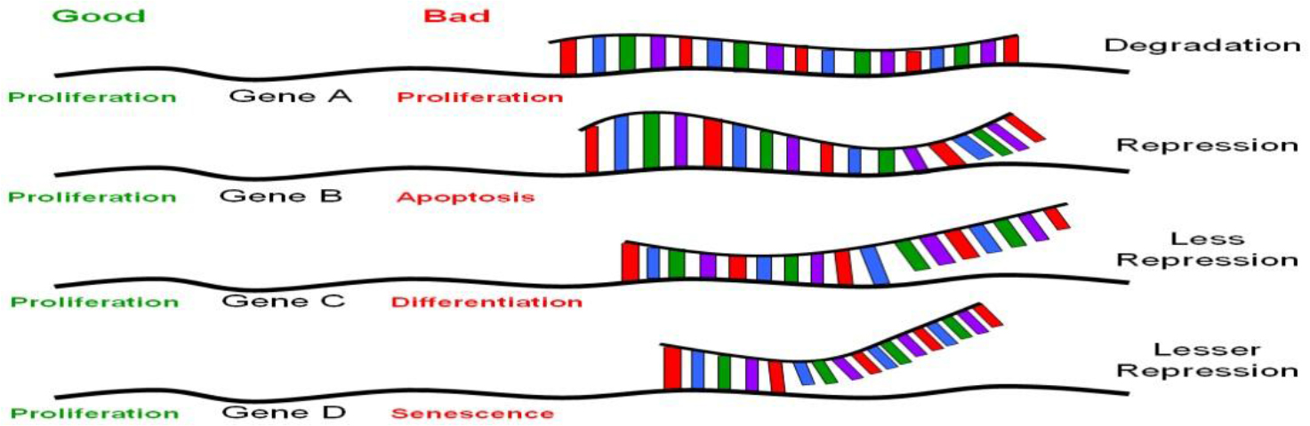
Figure 1. The nature of translational regulation by miRNAs or siRNAs.

The genome independent from the target genes, endogenous siRNAs often come from the target mRNAs. The processing of miRNAs comes from local hairpin formation of the target transcripts but that of siRNAs long bimolecular RNA duplexes. MiRNAs are single stranded RNAs, each 20–24nt long, and they do not encode for proteins. They have been found in all multicellular organisms, from plants to humans; they are abundant, estimated to be 3,000 – 40,000 molecules per cell. Computational and microarray analyses scanning for short, conserved repetitive sequences capable of forming secondary structures showed that genes encoding miRNAs composed of about 1% of the predicted genes in each species; hence, there are about 250 out of 30,000 genes in human genome, 100 in nematode and 80 in Drosophila [22].

MiRNAs are also unusual in their expression patterns. For example, lin-4 mentioned above is developmentally stage specific, so are many others [23]. Yet some are cell type or tissue-specific. For example, MiR-1 is preferentially expressed in mammalian heart [24], MiR-122 in the liver and MiR-124 in mouse brains, [25, 26], MiR-223 in granulocytes and macrophages of mouse bone marrow [27], and MiR-290 to MiR-295 in mouse embryonic stem cells [28]. Thus it appears that each developmental stage, cell type or tissue may have a distinctive MiR expression profile. Added to these unique expression profiles of MiR is the fact that more than 90% of all known MiR target genes encode for transcription factors, which regulate expression levels of many other structural or functioning genes, in plants and animals. Bartel interpreted these unique MiRs as “micromanagers” for gene transcription [17]. While this makes perfect sense, this unique expression profiling of MiR might pose significant concerns and require serious considerations when the siRNA technology is applied therapeutically. For example, what if the stem cell “micromanagers” are introduced through siRNAs and expressed in granulocytes or in livers? This issue would be related to the issues of true specificity of MiR targets, being discussed in the next paragraph.

Gene silencing via RNAi technology is achieved principally via base-pairing complementarity to target mRNA (some even to genomic DNA affecting transcription, which is beyond the scope of this article). Perfect sequence complementarity, as observed in siRNA-mediated gene silencing, results in degradation of the target mRNA (Figure 1, gene A). On the other hand, near perfect or incomplete complementation, as in the case of miRNA-mediated gene silencing, leads to inhibition without RNA degradation [16, 17, 19] (Figure 1, genes B-D). Mutational analyses on the known miRNAs and their target motifs indicated that the first 2–8nt of these miRNAs are often perfectly complementary to sequence motifs at the 3’-UTR involved in translational repression [29], and appear to be highly conserved among many different species [30, 31]. Unlike siRNAs, which usually target the genes from which they originate, miRNAs target many genes, perhaps a few thousand genes for each miRNA [16, 17]. Given that there are about 200–250 different miRNAs, the number of different species of mRNAs that are regulated by translational silencing can easily be in the thousands [22]. Taken together, there appear to be a set of genes to which a particular miRNA target, with a hierarchy of differential base-pairing affinities, as shown in Figure 1; the higher the complementarity in base-pairing, the more effective translational repression will be. Based on the above picture, introduction of a stem cell “micromanager” into a non-stem cell biological environment, as was raised in the last paragraph, may result in “mismanagement”, as would be the case of therapeutic applications using RNAi technology. This issue is hard to address without extensive knowledge on the nature of the set of genes that are target for any particular miRNA. One would not anticipate major problems if all the target genes of a particular miRs fall into the same category in biological functions. Conversely, major problems could appear if the target genes of a particular MiR are involved in various biological functions. This would not be true if there is a particular MiR that is ubiquitously expressed and is not “micromanaging” any specific cell type, tissue or developmental stage. Concerns have been raised regarding the recent observation of activation of a broad spectrum of non-specific siRNA-mediated immune responses in siRNA-transduced cells [32, 33]. These undesirable effects could be related to the concept of a particular “micromanager” being placed in a “wrong work environment”. The search for a non-micromanger MiR, if it exists, would be an important advancement from the standpoint of medical applications.

4. Rna Degradation

4.1. ARE elements

Messenger RNA turnover is a highly regulated process that involves both cis-acting sequence and trans-acting proteins. One of the ways in which mRNA turnover is controlled is through AU-rich motifs in the 3’UTR. These AU-rich elements (ARE) are comprised of a large class of cis-acting 3’UTR sequence that regulate RNA stability. These elements were first identified in the 3’UTR of mouse and human tumor necrosis factor (TNFa) mRNA, and then subsequently identified in other unstable RNAs [34]. This idea that AU-rich elements enhanced mRNA turnover was further supported when the 51 nucleotide ARE of granulocyte macrophage colony stimulating factor (GM-CSF) replaced the 3’UTR of the b-actin gene and caused rapid decay of the mRNA [35].

These ARE elements also appear to be highly conserved, more so than the coding regions of these genes. For example, in the c-fos gene the 3’UTR has 80% homology among diverse species (36). AU elements consist of either U-rich stretches of sequence, the pentamer AUUUA, or the nonamer, UUAUUUA(U/A)(U/A), and have been divided into three classes [see review in 36, 37]. Class I ARE control cytoplasmic deadenylation of mRNAs. All parts of the polyA tail are degraded at the same rate, resulting in intermediates that are completely degraded. They consist of 1–2 copies of AUUUA next to a U-rich region and examples of mRNAs include the transcription factors c-fos and c-myc. Class II ARE elements cause asynchronous cytoplasmic deadenylation resulting in degradation of the polyA tail at different rates in different transcripts. They consist of tandem AUUUA repeats and examples include GM-CSF, IL-2, TNFa, and IFNa. Class III ARE degrade mRNA in a similar mechanism as seen in Class I AREs, however, they do not contain an AU element. Class III elements contain U-rich segments and examples include c-jun and renin mRNA [37].

The exact mechanism by which AREs regulate mRNA stability has been unclear. Multiple repeats of the ARE sequence are necessary and both the structure and sequence in this region may be important for function. Evidence for secondary structure arises from RNA folding predictions of the VPR/VEGF ARES. There is 93 % homology between human and rat in forming two stem loops and hairpin region [38]. The similarity of the folding suggests that the structure is important and extremely conserved throughout diverse organisms.

AREs may also rely on other protein components and factors to regulate instability, and in some cases, stability of RNA transcripts. Evidence that ARE binding proteins play a role in regulating degradation has been seen in pathways that are involved in cellular responses to metabolic changes or environmental factors. These trans-acting proteins regulate decay in both gene specific and function specific mechanisms and are expressed differentially in different tissues at different times during development.

4.2. ARE binding proteins

The ARE binding proteins could influence rapid mRNA turnover by either direct recruitment or activation of RNAses, or through indirectly making contact with factors that bind ARE, independent of nucleases. These binding proteins can have either positive or negative effects, and can regulate stability, translation, or subcellular localization. Most of these experiments to identify putative AU-binding proteins have been performed using UV cross-linking and gel-shift assays [37, 38].

Seventeen proteins have been identified that bind to AU-rich elements. These include the heteronuclear ribonucleoproteins [hnRNP A1, hnRNP C [39], and hnRNP D [AUF-1,40] which bind these elements selectively and with different avidity. There are also RNQA binding proteins that display enzymatic activities, such as GAPDH [41] and AUH [42]. HuR proteins have also been identified and include hel-N1, HuC, HuD, and the ubiquitously expressed HuR [43]. Finally, there are other proteins that bind ARE including AU-A, AU-B, AU-C, tristetraprolin (TTP), butyrate response factor-1, KSRP, TIA-1, and TIAR [44].

The mechanism by which these proteins affect mRNA turnover remains unclear. One possibility is that the proteins bind the deadenylase and modulate degradation in this way. An alternative is that the proteins alter interactions for degradation by influencing the local structure providing better access for the ribonuclease. A third possibility is that these proteins recruit the exosome complex directly. Proteins that stabilize the mRNA trascript may do so by not allowing access of the exosome, while destabilizing AU binding proteins may directly recruit the exosome complex, resulting in rapid degradation [45].

4.3. Examples of proteins involved in mRNA turnover

Tristetraprolin (TTP) is a protein containing CCCH tandem zinc finger motifs [46] and has been demonstrated to destabilize class II ARE of TNFa and GM-CSF [39]. The destabilization requires two zinc fingers for binding activity, and TTP mutants that do not bind the GMCSF or TNFa ARE inhibit RNA turnover. TTP exists in many phosphorylated forms and may be a target in many signaling pathways. TTP is phosphorylated by p42 MAPK, p38, and MAPK activated protein kinase. TTP binds to 14–3-3 protein for cytoplasmic localization and has also been associated with the nuclear shuttle protein HuR in T-lymphocytes. The shuttling properties of this protein may contribute to cytoplasmic and nuclear decay of mRNA [47, 48].

K-homology type splicing regulatory protein (KSRP) binds c-fos and TNFa AREs and may contribute to exosome mediated degradation of mRNA [49]. KSRP is a shuttle protein that contains four copies of an RNA binding K homology domain. These domains are important in that mutations in these regions cause disease or differentiation defects [50].

AUF-1 is an AU-binding protein that is present as four isoforms. P42 and p45 are nuclear, while p37 and p40 are cytoplasmic [51]. The protein complex of p37 and p40 are able to destabilize c-myc in vitro. Phosphorylation appears to be involved as p38 inhibition stabilizes ARE through inactivation of AUF-1 [52]. Of these isoforms, in p37, two non-identical recognition motifs have been identified, an octameric RNP-1, and a hexameric RNP-2. Both are essential for the binding activity of AUF-1.

HuR is an AU-binding protein that stabilizes transcripts. It is a member of a family of proteins whose expression is developmentally regulated and tissue specific. HuR is ubiquitiously expressed. HuR is a nuclear/cytoplasmic shuttle protein that is predominantly nuclear. HuR contains RNA recognition motifs that bind RNA and shuttle it from the nucleus to cytoplasm. It may be this shuttling and the associated nuclear and cytpolasmic proteins that determine the stability of the RNA that is bound by HuR [43].

The regulation of these proteins is dependent on conditions and stimuli. In the cases of TTP and HuR, phophorylation by p38 (a mitogen activated protein kinase) could direct mRNA decay. PI-3 kinase and p38 MAPK independently stabilize IL-3 in NIH 3T3 cells. It may be the dynamic equilibrium between stabilization/degradation by TTP and HuR that determine the fate of this transcript. AUF-1 may act in a similar way with HuR. The exosome also plays an important role in degradation of transcripts and may be affected by accessibility and local topology of the 3’UTR ARE and the proteins that are bound [53].

5. Intracellular Localization

5.1. Cell motility and mobility

One way polarized cells establish their asymmetry is to localize mRNA transcripts to specific subcellular localization. RNA subcellular localization has been observed in oocytes and developing embryos of Frogs (Xenopus) and flies (Drosophila) [54–58]. In mammalian system, b-actin is expressed abundantly in all cells and it has many biological functions, including polarity and motility, protein synthesis, and various enzymatic biological processes. In motile chicken embryonic fibroblasts, b-actin mRNA has been shown to be highly expressed at the leading lamellae [59]. Rapid changes in actin polymerization at the leading edge drive extension of the lamellipodia, hence cell movement. Likewise, b-actin protein and mRNA colocalize to the leading lamellae of endothelial cells in response to wounding [60]. A 54-nt segment, called “zip-code” motif, located at the 3’-UTR of b-actin mRNA, is found to be responsible for such subcellular localization; this motif is thought to be distinct from those that mediate RNA stability or translational repression, as treatment of antisense oligonucleotides specific to the zip-code affected delocalization of b-actin mRNA but not RNA stability or suppression of actin synthesis. [61].

This zip code motif is highly conserved in primary sequence within b-actin mRNA from all species analyzed, including those of humans. Unlike the AU-rich sequence motif that affects RNA stability, there is, however, little sequence conservation among localization motifs that are present within the 3’-UTR of different mRNAs. Further studies identified ZBP1 (zip code binding protein) that binds to this zip-code with high affinity [62]. ZBP1 also colocalize with b-actin mRNA at the leading edge during cell movement, suggesting its involvement in binding b-actin mRNA, anchoring the mRNA to this region. To identify various components of the machinery responsible for this cell movement caused by polymerization of b-actin protein, similar co-localization studies of suspected molecules have been conducted. Elongation factor 1a(EF1a), involved in protein synthesis, and F-actin, a component of microtubule, have been shown to be involved in the binding and co-localization of b-actin mRNA at leading lamellae [63]. These studies suggest the involvement of an active protein translation apparatus and cytoskeleton in anchoring b-actin mRNA to the protrusion in crawling cells.

While the 54-nt zip code motif is clearly involved in its localization of b-actin mRNA to the leading edge of embryonic fibroblast, deletion of part or whole zip code reduces but does not eliminate its targeted localization activity, suggesting the involvement of additional motif other than the zip code motif [61]. Indeed, there is a homologous 43-nt zip code like element that also contributes to the full activity, albeit its activity is significantly less than that of the zip code. These results also suggest the presence of a number of genetic elements, within or outside the 3’-UTR, that are interacting with one another during localization of the target mRNA. The consequence of these interactions could be localized synthesis of proteins with optimal and effective execution of a function required at that location, such as the polymerization of b-actin being synthesized at leading edge of motile fibroblasts and myocytes, and growth cone of neuronal cells (see below). Consistent with this idea is that extremely intriguing observations are those reported by Elizabeth Gavis, who showed that in Drosophila oocytes, about 96% nanos mRNAs are unlocalized and distributed thoroughout the cytoplasm of the oocytes, and only the remaining portion of the mRNAs that are localized in the posterior end of the oocytes, via a 90-nt localization motif, are translationally active and functioning [64].

Not only more than one zip code motif can be involved in intracellular localization, more than one motif-binding protein can also be involved, each with different binding affinity to the target sequence. Recent work done by Singer and co-workers showed that as many as 5 proteins bind to the 54-nt zip code [65, 66]. ZBP1 binds with high affinity; mutational analyses showed that the protein contains domains responsible for granule formation, microtubule association, and of course, b-actin mRNA binding. Another protein is ZBP2, also an hnRNP protein like ZBP1, and has a human homologue. Unlike ZBP1, which contains a nuclear export signal and is predominantly cytoplasmic, ZBP2 contains a 47aa nuclear localization signal and is predominantly nuclear but it does shuttle in and out of the nucleus. Therefore mRNA localization process could begin in the nucleus: ZBP2 brings the b-actin mRNA out of the nucleus, and then ZBP1 takes over and escort the mRNA to the leading lamellae, through the microtubule assembly, where translation would begin. After protein synthesis, polymerization of b-actin would drive lamillipodia directionally and the cells move forward.

5.2. Neural transmission

Synaptic plasticity is a complex biological phenomenon that is associated with long-term potentiation (LTP), hence long-term memory storage in the brain. The strength of synapses appears to change in response to activation of neurotransmitter receptors present on cell surfaces of dendrites, and modulation of synaptic strength has been shown to be dependent on de novo protein synthesis, or localized protein synthesis in dendrites or synaptic sites as a function of synaptic activity [67–69]. Therefore, local protein synthesis within dendrites associated with mRNA intracellular localization represents an extremely interesting mechanism that can explain this activity dependent synaptic plasticity. Evidence supporting this model is quite clear.

In neurons, the majority of mRNAs are located in the cell body; however, a number of them are specifically located to the dendrites. Using high resolution in situ hybridization and image processing methods, Singer, Bassell and associates [70] showed that while b-actin mRNA and protein are distributed uniformly throughout the cytoplasm, b-actin mRNA and protein are localized in dendrites and growth cones of neurons. Likewise, the Arc mRNA and protein are also found to be localized to the synapse only in response to synaptic activation [71, 72].

The zip code motif has been shown to be responsible for b-actin mRNA localization in dendrites [65, 70, 73] Transport of specific mRNAs to dendrites is achieved through microtubules in the form of granules that apparently contain an active transport unit and translational machinery. The association of the zip code binding proteins, including ZBP1, with granules on microtubules has been shown to be dynamically dependent on KCl-induced neural depolarization and there is a rapid efflux of ZBP1 from the cell body to the dendrites [74]. This activity dependent trafficking of ZBP1 suggests selective targeting of b-actin mRNA to postsynaptic sites within dendrites in response to synaptic activity.

Localized mRNA and translational activation of protein kinases are also found to be present in dendrites or synaptic sites. One example is CaMKIIa (Calcium/ calmodulin-dependent protein kinase II alpha), whose induction and maintenance in LTP is known, and it has been implicated in dendritic mRNA transport. By in situ hybridization, CaMKIIa mRNA is localized in dendrites, and using GFP (green fluorescent protein) fusion constructs in the studies for visualization, CaMKIIa 3’-UTR has been shown to be responsible for such localization [75, 76]. The CaMKIIa mRNA is also found in microtubule granules, and synaptic activity, reflected by the degree of neuronal depolarization, increase the number of granules in the dendrites [76]. These data are consistent with in vivo observations. Mice lacking the CaMKIIa 3’-UTR exhibit disruption of CaMKIIa mRNA localization, reduced expression of this protein in dendrites, diminishing long-term potentiation and impairments of several forms of associative learning, suggesting that localized protein synthesis contributes to synaptic plasticity [77]. Full characterization of this localization motif and trans-acting factors binding to it has yet to be defined.

Another example in which localized translational activation is linked to synaptic sties is PKMz (protein kinase M zeta). PKMz is an atypical protein kinase C and is known to be involved in synaptic activity-dependent LTP and memory storage in the brain. Its mRNA has been shown to be rapidly transported and localized to synaptodendritic neuronal domain after induction [78]. Two localization motifs, called dendritic targeting elements (DTE), have been identified. One is at the 5’-UTR (DTE1) and the other is at the 3’-UTR (DTE2). DTE1 appears to direct somato-dendritic export of the mRNA, whereas DTE2 appears to deliver the mRNA to the dendrites.

6. The Uniqueness of Ran Gtpase

Ran GTPase is a unique member of a large family of G-proteins. It is expressed abundantly in all nucleated mammalian cells, and at steady state, most but not all of them reside in the nucleus. Ran GTPase is also (Figure 2) Various microbial pathogens invade cells using different Toll-like receptors (TLR) or other receptors on the cell surface, initiating one or more signaling transduction pathways many of which act through biochemical modification of NF-κB inhibitor IκB. This results in the release and activation of NF-κB, which translocates into the nucleus without the need to associate with Ran, binds to DNA sequence of immune response genes and activates transcription of these genes that encode for pro-inflammatory cytokines such as TNFa, IL-1 and IL-6. More nuclear Ran or higher N/C ratio of Ran, as in RanC/d-transduced cells, would enhance nuclear export of NF-κB via the Ran/Exp1 complex, reducing NF-κB transcriptional activation, hence down-modulating immune response. Conversely, more cytoplasmic Ran, hence lower N/C, as in RanT/n-transduced cells, would discourage nuclear export of NF-κB, hence no reduction of NF-κB transcriptional activity in the nucleus. More cytoplasmic Ran would also directly stimulate signal transduction pathways involved in immune response through binding to adaptor molecules such as RanBPM (see text).

JMG- P M Wong_F2

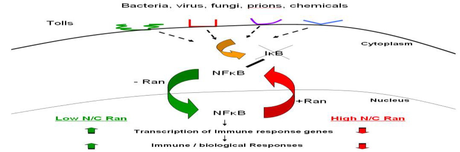
Figure 2. Ran-mediated immune modulation of host immune response.

Intimately involved in several very fundamental processes in cell cycle progression [5–7]. At interphase, it is a key molecule involved in the transport of a majority of all macromolecules in and out of the nuclear membrane. During mitosis, Ran is required for mitotic microtubule spindle formation and nuclear envelope assembly. It also prevents DNA re-replication during S phase [79]. Obviously then, it is amazing how Ran gets itself involved in all these important molecular events and how all these crucial regulatory events translate to biological phenomena. At the cellular and organismic levels in mammalian system, the clue comes from the identification of genetic changes at the 3’-UTR of Ran, producing corresponding and predictable changes in Ran’s RNA structure, in Ran’s nuclear/ cytoplasmic ratio, and in modulating Ran-mediated host immune response [8–10].

6.1. Single nucleotide change in 3’-UTR of Ran leading to changes in protein and RNA localization and other biological changes

By functional cDNA expression cloning, followed up with forward genetic analyses, we identified two Ran alleles, RanC/d and RanT/n, that differ from each other only by a single nucleotide, located at the 3’-UTR [8–12, 80–82]. This single base change leads to striking changes in RNA structure (12). As a result of this RNA structural change, we demonstrated difference in protein and RNA intracellular localization. Expression of RanC/d leads to a rapid nuclear localization and down-modulation of pro-inflammatory cytokine production, correlated with protection of mice from endotoxin-induced shock. Conversely, expression of the other allele, RanT/n, results in a predominant cytoplasmic localization of Ran proteins and an up-regulation of pro-inflammatory cytokine production, correlated with increase susceptibility to endotoxic shock. Subsequent studies provide more detailed biological changes in mechanistic terms as a result of this single nucleotide change and these details are strengthened by several other findings in the literature.

First, this single nucleotide change in Ran 3’-UTR correlates with changes in proteins and Ran RNA intracellular localization and is entirely consistent with the existence of localization sequence motif, the majority of which are located within the 3’-UTRs [54–56, 58]. Second, this difference in Ran intracellular localization results in changes in Ran’s nuclear/cytoplasmic (N/C) ratio Figure 2. In RanC/d-transduced cells, more nuclear Ran (Figure 3) The main principle of this model is the same as we predicted before – cytoplasmic Ran can initiate signal transduction events independent of nuclear Ran, by association with other signaling molecules in the cytoplasm [M and B in Figure 3 of [82]. Green ovals are RanGDP, blue ovals are RanGTP, BPM = Ran-BPM, GAP = RanGAP, LFA = LFA-1, AR = androgen receptor, Met = Met receptor for hepatic growth factor, RCC1 = Regulator of chromosome condensation, N/C ratio = nuclear/ cytoplasmic ratio. At steady state, most Ran GTPases reside in the nucleus, and some are located outside the nucleus by virtue of sumoylation of Ran-GAP that hydrolyses RanGTPs as soon as they are exported from the nucleus. Most cytoplasmic Ran there cycle back into the nucleus, some remain in the cytoplasm. Complex formation of cytoplasmic Ran, BPM and other molecules initiates signaling for cell motility and migration, proliferation, cell adhesion, modulation of immune response and other cytoplasmic functions. Complex formation of nuclear Ran and RCC1, or perhaps other molecules, would encourage nuclear events such as mitotic spindle formation, nuclear envelope assembly or DNA re-replication. These two sets of events could be opposing to each other; the final outcome is dictated by the absolute amounts of Ran in each compartment, as well as their relative amount, which is the N/C ratio of Ran.

JMG- P M Wong_F3

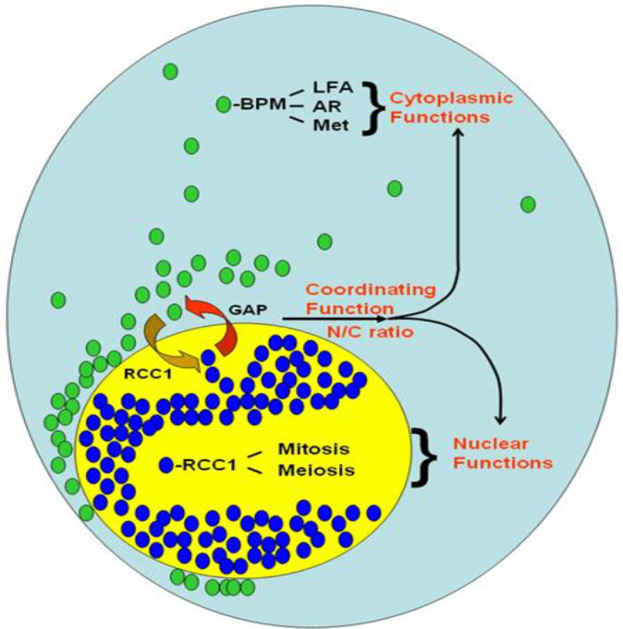
Figure 3. A model of Ran GTPase-mediated biological response.

Increases the N/C ratio; more nuclear Ran also increases the export of NF-κB from the nucleus to the cytoplasm because nuclear export of NF-κB is completely Ran dependent [8, 83–86]. Increase nuclear export of NF-κB results in decrease in its transcriptional activation of immune response genes, including those encoding for pro-inflammatory cytokines (manuscript submitted). Conversely, in RanT/n-transduced cells, more cytoplasmic Ran decreases the N/C ratio, resulting in reduced nuclear export of NF-κB, enhanced transcriptional activity of NF-κB, increased immune response and sensitivity to endotoxic shock. Third, the observed biological changes did not correlate perfectly with the N/C ratio of Ran, over-expression of either Ran allele did not result in overt growth damage (manuscript in preparation), yet in cells or mice transduced with RanT/n cDNA, increased immunological sensitivity as a result of RanT/n expression is apparent. More than likely, therefore, cytoplasmic Ran, in the form of RanGDP, possesses a function independent of the N/C ratio and that this function enhances the biological outcome induced by the change of N/C ratio. Indeed, cytoplasmic Ran has been shown to bind to Ran-BPM [87,88], an adaptor molecule that binds between Ran and a number of molecules involved in modulation of host immune response, cell migration, cell fate and wound healing; they include androgen receptor [89], LAF-1 [90], Met kinase receptor for hepatic growth factor [91,92], and others.

We propose that this interdependence between the absolute amount of Ran in each cellular compartment and their relative amount, i.e. N/C ratio, is the key to Ran’s role in the regulation of many fundamental aspects of mammalian cell growth and differentiation, a model we previously proposed [82], and is now more refined in (Figure 3). Because of this interdependent relationship, the interactions of Ran with other molecules must be characteristically dynamic. The abundance and ubiquity of Ran in all cells attest to the validity of this concept, as the endogenous Ran would be acting as a very strong buffer, biochemically speaking. Extension of this principle points to the immense potential of applying Ran as a genetic therapeutic vaccine in a variety of clinical applications in which the key fundamental molecular defect is Ran-dependent, be it a change in Ran nuclear/cytoplasmic ratio, or in RanGTP/GDP ratio. This clinical potential is further support by our recent data showing that over-expression of either RanT/n or RanC/d allele results in similar transient growth alterations but is non-toxic (manuscript in preparation).

6.2. The issue of C versus T

Sequence alignments have been performed in the past indicating that Ran GTPAse protein is highly conserved [9, 93–95]. Alignment of 3’-UTR Ran sequences from various species reveal the presence of a C residual at position 870 and the corresponding position for sequences of other species. What then is the origin of the RanT/n allele? RanT/n was first isolated and cloned from a cDNA library made from mRNAs of endotoxin-induced splenic B cells of C3H/HeOuJ, an LPS (lipopolysaccharide) responsive inbred mouse strain [9]. Identification of such cDNA was accomplished via functional conversion of B cells of C3H/HeJ, an LPS resistant mouse strain, from a state of hypo-responsiveness to a state of responsiveness upon LPS stimulation. Did RanT/n come from the genome of C3H/HeOuJ mice, or not? (Table 1).

Nucleotide position indicates the nucleotide regions in the 3’UTR that were mutagenized with single base pair substitutions. These regions are also indicated in the (figure 4). Significant structural changes were scored arbitrarily as being structure predictions determined by the M-fold program that disrupted the original structure of the region (i.e. alteration of the stem loop and bulge regions). Nucleotide clustering indicates the regions or specific nucleotides that were most apparent in altering the predicted secondary structure. This table is not intended to give a complete analysis of the 3’UTR, but rather a mean for modeling where single base changes may affect local secondary structure. The numbers in parentheses indicate the number of structures found in that particular region.

JMG- P M Wong_F4

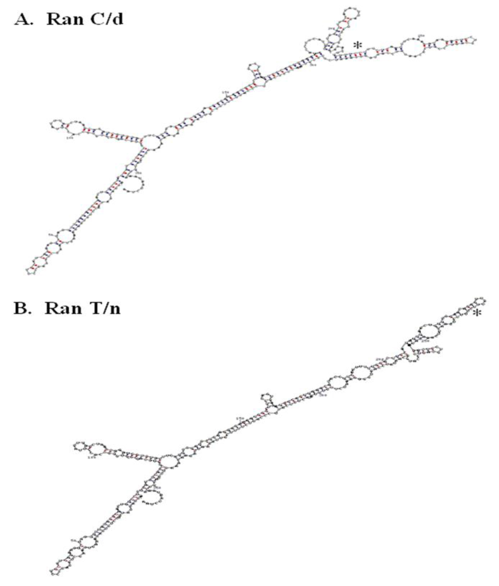
Figure 4. RNA secondary structure of RanC/d and RanT/n 3’-UTR. A.

Table 1. Summary of single base substitutions influencing change in predicted RNA secondary structures of mouse and human Ran 3’UTR.

Nucleotide Position (Mutagensis Regions)

Significant Structural Changes

Nucleotide Clustering

Mouse

767–807

5

767–807 (1)

787–805 (4)

832–867

13

832–844 (8)

859–867 (5)

Human

756–775

2

765, 771

825–925

20

887, 899, 902, 905,

908, 913–925, 993

The above alignment analysis would have provided a very clear answer had Ran gene existed in the mouse genome in single copy. This is obviously not the case. Blasting our Ran sequence deposited in GenBank (accession number AF159256) against the mouse genome showed that there are as many as 7 Ran isoform genes, each is located on a different chromosome. Existence of at least 7 Ran isoform genes was reported by D’Eutaschio, Rush and colleagues at NYU some years ago [93–95]. Among the 7 isoform genes, 4 sequences contain the “C” residue at position 870, one has a “G”, and two do not have sufficient information. Strictly speaking, therefore, the origin of RanT/n could not be ascertained one way or another until all Ran isoform sequences in the mouse genome have been clarified. Regardless of its origin, RanT/n was identified using functional cDNA expression cloning, where RanT/n expression in B cells from LPS-resistant C3H/HeJ mice restored their LPS responsiveness [9].

6.3. More sequence analysis predicting RNA structure

The single point mutation in Ran’s 3’-UTR studies suggest the presence of zip code motif to which specific proteins bind and escort the mRNA to specific intracellular locations. More RNA analysis will be helpful along this direction. Folding the whole 3’UTR of both ran alleles (870C and 870T) using the M-fold program and default parameters [96, 97] suggest that a single nucleotide change may change the folding in the local region (Figure 4). Previous folding using the GCG Squiggles program also suggests that there is a change in the secondary structure [12]. Such prediction of striking RNA structural difference between RanT/n and RanC/d was confirmed by digestion of in vitro transcribed RNAs from T/n and C/d templates with RNase T1, an RNA endonuclease that cleaves single-stranded RNA but not double stranded RNA portions preserved by stem-loops or hairpin structures [12].

Additional RNA secondary structures of Ran 3’UTRs were also generated by mutating, base by base, approximately 100 nucleotides 5’of the 870 position in both mouse and human Ran. Single nucleotide substitutions were inserted into the sequence and folded using the M-fold server [97]. The data from these analyses suggest that the region around 870 is very susceptible to single nucleotide changes in both human and mouse Ran mRNA (Table 1). Phylogenetic comparison of 3’UTR sequences from more organisms would help in identifying consensus motifs and to generate a consensus secondary structure. As reviewed earlier, since “RNA localization element” within the 3’-UTRs is not well conserved and multiple elements may exist in orchestrating localization of Ran to the nucleus periphery, systematic site-directed mutagenesis would aid in the identification of this Ran PNLE – “peri-nuclear localization element”.

6.4. Conserved regulatory element in 3’UTRs of “family member”

Conservation in the 3’UTR has also been observed in another ras family member, Rab1a. It is a regulatory protein necessary for the transport of vesicles from the endoplasmic reticulum to the Golgi apparatus. Sequence analysis of the 3’UTR in 27 different species yielded a 92% similarity between the most distant species (man and turtle), and 95% or greater between more closely related organisms such as mammals [98]. However, this sequence is not homologous to other Rab genes or Ras family members. This observation, along with the high sequence conservation of the 3’UTR of Rab1a, suggests that it has a specific function for location and/or stability of its RNA.

Alignment of the 3’UTR of Ran to other Ras family members does not yield a significant amount of homology (not shown). The Ran 3’UTR aligns best (~50% similar) to human n-Ras 3’UTR sequence as described by Hall & Brown [99]. This also suggests that the 3’UTR has a function specific for Ran mRNA localization. This lack of alignment does not preclude any undefined elements that may be present in the Ras family members that are present in their respective 3’UTR elements. This information will be used to identify potential motifs and tested for changes in localization of mRNA.

6.5. Ran 3’UTR has it all

The 3’-UTR has been thought of as a repository of many post-transcriptional regulators. Indeed, when the 3’-UTR of Ran was blasted against the UTR database, it provided two interesting observations. Plant and mouse Ran 3’-UTR contained an IRES element (internal ribosome entry site) whose functions is to allow independent translation of downstream sequences from this point on. Its role in Ran biology is presently unknown. In addition to the presence of IRES in the 3’- (Figure 4) A. RanC/d, B, RanT/n. The T/C change at position 870 in both structures is indicated with an asterisk. RNA structure was determined using the M-fold program with default parameters. C-G base pairing is indicated with red dots, while U-A base pairing is indicated with blue dots.

UTR region, mouse, human and songbird Ran 3’-UTRs also contain K box consensus sequences, TGTGAT. K boxes are highly conserved and ubiquitous, often found in 3’-UTR regions, and in genes affecting cell growth and development. It was initially found in the 3’-UTR of genes that regulate Notch receptors, which have been shown to play important roles in the development of neural and hematopoietic system. Genetic analyses in Drosophila show that a sequence motif negatively regulates translation of the mRNA in which K box resides, and deletion of this motif results in a gain of function [100,101]. Therefore, K box appears to function through translational repression. The significance of K box in the context of Ran biology is presently unknown.

Ran’s 3’-UTR therefore clearly has many post-transcriptional regulatory elements. Given that Ran GTPase is abundantly expressed and is expressed ubiquitously, the functions of these elements may operate in a coordinated yet flexible fashion to accommodate Ran’s diverse biological roles. Research emphasis on Ran’s 3’-UTR will certainly uncover important and fundamental principles illuminating its roles in cell cycle progression, nucleocytoplasmic transport, DNA re-replication, signal transduction, and Ran-mediated immune responses. Extension of these principles to clinical medicine will be highly beneficial. One example is our discovery of mutational changes within Ran’s 3’-UTR, resulting in predictable modulated host immune response.

7. Conclusion

The discovery of miRNAs and its renewed intense studies has revealed new roles of small RNAs in regulating cell growth and development. The “micromanagers” of miRNAs in specific tissues or cells during a particular stage of development attest to their important roles in many aspects of biology. Understanding of the significance of these biological regulations and how they work have been greatly facilitated by RNAi technology. Therapeutic applications of this technology, however, require more thorough understanding regarding the nature and the specificity of target genes, especially with the knowledge that 2–8nt of any particular miRNA, and therefore, very possibly, any particular siRNA, can have any where in the genome sequence complementation with recognition motifs of the intended target genes.

The 3’-UTR of Ran GTPase contains regulatory motifs involved in translational repression, RNA degradation and RNA intracellular localization. This unique feature emphasizes the importance of post-transcriptional regulation of Ran GTPase. This unique feature is consistent with its many important roles in many aspects of cell cycle progression, nucleocytoplasmic transport and immune response functions, but has apparent contradiction with its abundant and ubiquitous expression, which is more in line with functions of housekeeping proteins. Further over-expression appears to exert only a temporary growth alteration effects (manuscript in preparation). Mechanisms therefore must exist in adjusting not only its absolute levels within the cells through proteolytic degradation, but also its relative amount in each cellular compartment, reflected by either the nuclear/cytoplasmic ratio or the Ran GTP/GDP ratio. On this basis, the discovery of genetic mutants at its 3’-UTR capable of altering both the absolute and relative amounts of Ran, with expected corresponding and predictable functional and biological changes at the molecular, cellular and organismic levels, is invaluable. Such value has already been supported by the demonstration that RanC/d expression can down-modulate host immune response and confer resistance to septic shock, and that RanT/n expression has opposite effects. More such genetic studies in Ran will reveal more of its majestic beauty and its immense clinical applications including but not limited to cancer, AIDS, cardiovascular disorders, infectious diseases, neurologic, metabolic and genetic disorders.

8. Acknowledgement

Work done on Ran GTPase in our laboratory has been supported by NIH RO1 grants from NCI and NIAID.

References

  1. Fire A, S Xu, MK Montgomery, SA Kostas, SE Driver, et al. (1998) Potent and specific genetic interference by double-stranded RNA in Caenorhabditis elegans. Nature 391: 806–811.
  2. Napoli C, C Lemieux, R Jorgensen (1990) Introduction of a chimeric chalcone synthase gene into Petunia results in reversible co-Suppression of homologous genes in trans. Plant Cell 2: 279–289.
  3. van der Krol AR, Mur LA, Beld M, Mol JN, Stuitje AR (1990) Flavonoid genes in petunia: addition of a limited number of gene copies may lead to a suppression of gene expression. Plant Cell 2: 291–299. [crossref]
  4. Lee RC, Feinbaum RL, Ambros V (1993) The C. elegans heterochronic gene lin-4 encodes small RNAs with antisense complementarity to lin-14. Cell 75: 843–854. [crossref]
  5. Dasso M (2001) Running on Ran: nuclear transport and the mitotic spindle. Cell 104: 321–324. [crossref]
  6. Hetzer M, O. J. Gruss, IW Mattaj (2002) The Ran GTPase as a marker of chromosome position in spindle formation and nuclear envelope assembly. Nat Cell Biol 4: E177–184.
  7. Weis K (2003) Regulating access to the genome: nucleocytoplasmic transport throughout the cell cycle. Cell 112: 441–451.
  8. Wong PMC, SW Chung (2003) A functional connection between RanGTP, NF-?B and septic shock. J Biomed Sci 10: 468–474.
  9. Kang AD, PMC Wong, H Chen, R Castagna, SW Chung (1996) Restoration of lipopolysaccharide-mediated B-cell response after expression of a cDNA encoding a GTP-binding protein. Infect Immun 64: 4612–4617.
  10. Wong PMC, A Kang, H Chen, Q Yuan, P Fan, et al. (1999) Lpsd/Ran of endotoxin-resistant C3H/HeJ mice is defective in mediating lipopolysaccharide endotoxin responses. Proc Natl Acad Sci USA 96: 11543–11548.
  11. Yuan Q, F Zhao, SW Chung, P Fan, BM Sultzer, et al. (2000) Dominant negative down-regulation of endotoxin-induced tumor necrosis factor alpha production by Lpsd/Ran. Proc Natl Acad Sci USA 97: 2852–2857.
  12. Wong PMC, Q Yuan, H Chen, BM Sultzer, SW Chung (2001) A single point mutation at the 3’-untranslated region of Ran mRNA leads to profound changes in lipopolysaccharide endotoxin-mediated responses. J Biol Chem 276: 33129–33138.
  13. Ostareck DH, A Ostareck-Lederer, M Wilm, BJ Thiele, M Mann, et al. (1997) mRNA silencing in erythroid differentiation: hnRNP K and hnRNP E1 regulate 15-lipoxygenase translation from the 3’ end. Cell 89: 597–606.
  14. Ostareck-Lederer A, D H Ostareck, N Standart , BJ Thiele (1994) Translation of 15-lipoxygenase mRNA is inhibited by a protein that binds to a repeated sequence in the 3’ untranslated region. EMBO J 13: 1476–1481.
  15. Ostareck-Lederer A, D H Ostareck, C Cans, G Neubauer, K Bomsztyk, et al. (2002) c-Src-mediated phosphorylation of hnRNP K drives translational activation of specifically silenced mRNAs. Mol Cell Biol 22: 4535–4543.
  16. Novina CD, PA Sharp (2004) The RNAi revolution. Nature 430: 161–164.
  17. Bartel DP (2004) MicroRNAs: genomics, biogenesis, mechanism, and function. Cell 116: 281–297. [crossref]
  18. Bartel DP, CZ Chen (2004) Micromanagers of gene expression: the potentially widespread influence of metazoan microRNAs. Nat Rev Genet 5: 396–400.
  19. He Z, Sontheimer EJ (2004) “siRNAs and miRNAs”: a meeting report on RNA silencing. RNA 10: 1165–1173. [crossref]
  20. Wightman B, I Ha, (1993) G Ruvkun Posttranscriptional regulation of the heterochronic gene lin-14 by lin-4 mediates temporal pattern formation in C. elegans. Cell 75: 855–862.
  21. Rougvie AE (2001) Control of developmental timing in animals. Nat Rev Genet 2: 690–701. [crossref]
  22. Lim LP, Glasner ME, Yekta S, Burge CB, Bartel DP (2003) Vertebrate microRNA genes. Science 299: 1540. [crossref]
  23. Ambros V (2004) The functions of animal microRNAs. Nature 431: 350–355. [crossref]
  24. Lee RC1, Ambros V (2001) An extensive class of small RNAs in Caenorhabditis elegans. Science 294: 862–864. [crossref]
  25. Lagos-Quintana M, R Rauhut, A Yalcin, J Meyer, W Lendeckel, et al. (2002) Identification of tissue-specific microRNAs from mouse. Curr Biol 12: 735–739.
  26. Lagos-Quintana M, R Rauhut, W Lendeckel, T Tuschl (2001) Identification of novel genes coding for small expressed RNAs. Science 294: 853–858.
  27. Chen CZ, L Li, HF Lodish, DP Bartel (2004) MicroRNAs modulate hematopoietic lineage differentiation. Science 303: 83–86.
  28. Houbaviy HB, Murray MF, Sharp PA (2003) Embryonic stem cell-specific MicroRNAs. Dev Cell 5: 351–358.
  29. Lai EC (2002) Micro RNAs are complementary to 3’ UTR sequence motifs that mediate negative post-transcriptional regulation. Nat Genet 30: 363–364.
  30. Stark A, Brennecke J, Russell RB, Cohen SM (2003) Identification of Drosophila MicroRNA targets. PLoS Biol 1: E60. [crossref]
  31. Lewis BP, IH Shih, MW Jones-Rhoades, DP Bartel, CB Burge (2003) Prediction of mammalian microRNA targets. Cell 115: 787–798.
  32. Bridge AJ, Pebernard S, Ducraux A, Nicoulaz AL, Iggo R (2003) Induction of an interferon response by RNAi vectors in mammalian cells. Nat Genet 34: 263–264. [crossref]
  33. Sledz CA, Holko M, de Veer MJ, Silverman RH, Williams BR (2003) Activation of the interferon system by short-interfering RNAs. Nat Cell Biol 5: 834–839. [crossref]
  34. Caput D, B Beutler, K Hartog, R Thayer, S Brown-Shimer et al. (1986) Identification of a common nucleotide sequence in the 3’-untranslated region of mRNA molecules specifying inflammatory mediators. Proc Natl Acad Sci U S A 83: 1670–1674.
  35. Shaw G, R Kamen A (1986) conserved AU sequence from the 3’ untranslated region of GM-CSF mRNA mediates selective mRNA degradation. Cell 46: 659–667.
  36. Dalgleish G, Veyrune JL, Blanchard JM, Hesketh J (2001) mRNA localization by a 145-nucleotide region of the c-fos 3’–untranslated region. Links to translation but not stability. J Biol Chem 276: 13593–13599. [crossref]
  37. Mignone F, Gissi C, Liuni S, Pesole G (2002) Untranslated regions of mRNAs. Genome Biol 3: REVIEWS0004. [crossref]
  38. Levy NS, MA Goldberg, AP Levy (1997) Sequencing of the human vascular endothelial growth factor (VEGF) 3’ ubtranslational region (UTR) conservation of fice hypoxia-inducible RNA-protein binding sites. Biochem Biophys Acta 1352: 167–173.
  39. Hamilton BJ, E Nagy, JS Malter, BA Arrick, WF Rigby (1993) Association of heterogenous nuclear ribonucleoprotein A1 and C proteins with reiterated AUUUA sequences. J Biol Chem 268: 8881–8887.
  40. Zhang w, BJ Wagner, K Ehrenman, AW Schaefer, CT DeMaria, et al. (1993) Purification, characterization and cDNA cloning of an AU-rich element RNA-binding protein, AUF1. Mol Cell Biol 13: 7652–7665.
  41. Nagy E, WFC Rigby (1995) Glyceraldehyde-3-phosphate dehydrogenase selectively binds AU-rich RNA in the NAD+-binding region (Rossmann fold). J Biol Chem 270: 2755–2763.
  42. Nakagawa J, H Waldner, S Meyer-Monard, J Hofsteenge, P Jeno, (1955) AUH, a gene encoding an AU-specific RNA binding protein witn intrinsic enoyl-CoA hydratase activity. Proc Natl Acad Sci USA 92: 2051–2055.
  43. Brennan CM, Steitz JA (2001) HuR and mRNA stability. Cell Mol Life Sci 58: 266–277. [crossref]
  44. Piecyk M, Wax S, Beck AR, Kedersha N, Gupta M, et al. (2000) TIA-1 is a translational silencer that selectively regulates the expression of TNF-alpha. EMBO J 19: 4154–4163. [crossref]
  45. Chen CY, Gherzi R, Ong SE, Chan EL, Raijmakers R, et al. (2001) AU binding proteins recruit the exosome to degrade ARE-containing mRNAs. Cell 107: 451–464. [crossref]
  46. Carballo E, WS Lai, PJ Balckshear (1998) Feedback inhibition of macrophage tumor necrosis factor alpha production by tristetraprolin. Science 281: 1001–1005.
  47. Tchen CR, Brook M, Saklatvala J, Clark AR (2004) The stability of tristetraprolin mRNA is regulated by mitogen-activated protein kinase p38 and by tristetraprolin itself. J Biol Chem 279: 32393–32400. [crossref]
  48. Brooks SA, JE Connolly, WFC Rigby (2004) The role of mRNA turnover in the regulation of tristetraprolin expression: Evidence for an extracellular signal-regulated kinase-specific, AU-rich element-dependent, autoregulatory pathway. J Immunol 172: 7263–7271.
  49. Min H, CW Turck, JM Nikolic, DL Black (1997) A new regulatoru protein, KSRP, mediates exon inclusion through an intrionic splicing enhancer. Genes Dev 11: 1023–1036.
  50. Gherzi R, Lee KY, Briata P, Wegmüller D, Moroni C, et al. (2004) A KH domain RNA binding protein, KSRP, promotes ARE-directed mRNA turnover by recruiting the degradation machinery. Mol Cell 14: 571–583. [crossref]
  51. Wagner BJ, CT deMaria, Y Sun, GM Wilson, G. Brewer (1998) Structure and genomic organization of the human AUF1 gene: alternative pre-mRNA splicing generated four protein isoforms. Genomics 1: 195–202.
  52. Wilson GM, J Lu, K Sutphen, Y Sun, Y Huynh, et al. (2003) Regulation of A + U-rich element-directed turnover involving reversible phosphorylation of AUF1. J Biol Chem 278: 33029–33038.
  53. Dean JLE, G Sully, AR Clark, J Saklatvala (2004) The involvement of AU-rich element-binding proteins in p38 mitogen-activated protein kinase pathway-mediated mRNA stabilization. Cell Signal 16: 1113–1121.
  54. Kloc M, Zearfoss NR, Etkin LD (2002) Mechanisms of subcellular mRNA localization. Cell 108: 533–544. [crossref]
  55. Jackson RJ (1993) Cytoplasmic regulation of mRNA function: the importance of the 3’ untranslated region. Cell 74: 9–14. [crossref]
  56. Hazelrigg T (1998) The destinies and destinations of RNAs. Cell 95: 451–460. [crossref]
  57. Mowry KL, Cote CA (1999) RNA sorting in Xenopus oocytes and embryos. FASEB J 13: 435–445. [crossref]
  58. Kuersten S, Goodwin EB (2003) The power of the 3’ UTR: translational control and development. Nat Rev Genet 4: 626–637. [crossref]
  59. Lawrence JB, R. H (1986) Singer Intracellular localization of messenger RNAs for cytoskeletal proteins. Cell 45: 407–415.
  60. Hoock TC, PM Newcomb, IM Herman (1991) Beta actin and its mRNA are localized at the plasma membrane and the regions of moving cytoplasm during the cellular response to injury. J Cell Biol 112, 653–664.
  61. Kislauskis, E H, X Zhu, R H. (1994) Singer Sequences responsible for intracellular localization of beta-actin messenger RNA also affect cell phenotype. J Cell Biol 127: 441–451.
  62. Ross AF, Oleynikov Y, Kislauskis EH, Taneja KL, Singer RH (1997) Characterization of a beta-actin mRNA zipcode-binding protein. Mol Cell Biol 17: 2158–2165. [crossref]
  63. Liu G, Grant WM, Persky D, Latham VM Jr, Singer RH, et al. (2002) Interactions of elongation factor 1alpha with F-actin and beta-actin mRNA: implications for anchoring mRNA in cell protrusions. Mol Biol Cell 13: 579–592. [crossref]
  64. Bergsten SE, Gavis ER (1999) Role for mRNA localization in translational activation but not spatial restriction of nanos RNA. Development 126: 659–669. [crossref]
  65. Gu W, F Pan, H Zhang, GJ Bassell, RH Singer (2002) A predominantly nuclear protein affecting cytoplasmic localization of beta-actin mRNA in fibroblasts and neurons. J Cell Biol 156: 41–51.
  66. Farina KL, S Huttelmaier, K Musunuru, R Darnell, RH Singer (2003) Two ZBP1 KH domains facilitate beta-actin mRNA localization, granule formation, and cytoskeletal attachment. J Cell Biol 160: 77–87.
  67. Nguyen PV, Abel T, Kandel ER (1994) Requirement of a critical period of transcription for induction of a late phase of LTP. Science 265: 1104–1107. [crossref]
  68. Frey U, M Krug, KG Reymann, H Matthies (1988) Anisomycin, an inhibitor of protein synthesis, blocks late phases of LTP phenomena in the hippocampal CA1 region in vitro. Brain Res 452: 57–65.
  69. Stanton PK, Sarvey JM (1984) Blockade of long-term potentiation in rat hippocampal CA1 region by inhibitors of protein synthesis. J Neurosci 4: 3080–3088. [crossref]
  70. Bassell GJ, Zhang H, Byrd AL, Femino AM, Singer RH, et al. (1998) Sorting of beta-actin mRNA and protein to neurites and growth cones in culture. J Neurosci 18: 251–265. [crossref]
  71. Steward O, PF Worley (2001) Selective targeting of newly synthesized Arc mRNA to active synapses requires NMDA receptor activation. Neuron 30: 227–240.
  72. Steward O, Worley P (2001) Localization of mRNAs at synaptic sites on dendrites. Results Probl Cell Differ 34: 1–26. [crossref]
  73. Bassell GJ, Singer RH, Kosik KS (1994) Association of poly(A) mRNA with microtubules in cultured neurons. Neuron 12: 571–582. [crossref]
  74. Tiruchinapalli DM, YOleynikov, S Kelic, SM Shenoy, A Hartley, et al. (2003) Activity-dependent trafficking and dynamic localization of zipcode binding protein 1 and beta-actin mRNA in dendrites and spines of hippocampal neurons. J Neurosci 23: 3251–3261.
  75. Mayford M, D Baranes, K Podsypanina, ER Kandel (1996) The 3’-untranslated region of CaMKII alpha is a cis-acting signal for the localization and translation of mRNA in dendrites. Proc Natl Acad Sci USA 93: 13250–13255.
  76. Rook MS, M Lu, KS Kosik (2000) CaMKIIalpha 3’ untranslated region-directed mRNA translocation in living neurons: visualization by GFP linkage. J Neurosci 20: 6385–6393.
  77. Miller S, M Yasuda, J K Coats, Y Jones, M E Martone, et al. (2002) Disruption of dendritic translation of CaMKIIalpha impairs stabilization of synaptic plasticity and memory consolidation. Neuron 36: 507–519.
  78. Muslimov IA, V Nimmrich, A I Hernandez, A Tcherepanov, TC Sacktor, et al. (2004) Dendritic transport and localization of protein kinase Mzeta mRNA: Implications for molecular memory consolidation. J Biol Chem 279: 52613–52622.
  79. Yamaguchi R, Newport J (2003) A role for Ran-GTP and Crm1 in blocking re-replication. Cell 113: 115–125. [crossref]
  80. Chung SW, XY Huang, J Song, R Thomas, PMC Wong (2004) Genetic immune medulation of Ran GTPase against different microbial pathogens. Front BioSci 9: 3374–3383.
  81. Wong PMC (2002) Hypothesis: Ran GTPase-based potential therapeutic interventions against lethal microbial infections. The Scientific World JOURNAL 2: 684–689.
  82. Zhao F, Q Yuan, BM Sultzer, SW Chung, PMC Wong (2001) The involvement of Ran GTPase in lipopolysaccharide endotoxin-induced responses. J Endo Res 7: 53–56.
  83. Askjaer P, Jensen TH, Nilsson J, Englmeier L, Kjems J (1998) The specificity of the CRM1-Rev nuclear export signal interaction is mediated by RanGTP. J Biol Chem 273: 33414–33422.
  84. Lee SH, M Hannink (2001) The N-terminal nuclear export sequence of IkappaBalpha is required for RanGTP-dependent binding to CRM1. J Biol Chem 276: 23599–23606.
  85. Turpin P, RT Hay, C Dargemont (1999) Characterization of IkappaBalpha nuclear import pathway. J Biol Chem 274: 6804–6812.
  86. Tam WF, L H Lee, L Davis, R Sen (2000) Cytoplasmic sequestration of rel proteins by IkappaBalpha requires CRM1-dependent nuclear export. Mol Cell Biol 20: 2269–2284.
  87. Nakamura M, H Masuda, J Horii, K Kuma, N Yokoyama, et al. (1998) When overexpressed, a novel centrosomal protein, RanBPM, causes ectopic microtubule nucleation similar to gamma-tubulin. J Cell Biol 143: 1041–1052.
  88. Nishitani H, E Hirose, Y Uchimura, M Nakamura, M Umeda, et al. (2001) Full-sized RanBPM cDNA encodes a protein possessing a long stretch of proline and glutamine within the N-terminal region, comprising a large protein complex. Gene 272: 25–33.
  89. Rao MA, H Cheng, AN Quayle, H Nishitani, CC Nelson, et al. (2002) RanBPM, a nuclear protein that interacts with and regulates transcriptional activity of androgen receptor and glucocorticoid receptor. J Biol Chem 277: 48020–48027.
  90. Denti S, Sirri A, Cheli A, Rogge L, Innamorati G, et al. (2004) RanBPM is a phosphoprotein that associates with the plasma membrane and interacts with the integrin LFA-1. J Biol Chem 279: 13027–13034. [crossref]
  91. Wang D, Li Z, Messing EM, Wu G (2002) Activation of Ras/Erk pathway by a novel MET-interacting protein RanBPM. J Biol Chem 277: 36216–36222. [crossref]
  92. Wang D, Z Li, SR Schoen, EM Messing, G Wu (2004) A novel MET-interacting protein shares high sequence similarity with RanBPM, but fails to stimulate MET-induced Ras/Erk signaling. Biochem Biophys Res Commun 313: 320–326.
  93. Drivas G, Massey R, Chang HY, Rush MG, D’Eustachio P (1991) Ras-like genes and gene families in the mouse. Mamm Genome 1: 112–117. [crossref]
  94. Drivas GT, Shih A, Coutavas E, Rush MG, D’Eustachio P (1990) Characterization of four novel ras-like genes expressed in a human teratocarcinoma cell line. Mol Cell Biol 10: 1793–1798. [crossref]
  95. Coutavas EE, Hsieh CM, Ren M, Drivas GT, Rush MG, et al. (1994) Tissue-specific expression of Ran isoforms in the mouse. Mamm Genome 5: 623–628. [crossref]
  96. Mathews DH, J Sabina, M Zuker, DH Turner (1999) Expanded sequence dependence of thermodynamic parameters improves prediction of RNA secondary structure. J Mol Biol 288: 911–940.
  97. Zuker M (2003) Mfold web server for nucleic acid folding and hybridization prediction. Nucleic Acids Res 31: 3406–3415. [crossref]
  98. Wedemeyer N, T Schmitt-John, D Evers, C Thiel, D Eberhard, et al. (2000) Conservation of the 3’-untranslated region of the Rab1a gene in amniote vertebrates: exceptional structure in marsupials and possible role for posttranscriptional regulation. FEBS Lett 477: 49–54.
  99. Hall A, Brown R (1985) Human N-ras: cDNA cloning and gene structure. Nucleic Acids Res 13: 5255–5268. [crossref]
  100. Lai EC, C Wiel, GM Rubin (2004) Complementary miRNA pairs suggest a regulatory role for miRNA: miRNA duplexes. RNA 10: 171–175.
  101. Lai EC, C Burks, JW Posakony (1998) The K box, a conserved 3’ UTR sequence motif, negatively regulates accumulation of enhancer of split complex transcripts. Development 125: 4077–4088.