Archives

Chronic Takotsubo Cardiomyopathy after Coronary Bypass Surgery Caused by Hypothyroidism on Levothyroxine Replacement Therapy

DOI: 10.31038/JCCP.2018113

Case Report

A meanwhile 81-year old female patient was recurrently admitted to hospital because of dyspnoea and bilateral edema.

13 years ago (in 2005) the patient had coronary bypass surgery with normally contracting left ventricle with an ejection fraction of 64%.

She had severe hypothyroidism and was on levothyroxine replacement therapy.

In 2014 the patient developed atrioventricular node tachycardia and was treated with beta blocking agents and amiodarone. In the same year she developed high-grade atrioventricular block and DDD pacemaker was implanted.

At the end of 2014 echocardiography revealed asynchronous septal left ventricular hyokinesia due to continuous right ventricular stimulation with an ejection fraction of 58%.

In the next hospital stay in february 2015 the patient presented with massive dyspnoea and massive bilateral edema. Echocardiography revealed reduction of left ventricular function with an ejection fraction of 40%, and apical ballooning. Because of the echocardiographic finding coronary angiography was initiated and showed normal bypass morphology without any stenoses. She was still on levothyroxine replacement therapy at a dosage of 125 ug oral medication.

In the next years she was recurrently hospitalised with biventricular heart failure and echocardiographic reduction of left ventricular function, and continuous apical ballooning.

In conclusion, in the first view DDD pacemaker implantation seemed to contribute to reduction of echocardiographic left ventricular function but is limited to asynchronous septal hyokinesia. In a second view chronic takotsubo cardiomyopathy despite well functioning bypass grafting caused by severe hypothyroidism on current levothyroxine replacement therapy was debated.

Takotsubo cardiomyopathy caused by pheochromocytome [1] or hyperthyroidism [2] is well known due to cathecholamine increase.

Severe hypothyroidism seems to be a reason for takotsubo cardiomyopathy [3, 4] or on levothyroxine replacement therapy as well [5]. Due to ongoing hypothyroidism takotsubo cardiomyopathy is in a chronic state and left ventricular recovery is not yet reported during a long period of time from 2015 until 2018.

The theory is that reversible or continuous left ventricular dysfunction is caused by intense, neuroadrenergic myocardial stimulation, as it appears in severe hypothyroidism although on levothyroxine replacement therapy. This phenomenon appears to be the main trigger despite multiple described mechanisms.

References

  1. Gagnon N, Mansour S, Bitton Y, Bourdeau I (2017) Takotsubo-like cardiomyopathy in a large cohort of patients with pheochromocytoma and paraganglioma. Endocr Pract. 23: 1178–1192
  2. Miyazaki S, Kamiishi T, Hosokawa N, Komura M, Sagai H, Takamoto T (2004) Reversible left ventricular dysfunction takotsubo cardiomyopathy associated with hyperthyreoidism. Jpn Heart J 45: 889–94
  3. Aggarwal S, Papani R, Gupta V. Can thyroid break your heart? (2015) Role of thyroid in takotsuvo cardiomyopathy: A single center retrospective study. In J Cardiol 184: 545–6
  4. Micallef T, Gruppetta M, Cassar A, Fava S (2011) Takotsubo cardiomyopathy and severe hypothyroidism. J Cardiovasc Med (Hagerstown) 12: 824–827. [crossref]
  5. Madias JE (2015) Is hypothyroidism (on levothyroxine replacement) a precipitant of Takotsubo syndrome? Int J Cardiol 187: 29–30. [crossref]

A Possible Role for Midkine in the Pathogenesis of Behçet’s Syndrome

DOI: 10.31038/IMROJ.2018323

Abstract

Midkine (MK), a heparin-binding cytokine, is considered to be involved in disease mechanisms of several autoimmune (e.g rheumatoid arthritis, systemic lupus erythematosus and multiple sclerosis) and autoinflammatory (e.g Crohn’s disease and ulcerative colitis) diseases. Behçet’s syndrome (BS) is accepted as a mixed pattern disease with evidence of both acquired autoimmune component and autoinflammatory components. Therefore, our hypothesis is that MK might be overexpressed in BS patients and if so, it might serve as a disease marker in BS. Furthermore, inhibition of the hypothesized MK overexpression using MK inhibitors such as MK-aptamer might contribute to the management of BS.

Introduction

Behçet’s syndrome (BS) known also as Behçet‘s disease (BD), was first described by Prof. Dr. Hulusi Behçet, a Turkish dermatologist in 1937 [1]. The classic trisymptom complex of this syndrome is recurrent aphthous stomatitis, genital aphthous ulcers and hypopyon-uveitis [1]. It is a chronic, relapsing-remitting inflammatory vascular disease with no pathognomonic tests. In addition to oral, genital and eye involvement multiple organ systems, including skin, gastrointestinal, vascular and neurological systems are affected [2]. The prevalence of BS is significantly higher in the Mediterranean, the Far East and Central Asia (therefore called “Silk Road Disease”) compared to Europe and the United States [3-6]. Although nearly 80 years have passed since the first description of BS, the etiology and pathogenesis has not yet fully clarified. Several mechanisms, including neutrophil hyperfunction [7] and T cell hypersensitivity to several bacterial antigens may play a central role in the pathogenesis of BS [8].

Midkine (MK) is a growth factor (heparin-binding cytokine) that promotes a number of functions in target cells such as migration, proliferation, survival, growth, reproduction and repair, angiogenesis and gene expression [9]. MK is involved in the onset and/or progression of many cancers and inflammatory diseases. Therefore, it has been suggested that both MK and MK inhibitors are expected to contribute in the treatment of various diseases [10]. In addition, MK may serve as an indicator and marker in certain disorders such as rheumatoid arthritis [11].

Our proposal

Our proposal is that MK may play a role in the pathogenesis of BS. Furthermore, if MK is acting as a proinflammatory cytokine, it could be assumed that MK inhibitors may contribute to the treatment of BS.

Evaluation of the proposal

BS is a chronic inflammatory disorder caused by vasculitis that results in damage to both arteries and veins. Although the pathogenesis is not yet known, a Th1-type inflammatory reaction is seen like in some other primary vasculitides [12]. There are no biochemical tests that are specific for the diagnosis of BS, therefore the syndrome is diagnosed clinically. Some laboratory tests and imaging is done to rule out other conditions that may mimic BS. HLA B51 is used as a genetic marker for the diagnosis of BS, however it is also seen in up to 20% of the general population. The diagnosis of BS depends mostly on a good physical examination, a detailed history and presence of the typical symptoms and signs.

Recent studies showed a significant association between MK and autoimmune and autoinflammatory diseases. One of these studies showed that the plasma levels of MK and the other heparin-binding growth factor pleiotrophin were significantly higher in systemic lupus erythematosus (SLE), rheumatoid arthritis (RA) and Sjögren’s syndrome (SS) patients compared with healthy controls (HCs) [13]. Furthermore, it has been demonstrated that elevated plasma midkine and pleiotrophin levels were associated with rash, anti-SSA and IL-17 in SLE patients [13].

MK participates in the migration of inflammatory leukocytes and osteoclast differentiation in RA and may be a key molecule in the pathogenesis of the disease [14]. Shindo et al. have shown that RA patients had a significantly higher serum MK level than HCs. In addition, the serum levels of MK tended to be decreased by anti-TNF therapy. They suggested that the serum MK level could be a marker of disease activity in RA and an indicator of a poor prognosis and that MK may have a role in the pathogenesis of RA via induction of inflammatory mediators [11].

It has been shown that normal synovial fluid and noninflammatory synovial tissue did not contain detectable MK, whereas in the inflammatory synovitis of RA and osteoarthritis (OA), MK was detected in synovial fluid, synoviocytes, and endothelial cells of new blood vessels [15]. Therefore, MK showed inflammation-associated expression in patients with RA and OA. Furthermore, MK has been demonstrated to promote chemotaxis of neutrophils and promote fibrinolysis in these cases [15].

Another study on RA showed that a chimeric-type siRNA for MK strongly inhibited postsurgical adhesion and moderately attenuated the antibody- induced arthritis in mice. Therefore, the authors suggested that MK may be an important molecular target in the treatment or prophylaxis of RA [16].

MK levels were found to be high in multiple sclerosis (MS), which is also an autoimmune disease characterized by inflammatory demyelination and neuronal damage in the central nervous system (CNS) [17]. In the review of “midkine and multiple sclerosis”, Takeuchi H. suggests that MK negatively regulates autoimmune tolerance by suppressing the development of DCreg and the expansion of Treg cells. Pharmacological inhibition of MK by an RNA aptamer significantly increases DCreg and Treg and ameliorates experimental autoimmune encephalomyelitis (EAE) without any detectable adverse effects. Thus, blockade of MK signaling may provide an effective therapeutic strategy against autoimmune diseases including MS [18].

Besides autoimmune diseases, MK is implicated also in inflammatory diseases. It has been reported that circulating MK was elevated both in quiescent and active Crohn’s disease (CD) and that this elevation of MK corresponds well with disease activity and reflects the severity of inflammatory response and exacerbation of pathological angiogenesis. Furthermore, MK as a biomarker was found slightly better than that of CRP in CD [19]. Additionally, high expression of MK has been found in bowel inflammation in ulcerative colitis (UC) [20]. Interestingly inflammatory bowel disease is a major manifestation of BS. In fact, it can be very difficult to tell a BS patient from a patient with Crohn’s disease unless extraintestinal lesions are also present in that patient [21].

In conclusion, MK plays important roles as a disease marker and as an indicator of prognosis in certain autoimmune and autoinflammatory diseases (Table 1). In addition, blockade of MK signaling may provide an effective therapeutic strategy against such disorders. We suggest that knowing the role of MK in the pathogenesis and treatment of BS, may offer new insides to the difficult management of this syndrome.

Table 1. High MK serum levels in autoinflammatory and autoimmune diseases.

Autoimmune diseases

Autoinflammatory diseases

Rheumatoid arthritis

Chron’s disease

Systemic lupus erythematosus

Ulcerative colitis

Sjögren’s syndrome

Multiple sclerosis

 Behçet’s syndrome?

Funding: None.

Conflict of interest statement: We declare that there are no conflicts of interest.

Acknowledgement: We would like to thank to Professor Hasan Yazici from the Department of Rheumatology, Academic Hospital, Istanbul, Turkey for valuable guidance, suggestions, and comments.

References

  1. Behçet H (1937) Über rezideiverende, aphthöse, durch ein Virus verursachte Geschwüre am Mund, am Auge und an den Genitalien. Dermatol Wochenschr 46: 414–9.
  2. Yurdakul S, Yazici H (2008) Behçet’s syndrome. Best Pract Res Clin Rheumatol 22: 793–809. [Crossref]
  3. Azizlerli G, Kose AA, Sarica R (2003) Prevalence of Behçet’s disease in Istanbul, Turkey. Int J Dermatol 42: 803–806.
  4. Cakir N, Dervis E, Benian O, Pamuk ON, Sonmezates N, et al. (2004) Prevalence of Behçet’s disease in rural western Turkey: a preliminary report. Clin Exp Rheumatol 22: S53–55. [Crossref]
  5. Zouboulis CC, Kotter I, Djawari D (1997) Epidemiological features of Adamantiades-Behçet’s disease in Germany and in Europe. Yonsei Med J 38: 411–422.
  6. Calamia KT, Wilson FC, Icen M, Crowson CS, Gabriel SE, et al. (2009) Epidemiology and Clinical Characteristics of Behçet’s Disease in the US: A Population-Based Study. Arthritis Rheum 61(5): 600–604.
  7. Matsumura N, Mizushima Y (1975) Leucocyte movement and colchicine treatment in Behcet’s disease. Lancet 2: 813. [Crossref]
  8. Hirohata S, Oka H, Mizushima Y (1992) Streptococcal-related antigens stimulate production of IL6 and interferon-gamma by T cells from patients with Behcet’s disease. Cell Immunol 140(2): 410–419.
  9. Muramatsu T (2014) Structure and function of midkine as the basis of its pharmacological effects. Br J Pharmacol 171(4): 814–826.
  10. Muramatsu T (2011) Midkine: A Promising Molecule for Drug Development to Treat Diseases of the Central Nervous System. Current Pharmaceutical Design 17: 410–423.
  11. Shindo E, Nanki T, Kusunoki N, Shikano K, Kawazoe M, et al. (2017) The growth factor midkine may play a pathophysiological role in rheumatoid arthritis. Mod Rheumatol 27: 54–59. [Crossref]
  12. Melikoglu M, Kural-Seyahi E, Tascilar K, Yazici H (2008) The unique features of vasculitis in Behçet’s syndrome. Clin Rev Allergy Immunol 35: 40–46. [Crossref]
  13. Wu (2017) Elevated plasma midkine and pleiotrophin levels in patients with systemic lupus erythematosus.; 8(25): 40181–40189.
  14. Maruyama (2004) Midkine, a heparin-binding growth factor, is fundamentally involved in the pathogenesis of rheumatoid arthritis. Arthritis and Rheumatism 50(5): 1420–1429.
  15. Takada T, Toriyama K, Muramatsu H, Song XJ, Torii S, et al. (1997) Midkine, a retinoic acid-inducible heparin-binding cytokine in inflammatory responses: chemotactic activity to neutrophils and association with inflammatory synovitis. J Biochem 122(2): 453–8.
  16. Yamamoto (2006) Midkine as a molecular target: Comparison of effects of chondroitin sulfate E and siRNA. Biochemical and Biophysical Research Communications 351: 915–919.
  17. Hemmer B, Archelos JJ, Hartung HP (2002) New concepts in the immunopathogenesis of multiple sclerosis. Nat Rev Neurosci 3: 291–301. [Crossref]
  18. Takeuchi H (2014) Midkine and multiple sclerosis. Br J Pharmacol 171: 931–935. [Crossref]
  19. Krzystek-Korpacka (2010) Circulating Midkine in Crohn’s Disease: Clinical Implications. Inflamm Bowel Dis 2010; 16: 208–215.
  20. Krzystek-Korpacka M, Gorska S, Diakowska D, Kapturkiewicz B, Podkowik M, et al. (2017) Midkine is up-regulated in both cancerous and inflamed bowel, reflecting lymph node metastasis in colorectal cancer and clinical activity of ulcerative colitis. Cytokine 89: 68–75. [Crossref]
  21. Yazici H, Seyahi E, Hatemi G, Yazici Y (2017) Behçet syndrome: a contemporary view. Nat Rev Rheumatol. 208.

How temperature affects equine semen: refrigeration versus cryopreservation. A simple method to select high quality spermatozoa

DOI: 10.31038/IJVB.2018221

Abstract

Cooled and frozen equine semen shows a reduction in fertility, compared to fresh one. In this study, cooled and frozen-thawed equine spermatozoa were compared and analyzed for plasma and acrosomal membrane integrity and mitochondrial membrane potential, combining three fluorescent probes:  H258, CTC, JC-1 with a micro-spectrofluorimetric analysis (Quanticell equipped with a digital system for color images acquisition). Total and progressive motility, average path velocity (VAP), straight-line velocity (VSL), curvilinear velocity (VCL) and amplitude of lateral head displacement (ALH) were measured by CASA system.

We employed an innovative approach to study the reproductive potential of the male gamete subjected to cooling protocols for semen storage. In fact, we evaluated the modifications of equine sperm physiology induced by temperature during cooling and freezing treatments looking at the modifications of different functional sperm characteristics by a simultaneous analysis of different sperm markers with the aim of selecting those that are the most efficient signs for sperm fertility. We identified the mitochondrial membrane potential because it provides useful information on equine sperm quality strictly correlated with fertility. We consider it a useful marker for sperm fertility to be used as a guide to select high-quality semen to be employed in equine breeding farmers.

Keywords

Stallion; Sperm; Cooled semen; frozen semen; Sub-lethal damages

Introduction

Nowadays reproductive technologies are more attractive for the equine industry [1] because of the increasing use of both refrigerated and frozen sexed spermatozoa. Despite the high number of published papers, further knowledge on stallion sperm cryobiology still requires investigations. In the equine species, sperm quality is affected by inter-individual variability, because stallion selection is often based on performance and phenotype more than on sperm quality [2].

Short- and long-term sperm storage is a pre-requisite for the success of artificial insemination. Cooled-stored stallion semen is usually kept at a temperature of about 4-6°C for about 24h. This range of temperature has been evaluated to be efficacious for maintaining motility and consequently fertility near to fresh semen [3] Long-term storage is useful to preserve gametes from high merit animals, to check the health status of semen samples and to share superior genetics among international distributors [4]. Survival of the sperm cell to refrigeration and cryopreservation depends on its shape and size, on its hydration level and on the permeability of its cell membrane. Refrigerated and frozen-thawed semen, used in different artificial insemination programs, show some limits regards viability compared to fresh semen, due to changes in temperature. Temperature variations during a rapid cooling or during cryopreservation of spermatozoa are in fact known to exert deleterious effects on the survival of spermatozoa, resulting in lower conception rates following artificial insemination. Fertility decrease is the result of the reduction of the percentage of motile sperm, or the consequence of morphological abnormalities induced by cryopreservation, as well as of damages of sperm cell plasma membrane [5], that are responsible of the induction of cell death and sub-lethal damages in the surviving population of spermatozoa [6]. Semen evaluation represents a useful tool to investigate male infertility [7] and in many circumstances, a prospective test is desirable to identify an infertile stallion before it embarks on its breeding career [8]. In horse breeding, fertility trials associated to artificial insemination techniques are hampered by the low number of fertilized mares and by the high difficulties in management and insemination [9]. Given the limitations of the standard examination procedures in predicting horse fertility or even in identifying all infertile stallions, many approaches were employed with the hope of defining a relatively straightforward and inexpensive test closely correlated with fertility [10]. The limitation of these approaches is that most tests evaluate only a limited number of those characteristics  necessary for the assessment of sperm fertility rate. For this reason, the combined use of different tests could be more promising to achieve a reliable evaluation of the functional characteristics related to sperm quality [8, 11, 12].

The integrity of sperm membrane is crucial for the maintenance of sperm fertilizing capacity [7] in fact, it is a fundamental requisite for sperm viability and for the success of fertilization. Viable spermatozoa are defined as cells that possess an intact plasma membrane [9]. Several viability assays evaluate the integrity of different plasma membrane compartments by a microscopic or cytofluorimetric approach after cell staining [9, 13]. These techniques, based on the use of viable (SYBR-14) and non-viable propidium iodide (PI) and Hoechst 33258 (H258) dyes allow detecting sperm viability [14].

Acrosome integrity is essential for oocyte fertilization [7, 15], different fluorescent staining such as Pisum sativum agglutinin (PSA) and chlortetracycline (CTC) perform its evaluation. Besides plasma membrane and acrosome integrity, the mitochondrial status plays an important role in determining sperm cell fertility competence because of its relationship with the energetic status of the cell and with motility [9]. The functionality of mitochondria is often studied by rhodamine 123 and MitoTracker fluorochromes but these techniques do not possess the ability to differentiate mitochondria with low or high membrane potential [16]. The lipophilic cationic compound, 5,5’,6,6’-tetrachloro-1,1’,3,3’-tetraethylbenzimidazolyl carbocyanine iodide (JC-1) allows for a distinction between spermatozoa with low and high functional mitochondria since this molecule possesses the ability to form aggregates or monomers, each endowed with a different emission spectrum. JC-1 is maintained as a monomer in mitochondria with low membrane potential, emitting green fluorescence, whereas it aggregates in mitochondria with high membrane potential, emitting orange fluorescence. Several authors still employ the sperm chromatin structure assay test (SCSA), based on the metachromatic properties of the acridine orange, to evaluate breaks in the DNA that may have escaped repair during the last steps of spermatogenesis.

Sperm motility is a very important parameter for sperm quality evaluation and it can be assessed using Computer Assisted Sperm Analysis (CASA) system that captures and digitized successive microscopic images [9, 17].

Fluorescent probe association is able to perform a simultaneous evaluation of several sperm cell compartments [18]. It is intuitive that the higher the number of sperm characteristics, the better is the in vitro fertility prognosis [19].

The aim of this study is to evaluate damages, induced by temperature during refrigeration and cryopreservation, on the plasma, acrosomal and mitochondrial membrane of equine sperm cells and to correlate them in order to select the best characteristics useful for a reliable prediction of the fertilizing competence of each semen sample employed in equine breeding.

Materials and methods

Ethical statement

The experiment was approved by the Ethical and Scientific Committee of “Alma Mater Studiorum”, University of Bologna”

Media and Reagents

A modified Tyrode’s medium without bicarbonate was used for incubating sperm in ‘‘non-capacitating’’ conditions. The Tyrode’s medium contained 111 mM NaCl, 3.1 mM KCl, 2 mM CaCl2, 0.4 mM MgSO4, 0.3 mM KH2PO4, 50 mg kanamycin/ml, 20 mM HEPES, 5 mM glucose, 21.7 mM sodium lactate, 1 mM sodium pyruvate. Stock solutions minus CaCl2 and pyruvate were prepared, filtered through a 0.2 mm membrane and stored at +4°C [20]. The remaining two ingredients were added 20-24 h before the experiment and the medium maintained under 5% CO2 in air at +37°C until the beginning of the experiment; pH and osmolality of the medium were maintained at 7.4 and 300 mOsm/kg, respectively.

All chemicals were purchased from Sigma-Aldrich (Milano, Italy) unless otherwise stated.

Semen collection and processing

Semen (eight ejaculates per stallion) was obtained from two individually housed stallions (Standarbred, 6 and 7 years old), routinely used in the artificial insemination programs at AUB-National Institute for Artificial Insemination, located in Cadriano (Bologna, Italy). The management of the stallions and the collection of semen samples were performed in accordance with health and welfare institutional and European regulations.

Ejaculates were collected regularly (three times/week) during the breeding season of 2011, using a Missouri artificial vagina with an in-line filter (Nasco, Fort Atkinson, WI, USA). Gel-free semen volume was measured with a graduated cylinder, semen concentration was determined using a Bürker chamber (Saaringia, Germany) and motion characteristics were estimated with CASA system. Subsequently, the filtered semen was diluted with Kenney’s extender [21] supplemented with a Tyrode medium, to a final concentration of about 20-25 × 106 spermatozoa ⁄ml. One ml aliquot of each fresh semen sample was evaluated by CASA system. After a 10 min incubation period at +37°C, 2 µl of the suspension was loaded onto a pre-warmed (37°C) Leja 20 µm four chamber slide (IMV Technologies, Piacenza, Italy) and seven fields per chamber were analyzed with CASA (HTM IVOS Version 12; IMV Technologies) using the standard setup for equine. The system parameter settings were:  45 frames acquired at 60 frames ⁄s; minimum contrast 70, minimum cell size 4 pixels; lower VAP cut-off 20 µm ⁄s, VAP cut-off for progressive cells 50 µm ⁄s and straightness 75%. Total motility, progressive motility, average path velocity and the number of rapid spermatozoa were recorded [22]. Those ejaculates containing >60% motile spermatozoa after dilution were used for subsequent assessment. Sixteen ejaculates were analyzed. Subsequently, each sample was fractionated in two aliquots:  one was chilled at 4°C, the other one was frozen in 0.5 ml straw. The cooled-semen was placed in commercial styrofoam box and immediately shipped to the laboratory of Department of Emergency and Organs Transplantation (DETO) at the University of Bari, for immediate evaluation, while straws of frozen semen were stored in liquid nitrogen for successive evaluations.

Evaluation of plasma, acrosomal membrane and mitochondrial function

The fluorescent probes:  H258 and CTC allow to evaluate the integrity of the plasmatic and mitochondrial membranes respectively. The first dye enters the cell and stains the nucleus when plasma membrane is damaged while the second enters the cell when the acrosomal membrane is damaged. Mitochondrial status was assessed by the lipophilic cationic JC-1 that differentiates mitochondria with low or high membrane potential by colors.

A stock solution of H258 was prepared to dissolve 10 mg in 100 µl distilled water. It was wrapped in foil and kept at 4°C. For use, 1 µl of the stock solution was diluted with 10 ml protein-free medium (PBS) and kept at +4°C. The fixative was made by mixing 1: 1 (v/v) 25% glutaraldehyde and 1 M Tris, pH 7.4 [23].

The CTC solution was made of 0.75 mM CTC and 5 mM L-cysteine in chilled CTC buffer containing 20 mM Tris and 130 mM NaCl, pH 7.8 [24].

The stock solution of 1.53 mM JC-1 in DMSO was prepared prior to use [16]. Different fluorescent probes with characteristics of excitation/emission were used simultaneously.

For each sample, aliquots of refrigerated and cryopreserved semen, pre-warmed at 37°C and to a final concentration of about 20×106 spermatozoa/ml, were suspended in 1 ml of Tyrode’s medium and JC-1 solution (2 µM final concentration) was added. Cells were incubated for 15 min at 37°C, then washed in PBS and incubated again for 2 min with 2 µl H258 solution. Unbound dye was removed centrifuging (900xg for 5 min) the stained suspension on 500 µl of 2% (w/v) polyvinylpyrrolidone in PBS.

The supernatant was discarded and the pellet re-suspended in 45 µl Tyrode’s medium. To each sample, 45 µl of the CTC solution and 4 ml of fixative solution were added and mixed. Few droplets of this suspension containing fixed and stained spermatozoa were placed between two coverslips and visualized by a Quanticell micro spectrofluorimetric system (VisiTech International, Sunderland, UK), consisting of a video digital imaging apparatus for the acquisition of color images (Nikon Instruments).

Plasma membrane integrity was evaluated using a 346-460 nm wavelength excitation/emission filters. Acrosomal membrane integrity was assessed by CTC at the 392-536 nm wavelength (excitation/emission). In mitochondria with high membrane potential, JC-1 forms multimeric aggregates and when excited at 480 nm, it emits light at 590 nm (high orange), on the contrary within mitochondria with low membrane potential, JC-1 forms monomers emitting at 525-530 nm (green) when excited at 488 nm wavelength.

For each ejaculate, about two hundred spermatozoa were analyzed and classified according to their florescence emission.

Sperm motility

Sperm kinetic was evaluated using a Sperm Analyzer (CASA-system; HTM-IVOS, Version 12.3, Hamilton-Thorne, Biosciences, MA, USA). Each ejaculate was analyzed for the cooled and frozen aliquots. Briefly, a 2 µl drop was recovered and placed in a Leja 4 analysis chamber, 20 micron depth (Leja Products B.V., The Netherlands). The analysis was performed at 37°C. The following motility characteristics were measured for each sample:  percentage of motile spermatozoa; percentage of spermatozoa with a progressive motility; average path velocity (VAP):  the velocity of the smoothed cell path (µm/s); straight line velocity (VSL):  the average velocity measured in a straight line from the beginning to the end of the track (µm/s); curvilinear velocity (VCL):  the average velocity measured over the actual point to point track followed by the cell (µm/s); amplitude of lateral head displacement (ALH); beat cross frequency (BCF); linearity (LIN); straightness (STR) and the percentage of rapid, medium, slow and static spermatozoa.

It has been necessary to set up the software as previously indicated, to clearly identify all spermatozoa, moreover to be sure that all sperm trajectories were correctly analyzed by CASA, the playback mode was also activated. Seven microscopic fields, selected randomly, were scanned.

Statistical analysis

The analysis of variance (ANOVA) was used to evaluate all recorded parameters. Differences were considered significant when P< 0.05. Linear regression and correlations among variables were also calculated.

Results

Evaluation of plasma, acrosomal membrane and mitochondrial function

At first, in each sample (cooled and frozen), the integrity of both the plasmatic and acrosomal membrane were evaluated recording the percentage of male gametes with intact or damaged membrane, moreover the percentage of spermatozoa with high or low mitochondrial potential was also recorded (Figure 1).

IJVB 2018-105 - Maria Italy_F1

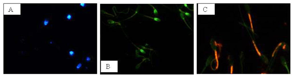
Figure 1. Photomicrography of equine spermatozoa. A Spermatozoa stained with H258 (Hoechst 33258), B Spermatozoa stained with CTC (chlortetracycline), C Spermatozoa stained with JC-1 (5,5’,6,6’-tetrachloro-1,1’,3,3’ tetraethylbenzimidazolyl carbocyanine iodide).

NIS-Elements F 3.0 (Nikon Instrument) software was employed to evaluate simultaneously all fluorescent probes. The results were used to classify spermatozoa in eight classes (Table 1), according to the morpho-functional state of plasmatic and acrosomal membranes and to mitochondrial function (Figure 2).

Table 1. Classification of equine sperm cells according to fluorescence emitted by H258, CTC and JC-1 probes.

Sperm Cell Category

aH258

bCTC

cJC-1

Intact plasma membrane, intact acrosome and high mitochondrial potential

Intact plasma membrane, intact acrosome and low mitochondrial potential

Intact plasma membrane, damage acrosome and high mitochondrial potential

Intact plasma membrane, damage acrosome and low mitochondrial potential

Damage plasma membrane, intact acrosome and high mitochondrial potential

Damage plasma membrane, intact acrosome and low mitochondrial potential

Damage plasma membrane, damage acrosome and high mitochondrial potential

Damage plasma membrane, damage acrosome and low mitochondrial potential

+

+

+

+

+

+

+

+

 +

+

+

+

 aH258 positive (+) = blue stained nucleus. bCTC positive (+) = green acrosome region. cJC-1 positive (+) = red in mid-piece region; negative (-) = green in mid-piece region.

IJVB 2018-105 - Maria Italy_F2

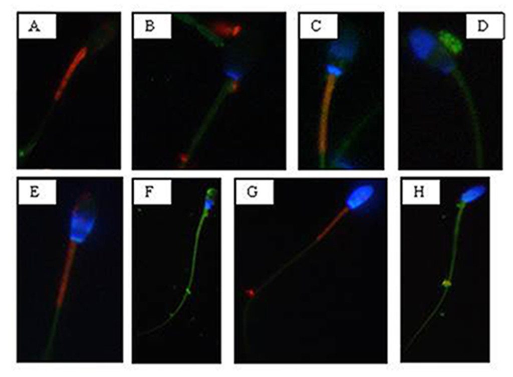
Figure 2. Photomicrography of equine spermatozoa stained with H258,CTC and JC-1. A) Intact plasma membrane, intact acrosome and high mitochondrial potential, B) Intact plasma membrane, intact acrosome and low mitochondrial potential, C) Intact plasma membrane, damaged acrosome and high mitochondrial potential, D) Intact plasma membrane, damaged acrosome and low mitochondrial potential, E) Damaged plasma membrane, intact acrosome and high mitochondrial potential, F) Damaged plasma membrane, intact acrosome and low mitochondrial potential, G) Damaged plasma membrane, damaged acrosome and high mitochondrial potential, H) Damaged plasma membrane, damaged acrosome and
low mitochondrial potential.

The analysis revealed that both cooled and frozen-thawed samples had similar amount of spermatozoa with intact or damaged plasma membrane, also the percentage of cells with intact and damaged acrosomal membrane was not statistically different; on the contrary a dramatic decrease in the percentage of spermatozoa with high mitochondrial potential was observed in frozen samples (Table 2).

Table 2. Recovery rate:  cooled versus frozen semen

Sperm parameters

cooled semen

Frozen semen

Intact plasma membrane (%)

Damage plasma membrane (%)

64

36

62

38

Intact acrosome (%)

Damage acrosome (%)

55

45

50

50

High mitochondrial potential (%)

Low mitochondrial potential (%)

47

53

24

76

Recovery rate (%) of spermatozoa showing different morphology of the acrosome and plasma membranes and mitochondrial potential in cooled and frozen semen.

Compared to cooled semen, in cryopreserved samples we recorded a reduction in the number of spermatozoa with intact plasma membrane, intact acrosome and high mitochondrial potential (6%); spermatozoa with intact plasma membrane, damaged acrosome and high mitochondrial potential were also lower (16%), as well as those with damaged plasma membrane, intact acrosome and high mitochondrial potential (1%). Moreover, also the number of spermatozoa with the damaged plasma membrane, intact acrosome and low mitochondrial potential were slightly reduced in frozen samples. While differences described above were not statistically significant, the reduction, caused by cryopreservation, in the class of intact plasma membrane, damaged acrosome and high mitochondrial potential spermatozoa was statistically significant (P< 0.001) as well as in the class of damaged plasma membrane, intact acrosome and low mitochondrial potential spermatozoa (P< 0.05) (Table 3).

Table 3. Cooled versus frozen semen.

Sperm Cell Category

Cooled semen

Frozen semen

Intact plasma membrane, intact acrosome and high mitochondrial potential

21%

15%

Intact plasma membrane, intact acrosome and low mitochondrial potential

9%

18%

Intact plasma membrane, damage acrosome and high mitochondrial potential

21%

5%

Intact plasma membrane, damage acrosome and low mitochondrial potential

13%

26%

Damage plasma membrane, intact acrosome and high mitochondrial potential

3%

2%

Damage plasma membrane, intact acrosome and low mitochondrial potential

22%

14%

Damage plasma membrane, damage acrosome and high mitochondrial potential

2%

2%

Damage plasma membrane, damage acrosome and low mitochondrial potential

9%

18%

Recovery rate of spermatozoa (%) for each of the eight morphological cell classes.

Cryopreservation led to an increase of spermatozoa with intact plasma membrane, intact acrosome and low mitochondrial potential spermatozoa (9%), of intact plasma membrane, damaged acrosome and low mitochondrial potential spermatozoa (13%), of damaged plasma membrane, damaged acrosome and low mitochondrial potential spermatozoa (9%), compared to refrigeration. These increases due to cryopreservation were statistically significant for intact plasma membrane, intact acrosome and low mitochondrial potential spermatozoa (P<0.05), for intact plasma membrane, damaged acrosome and low mitochondrial potential spermatozoa (P< 0.05) and for damaged plasma membrane, damaged acrosome and low mitochondrial potential spermatozoa (P< 0.05).

Sperm motility

Motility analysis was also conducted on each ejaculate of fresh semen; the recorded mean values of total and progressive motility were 79.06% ± 3.8 and 63.87% ± 2.16, respectively.

Sperm motility values of cooled and frozen-thawed semen are shown in Table 4. Semen cryopreservation affected sperm motility. Refrigerated semen displayed higher total and progressive motility than frozen-thawed semen (P< 0.001). The estimated reduction in the percentage of total and progressive motile spermatozoa related to cryopreservation was 20.33% and 19.57%, respectively.

Table 4. Motility parameters:  cooled versus frozen semen.

Motility parameters

cooled semen

frozen semen

Total motility (%)

Progressive motility (%)

VAP (mm/s)

VSL (mm/s)

VCL (mm/s)

ALH (mm)

BCF (Hz)

STR (%)

LIN (%)

Rapid spermatozoa (%)

Medium spermatozoa (%)

Slow spermatozoa (%)

Static spermatozoa (%)

74,42 ± 3,69 **

59,37 ± 1,76 **

108,71 ± 17,10 **

58,31 ± 13,04

224,44 ± 30,70 **

8,24 ± 0,94 **

34,61 ± 5,18

53,75 ± 10,66

26,86 ± 6,26

45,61 ± 11,05 **

7,36 ± 3,43

7,16 ± 3,32

39,88 ± 11,69

54,09 ± 3,43

39,8 ± 3,09

73,48 ± 15,37

57,8 ± 12,87

149,83 ± 21,24

6,21 ± 0,65

39,28 ± 3,24

71,91 ± 5,71

38,96 ± 4,41

16,79 ± 10,78

8,41 ± 5,95

13,99 ± 7,47 *

61,22 ± 20,84 *

Motility parameters of equine cooled or frozen sperm samples:  VAP (average path velocity); VSL (straight line velocity); VCL (curvilinear velocity); ALH (amplitude of lateral head displacement); BCF (beat cross frequency); STR (straightness); LIN (linearity). * P < 0.05, ** P < 0.001.

Evaluated VAP and VCL were lower in frozen-thawed semen than in refrigerated one (P< 0.001).

The VSL of refrigerated spermatozoa did not differ significantly from that of cryopreserved sperm.

The ALH was higher in cooled than in frozen-thawed semen (P<0.001). The BCF of cooled spermatozoa did not differ significantly from that of frozen sperm cells.

Cooled semen displayed a higher percentage of rapid spermatozoa (P<0.001) while a 28.82% reduction was induced by cryopreservation. The percentage of medium spermatozoa was similar in cooled and frozen semen. Slow and static spermatozoa were higher in frozen-thawed than in cooled semen (P<0.05) the percentage increase, probably due to the cryopreservation process, was 6.83% and 21.34%, respectively.

Correlations

Significant correlations were observed between mitochondrial energy level and different sperm membrane conditions. In cooled semen a high correlation between high mitochondrial potential and intact plasma membrane (r=0.91), high mitochondrial potential and intact acrosome (r=0.93), low mitochondrial potential and damage acrosome (r=0.90), were found. In frozen semen, a high correlation between low mitochondrial potential and damaged plasma membrane (r=0.87), low mitochondrial potential and damaged acrosome (r=0.92) were found. On the contrary, no correlations between mitochondrial membrane potential and different motility characteristics were evidenced.

Discussion

Severe changes in temperature are a common feature of semen preservation protocols but are not a biological phenomenon to which the sperm cell is adapted [25]. Temperature transitions associated with semen fast chilling or freezing are in fact well known for their production of deleterious effects on sperm survival and consequently, lower conception rates following artificial insemination [5].

The reduction in fertilizing ability has typically been attributed to a reduced rate of sperm motility and to the induction of morphological abnormalities [5]. Damages to the plasma membrane, to the acrosomal membrane and to the mitochondrial function of spermatozoa undergoing cooling and/or freezing depend on changes of temperatures and osmolarity, which cause morphological alterations in the organization and composition of proteins and lipids of the sperm surface [26].

Sperm quality related to functional characteristics has been investigated by the association of different fluorescent probes in ram [27], bull [28] and stallion [13, 16, 18].

In this study we validated the association of three fluorescent probes H258, CTC and JC-1 to evaluate the state of plasma and acrosomal membranes, as well as the mitochondrial function both in cooled and frozen equine sperm, using the inverted fluorescent microscopy highlighting the prominent role of the mitochondrion as marker for equine sperm quality.

We found both in cooled and frozen equine semen samples a higher number of spermatozoa with intact plasma membranes compared to those with destroyed membranes, this result is in line with the observations reported by Vasconcelos et al., [29] and by De Leeuw et al., [30].

It is reasonable to attribute the poor fertility of frozen semen to ultrastructural modifications of the cell membranes and to phase transitions occurring during cooling and rewarming. It seems reasonable that the reorganization of sperm membrane bilayer alters the interactions among lipids and between lipids and proteins required for normal membrane functions, determining a re-modelling of the membrane components [31, 32] and the decreased amount of polyunsaturated fatty acids and cholesterol [33]; [34]. Plasma membrane disruption due to cooling or freezing determines the loss of cations and enzymes from the sperm cells, providing some explanation for the reduction in sperm motility and metabolic activity observed in cold-shock spermatozoa [35]. Cold shock also destroys the selective permeability of sperm membranes to calcium, thus leading to its excessive intracellular accumulation, and consequently, to a reduction in motility and cell necrosis. These processes result in an advanced stage of sperm maturation characterized by capacitation, hyperactivation and acrosome reaction [35].

For what concerns the acrosome membrane, in the present study cooled spermatozoa displaying their integrity, were more numerous than those with a damaged acrosome membrane. As to mitochondrial membrane potential, it is a sensitive indicator of the functional status of the organelle; in the present study, cooled spermatozoa with intact plasma membrane showed a high mitochondrial potential. Whereas a high percentage of spermatozoa with an intact plasma membrane and low mitochondrial potential was observed among cryopreserved ones, suggesting that cryopreservation may result in a significant loss in mitochondrial potential.

Cryopreservation amplified the mitochondrial damage compared to cooling, probably because of the block in ATP synthesis and consequently in the reduction in the activity of ATP-dependent pumps [36], a tendency to make the plasma membrane unstable. This may lead to an increase in intracellular Na+ and a reduction in intracellular K+, with reduced Ca2+ efflux due to inhibition of the Ca2+ ATP-ase pumps, increased leakage of Ca2+ across the membrane due to the instability caused by removal of cholesterol, and/or increased Ca2+ influx due to the activation of unidentified channels [37]. Changes associated with this process include an increase of capacitated and acrosome-reacted spermatozoa, as reported by Felix [37]. These results are in agreement with those reported by Albrizio et al., [38]; they showed that cryopreservation increases the percentage of capacitated and acrosome reacted spermatozoa, increase the intracellular Ca2+ concentration  and leads to a higher functional response of L-type Ca2+-channels to both the agonist (Bay K-8644) and antagonist (Nifedipine). Moreover, cryopreservation was found to determine the activation of lytic enzymes and to increase mitochondrial permeability, leading to cell death [10]. This is in accordance with the results of the present study, in which the percentage of spermatozoa with damaged acrosome membrane and low mitochondrial potential is higher in frozen than in cooled ones.

It has been suggested that plasma membranes and mitochondria in bovine sperm are both affected by cryopreservation [39]. In accordance with the above-mentioned study, we found that plasma membrane and mitochondrial membrane were equally vulnerable to the freezing and thawing process, because of their high thermolability [40]. Moreover, in this study, plasma membrane and mitochondrial membrane conditions are highly correlated demonstrating that these structures depend on each other, as reported by Celeghini et al., [7]. The mitochondrial plasma membrane is a good predictor to determine and assess plasma membrane and acrosome membrane damages because of thermic treatment. Besides structural sperm characteristics, conflicting opinions concerning the relationship between motility and fertility in the stallion exist [41, 42] although sperm cell motility and the quality of motility are still the most reliable indicators of sperm viability. In this study, total motility recorded in cooled semen was in agreement with similar data reported by Love et al.,[13] and Heckenbichler et al., [43], whereas, the same parameter found in frozen semen was similar to that found by Wrench et al., [44] and Salazar et al., [45]. The progressive motility found in cooled semen was similar to that reported by Cocchia et al., [46] and Heckenbicher et al., [43], whereas the same parameter found in frozen semen was similar to that found by Wrench et al., [44]. It is well known the functional relationship between the mitochondrion and the cellular apparatus that drives sperm movements; in fact mitochondria are well known for producing energy in the form of ATP and sperm motility depends upon the energy produced by the oxidative phosphorylation. Mitochondria are a major source of Reactive Oxygen Species (ROS) and appear to be the cellular structures most susceptible to damages during cooling and cryopreservation [47], leading to a loss of motility in frozen semen.

As to VCL, VAP, VSL and ALH parameters, the results found in cooled semen were in agreement with those of Aurich et al., [48], whereas those found in frozen semen were similar to the results of Salazar et al., [45].

This study demonstrates that the quality of motility is different in cooled and frozen semen, in fact, cooled samples showed higher VAP and VCL values than frozen ones. These results suggest that motile cells, that are numerically higher in cooled than in frozen semen, are also characterized by higher speed as deduced by their higher VAP and VCL, on the contrary, linear characteristic of sperm motility is not affected by temperature reduction. We found that ALH values were higher in refrigerated semen than in the cryopreserved one. In general, higher ALH values are unwanted, because they can interfere with cell progression, as demonstrated by [18], which stand for reduced sperm quality. However until now a cutoff value for ALH has not been defined so that is not clear the influence of this parameter on the progressive motility and on the ability of the spermatozoon to go forward into the female reproductive tract [7].

As to motility characteristics, other authors found opposite results for refrigerated and cryopreserved semen [42]; [49]; [50]. This could be only an apparent discordance considering that many factors may have a role such as season of semen recovery, physiological differences among stallions, frequency in collecting semen, different extenders for semen dilutions, different settings of the CASA system (threshold setting, specimen concentration, video digitalization rate) [9, 17, 51-53].

We, as several authors working on other animal species, didn’t find a direct correlation between mitochondrial function and a specific kinetic parameter, probably because equine sperm motility is modulated by different factors not only by mitochondria [14, 23].

In conclusion, our study, reporting the simultaneous evaluation of different functional characteristics of equine spermatozoa subjected to temperature changes, increases the information on semen quality helping the selection of those characteristics more correlated with semen fertility potential. In particular, we demonstrated that mitochondria are good indicators of sperm survival and can be used to discriminate between good and poor semen quality. Moreover, they are strictly correlated to the state of cellular membrane, therefore, a proper evaluation of mitochondria sounds necessary when analyzing sperm samples. Standing on the present study, mitochondrial membrane potential provides useful information on equine sperm quality so that it may be exploitable as a marker of sperm fertility; moreover, the high mitochondrial sensitivity to temperature variations observed in this study highlights the importance of developing new strategies to protect the functionality of this organelle to preserve sperm cell physiology.

Disclosure

None of the authors of this paper has a financial or personal relationship with other people or organizations that could inappropriately influence or bias the content of the paper.

This paper was concepted by GM Lacalandra, G. Mari and A. Zarrilli; written by M. Albrizio, G. Mari, GM Lacalandra and A. Zarrilli. Data were acquired by AC Guaricci, A. Moramarco and E. Micera; B. Mislei, and G. Rizzato analysed and interpreted the data. Albrizio revised the manuscript and cared the journal submission.

All authors approved the final article.

Acknowledgements: This work was supported by Italian University funds

References

  1. Allen WR (2005) The development and application of the modern reproductive technologies to horse breeding. Reprod Domest Anim 40: 310–29.
  2. Macías García B, González Fernández L, Ortega Ferrusola C, Morillo Rodríguez A, Gallardo Bolaños JM, et al. (2011) acids and plasmalogens of the phospholipids of the sperm membranes and their relation with the post-thaw quality of stallion spermatozoa. Theriogenology 75: 811–8.
  3. Aurich C (2005) Factors affecting the plasma membrane function of cooled-stored stallion spermatozoa. Anim Reprod Sci 89: 65–75.
  4. Leahy T(2011) Gadella BM. Sperm surface changes and physiological consequences induced by sperm handling and storage. Reproduction 142: 759–78.
  5. Holt WV (2000) Basic aspects of frozen storage of semen. Anim Reprod Sci 62:  3–22. [crossref]
  6. Anzar M, He L, Buhr MM, Kroetsch G, Pauls KP (2002) Sperm apoptosis in fresh and cryopreserved bull semen detected by flow cytometry and its relationship with fertility. Biol Reprod 66: 354–60.
  7. Celeghini ECC, de Andrade AFC, Raphael CF, Nascimento J, Ticianelli JS, et al. (2010) Damage assessment of the equine sperm membranes by fluorimetric tecnique. Braz Arch Biol Techn 53: 1285–92.
  8. Colenbrander B, Gadella BM, Stout TAE (2003) The predictive value of semen analysis in the evaluation of stallion fertility. Reprod Dom Anim 38: 305–11.
  9. Giannoccaro A, Lacalandra GM, Filannino A, Pizzi F, Nicassio M, et al. (2010) Assessment of viability, chromatin structure stability, mitochondrial function and motility of stallion fresh sperm by using objective methodologies. J Cell Anim Biol 4: 34–41.
  10. Neild DM, Gadella BM, Chaves MG, Miragaya MH, Colenbrander B, Aguero A (2003) Membrane changes during different stages of a freeze-thaw protocol for equine semen cryopreservation. Theriogenology 59: 1693–705.
  11. Rodriguez-Martines H (2003) Laboratory semen assessment and prediction of fertility:  still utopia? Reprod Dom Anim 38: 312–8.
  12. Gillan L, Evans G, Maxwell WMC (2005)  Flow cytometric evaluation of sperm parameters in relation to fertility potential period. Theriogenology 63: 445–57.
  13. Love CC, Thompson JA, Brinsko SP, Rigby SL, Blanchard TL, Lowry VK Varner DD (2003) Relationship between stallion sperm motility and viability as detected by two fluorescence staining techniques using flow cytometry. Theriogenology 60: 1127–38.
  14. Martinez-Pastor F, Johannisson A, Gil J, Kaabi M, Anel L, Paz P, Rodriguez-Martinez H (2004) Use of chromatin stability assay, mitochondrial stain JC-1, and fluorometric assessment of plasma membrane to evaluate a frozen-thawed ram semen. Anim Reprod Sci 84121–33.
  15. Flesch FM, Gadella BM (2000) Dynamics of the mammalian sperm plasma membrane in the process of fertilization. Biochim Biophys Acta 1469: 197–235.
  16. Gravance CG, Garner DL, Baumber J, Ball BA (2000) Assessment of equine sperm mitochondrial function using JC-1. Theriogenology 53: 1691–703.
  17. Ball BA, Medina V, Gravance CG, Baumber J (2001) Effect of antioxidants on preservation of motility, viability and acrosomal integrity of equine spermatozoa during storage at 5 °C. Theriogenology 56: 577–89.
  18. Arruda RP, Ball BA, Gravance CG, Liu IKM (2003) Flow cytometric membrane and acrosomal integrity of the stallion spermatozoa. Acta Sci Vet 31: 226–7.
  19. Tartaglione CM, Ritta MN (2004) Prognostic value of spermatological parameters as predictors of in vitro fertility of frozen-thawed bull semen. Theriogenology 62: 1245–52.
  20. Rathi R, Colenbrander B, Bevers MM, Gadella BM (2001) Evaluation of in vitro capacitation of stallion spermatozoa. Biol Reprod 65: 462–70.
  21. Kenney RM, Bergman RV, Cooper WL, Morse GW (1975) Minimal contamination techniques for breeding mares:  techniques and preliminary findings. Proc Am Ass Equine Pract 21: 327–36.
  22. Mari G, Rizzato G, Merlo B, Iacono E, Seren E, Tamanini C, et al. (2008) uality and fertilizing ability in vivo of sex-sorted stallion spermatozoa. Reprod Dom Anim 45: 331–5.
  23. Albrizio M, Guaricci AC, Maritato F, Sciorsci RL, Mari G, et al. (2005) Expression and subcellular localization of the mu-opioid receptor in equine spermatozoa:  evidence for its functional role. Reproduction 129: 39–49.
  24. Neild DM, Gadella BM, Aguero A, Stout TAE, Colenbrander B (2005) Capacitation, acrosome function and chromatin structure in stallion sperm. Anim Reprod Sci 89: 47–56.
  25. Lopez-Fernandez C, Crespo F, Arroyo F, Fernandez JL, Arana P, et al. (2007) Dynamics of sperm DNA fragmentation in domestic animals II. The stallion. Theriogenology 68: 1240–50.
  26. Amann RP, Pickett BW (1987) Principles of cryopreservation and a review of cryopreservation of stallion spermatozoa. J Equine Vet Sci 7: 145–73.
  27. Windsor DP (1997) Mitochondrial function and ram sperm fertility. Reprod Fertil Dev 9: 279–84.
  28. Thomas CA, Garner DL, Dejarnette JM, Marshall CE (1997) Fluorometric assessments of acrosomal integrity and viability in cryopreserved bovine spermatozoa. Biol Reprod 56: 991–8.
  29. Vasconcelos AB, Santana MA, Santos AMC, Santoro MM, Lagares MA (2011) Metabolic evaluation of cooled equine spermatozoa. Andrologia 42: 106–11.
  30. Heise A, Thompson PN, Gerber D (2011) Influence of seminal plasma on fresh and post-thaw parameters of stallion epididymal spermatozoa. Anim Reprod Sci 123:  192–201. [crossref]
  31. De Leeuw FE, Chen H-C, Colenbrander B, Verkleij AJ (1990) Cold-induced ultrastructural changes in bull and boar sperm plasma membranes. Cryobiology 27: 171–83.
  32. Drobnis EZ, Crowe LM, Berger T, Anchordoguy TJ, Overstreet JW, Crowe JH (1993) Cold shock damage is due to lipid phase transitions in cell membranes:  a demonstration using sperm as a model. J Exp Zool 265: 432–7.
  33. Maldjian A, Pizzi F, Gliozzi T, Cerolini S, Penny P, et al. (2005) Changes in sperm quality and lipid composition during cryopreservation of boar semen. Theriogenology 63:  411–421. [crossref]
  34. Chakrabarty J, Banerjee D, Pal D, De J, Ghosh A Majumder GC (2007) Shedding off specific lipid constituents from sperm cell membrane during cryopreservation. Cryobiology 54: 27–35.
  35. Bailey JL, Bilodeau JF, Cormier N (2000) Semen cryopreservation in domestic animals:  a damaging and capacitating phenomenon. J Androl 21:  1–7. [crossref]
  36.  Silva PF, Gadella BM (2006) Detection of damage in mammalian sperm cells. Theriogenology 65:  958–978. [crossref]
  37. Felix R (2005) Molecular physiology and pathology of Ca2+-conducting channels in the plasma membrane of mammalian sperm. Reproduction 129: 251–62.
  38. Albrizio M, Lacalandra GM, Micera E, Moramarco AM, Surdo NC, et al. (2010) Effects of cryopreservation on capacitation, acrosome reaction and activity of L-type voltage-dependent calcium channels of equine sperm cells. Proceedings of the Tenth International Symposium on Equine Reproduction, Lexington; KY, USA. Anim Reprod Sci 121: 218–19.
  39. Bollwein H, Fuchs I, Koess C (2008) Interrelationship between plasma membrane integrity, mitochondrial membrane potential and DNA fragmentation in cryopreserved bovine spermatozoa. Reprod Dom Anim 43: 189–95.
  40. Breininger E, Beorlegui NB, O’Flaherty CM, Beconi MT (2005) Alpha-tocopherol improves biochemical and dynamic parameters in cryopreserved boar semen. Theriogenology 63: 2126–35.
  41. Jasko DJ, Hataway JA, Schaltenbrand VL, Simper WB, Squires EL (1992) Effect of seminal plasma and egg yolk on motion characteristics of cooled stallion spermatozoa. Theriogenology 37: 1241–52.
  42. Kuisma P, Andersson M, Koskinen E, Katila T (2006) Fertility of frozen-thawed stallion semen cannot be predicted by the currently used laboratory methods. Acta Vet Scand 48: 14–22.
  43. Heckenbichler S, Deichsel K, Peters P, Aurich C (2011)  Quality and fertility of cooled-shipped stallion semen at the time of insemination. Theriogenology 75: 849–56.
  44. Wrench N, Pinto CR, Klinefelter GR, Dix DJ, Flowers WL, et al. (2010) Effect of season on fresh and cryopreserved stallion semen. Anim Reprod Sci 119:  219–227. [crossref]
  45.  Salazar JL Jr, Teague SR, Love CC, Brinsko SP, Blanchard TL, et al. (2011) Effect of cryopreservation protocol on postthaw characteristics of stallion sperm. Theriogenology 76:  409–418. [crossref]
  46. Cocchia N, Pasolini MP, Mancini R, Petrazzuolo O, Cristofaro I, et al. (2011) Effect of sod (superoxide dismutase) protein supplementation in semen extenders on motility, viability, acrosome status and ERK (extracellular signal-regulated kinase) protein phosphorylation of chilled stallion spermatozoa. Theriogenology 75: 1201–10.
  47. Ortega-Ferrusola C, Garcia BM, Gallardo-Bolanos JM, Gonzalez-Fernandez L, Rodriguez-Martinez H, et al. (2009) Apoptotic markers can be used to forecast the freezeability of stallion spermatozoa. Anim Reprod Sci 114: 393–403.
  48. Aurich C, Seeber P, Muller-Schlosser F (2007) Comparison of Different Extenders with Defined Protein Composition for Storage of Stallion Spermatozoa at 5°C. Reprod Dom Anim 42: 445–8.
  49. Rota A, Furzi C, Panzani D, Camillo F. Studies on Motility and Fertility of Cooled Stallion Spermatozoa. Reprod Dom Anim 39: 103–9.
  50. Spizziri BE, Fox MH, Bruemmer JE, Squires EL, Graham,JK (2010) Cholesterol-loaded-cyclodextrins and fertility potential of stallions spermatozoa. Anim Reprod Sci 118: 255–64.
  51. Brinsko SP, Crockett EC, Squires EL (2000) Effect of centrifugation and partial removal of seminal plasma on equine spermatozoal motility after cooling and storage. Theriogenology 54: 129–36.
  52. Sieme H, Katila T, Klug E (2004) Effect of semen collection practices on sperm characteristics before and after storage and on fertility of stallions. Theriogenology 61: 769–84.
  53. Pena FJ, Macías García B, Samper JC, Aparicio IM, Tapia JA, et al. (2011) Dissecting the molecular damage to stallion spermatozoa:  The way to improve current cryopreservation protocols? Theriogenology 76: 1177–86.

Integrin Inhibition in the Tumor Microenvironment – more complex than expected

DOI: 10.31038/CST.2018323

Abstract

Tumor cell migration and invasion are critical steps in the metastatic cascade and depend on the interaction between tumor cells, the extracellular matrix (ECM) and the endothelial cells. Integrins are key receptors that link cells and ECM, acting as mechanical sensors of the cell microenvironment. Particularly, Arg-Gly-Asp (RGD)-binding integrins such as αvβ3 and α5β1 integrins are of special interest in cancer progression. Integrins also interact with growth factor receptors resulting in an important cross talking between intracellular signaling pathways. Studies have provided evidence of distinct roles for αvβ3 and α5β1 integrins during migration. Therefore, αvβ3 and α5β1 integrins became an attractive target for pharmacological inhibition in cancer therapy and metastasis prevention. Cilengitide, the first integrin inhibitor based on the RGD motif, is currently under clinical trial in cancer patients with limited success. Monoclonal antibodies to integrins also presented modest results. Therefore, efforts to achieve a better understanding on the integrin roles in cancer progression are needed. In the last few years, new mechanisms that may help to explain the lack of success of integrin inhibitors were described and are commented here.

Keywords

integrin, cancer, disintegrins

Introduction

Cancer is one of the major concerns related to human health, and metastasis, when occurs, is the main cause of deaths in patients with cancer. Despite all efforts of academic, governmental and private institutions, there are very few options to prevent or treat metastasis. [1] One of the reasons for this lack of success relays on the current limited knowledge on the cellular programs driving the process of metastasis. Recently, new mechanisms that allow prolonged tumor cell survival after loss of attachment to the ECM have been reported, including autophagy and entosis [2]. In addition to shedding some light in the knowledge of tumor progression, these mechanisms provided new targets and options for metastasis treatment. Here we review some key aspects of cell attachment/detachment to ECM by integrins in the context of tumor microenvironment. We will also comment on the results obtained so far with integrin-targeted anticancer therapy.

Tumor Microenvironment and the Integrins

In the past ten years, the microenvironment where tumor cells develop has achieved special importance mostly due to its pivotal role in tumor progression. The tumor microenvironment (TME) provides signals and several kinds of support from distinct cell types and from the extracellular matrix (ECM) [3, 4]. Signals from the TME comprise soluble factors released by stromal cells such as fibroblasts, stem and immune cells, blood vessels, products from proteolysis of ECM components and cytokines [5]. On the other hand, tumor cells release proteases, microvesicles and growth factors that affect and modify the surrounding cells and the ECM. The TME also has a key role in the resistance to therapy due to a continuous communication with tumor cells and modulation of their responses [6, 7]. Integrins are among the crucial cell surface receptors in supporting the cross talk between cells and ECM, and therefore are critically involved in tumor progression [8].

Integrins are transmembrane receptors that support the adhesion of cells to the ECM [8]. Loss of integrin adhesion usually induces cell death unless cells can find new adhesion sites. Integrin are formed by heterodimers containing one α and one β subunits with the ability to recognize ECM components such as collagen (Col), fibronectin (FN) and laminin (LM) with high affinity [9]. There are 18 α subunits and 8 β subunits that can be combined in several ways to form distinct receptors with different specificity to ECM components. For instance, α5β1 integrin is the main receptor for FN whereas vitronectin (VN) binds preferentially to αvβ3 integrin. Both α5β1 and αvβ3 integrins recognize the tripeptide RGD motif within ECM proteins; however other integrins also binds to the RGD motif such as αvβ1, αvβ5, αvβ6, αvβ8, α8β1, and the platelet fibrinogen receptor, αIIbβ3 integrin [10]. One of the most interesting feature of integrins is the fact that, despite binding the same ligand, each one has its own cell-dependent pattern of expression and plays distinct roles in cell adhesion and migration [11].

Integrins in cell adhesion and migration

Integrin clustering and activation upon ligand binding triggers intracellular signaling pathways including the activation of several kinases like the focal adhesion kinase (FAK), mitogen activated protein kinase (MAPK) and extracellular signal-regulated kinase (ERK) resulting in changes in cell behavior [8, 12]. Cell adhesion and migration are among the main cell abilities that are under strict integrin control and are crucial steps during tumor progression. Moving cells may change their integrin profile in response to modifications on the ECM milieu. In a FN-rich microenvironment, cells will depend mostly on integrins such as α5β1 and αvβ3 for motility and, despite their ability in binding to the same ligand, these two integrins have distinct and specific roles in cell migration [13].

Adhesion to fibronectin by αvβ3 integrin supports extensive actin cytoskeletal reorganization resulting in a single large lamellipod with static cell–matrix adhesions at the leading edge [14]. On the other hand, cell adhesion by α5β1 generates thin protrusions containing highly dynamic cell–matrix adhesions in multiple directions [14]. Therefore, these authors concluded that β1 integrins support random migration, whereas β3 integrins are related to persistent migration. In agreement with these data, we have demonstrated that blockage of αvβ3 integrin by a DisBa-01, a recombinant RGD-disintegrin from snake venom, resulted in loss of directionality and decrease of speed migration [15]. Despite having the RGD adhesive motif, which is recognized by both αvβ3 and α5β1 integrins, the dissociation constant of DisBa-01 for the αvβ3 integrin is 100 times higher than for α5β1 integrin [15]. These results confirm the role of αvβ3 integrin in defining directionality of the cell movement.

Rosa-Cusachs et al., 2009 also provided evidence of distinct roles for α5β1 and αvβ3 integrin during cell adhesion/migration processes, with the demonstration that α5β1 integrin is responsible for supporting high adhesion forces while αvβ3 integrin starts talin-dependent mechanotransduction. However, these effects depend on the substrate where cells attach. Fibroblasts exhibit persistence migration on FN-coated surfaces [16]. Ligand-specific activation of αvβ3 e α5β1 integrins also confirmed the distinct roles of each integrin in cell adhesion. Activation of αvβ3 integrins led to stabilization of peripheral focal adhesions while fibrillary structures were observed when α5β1 integrins are activated [17]. Moreover, αv-class of integrins such as αvβ3 were demonstrated to outcompete α5β1 integrins in FN binding; however, after engagement, αv-class integrins cooperate with α5β1 integrins to form additional adhesion sites consequently strengthening cell adhesion to FN [18].

Although they may bind the same ligand, β1 and β3 integrins have distinct and cooperative roles in mechanotransduction. By means of traction force microscopy, Millioud and colleagues demonstrated that deleting β3 subunit increases traction forces, whereas the deletion of β1 subunit results in a strong decrease of contractile forces [19].

Integrins and Growth Factor Receptors

The cross talk between integrin and growth factor receptor (GFR) signaling pathways is well documented in the literature in both normal cells and tumor cells. Endothelial cells seem to be highly sensitive to integrin activation of GFRs including the vascular endothelial growth factor receptor (VEGFR), epidermal growth factor receptor (EGFR), and the platelet-derived growth factor receptor (PDGFR). This cooperative process is crucial in physiological and tumor angiogenesis and it is considered one key factor in tumor progression. The α5β1 integrin was demonstrated to induce tumor cell proliferation by two pro-survival mechanisms including direct activation the EGFR signaling cascade and by signaling via continuous integrin-dependent activation of protein kinase B (PKB, also known as AKT) [20]. Moreover, EGFR overexpression led to inactivation of α5β1 integrin in A431 squamous carcinoma cells, which would be interesting in order to prevent cell interaction with the ECM and therefore to avoid tumor cell proliferation. However, treatment of cells with EGFR kinase inhibitor resulted in reactivation of the integrin [21]. Since GFRs are usually overexpressed in many types of cancer cells, pro-survival signaling pathways from both GFR and integrins would be more efficiently impaired upon association of anti-integrin/anti-GFR treatments than either treatment alone [22].

VEGF binding to its receptors and co-receptors induce receptor homodimerization and heterodimerization, followed by activation of tyrosine kinases and signaling cascades, with the association of a set of adaptor proteins. The activation of these signaling pathways results in activation, migration and proliferation of endothelial cells, crucial steps for neoangiogenesis. In addition, mechanical forces such as shear stress may activate VEGRF2 similarly to ligand binding, leading to the activation of intracellular signaling pathways [23]. Integrins are proposed to act as co-receptors upon VEGF binding to VEGFRs, similarly to neuropilins and heparin sulfate proteoglycans in endothelial cells [24]. However, integrins seem to participate actively in the control of VEGFR signaling. Blocking αvβ3 integrin by a RGD-antagonist downregulated the expression of VEGF, VEGFR1 and VEGFR-2 in endothelial cells but not in MDA-MB-231 breast tumor cells [25]. Contrastingly, we observed an increase in VEGF protein levels by human fibroblasts after treatment with the αvβ3 integrin antagonist. In addition, we have also demonstrated that this RGD-antagonist inhibits endothelial in vitro cell migration and in vivo angiogenesis in mice [26, 27]. These results suggest that the αvβ3 integrin is not only a co-receptor; instead, it is an active partner in the control of VEGF signaling in endothelial cells.

A key role for α1β1 and α2β1 integrins in the process of angiogenesis triggered by vascular endothelial growth factor (VEGF) was previously reported. Antibody antagonism of either integrin resulted in potent inhibition of VEGF-driven angiogenesis in mouse skin, reduced tumor growth and angiogenesis of human squamous cell carcinoma xenografts. [28]

Integrins and MMPs

Matrix metalloproteases (MMP) comprise a class of zinc-dependent enzymes responsible for ECM remodeling and degradation. MMPs are also involved in the processing of growth factors, cytokines and surface transmembrane proteins. MMPs are produced as inactive zymogens that can be activated by proteolytic removal of the pro-peptide domain by furin, or by autoproteolysis. To date, more than 24 MMPs are known, including secreted or membrane anchored forms, (membrane-type MMPs, MT-MMPs) that play crucial roles in the process of MMP activation. For instance, activation of pro-MMP-2 at the cell surface involves the formation of a trimolecular complex with membrane type-1 metalloprotease (MT-1-MMP) and tissue inhibitor of metalloproteases-2 (TIMP-2). MMP-2 and MMP-9 are also known as gelatinases A and B, respectively, due to the ability to digest degraded forms of collagen. Gelatinases are characterized by the presence of a fibronectin-like domain that drives the enzyme to its ECM substrates. MMP-2 and MMP-9 are of particular interest in cell migration and consequently, in metastasis development. MMP-2 is constitutively produced by most cells; however, MMP-9 is expressed by only a few types of cells including neutrophils, macrophages and tumor cells, being induced in several pathological conditions such as tumor invasion [29, 30].

MMPs play key roles in tumor progression such as degrading ECM to allow migration of tumor cells to distant secondary sites, allowing endothelial cell migration and proliferation to produce tumor angiogenesis, and releasing growth factors from the ECM to provide constant survival and proliferation signals.

The role of integrins in controlling MMP activity is less studied. Previous studies demonstrated that interaction of fibronectin with α4β1 integrin upregulates MMP-9 expression by infiltrating leukocytes in damaged livers and the blockade of this interaction disrupted leukocyte migration [30]. MMP-2 activation upregulates VEGF-A expression in melanoma cells via an αvβ5 integrin/phosphoinositide-3-kinase-dependent pathway [31]. The αvβ3 integrin has been closely related to tumor progression and reduced patient survival rates in melanoma, colon carcinoma and breast cancer, increasing migration and invasion of tumor cells [32, 33]. Blocking αvβ3 integrin inhibited MT-1-MMP-dependent activation of MMP-2 induced by collagen I; however, cells expressing high levels of β3 integrin subunit have increased abilities of adhesion and migration [34]. Blocking αvβ3 integrin in endothelial cells by a RGD-based antagonist completely abolished MMP-2 activity; in contrast, the same treatment increased almost twice the MMP-9 levels in the conditioned media from MDA-MB-231 breast tumor cells [25]. These results demonstrated that one single specific integrin inhibitor might induce different cell-dependent effects.

Integrins and tumor progression

The correlation of expression levels of αvβ3, αvβ5, α5β1, α6β4, α4β1, αvβ6 and αvβ8 integrins with metastasis and poor patient prognosis is well documented (reviewed by Nieberler et al., 2017). [35] One of the most studied integrin in tumor progression is the α5β1 integrin. Higher levels of α5 subunit were found than in normal tissue in patients with advanced renal cell carcinoma and correlated with tumor grade, metastasis development and reduced patient survival [36]. Expression of αvβ6 is significantly associated with the progression of breast ductal carcinoma to an invasive form by a mechanism involving upregulation of MMP-9 and transforming growth factor-β (TGF-β) [37].

Previous reports associated β1 and β3 integrins with TGF-β stimulation of epithelial–mesenchymal transition (EMT) and breast tumor metastasis by means of a compensatory mechanism [38]. Inactivation of β1 integrin impairs the TGF-β effect in promoting tumor cell migration; however, a strong compensatory upregulation of β3 integrin restores the induction of the EMT phenotypes by TGF-β, indicating that the two integrins must be targeted to prevent tumor progression [38].

Integrin-targeted therapy

Cilengitide was one of the first antiangiogenic drugs directed to blockade of cell adhesion to the ECM by antagonizing αvβ3 and α5β1 integrins (IC50 for inhibition of cell adhesion of 0.2 and 11 nM, respectively). In phase I studies, patients with advanced solid tumors were treated with cilengitide without consistent results [39]. In another study, cilengitide was tested in association with cediranib, an inhibitor of VEGFR-associated tyrosine kinase. The association of cilengitide with the two drugs was well tolerated, however there were no changes in the survival rates [40, 41]. Cilengitide combined with temozolomide, an oral alkylating agent, did not increase the survival rates of glioblastoma patients [42]. Among antiangiogenic drugs, only the anti-VEGF-A bevacizumab increased disease-free survival time in patients with glioblastoma [43]. Recently, 12 patients with solid tumors such as breast cancer were treated with cilengitide, with partial positive response to the treatment and 05 had stable disease as the best response [44]. In summary, after 10 years of clinical assays with cilengitide for different tumors, results are still disappointing. Reasons for the lack of success may be related to the determination of the effective doses and time of administration. [45- 47] Other reason for lack of success may be the dose-dependent opposing effects of cilengitide related to tumor angiogenesis, with low doses being pro-angiogenic in contrast with anti-angiogenic effect of higher doses [48]. However, a better comprehension on the distinct roles of integrins in the context of complexity of the tumor microenvironment is needed before discarding integrin-targeted therapy.

Volociximab, a monoclonal antibody anti-α5β1 integrin, has been tested in at least 10 phase I and II clinical trials to treat some types of tumors, including advanced non-small cell lung cancer (NSCLC) and metastatic melanoma, among others. Volociximab was used either as single therapy or in combination with classical drugs such as carboplatin and paclitaxel. Preliminary results showed modest but relevant results such as an increase in median progression-free survival of 6.3 months and decreased levels of potential biomarkers of angiogenesis or metastasis after six cycles of treatment [49].

Etaracizumab, an anti-αvβ3 integrin monoclonal antibody, decreased SKOV3ip1 ovarian tumor cell proliferation and invasion in vitro and resulted in about 50% of tumor weight decrease in mice [50]. Combination therapy with paclitaxel gave better results in decreasing tumor weight, and tumors showed reduced levels of p-Akt and p-mTOR; however, microvessel density of resected tumors after therapy were not decreased [50]. The β1 integrin subunit has been associated to therapeutic resistance to trastuzumab (anti-EGFR/HER2 monoclonal antibody) and to lapatinib (an EGFR/HER2 tyrosine kinase inhibitor) of human epidermal growth factor receptor (HER)-2-positive breast tumor cells [51].

One of the most intriguing question in integrin-based anti-cancer therapy is the fact that the strong and positive results on inhibition of tumor progression in pre-clinical assays did not translate to clinical assays. Integrin inhibitors including monoclonal antibodies and synthetic molecules showed disappointed results in patient survival time, disease stabilization and the development of metastasis [42, 48, 52]. One reason for the negative results may rely on the complexity of the mechanism of action of integrins, their ability to compensate each other and inducing an even worse phenotype. Deleting β1 integrin was compensated by β3 integrin, which stimulated metastasis in murine model of breast cancer [38].

Besides the compensatory mechanism, integrin inhibition should induce cell death by anoikis, a kind of apoptotic cell death that occurs due to the loss of cell attachment to the ECM [53]. However, ECM detachment results in antiapoptotic signals, as a defense mechanism against anoikis until cells be able to find a new place to attach again. Meantime, cells undergo autophagy, a cell process mostly triggered by the loss of integrin-mediated adhesion that allow cell survival during some time. However, prolonged detachment will later induce apoptosis by anoikis [54]. One of the most critical finding is that tumor cells usually develop resistance to anoikis due to a sustained autophagic response [54].

Entosis, an even more sophisticated cell survival mechanism, was described [55]. Entosis, also referred to as cell-in cell structures, or cell cannibalism, is triggered by loss of attachment to ECM, similarly to the process of autophagy. Entotic cells have been observed in many types of tumors and may be one of the reasons for the abnormal number of chromosomes found in most tumors [2]. Engulfed cells are alive and may divide inside the host, and in case of tumor cells, such process may occur indefinitely. Such mechanisms of tumor cell survival that happen upon ECM detachment may certainly contribute for the lack of success of integrin inhibitors in clinical trials.

More recently, the mechanism of vessel co-option was described as a mediator of resistance to anti-angiogenic therapy of breast tumor liver and lung metastasis [56]. Vessel co-option is an alternative pathway of tumor cells for obtaining nutrients from blood using the pre-existing vasculature without producing new vessels. Blocking VEGF/VEGFR signaling induces co-option and tumor growth in glioblastoma patients. [57] Since there is a close reciprocal stimulatory relationship between VEGF and αvβ3 integrin [58], one might expect a role for integrin inhibition in supporting the mechanism of co-option. This possibility remains to be elucidated. These results demonstrate the complexity of the TME and its relevance in tumor progression.

New insights on integrin targeting

In spite of being extensively described, integrin inhibition in tumor microenvironment is still challenging and attractive. The failure of targeted therapies so far leads to deeper investigations about signaling pathways, endocytic trafficking and recycling of integrins [59, 60].

As a result, the attention that before was outside cell, turned to intracellular integrin fate affecting the overall cell behavior. Its known that integrin trafficking dictates the nature of Rho GTPase signaling during cytokinesis and cell migration [61]. FN binding promotes both accelerated internalization and ubiquitination of α5β1 receptors. Subsequently, alterations in pH inside endossomal compartments will define either recycling or degradation of integrins [59, 62] When α5 integrin suffers ubiquitination upon FN binding, the ESCRT machinery acts sorting this receptor to either multivesicular bodies, recycling or lysosomal degradation [59]. It was previously demonstrated that mutation on the ubiquitination site in cytoplasmic tail of α5 integrin causes a different sorting of fibronectin on cell, affecting fibroblast migration [63].

The multivesicular bodies are cell compartments containing intraluminal vesicles named exosomes that are secreted to the extracellular space by shedding from the plasma membrane, improving cell communication with the TME [64]. The presence of both integrin and FN inside multivesicular bodies had light to the hypothesis that these receptors might be present in vesicles in the extracellular environment. Sung et al demonstrated that in fact α5 integrin is secreted as an exosome cargo, and more than that, FN was also secreted in these vesicles. Thus, a new and promising science of integrins as a target has emerged from the microvesicles field. Studies on cell communication had proved that integrin transfer can occur through exosomes delivery in both TME and the pre-metastatic niche [65]. In addition, the integrin content of exosomes can change the types of integrin expression in the new tumor focus [66]. The real contribution of integrins on cell communication using cell-derived vesicles is still unclear, as well as the effects of blockage of these receptors. However, this can be the missing clue that can explain the failure of earlier integrin inhibitors in drug development.

Conclusions

Cell attachment to the ECM via integrins is a key factor for tumor progress; however, integrin inhibition may be carefully considered as a target for drug development. Combination of anti-growth factors antibodies, tyrosine kinase inhibitors and integrin inhibitors may be an interesting choice but must be first evaluated in pre-clinical assays considering all the possible escape mechanisms that tumor cells can develop. Most of the studies so far have considered the signaling in a cellular level, however, in complex organisms, several other factors might systemically interfere on integrin inhibition, making the drug development even more challenging. The integrins may have won some battles but not the war.

Acknowledgements: This work was supported by Fundação de Amparo à Pesquisa do Estado de São Paulo (FAPESP, 2013/00798-2 and 2014/18747-8) and by the Conselho Nacional de Desenvolvimento Científico e Tecnológico (CNPq 308096/2013-4), Brazil.

References

  1. Steeg PS (2016) Targeting metastasis. Nat Publ Gr 16.
  2. Janssen A, Medema RH (2011) Entosis: aneuploidy by invasion. Nat Cell Biol 13: 199–201.
  3. Desgrosellier JS, Cheresh DA (2010) Integrins in cancer: biological implications and therapeutic opportunities. Nat Rev Cancer 10: 9–22.
  4. Brücher BLDM, Jamall IS (2014). Cell-Cell Communication in the Tumor Microenvironment, Carcinogenesis, and Anticancer Treatment. Cell Physiol Biochem 34: 213–43.
  5. Korkaya H, Liu S, Wicha MS (2011) Breast cancer stem cells, cytokine networks, and the tumor microenvironment. J Clin Invest 121: 3804–9.
  6. Wu T, Dai Y (2017) Tumor microenvironment and therapeutic response. Cancer Lett 387: 61–8.
  7. Longmate W, DiPersio CM (2017) Beyond adhesion: emerging roles for integrins in control of the tumor microenvironment. F1000Research 6: 1612.
  8. Hynes RO (2002) Integrins: bidirectional, allosteric signaling machines. Cell 110: 673–87.
  9. Hynes RO (1987) Integrins: a family of cell surface receptors. Cell 48: 549–554. [crossref]
  10. Nieberler M, Reuning U, Reichart F, Notni J, Wester H-J, et al. (2017) Exploring the Role of RGD-Recognizing Integrins in Cancer. Cancers (Basel) 9: 116.
  11. Montenegro CF, Casali BC, Lino RLB, Pachane BC, Santos PK, et al. (2017) Inhibition of avß3 integrin induces loss of cell directionality of oral squamous carcinoma cells (OSCC). Languino LR, editor. PLoS One 12: e0176226.
  12. Miranti CK, Brugge JS (2002) Sensing the environment: a historical perspective on integrin signal transduction Early studies on the regulation of cell behaviour by adhesion. Nat Cell Biol
  13. Roca-Cusachs P, Gauthier NC, del Rio A, Sheetz MP (2009) Clustering of 5 1 integrins determines adhesion strength whereas v 3 and talin enable mechanotransduction. Proc Natl Acad Sci 106: 16245–50.
  14. Danen EHJ, van Rheenen J, Franken W, Huveneers S, Sonneveld P, et al. (2005) Integrins control motile strategy through a Rho–cofilin pathway. J Cell Biol 169: 515–26.
  15. Montenegro CF, Casali BC, Lino RLB, Pachane BC, Santos PK, et al. (2017) Inhibition of avß3 integrin induces loss of cell directionality of oral squamous carcinoma cells (OSCC). Languino LR, editor. PLoS One 12: e0176226.
  16. Missirlis D, Haraszti T, Scheele C v. C, Wiegand T, Diaz C, et al. (2016) Substrate engagement of integrins a5ß1 and avß3 is necessary, but not sufficient, for high directional persistence in migration on fibronectin. Sci Rep 6: 23258.
  17. Schaufler V, Czichos-Medda H, Hirschfeld-Warnecken V, Neubauer S, Rechenmacher F, et al. (2016) Selective binding and lateral clustering of a 5 ß 1 and a v ß 3 integrins: Unraveling the spatial requirements for cell spreading and focal adhesion assembly. Cell Adh Migr 10: 505–15.
  18. Bharadwaj M, Strohmeyer N, Colo GP, Helenius J, Beerenwinkel N, et al. (2017) aV-class integrins exert dual roles on a5ß1 integrins to strengthen adhesion to fibronectin. Nat Commun.
  19. Milloud R, Destaing O, de Mets R, Bourrin-Reynard I, Oddou C, et al. (2017) avß3 integrins negatively regulate cellular forces by phosphorylation of its distal NPXY site. Biol Cell 109: 127–37.
  20. Morozevich GE, Kozlova NI, Ushakova NA, Preobrazhenskaya ME, Berman AE (2012) Integrin a5ß1 simultaneously controls EGFR-dependent proliferation and Akt-dependent pro-survival signaling in epidermoid carcinoma cells. Aging (Albany NY) 4: 368–74.
  21. Vial D, McKeown-Longo PJ (2016) Role of EGFR expression levels in the regulation of integrin function by EGF. Mol Carcinog 55: 1118–23.
  22. Eke I, Zscheppang K, Dickreuter E, Hickmann L, Mazzeo E, et al. (2015) Simultaneous β1 integrin-EGFR targeting and radiosensitization of human head and neck cancer. J Natl Cancer Inst 107. [crossref]
  23. Jin Z-G, Ueba H, Tanimoto T, Lungu AO, Frame MD, Berk BC (2003) Ligand-Independent Activation of Vascular Endothelial Growth Factor Receptor 2 by Fluid Shear Stress Regulates Activation of Endothelial Nitric Oxide Synthase. Circ Res 93: 354–63.
  24. Simons M, Gordon E, Claesson-Welsh L (2016) Mechanisms and regulation of endothelial VEGF receptor signalling. Nat Rev Mol Cell Biol 17: 611–25.
  25. Montenegro CF, Salla-Pontes CL, Ribeiro JU, Machado AZ, Ramos RF, et al. (2012) Blocking avß3 integrin by a recombinant RGD disintegrin impairs VEGF signaling in endothelial cells. Biochimie. 94: 1812–20.
  26. Ramos OHP, Kauskot A, Cominetti MR, Bechyne I, Salla Pontes CL, et al. (2008) A novel alpha(v)beta (3)-blocking disintegrin containing the RGD motive, DisBa-01, inhibits bFGF-induced angiogenesis and melanoma metastasis. Clin Exp Metastasis 25: 53–64.
  27. Cassini-Vieira P, Deconte SR amos, Tomiosso TC arla, Campos PP eixoto, Montenegro C de F, et al. (2014) DisBa-01 inhibits angiogenesis, inflammation and fibrogenesis of sponge-induced-fibrovascular tissue in mice 92: 81–9.
  28. Senger DR, Perruzzi CA, Streit M, Koteliansky VE, de Fougerolles AR, Detmar M (2002) The alpha(1)beta(1) and alpha(2)beta(1) integrins provide critical support for vascular endothelial growth factor signaling, endothelial cell migration, and tumor angiogenesis. Am J Pathol 160: 195–204.
  29. Egeblad M, Werb Z (2002) New functions for the matrix metalloproteinases in cancer progression. Nat Rev Cancer 2: 161–174. [crossref]
  30. Hamada T, Fondevila C, Busuttil RW, Coito AJ (2007) Metalloproteinase-9 deficiency protects against hepatic ischemia/reperfusion injury. Hepatology 47: 186–98.
  31. Desch A, Strozyk EA, Bauer AT, Huck V, Niemeyer V, et al. (2012) Highly Invasive Melanoma Cells Activate the Vascular Endothelium via an MMP-2/Integrin avß5–Induced Secretion of VEGF-A. Am J Pathol 181: 693–705.
  32. Cooper CR, Chay CH, Pienta KJ (2002) The role of alpha(v)beta(3) in prostate cancer progression. Neoplasia. 4: 191–4.
  33. Pécheur I, Peyruchaud O, Serre C-M, Guglielmi J, Voland C, et al. (2002) Integrin avß3 expression confers on tumor cells a greater propensity to metastasize to bone. FASEB J 16: 1266–8.
  34. Borrirukwanit K, Lafleur MA, Mercuri FA, Blick T, Price JT, et al. (2007) The type I collagen induction of MT1-MMP-mediated MMP-2 activation is repressed by aVß3 integrin in human breast cancer cells. Matrix Biol 26: 291–305.
  35. Nieberler M, Reuning U, Reichart F, Notni J, Wester H-J, et al. (2017) Exploring the Role of RGD-Recognizing Integrins in Cancer. Cancers (Basel) 9: 116.
  36. Breuksch I, Prosinger F, Baehr F, Engelhardt F-P, Bauer H-K, et al. (2017) Integrin a5 triggers the metastatic potential in renal cell carcinoma. Oncotarget 8: 107530–42.
  37. Allen MD, Thomas GJ, Clark S, Dawoud MM, Vallath S, et al. (2014) Altered Microenvironment Promotes Progression of Preinvasive Breast Cancer: Myoepithelial Expression of v 6 Integrin in DCIS Identifies High-risk Patients and Predicts Recurrence. Clin Cancer Res 20: 344–57.
  38. Parvani JG, Galliher-Beckley AJ, Schiemann BJ, Schiemann WP (2013) Targeted inactivation of 1 integrin induces 3 integrin switching, which drives breast cancer metastasis by TGF-. Mol Biol Cell. 24: 3449–59.
  39. Hariharan S, Gustafson D, Holden S, McConkey D, Davis D, et al. (2007) Assessment of the biological and pharmacological effects of the a?ß3 and a?ß5 integrin receptor antagonist, cilengitide (EMD 121974), in patients with advanced solid tumors. Ann Oncol 18: 1400–7.
  40. Friess H, Langrehr JM, Oettle H, Raedle J, Niedergethmann M, et al. (2006) A randomized multi-center phase II trial of the angiogenesis inhibitor Cilengitide (EMD 121974) and gemcitabine compared with gemcitabine alone in advanced unresectable pancreatic cancer. BMC Cancer 6: 285
  41. Gerstner ER, Ye X, Duda DG, Levine MA, Mikkelsen T, et al. (2015) A phase I study of cediranib in combination with cilengitide in patients with recurrent glioblastoma. Neuro Oncol 17: 1386–92.
  42. Stupp R, Hegi ME, Gorlia T, Erridge SC, Perry J, et al. (2014) Cilengitide combined with standard treatment for patients with newly diagnosed glioblastoma with methylated MGMT promoter (CENTRIC EORTC 26071-22072 study): a multicentre, randomised, open-label, phase 3 trial. Lancet Oncol 15: 1100–8.
  43. Lombardi G, Pambuku A, Bellu L, Farina M, Della Puppa A, et al. (2017) Effectiveness of antiangiogenic drugs in glioblastoma patients: A systematic review and meta-analysis of randomized clinical trials. Crit Rev Oncol Hematol 111: 94–102.
  44. Haddad T, Qin R, Lupu R, Satele D, Eadens M, et al. (2017) A phase I study of cilengitide and paclitaxel in patients with advanced solid tumors. Cancer Chemother Pharmacol 79: 1221–7.
  45. Steri V, Ellison TS, Gontarczyk AM, Weilbaecher K, Schneider JG, et al. (2014) Acute depletion of endothelial ß3-integrin transiently inhibits tumor growth and angiogenesis in mice. Circ Res 114: 79–91.
  46. Atkinson SJ, Ellison TS, Steri V, Gould E, Robinson SD (2014) Redefining the role(s) of endothelial avß3-integrin in angiogenesis: Figure 1. Biochem Soc Trans 42: 1590–5.
  47. Blandin A-F, Renner G, Lehmann M, Lelong-Rebel I, Martin S, Dontenwill M (2015) ß1 Integrins as Therapeutic Targets to Disrupt Hallmarks of Cancer. Front Pharmacol 6: 279.
  48.  Chinot OL1 (2014) Cilengitide in glioblastoma: when did it fail? Lancet Oncol 15: 1044–1045. [crossref]
  49. Besse B, Tsao LC, Chao DT, Fang Y, Soria J-C, et al. (2013) Phase Ib safety and pharmacokinetic study of volociximab, an anti-a5ß1 integrin antibody, in combination with carboplatin and paclitaxel in advanced non-small-cell lung cancer. Ann Oncol 24: 90–6.
  50. Landen CN, Kim T-J, Lin YG, Merritt WM, Kamat AA, et al. (2008) Tumor-selective response to antibody-mediated targeting of alphavbeta3 integrin in ovarian cancer. Neoplasia 10: 1259–67.
  51. Huang C, Park CC, Hilsenbeck SG, Ward R, Rimawi MF, et al. (2011) ß1 integrin mediates an alternative survival pathway in breast cancer cells resistant to lapatinib. Breast Cancer Res 13: R84.
  52. Kobayashi M, Sawada K, Kimura T (2017) Potential of integrin inhibitors for treating ovarian cancer: A literature review. Cancers (Basel).
  53. Frisch SM, Ruoslahti E (1997) Integrins and anoikis. Curr Opin Cell Biol 9: 701-706. [crossref]
  54. Vlahakis A, Debnath J (2017) The Interconnections between Autophagy and Integrin-Mediated Cell Adhesion. J Mol Biol 429: 515–30.
  55. Overholtzer M, Mailleux AA, Mouneimne G, Normand G, Schnitt SJ, et al. (2007) A Nonapoptotic Cell Death Process, Entosis, that Occurs by Cell-in-Cell Invasion. Cell 131: 966–79.
  56. Frentzas S, Simoneau E, Bridgeman VL, Vermeulen PB, Foo S, et al. (2016) Vessel co-option mediates resistance to anti-angiogenic therapy in liver metastases. Nat Med 22: 1294–302.
  57. Rubenstein JL, Kim J, Ozawa T, Zhang M, Westphal M, et al. (2000) Anti-VEGF antibody treatment of glioblastoma prolongs survival but results in increased vascular cooption. Neoplasia 2: 306–14.
  58. Brakenhielm E (2007) Substrate Matters: Reciprocally Stimulatory Integrin and VEGF Signaling in Endothelial Cells. Circ Res 101: 536–8.
  59. Lobert VH, Stenmark H (2010) Ubiquitination of a-integrin cytoplasmic tails. Commun Integr Biol. 3: 583–5.
  60. Paul NR, Jacquemet G, Caswell PT. Endocytic Trafficking of Integrins in Cell Migration. Curr Biol 25: R1092–105.
  61. Caswell PT, Vadrevu S, Norman JC. Integrins: masters and slaves of endocytic transport. Nat Rev Mol Cell Biol. 10: 843–53.
  62. Kharitidi D, Apaja PM, Manteghi S, Suzuki K, Malitskaya E, et al. (2015) Interplay of Endosomal pH and Ligand Occupancy in Integrin?a5b1 Ubiquitination, Endocytic Sorting, and Cell Migration. Cell Rep 13: 599–609.
  63. Brech A, Pedersen NM, Wesche J, Oppelt A (2010) Article Ubiquitination of a 5 b 1 Integrin Controls Fibroblast Migration through Lysosomal Degradation of Fibronectin-Integrin Complexes 148–59.
  64. Raposo G, Stoorvogel W (2013) Extracellular vesicles: exosomes, microvesicles, and friends. J Cell Biol 200: 373–383. [crossref]
  65. Costa-Silva B, Aiello NM, Ocean AJ, Singh S, Zhang H, et al. (2015) Pancreatic cancer exosomes initiate pre-metastatic niche formation in the liver. Nat Cell Biol 17: 816–26.
  66. Hoshino A, Costa-Silva B, Shen T-L, Rodrigues G, Hashimoto A, et al. (2015) Tumour exosome integrins determine organotropic metastasis. Nature 527: 329–35.

Sweet Syndrome Leading to Diagnosis of Myelodysplastic Syndrome during Pregnancy

DOI: 10.31038/IGOJ.2018112

Case Report

Background

Myelodysplastic syndromes (MDS) are defined as a series of malignant hematological conditions in which hematopoiesis is dysplastic and ineffective. MDS have the potential to progress to Acute Myeloid Leukemia (AML), which is often refractory to treatment. MDS causes chronic cytopenias, such as neutropenia, thrombocytopenia and anemia. Patients affected by MDS are often asymptomatic, however, some may present with infection or symptoms of chronic anemia. Cutaneous manifestations, such as Sweet Syndrome and Myeloid Sarcoma are rare, and may herald a transition to AML. In the pregnant patient, many overlapping symptoms may be seen, including fatigue, anemia and immunocompromise. When determining delivery planning in the affected pregnant patient, the risks of neonatal prematurity should be weighed against the risk of delaying maternal treatment and transformation to AML.

Case Description

Patient is a 24 year old G2P1001 at 28 weeks gestation who initially presented to her PCP with complaint of new onset blistering rash on her hands and feet. She was treated with PO antibiotics, and topical steroid and antifungal creams without improvement. She was therefore referred to Dermatology. At the time of her referral visit, the rash was noticed to be spreading from hands/feet to elbows, ankles and knees.  A punch biopsy was performed that revealed deep perivascular lymphohistiocytic infiltrate, neutrophils, and interstitial dermal mucin.  A CBC was performed and found 7% blasts in circulation. Therefore, the rash was thought to be Sweet Syndrome, with an underlying systemic hematologic process. The patient was referred to Hematology/Oncology, and a bone marrow biopsy confirmed high grade myeloid neoplasm with high risk features, specifically chronic myelomonocytic leukemia (CMML-2) and refractory anemia with excess blasts (RAEB-2).  A multidisciplinary conference was held at the time of diagnosis when the patient was 32w0d with Hematology/Oncology, Maternal Fetal Medicine, and Neonatology. A treatment plan was developed, and a scheduled induction of labor (IOL) was planned at 34w0d to allow for expedited treatment of maternal MDS. If any evidence of disease progression were to occur, the plan was for immediate delivery.  She underwent a normal spontaneous vaginal delivery after IOL of a 1745 g (3 lb 13 oz) baby girl with APGARs of 8 and 9 at 1 and 5 minutes, respectively. At seventeen days postpartum, she began her chemotherapeutic regimen of Azacitidine and Lenolidomide.

Discussion

The estimated incidence of MDS is 4.1/100,000 in the United States, with the median age at diagnosis of 65 years. Age appears to be an independent risk factor for the development of disease, with an incidence of 89 per 100,000 for those aged >80.  Although the pathogenesis of MDS is unclear, it is thought, that like most malignancies, a series of oncogenic mutations develop that result in malignant process. These oncogenic mutations may result de novo, or as a consequence of exposure to prior chemotherapy (specifically alkylating agents), radiation, or environmental exposure to benzenes. Benzenes can be found in high concentrations in cigarette and second hand smoke, oil refineries and petroleum based fumes, rubber manufacturers, and chemical or plastic manufacturing plants. Autoimmune connective tissue disorders have also been associated with diagnosis of MDS, including Sjogren’s syndrome, polyarteritis nodosa, polymyalgia rheumatic, Behçet’s syndrome, inflammatory bowel disease and pyoderma gangrenosum, but a causal link between these disorders and subsequent MDS has not been established.

Most patient’s diagnosis of MDS comes as a result of abnormalities found routine laboratory draws prompting further evaluation. If symptoms are present, they are often attributed to resultant cytopenias. Anemia is the most common, and presents as fatigue, shortness of breath, tachycardia, and pallor. Physical findings of underlying malignancy are rare. Cutaneous manifestations, such as Sweet Syndrome or Myeloid sarcoma, are uncommon and typically represent transformation from MDS to AML.  Diagnosis of MDS following abnormal blood count is confirmed by a large blast burden and dysplastic cells seen on bone marrow biopsy.

In the pregnant population, non-specific symptoms of anemia are heralded as gestation related. Dilutional anemia of pregnancy is the most common cause for anemia in the pregnant patient, followed closely by iron deficiency.  Only 25 cases of MDS diagnosed during pregnancy have been reported.  It is anticipated, that as the survival rate of childhood cancers increases, there will be an uptick in reproductive aged women diagnosed with MDS secondary to prior chemotherapy exposure.  Due to rarity of the diagnosis, evidence based treatment algorithms in this population are lacking. In addition, antineoplastic agents, such as Lenalidomide (a thalidomine analog) and azacitidine, are contraindicated during pregnancy.

Prognosis of MDS is based on multiple factors. The Revised International Prognosis Scoring System (IPSS-R) in MDS can be used as a resource to determine mean survival. The IPSS-R takes into account the percent of blasts in the bone marrow, genetic alterations, and presence and severity of cytopenias. Based on this calculator, our patient’s score is 6.5 which confers a “high” risk disease with median time to transformation of AML of 1.4 years and median survival of 1.6 years from time of diagnosis.

Treatment of MDS is indicated in any patient with symptomatic cytopenias. For high risk disease, aggressive chemotherapy with Azacitidine or Decitabine followed by allogenic hematopoietic cell transplant (HCT) can be offered. Pretreatment with intense chemotherapeutic regimens is recommended to decrease bone marrow blast burden to <5% at the time of bone marrow transplant. HCT is the only treatment available with a potential for a cure.  Supportive therapy with transfusions and antibiotic treatment is necessary for improved survival. Complete remission of the disease is the goal of therapy and is defined as bone marrow with < 5% blasts and normal maturation of all cell lines.

Conclusions

Diagnosis and treatment of maternal conditions becomes complicated when a viable pregnancy is involved. In the case of maternal malignancies, the initiation of chemotherapeutic regimens must be weighed against the potentially lifelong complications of neonatal prematurity.  In cases of maternal malignancy diagnosed at early gestational ages, the ethical controversy of pregnancy termination versus continued gestation remains. In this case, the diagnosis of maternal MDS occurred during the third trimester after viability, allowing for administration of antenatal corticosteroids and sufficient fetal development in utero prior to scheduled induction of labor. In women with high risk malignancies, delivery and treatment is often recommend, but the burden of the decision rests on the shoulders of the mother, as she is forced to tally the costs of both physical and emotional ramifications of her choice on herself, her family, and the unborn child.

References

  1. Greenberg P, Cox C, LeBeau MM, Fenaux P, Morel P, et al. (1997) International scoring system for evaluating prognosis in myelodysplastic syndromes. Blood 89(6): 2079–2088. [Crossref]
  2. Rosai J, Lamps LW, McKenney JK, Myers JL (2011) Rosai and Ackerman’s surgical pathology e-book Elsevier Health Sciences.
  3. Shinde GR, Vaswani BP, Patange RP, Laddad MM, Bhosale RB (2016) Diagnostic performance of ultrasonography for detection of abruption and its clinical correlation and maternal and foetal outcome. J Clin Diagn Res 10(8): QC04-7. [Crossref]
  4. Steensma DP, Tefferi A (2001) Myelodysplastic syndrome and pregnancy: The mayo clinic experience. Leuk Lymphoma 42(6): 1229–1234. [Crossref]
  5. Volpicelli P, Latagliata R, Breccia M, Carmosino I, Stefanizzi C, et al. (2008) Pregnancy in patients with myelodysplastic syndromes (MDS). Leuk Res 32(10): 1605–1607. [Crossref]
  6. Wood BL (2007) Myeloid malignancies: Myelodysplastic syndromes, myeloproliferative disorders, and acute myeloid leukemia. Clin Lab Med 27(3): 551–575 [Crossref]

Assessing the Radiation Doses Received by Patients Undergoing Mammography Imaging at Mulago National Referral and Teaching Hospital

DOI: 10.31038/CST.2018322

Abstract

Introduction: The paper presents the assessment of radiation doses received by patients undergoing mammography imaging at Mulago National Referral and Teaching Hospital for a period of five months.

Materials and Methods: Mammography polymethyl methacrylate (PMMA) phantoms of different sizes were used in the determination of the doses to the breast of patients. Mean glandular doses (MGDs) of 60 patients who presented for mammography within the study period were determined from their exposure data, the tube output, and half-value layer (HVL) of the mammography unit. The  kVp, mAs, compressed breast thickness (CBT), and projection view (Mediolateral oblique projection, MLO /Cranial-caudal projection, CC) for each patient were then used to expose the phantoms simulating the different patients’ CBT. The MGD was calculated as the product of the entrance surface dose (ESD) at the surface of the phantom and the conversion factors extracted from the European protocol on dosimetry in mammography, EUR 16263, 1996. Thermoluminescent dosimeters (TLDs) were used in the measurement of the ESD.

Results: The average MGD was 1.6 ± 0.5 (range 0.8 – 2.5) mGy for CC projection and 1.7 ± 0.3 (range 1.1 – 2.4) mGy for the MLO projection. The MGD for a 4.5 cm CBT was 1.6 ± 0.1 mGy and 1.8 ± 0.01 mGy for CC and MLO projections respectively which were both less than 3.0 mGy recommended by the American College of Radiology.

Conclusion: The values of MGD found in this study were therefore acceptable and comparable to the recommended standards by the American College of Radiology.

Key words

mammography, mean glandular dose, phantom, thermoluminescent dosimeter.

Introduction

Breast cancer cases are rising worldwide especially across the developing world [1]. Women breast cancer is the main cause of cancer mortality in women globally. In Uganda, it comes third after cervical cancer and Kaposi’s sarcoma [2]. Presently, breast cancer has no effective primary prevention. Early detection of breast cancer can help treat and cure the disease. International Atomic Energy Agency (IAEA) report on radiation protection of patients shows that mammography is very good at early identification and diagnosing of breast cancer and has decreased mortality mostly in women of age 50-69 years to up to 20% – 35% reductions (IAEA-RPOP, 2014).  It therefore implies that efforts need to be made to detect breast cancer while still in its early and treatable stages [2].

Mammography helps in identifying very small breast tumors with the aim of detecting breast cancer when it is still in its treatable conditions. Mammography necessitates control over management of patient dose and reduction of possible risk since the breast glandular tissue is very sensitive to ionizing radiation [3]. Mammography calls for use of X-rays that have the potential to cause cancer, but the advantages of mammography weigh more than any possible harm that may result from radiation exposure [4]. A balance between image information and the absorbed dose to the patient’s breast always needs to be taken into account i.e. the dose should be kept as low as reasonably achievable [5].

The radiation exposure of a mammography patient is expressed in terms of the mean glandular dose (MGD). The MGD is the mean dose to the breast glandular tissue. It is taken to be an important quantity for establishing risk from different mammography procedures [6]. However, the MGD cannot be measured directly as it occurs within the breast. MGD for each patient is got from calculations involving exposure parameters used to obtain the mammogram and the measurement of tube output [6]. It is very essential that the dose experienced by patients as a result of  exposure to radiation is optimum, therefore  the MGD is pertinent ( Elsie et al., 2010) to Mammography. The American College of Radiology, 2014 recommendation on MGD for a 4.5 cm compressed breast thickness is less than 3 mGy for screen/film with grid.

This study aims at determining and assessing the radiation doses received by patients undergoing mammography imaging at Mulago National Referral and Teaching Hospital.

Materials and Methods

Study Design

The performance of the mammography machine was tested by investigating the accuracy and reproducibility of the kVp settings and measuring the half value layer (HVL).

Patients’ consent was sought and their data and exposure parameters used for mammography examination were recorded by the Mammographer on the exposure data form. The exposure parameters recorded included the patient’s age, Tube potential (kVp), Tube loading (mAs), compressed breast thickness (CBT), projection view, and whether the right or left breast was exposed. Since radiation exposure of humans for medical research is deemed to be unjustified [7] mammography polymethyl methacrylate (PMMA) phantoms of different sizes were used to simulate the different patient breast sizes.  The PMMA phantoms were exposed using the same exposure parameters as were used on different patients to obtain the entrance surface dose (ESD) that was received by each patient in each projection view.

Calculations were then done to determine the mean glandular doses (MGDs) that were received by the patients.

Materials

The equipment and tools used for collecting data consisted of the mammography machine, a digital kVp meter (Unfors Mult-O-Meter) type 535L, PMMA mammography phantoms that were used to evaluate radiation dose and were tested to ensure that they simulated radiographic features of the breast tissue,  Thermoluminescent dosimeters (TLD badges) that were used in measurement of entrance surface dose (ESD). Lithium Fluoride TLD 100 (LiF TLD-100) badges with two chips were used and the average reading of both chips was taken [8].

The TLD badges were calibrated using caesium 137 Irradiator, and annealed before they were exposed to radiation. The TLD badges were read from the laboratory of the Physics Department of Makerere University using the Harshaw 4500 TLD reader, which was calibrated using the Strontium-90 (Sr-90) Irradiator (model 2000) [9].

The Mammography Machine

The mammography machine used was a Philips Mammo Diagnost UC with serial number 885538968. Its focal spot size was 0.3 mm and it used a high frequency X-ray generator.  The machine’s exposure voltage ranged from 23 to 35 kV, adjustable in increments of 1 kV. The filter used was molybdenum (Mo) and the target/filter combination was Mo/Mo. The compressed breast thickness (CBT) was measured by an inbuilt device (ruler) whose accuracy was verified. Calibration and full quality control measurements were also made on the machine according to the requirements of the regulatory authority. Preliminary tests were done on the machine to ensure proper function for example testing for accuracy and reproducibility of kVp settings, and measurement of half value layer (HVL). The instrument used for these measurements was the Unfors Mult-O-Meter (Model 535L, Serial Number 147389), which was used for both half value measurements (as a dosimeter) and for kVp measurements. The Unfors Mult-O-Meter was calibrated using a Siemens Mammomat 3000 with a Mo anode and 30 µm Mo added filtration [10].

Determining Radiation Doses Received by Patients

Doses of 60 patients who presented for mammography within a period of five months were calculated from patient exposure data and from the measurements of tube output and HVL of the mammography unit. For every mammogram obtained, an assessment for image quality compliance was carried out by qualified radiologists. The MGD was determined for different patients using polymethyl methacrylate (PMMA) phantoms of different sizes, imaged using the same parameters as were used on patients. The tube output was determined as the ratio of entrance surface dose (ESD) and mAs at a distance of 0.6 m. I.e.

CST 2018-108 - Layila Uganda_F3

TLDs were used in the measurement of ESD. In measurement of ESD, the mammography machine was set up for either cranial-caudal (CC) or mediolateral oblique (MLO) projection view with the compression plate present. The MLO projection was performed at 45o. For each patient’s breast thickness, a representative phantom was positioned on the breast support and a TLD placed on top of the phantom (at the reference point). An exposure was made using the same parameters as used clinically on patients. The TLDs were then kept away from radiation and later taken to the laboratory for reading [11-13].

The MGDs were calculated according to the European protocol on dosimetry in mammography, 1996 as;

MGD = KPMMA × g × c

Where KPMMA is the incident air kerma at the surface of the phantom, (KPMMA = Tube output × Tube loading, mAs), g-factors convert air kerma into dose, c-factors correct for different glandularity than 50%, and both g- and c-factors depend on the HVL and CBT. The HVL was determined and found to be 0.42 mm.

The distribution of MGDs for both CC and MLO views for the different breast sizes was determined.

Results

The distribution of entrance surface dose (ESD) of both CC and MLO views for the different tube potentials were as shown in Table 1, and represented in a histogram in Figure 1.

CST 2018-108 - Layila Uganda_F1

Figure 1. Distribution of ESD for different tube potentials (kVp) for both CC and MLO views.

Table 1. ESD for the different tube potentials for both CC and MLO views

ESD ± 0.01(mGy)

Tube Potential (kVp)

CC

MLO

22

3.76

3.85

23

4.96

4.64

24

5.90

5.28

25

7.29

6.95

26

8.94

7.93

27

10.11

9.53

28

11.24

10.16

29

13.41

12.05

Table 2 shows the kVp values that were used for the different CBT. The CBT was measured as the distance between the bottom of the compression plate and the table upon which the breast rested. This was done using an in built device (ruler) whose accuracy was verified.

Table 2. kVp range used for the various CBT

CBT (cm)

2.5

3.0

3.5

4.0

4.5

5.0

5.5

6.0

6.5

7.0

kVp used

22-25

22-25

22-26

22-28

23-26

22-28

25-29

25-26

26-28

27-29

The MGD per projection view for each patient was determined by multiplying the ESD by the conversion factors which are a function of half value layer (HVL) and compressed breast thickness (CBT). The conversion factors used in this study were those by Dance et al, extracted from the European protocol on dosimetry in mammography, 1996. The average value of MGD per CBT for the two projection views was determined and plotted as shown in Figure 2 below.

CST 2018-108 - Layila Uganda_F2

Figure 2. Average of MGD for the different CBT for the two projection views.

The average MGD was 1.6 ± 0.5 (range 0.8 – 2.5) mGy for the CC projection and 1.7 ± 0.3 (range 1.1 – 2.4) mGy for the MLO projection. The average MGD for a 4.5 cm CBT was found to be 1.6 ± 0.1 mGy and 1.8 ± 0.01 mGy for the CC and MLO projections respectively.

Discussion

From figure 1 above, the values of ESD for CC view for a given kVp value were found to be generally higher than those for MLO view. This is because with MLO view, more breast tissue is imaged thereby providing extra tissue without extra exposure, hence lower values of ESD.

There was some non uniformity in the distribution of kVp values for the various CBT. E.g.  Patients with 2.5 cm, 3.0 cm and 3.5 cm CBT were exposed using almost the same kVp range, some patients of 5.0 cm CBT were exposed using kVp as low as 22 kVp. This implies therefore that some patients with lower CBT could be found to have received higher MGD as compared to some with higher CBT. This was however a result of difference in patient breast density.

There was a fluctuation in the distribution of MGD for the different CBT (Figure 2). This was due to the difference in kVp used for the different CBT. The average dose values obtained of 1.6 ± 0.5 mGy for CC projection and 1.7 ± 0.3 mGy for MLO projection view were found to be less than those found in the study done in Kenya by JS Wambani et al; 2011, where the average MGD was 2.14 and 2.44 mGy for CC and MLO projection views respectively and also less than those obtained in a study done in Bulgaria by Simona and Jenia; 2008, on two mammography units where the MGD for unit 1 was 2.0 ± 1.0 mGy for CC projection and 2.6 ± 1.8 mGy for MLO, and for unit 2, the MGD was 2.1 ± 1.0 mGy and 2.2 ± 1.0 mGy for CC and MLO projection views respectively.

The MGD for CC projection was found to be less than for MLO projection as in other studies done in Kenya and Bulgaria. The MGD for a 4.5 cm CBT for CC and MLO projections of 1.6 ± 0.1 mGy and 1.8 ± 0.01 mGy respectively were both less than 3.0 mGy recommended by the American college of radiology and the U.S Food and Drug Administration, 2005.

Conclusion

The MGD for a 4.5 cm CBT of 1.6 ± 0.1 mGy and 1.8 ± 0.01 mGy for CC and MLO projections respectively were both less than 3.0 mGy recommended by the American college of radiology and the U.S Food and Drug Administration, 2005. Hence the doses obtained were acceptable. Further studies should consider carrying out the study for a longer period to have a bigger number of patients, and therefore a wider variation in parameters, and also consider mammography units of different technical characteristics from different mammography centers for the establishment of national diagnostic reference levels.

References

  1. Wheeler Celeste. (2003, April). The Mammography Quality Standards Act:  Misread Mammograms, Malpractice, and the Politics of Regulation.
  2. Uganda Breast Cancer Working Group (2003) Breast cancer guidelines for Uganda. Afr Health Sci 3:  47–50. [crossref]
  3. Rothenberg LN (1990) AAPM tutorial. Patient dose in mammography. Radiographics 10:  739–746. [crossref]
  4. American Cancer Society (ACS), 2014. Mammograms and Other Breast Imaging Tests.
  5. IAEA-TECDOC-1447 (2005) Optimization of Radiological Protection of the Patients:  Image Quality and Dose in Mammography. ISBN:  92-0-102305-7
  6. American College of Radiology (ACR) (2014) Practice parameter for the performance of screening and diagnostic mammography
  7. IAEA-TECDOC-1067 (1999) Organization and Implementation of a National Regulatory Infrastructure Governing Protection against Ionizing Radiation and Safety of Radiation Sources. ISSN 1011-4289
  8. Dance DR, Skinner CL and Carlsson GA (1999) Breast dosimetry. Appl. Radiat. Isot. 50:  185–203.
  9. Elsie K, Gonzaga M, Francis B, Michael K, Rebecca N, Rosemary B, Zeridah M (2010). Current knowledge, attitudes and practices of women on breast cancer and mammography at Mulago hospital. Pan African Medical Journal. 5: 9
  10. IAEA Radiation Protection of Patients (RPOP).
  11. JS Wambani, GK Korir, MN Shiyanguya, & IK Korir (2011). Assessment of patient doses during mammography practice at Kenyatta National Hospital. East African Medical Journal.
  12. Simona Avramova-Cholakova and Jenia Vassileva.(2008). Pilot study of patient and phantom breast dose measurements in Bulgaria. Pol J Med Phys Eng 14: 21–32
  13. U.S Food and Drug Administration, (2005, September). Mammography Facility Surveys, Mammography Equipment Evaluations, and Medical Physicist Qualification Requirements under MQSA.

Gerhard Domagk (1895-1964) and the Origin of Anti- Bacterial Therapy

DOI: 10.31038/IMROJ.2018322

Abstract

This article presents the story of Gerhard Domagk, who was responsible for developing Prontosil, the first effective antibacterial agent. Although penicillin was serendipitously discovered by Alexander Fleming before Prontosil, because of expert chemical support, the forerunner of the sulfonamides was the first antibiotic produced commercially and patented.

Even though his work created the age of anti-bacterial therapy, in my view Domagk did not receive the acclaim he deserved. This essay examines the circumstances surrounding Domagk’s work, as well as focusing on the other individuals involved in the project who ultimately contributed to its success. The paper also explains how biomedical scientists and chemists integrated their respective expertise to achieve their respective goals. Prior to the work of Domagk, the concept that a bacterial infection could be cured by the systemic administration of a chemical substance was thought by most clinicians to be unrealistic and even foolhardy

Although Gerhard Domagk was awarded the Nobel Prize in 1939, in my view, the scientific community tends to view him as a marginal figure, as compared to Alexander Fleming and Selman Waksman, the discoverer of streptomycin. Domagk not only ushered in the new era of chemotherapy, but he set the landscape by demonstrating to other investigators that infection could be cured. Because there has been no greater advancement in clinical medicine than the advent of anti-bacterial therapy, Domagk’s monumental contributions to the welfare of mankind should continue to be revisited and celebrated with the highest accolades.

Keywords

Gerhard Domagk; Heinrich Horlein; Prontosil; Anti-bacterial therapy; Sulfanilamide; Bayer; Fritz Mietzsch; Josef Klarer; Pasteur Institute

Background

The early development of antibacterial agents was predicated upon Paul Ehrlich’s germ theory [1] (Figure 1), which postulated that infectious diseases were caused by bacteria, and that an infection could be cured if a given drug was selectively taken up by invading microorganisms. But proof of this theory would have to await the ability to isolate bacteria in culture.

In the early 1900’s Ehrlich made further advances in chemotherapy when he introduced arsenicals to cause the reduction of syphilis by 50%. However, results with various metals, such as gold salts and antimony, proved to be inconsistent. Ehrlich misguidedly continued to concentrate on the possible effects of chemical compounds on microorganisms in test tubes or on culture media, rather than in animals or humans. Nevertheless, the pioneering work of Ehrlich would provide Gerhard Domagk with the motivation and commitment to eradicate invading bacteria in laboratory animals and humans by the discovery of Prontosil [2].

IMROJ-2018-102_Ronald_USAf1

Figure 1. Paul Ehrlich. Taken from Wikiwand. www.wikiwand.com/gl/Paul_Ehrlich.

Early years

Gerhard Domagk was the son of a teacher and assistant Headmaster of a school in Germany [3] (Figure 2). Educated in schools that emphasized science, Domagk made an early decision to become a physician. He began a course in Medicine at the University of Kiel in 1914; but was forced to interrupt his studies because of the outbreak of the first world war. After serving as a medical corpsman, he completed his medical degree in 1921. Wounded during the war, Domagk was exposed first hand to the limits of medicine to treat the wounded by utilizing the antiseptic properties of chlorine water and carbonic acid. At the time, this treatment did not eradicate fatal diseases such as gangrene and usually ended in the amputation of limbs. Domagk observed how easily wounded soldiers contracted gas gangrene, which spawned his enduring interest in curing these infections [4].

IMROJ-2018-102_Ronald_USAf2

Figure 2. Gerhard Domagk (taken from L. Colebrook. Biogr. Mem. Royal Soc. 1964; 10: pg. 38).
www.jstor.org/stable/769310.

As a disciple of Paul Ehrlich, a giant of nineteenth century science, Domagk viewed that the action of a drug was attributed to its effect on the immune system [5]. Toward this end, he learned how to inoculate animals, dissect their organs, and analyze tissues under the microscope. After serving for short a period of time in the laboratory of Professor Hoppe Seyler and then in 1923 in the Pathological Institute at Greifswald, Domagk decided to pursue a research career in experimental pathology and incorporate physiology and chemistry in his work [6]. He assumed a faculty position in the Department of Pathology at the University of Munster.

However in 1927, faced with an inadequate source of funds and the lack of a clear path to career advancement, Domagk was recruited by the pharmaceutical division of I.G. Farbenindustrie, a giant conglomerate composed of leading German chemical companies that included Bayer and Hoechst. The person who hired Domagk was Heinrich Horlein, who had read Domagk’s articles on the immune system and was impressed by his ideas about bacteria and the infectious process. Horlein, who came to Bayer in 1909, was eventually put in charge of chemical, bacteriological, and pharmaceutical research at Bayer’s Elberfeld-Wuppertal facility [7]. (Figure 3).

IMROJ-2018-102_Ronald_USAf3

Figure 3. Philipp Heinrich Horlein- 1953. (Taken from Wikipedia; 25 Nov-2017.)

Horlein, like Domagk, was an admirer of Paul Ehrlich and was responsible for the discovery of Luminal, an anti-epileptic. Although the scientific world had become resigned to the idea that magic bullets involving synthetic agents were not a realistic goal, Horlein proposed to utilize dyes to study infectious diseases and develop agents that would eradicate invading microorganisms. To carry out this project, he recruited Domagk.

The early development of sulfonamides

The synthesis of sulfanilamide was first reported in 1909 by a chemistry student named Paul Gelmo at the University of Vienna in his thesis on aniline dyes [8]. However, no mention was made of its potential usefulness as a medical tool. Discovery of its potential therapeutic value came in 1909 when Hoerlein and co-workers at I.G. Farben synthesized the first azo dyes [9] containing sulfonamides and noted them to be particularly effective in forming a firm combination with proteins of wool and silk. At the time, Farben was involved in investigating dyes for textile purposes. Although Horlein patented sulfonamides in 1909 [10], he never considered the possibility that the compound could serve as an antibacterial agent. Prontalbin thus became the first version of sulfanilamide produced by Bayer without the company realizing its potential as a clinically effective agent.

The second synthesis of sulfanilamide came in 1915 by Michael Heidelberger, a highly regarded immunologist and winner of the Lasker Award for his work on pneumococcal polysaccharides. He had read about the discovery of sulfanilamide by Gelmo and thought that it may have some antibacterial activity against streptococcus and pneumococcal infections. In 1915, working at Rockefeller University with a chemist named Walter Jacobs, Heidelberger synthesized sulfanilamide and found that bacteria were not eradicated by sulfanilamide in vitro. Even though Heidelberger and Jacobs went on to convert sulfanilamide into a substance which was toxic to infected mice, this investigation was terminated because the authors were unable to envision that a compound with a structure as simple sulfanilamide would be effective in combatting infection. As a result, the development of sulfa drugs was delayed for twenty years and the opportunity to save thousands of lives was temporarily lost. Years later, Heidelberger expressed his profound regrets about not following up on his initial work with sulfanilamide [11].

Development of Prontosil

During the first half of the 20th century, IG Farben was the largest chemical and pharmaceutical company in the world. Although only 32, Gerhard Domagk was appointed director of research in Experimental Pathology and Bacteriology by Heinrich Horlein [12]. Domagk’s task was to establish a renaissance in pharmacotherapy by investigating potential anti-bacterial properties of azo dyes. Domagk had generous financial resources, expanded facilities and expert chemists who were able to synthesize a large number of compounds. He would spend the remainder of his career there.

Paul Ehrlich had used synthetic dyes to stain specific molecules to discover the organic arsenical Salvarsan for the treatment of syphilis; this discovery was followed a few years later by his development of anti-malarials. However, the most life-threatening infections, including streptococcal septicemia, meningitis, and tuberculosis, still remained untreatable by pharmacotherapy In 1932, while searching for a way to combat bacterial infections by chemical means, Domagk by chance discovered a surface-active compound which was marketed under the trade name of Zephiro [13]. This discovery provided him with encouragement to undertake his investigations on azo dyes.

A major hurdle to the success of Domagk’s undertaking was to find a suitable test for antibacterial activity. He placed emphasis on testing drugs in living systems, even if they failed to be effective n bacterial cultures in vitro. In forging a strategy, he developed a unique test for screening the survival of mice that had been inoculated with Streptococcus pyogenes. The enhanced virulence of this strain of streptococcus convinced Domagk that the assay would identify only the most effective agents. After screening gold compounds and acridine dyes, Domagk and his team began to obtain more positive results with dyes that were derivatives of sulfonamide containing the sulfa group p-position relative to nitrogen.

Encouraged by the confident and determined approach employed by Domagk, Josef Klarer and Fritz Mietzsch began synthesizing sulfonamide-containing dyes for Domagk to test. The general methodology employed to systematically synthesize and screen a large number of chemically related compounds was similar to that employed by Paul Ehrlich to produce Salvarsan.

The breakthrough came in the mid 1930’s after a laborious process that took several years. One of these compounds (KL 695), though inactive in vitro, was found to possess weak activity in mice infected with streptococcus. Buoyed by this minor success, Klarer and Mietzsch continued to synthesize azo compounds and eventually produced KL 730, which was named Prontosil rubrum (sulfonamidochrysoidine). This compound had a remarkable antibacterial action in mice and a relatively broad effect against gram positive cocci; however, it was ineffective against enterobacteria. Domagk and his team also found that although Prontosil was effective against streptococcal infections in laboratory mice even at low doses, it had no effect in the test tube against microorganisms.

IG Farben applied for a patent in 1932 with the chemists Josef Klarer and Fritz Mietzsch as inventors, covering Prontosil and several other azo dyes. Its new trade name was Streptozon. Ironically, this compound had originally been developed as an industrial dye for wool and leather [14]. It is of interest to note that Klarer and Mietzsch received money for the synthesis of Prontosil, while Domagk, although subsequently being afforded the honor of its discovery, did not.

After Domagk successfully treated rabbits infected with H. streptococcus, I.G. Farben began to distribute the drug to physicians. However, Domagk knew that the drug might have a different effect on a bacterial infection in humans; and he began collaborating with a local hospital to test prontosil in humans. By the summer of 1933, Streptozon was demonstrated to be effective and safe in both animal experiments and in a relatively few patients. Although the results were initially received with some skepticism by the medical community, the drug was able to treat even the most severe infections, and the death rate from such diseases as cholera, pneumonia, and meningitis fell sharply.

Although Domagk’s work provided the groundwork for the explosive developments in the field of pharmacotherapy that ensued, he was a man dedicated to thoroughness. He was not convinced that Prontosil would be effective in humans until February 1935 when the drug was successfully administered to his four year-old daughter, who had developed septicemia after pricking her finger with a needle [15]. Even then, Domagk waited until clinical reports verified the effectiveness of the drug at a local hospital and Dusseldorf University Hospital, and finally reached the market.

Domagk published his laboratory and clinical findings in February 1935 in the Deutsche Medizinische Wochenschrift, the then preeminent medical journal in Germany [16]. These two papers can be described as paradigmatic. Domagk authored a number of additional articles to keep his name in the limelight much to the dismay of Klarer and Mietzsch, who felt that they deserved more credit for the discovery. In any case, French scientists would render the new miracle drug obsolete.

A patent on the drug was granted to Bayer early in 1935, a few months after the French patent. Although the results were first received with some skepticism by the medical community, the findings were quickly reproduced in a British hospital where Prontosil was shown to cure puerperal fever, a now virtually non-existent infection associated with childbirth. Later that year, Prontosil received further recognition when the son of United Staes President Franklin Roosevelt was successfully treated for a severe streptococcal infection. After that, Horlein orchestrated tests of Prontosil rubrum throughout Europe, and after it was patented in 1936, Prontosil began to be widely employed on the European continent [17].

In 1935, Alexander Weech (1895-1977), a pediatrician working at Columbia University, treated the first patient in the United States with prontosil [18]. After translating and reading the publications of Domagk, Weech obtained a supply of the drug from a pharmaceutical company and successfully treated a young girl with a severe streptococcal infection, who happened to be the daughter of a colleague [19].

However, the quantitative metabolic and toxicological studies carried out at John Hopkins by Perrin Long and Eleanor Bliss in 1936 did more than any other center in the United States to elucidate how sulfanilamide worked in both animals and humans [20]. They confirmed that para-aminobenzene-sulfonamide was the effective moiety of the prontosil molecule and that it exerted a bacteriostatic rather than a bacteriocidal effect. By the early 1940’s the American companies were producing vast quantities of the drug. It should be emphasized that although penicillin is often credited for being the first antibiotic, Prontosil and other sulfa drugs were employed clinically almost ten years before penicillin became available.

Although Domagk was successful by the tedious and large scale testing of numerous compounds, plus the large investment of funds, a decade later there was a radical change in how drug research was done. James Black and then Gertrude Elion and George Hitchings utilized a more efficient method of drug development by employing basic physiologic and pharmacologic principles to formulate a rationale for achieving drug selectivity, rather than by arbitrarily modifying natural products and testing results empirically [21].

The French participation

Because of the work of Domagk, scientists, no longer fettered by preconceived biases, began to find great interest in this project. While Bayer chemists continued to pursue unsuccessful studies with azo dyes, in late 1935 a group working at the Pasteur Institute in Paris that was led by the highly regarded chemist Ernest Fourneau, and included the future Nobelist Daniel Bovet, became interested in Domagk’s discovery. When Heinrich Horlein received a letter from the Pasteur Institute requesting a sample of the newly discovered drug, he replied positively [22].

The French group staked a claim to the drug when they found that Prontosil was metabolized to sulfanilamide by testing an agent common to all of the previous products, para-amino-phenyl-sulfonamide [23]. In other words, simple sulfanilamide would have been just as effective as the drug produced by Domagk and his chemists! The findings of the French team derailed any hopes that Bayer might have had to derive a prodigious profit from Domagk’s work. Sulfanilamide, the active molecule, had been synthesized and patented in 1909, and the patent had expired. Therefore, the agent was available to anyone.

The French manufactured the compound under the name Rubiazol. It was ironical that chemical manufacturing companies had already produced vast amounts of sulfanilamide as an intermediate in the process of dye manufacturing. In addition to finding that the agent was effective in animals, the French group began distributing the drug to French physicians. Being much less expensive and not causing the skin to turn red, sulfanilamide was superior to the compound produced by Bayer.

The principal significance of the work carried out by the French is that it answered the question as to why Prontosil did not kill bacteria grown outside the living body, as well as to why attaching sulfa to dyes produced a pharmacologically active substance. The French scientists not only discovered that sulfanilamide was active both in vivo and in vitro, but that intestinal enzymes converted Prontosil into sulfanilamide. They were thus able to explain why Prontosil was inactive in vitro.

Because Prontosil had first been patented in 1909, the patent had expired by the time the French team made its discovery. Because any firm could produce the drug and market it, a second communication by Domagk soon after described the new chemotherapeutic dye and gave it the name of prontosil-s (neoprontosil) [24]. Bayer then introduced a group of new agents to the market in Germany which was highly successful.

The scientists at the Pasteur Institute acrimoniously accused Bayer and Domagk of having discovered sulfanilamide in the time period between 1932 and 1935, but purposefully delayed publication until they could find a similar drug that they could patent. The French scientists argued that if Bayer and Domagk had made the results public earlier, then countless lives would have been saved. However, Domagk appears never to have tested sulfanilamide and in fact was unaware that sulfanilamide was its active component. More likely, he delayed publication until he was convinced that the drug was safe for clinical use.

While Domagk delayed publication of his findings for three years, the scientists at the Pasteur Institute began producing a number of sulfa-containing compounds and assessed their effects, which were quickly published. Soon after, the French firm, Rhone-Poulenc, manufactured Septazine that was sufficiently different in structure to enable patenting.

In addition, Sir Henry Dale, Great Britain’s most renowned pharmacologist, quickly became interested in Prontosil even before information about it appeared in the scientific literature [25]. Dale asked for a supply from Horlein and placed it in the hands of Leonard Colebrook (1883-1967) at Queen Charlotte’s Hospital in London. At first, Colebrook found minimal effects of the drug in vitro or in mice, but his subsequent tests on puerperal (child bed) fever quickly achieved very positive results and facilitated the acceptance of the new drug [26].

The Aftermath of Prontosil

By the end of 1936, Prontosil, the German version and Septazine, the French version, were employed wherever streptococcal infections were rampant. The prontosil line was replete with a large number of trade name variations that flooded the market. Sulfanilamide was officially adopted as the name for the new agent in 1937. One year later sulfapyridine, which possessed a wide spectrum of action, was marketed and became the preferred treatment of pneumococcal pneumonia. Although sulfapyridine was the sulfa drug of choice for a brief period, it was soon followed by sulfathiazole and then sulfadiazine.

During the second world war, Bayer was deeply involved in wartime research. While Domagk’s work continued unhindered, he diligently attempted to persuade the military to employ the sulfa drugs more aggressively. He traveled through various parts of occupied Europe to apprise the German doctors of the benefits of using these drugs. The British army was cautious in employing the new agents before Dr. Leonard Colebrook convinced the military authorities to use them more extensively. By 1943, every German, British, and American soldier would carry a supply a sulfa drug or had ready access to it. The marked impact of sulfa drugs extended beyond war wounds to treating dysentery.

Domagk eventually shifted the focus of his work to the treatment of tuberculosis and cancer chemotherapy. Tuberculosis afflicted mainly the poor and was not considered particularly important for the army, so support for Domagk’s work at Bayer began to wane. Nevertheless, his efforts to combat tuberculosis led to the development of semithiocarbazones, which are versatile compounds employed for treating antibacterial, antiviral, antifungal, and antimalarial disorders.

Sulfa drugs became generally available in the United States just five years prior to the Japanese attack on Pearl Harbor. Although supplies were restricted during the war for civilians, they were instrumental in reducing the morbidity and mortality of wound infections and gas gangrene in the military. The excitement created by the advent of these then novel drugs was to last for only another ten years, before they were superseded by penicillin and a plethora of other antibiotics. Bacterial resistance has limited the present day use of sulfa drugs; however they are still employed to treat urinary tract infections, ear infections, allergies, and skin disorders.

Political aspects

When the Nazis came into power, the leaders at IG Farben decided to concentrate on business and tried to remain apolitical. Although Horlein was sympathetic to the credos of Nazism and even joined the Nazi party [27], Domagk, on the other hand, did not support the policies of the Fascist regime. Although a patriotic German, he also had disdain for the edicts that prompted the exodus of colleagues from Germany and greatly diminished the quality of scientific research in Germany during the 1930’s. In addition, his own experiences during World War I tempered his enthusiasm for the war effort and he became an avid supporter of the use of sulfa drugs to treat victims of the global conflict.

After the war, IG Farben was dismantled and the Bayer Company became a separate division. Directors of the conglomerate were tried by the Nuremberg War Crimes Tribunal for utilizing slave labor, confiscating property, and manufacturing poisonous gases [28]. After many years, it is still difficult to reconcile opposing perspectives on an organization which did so much to improve the human condition with the one that would forever be regarded as a scourge on mankind.

While Domagk continued to pursue his work, Heinrich Horlein was put on trial for complicity with the Nazi regime. However, it could not be proven that he had been aware of the use of poison gas and medical experiments in the concentration camps. After being acquitted of all charges, he returned to Bayer in 1949, given a pension, and served as director of the company’s advisory board. He died in Wuppertal in 1954.

The Nobel Prize

Despite the wartime transgressions of the company, IG Farben spawned a new era in medicine, and Gerhard Domagk was awarded the Nobel Prize in 1939 “for the discovery of the antibacterial effects of prontosil” [29]. However, the German government forced him to decline the award as a consequence of moral stands taken by political dissidents such as Carl von Ossietzky, who had been awarded the Nobel Peace Prize. Domagk was even imprisoned for a short time. This edict delayed the formal presentation of the award until 1947. However, by this time the monetary award of the Prize had reverted to the Nobel Foundation [30].

While the Nobel Prize was given only to Domagk, one may argue that the award was deserved by others who contributed to the development of the sulfa drugs. They include Klarer, Mietzsch, Horlein, as well as members of the French group. Klarer’s work was particularly valuable, as was the French team which discovered the active agent. However, the Prize was awarded to Domagk for the initial discovery of Prontosil, which provided the impetus for the discoveries of the many antibiotics that soon followed.

Awards and Honorary Degrees

Despite being rebuffed by the Nazi government, Domagk held many honorary doctorates from a number of European and South American Universities. He also was the recipient of the Paul Ehrlich Gold Medal and the Paul Ehrlich Prize; and served as a Foreign Member of the British Academy of Science, the Royal Society, and the Japanese Order of Merit of the Rising Sun [31].

Epilogue

Although the accomplishments of Gerhard Domagk were less celebrated than many other Nobelists, there has been no greater advancement in the annals of clinical medicine than the advent of anti-microbial chemotherapy. Prior to the work of Domagk, the concept that a bacterial infection could be cured by the systemic administration of a chemical substance was thought by most clinicians to be a fantasy and out of touch with reality. By demonstrating to other investigators that this goal could be achieved, Gerhard Domagk’s contributions to the welfare of mankind earn him the highest accolades.

References

  1. Chemical Heritage Foundation. Paul Ehrlich. August 2015. https://www.chemheritage.org/historical-profile/paul-ehrlich.
  2. (1964) A complete bibliography of Gerhard Domagk can be found in: Colebrook L. Gerhard Domagk 1895–1964. Biogr Mem Fell Royal Soc 10: pg. 44–50.
  3. Domagk G (1965) Biographical. The Nobel Prize in Physiology or Medicine 1939. Elsevier Publishing Company, Amsterdam, 1965. Gerhard Domagk. Chemical Heritage Foundation. We Tell the Story of Chemistry. Chemical Heritage Press; Philadelphia. 2003.
  4. Cotton Simon (2011) Sulfanilamide (and its relatives). Molecule of the Month web page. July 2011. http://www.chm.bris.ac.uk/motm/sulfanilamide2/sulfanilamideh.htm. (retrieved May 30, 2017).
  5. Domagk G (2003) Chemical Heritage Foundation. We Tell the Story of Chemistry. Chemical Heritage Press; Philadelphia.
  6. Colebrook L. Gerhard Domagk 1895–1964. Biogr Mem Fell Royal Soc 1964; 10: (Nov. 1964). pp. 38–40.
  7. Hager T (2006) The Demon under the Microscope: From Battlefield Hospitals to Nazi Labs. Chapter 7. ISBN 1-4000-8214-5.
  8. Gelmo P (1999) Sulphamides of p-aminobenzenesulphonic acid. Jf Prakt Chem 77: 369–382.
  9. Azo dyes are defined as a class of synthetic organic compounds that contain nitrogen as the azo group (-N=N)-. http://www.britannica.com/science/azo-dye.
  10. Horlein H (1909) Deutsches Reich Patentschrift. Number 226239.
  11. Comroe, Julius H (1976) Missed Opportunities. Annu Rev Resp Diseases 114: (6) 1167–1168.
  12. Hager T (2006) The Demon under the Microscope: From Battlefield Hospitals to Nazi Labs. Chapter 6. ISBN 1-4000-8214-5.
  13. Horlein H (1936) The chemotherapy of infectious diseases caused by protozoa and bacteria. Proc Roy Soc Med 23: 319–324.
  14. Biographies. Gerhard Domagk. Bayer AG. https://www.bayer.com. (Last updated January 4, 1916. (Retrieved June 21, 2017).
  15. Cotton Simon (2011) Sulfanilamide (and its relatives). Molecule of the Month web page. July 2011. http://www.chm.bris.ac.uk/motm/sulfanilamide2/sulfanilamideh.htm. (Retrieved May 30, 2017).
  16. The Making of a Miracle Drug. Finding the first Antibiotic. Smells like Science. (Part 3 of a 4 part series about Scientific Discovery). 5/19/2017. http://smellslikescience.com/the-making-of-a-miracle-drug/ (retrieved May 31, 2017).
  17. Domagk G (1935) Ein Beitrag zur Chemotherapie der bakteriellen Infektionen. Deutsche med Wchnschr 61: 250–253. Experimental details of Domagk’s work can also be found in: Horlein Professor H. The chemotherapy of infectious diseases caused by protozoa and bacteria. Section of Tropical Diseases and Parasitology. Proc. Royal Soc. Med. 1935; 29: pp. 319–324.
  18. Horlein, Heinrich (1937) The development of chemotherapy for bacteriological diseases. Practitioner 139: 635–649.
  19. Forbes GB (1972) A. Ashley Weech M.D. Amer J Dis of Children 124(6): 818–819.
  20. Weech AA (1970) The guiding lamp of history. Amer Dis of Children 119(3): pg. 199.
  21. Perrin L, Eleanor B (2017) The Clinical and Experimental Use of Sulfanilamide. Sulfapyridine and Allied Compounds. (1939). New York; The Macmillan Company. 318 pages. See also: Prontosil- from Wikipedia. May 2017.
  22. Rubin RP (2007) Great Discoveries in Pharmacology. Pharmacol Rev 59: 308–314.
  23. Hager T (2006) The Demon under the Microscope: From Battlefield Hospitals to Nazi Labs. 164–166. ISBN 1-4000-8214-5.
  24. Fourneau E, Trefouel JT, Nitti F, Bovet D (1936) Chimiotherapie des infections streptococciques par les derives du p-aminophenylsulfamide. Compt Rend Soc de biol 122: 652–654.
  25. Domagk G (1935) Eine neue Klasse von Desinfektionsmitteln. Deutch. Med. Wochenschrift. 61(7): 250–253. Ibid. Chemotherapie der bakteriellen Infektionen 61(21): 829–832.
  26. Hager T (2006) The Demon under the Microscope: From Battlefield Hospitals to Nazi Labs. Chapter 12. ISBN 1-4000-8214-5.
  27. Colebrook L, Kenny M (1936) Treatment of puerperal infections and experimental infection in mice, with prontosil. Lancet 16: 1279–1286.
  28. Hager T (2006) The Demon under the Microscope: From Battlefield Hospitals to Nazi Labs. 180–181. ISBN 1-4000-8214-5.
  29. Donohoe, Gabriel (2008) Nuremberg Trials: Big Pharma’s Crimes Against Humanity. Natural News.com http: //www.naturalnews.com/024534_europe_health_WHO.hml.
  30. Rubin RP (2007) A Brief History of Great Discoveries in Pharmacology. 59: pg. 318.
  31. Colebrook L. Gerhard Domagk 1895–1964. Biogr Mem Fell Royal Soc 1964. 10: (Nov. 1964). pg. 44. Gerhard Domagk- Biographical. The Nobel Prize in Physiology or Medicine 1939. Elsevier Publishing Company, Amsterdam, 1965.

The Akt signaling pathway in skin cancer cell lines treated with RRM-MV, a short peptide analogue of the MT5 protein of myxoma virus

DOI: 10.31038/CST.2018321

Abstract

In silico methods can help in identifying drug targets via bioinformatics tools. In our previous studies, a 2.34 kDa short linear peptide analogue to myxoma virus M-T5 protein was computationally designed using the resonant recognition model (RRM). Quantitative and qualitative cell cytotoxicity assays showed that the peptide analogue can induce a dose and time dependent tumoricidal effect on skin cancer cells but not on normal cells. It has been reported that the viral protein (M-T5) binds with p-Akt to regulate Akt signaling in some human cancer types. Hence in this study we investigated the effects induced by the peptide analogue RRM- MV on the Akt pathway in skin cancer and normal cells. Akt expression levels in cells treated with RRM-MV, in the presence or absence of PI3K/Akt inhibitor, were detected in mammalian skin cancer and normal cells using immunoblotting. The results revealed that different endogenous levels of p-Akt were expressed in the human melanoma and carcinoma cells following treatment with RRM-MV. Yet, it did not appear to affect the p-Akt expression in the mouse melanoma cells. Furthermore, RRM-MV treatment did not seem to affect the level of total Akt in any type of cancer cells under the same experimental conditions. Hence, the bioactive peptide RRM-MV appears to be targeting the Akt pathway in some types of skin cancer cells. Elucidating the mechanism of RRM-MV effects on other cellular apoptosis pathways requires further investigation.

Keywords

Akt, RRM-MV, Cell death pathway, Carcinoma, Melanoma, Myxoma virus.

Background

Malignant melanomas can be resistant to conventional therapies such as chemotherapy, immunotherapy, radiation therapy and/or surgery, and patient’s chances of survival often depend on early diagnosis and treatment [1]. The discovery and development of novel cancer therapeutic agents is an important research field as cancer is an international health problem with the current treatments achieving only limited success. Oncolytic virotherapy is a novel therapy that can be applied solely or in conjunction with the conventional therapies. In particular, some viruses have the ability to target and destroy malignant cancer cells without harming the normal cells [2- 5]. Myxoma virus (MYXV) is a new poxvirus oncolytic candidate that has been added to the list of oncolytic viruses used in virotherapy. MYXV is a member of Poxviridae family and the Chordopoxvirinae subfamily [6-8]. It is a rabbit-specific pathogen that causes myxomatosis in European rabbits (Oryctolagus cuniculus), which is considered as a fatal disease [8]. However, MYXV is non-pathogenic in humans and all other vertebrate species [6, 9-13].

Previously, MYXV was shown to be able to selectively kill human cancer cells [14-17]. Intense in vitro investigations have indicated that replication of MYXV in human cancer cells is associated with the hyperactivation of the serine/threonine kinase Akt in cancer cells [17-27]. In addition, human cancer cells have been grouped into three types depending on the ability of being productively infected with MYXV, and the capability of MYXV to activate Akt through the action of a M-T5 viral protein [26, 28]. M-T5 is an ankyrin-repeat host range protein that is shown to be required for virus replication in rabbit lymphocytes, and is critical for virus replication in the majority of human tumor cells [29]. M-T5 interacts with two human cellular proteins. The first cellular protein is Cullin-1, an E3 ubiquitin ligase that is involved in the progression through the cell cycle. The interaction of M-T5 with Cullin-1 inhibits MYXV- infected cells from cell cycle arrest and stress-induced cell death [30]. The second cellular protein is Akt-1/protein kinase B (PKB) that directly interacts with M-T5 [26, 31]. Akt is a serine/threonine kinase essential for normal cell processes such as programmed cell death, proliferation, cell cycle progression, angiogenesis and metabolism [22]. It is activated via the phosphorylation of its central component at ser-473/474 and thr-308/309 [32]. Akt dysregulation is commonly detected in an extensive spectrum of human cancers [23, 33-39]. It was found that many human cancer cells exhibit a high endogenous activation of Akt in vitro [18, 33, 34, 40]. Akt is a central regulator of cellular signaling; hence, it is not surprising that many viruses have developed new strategies to control the activation of Akt [41].

Therapeutic peptides have been emerging as a novel class of drugs for cancer therapy and are being intensively studied for their role in developing novel cancer therapeutics [2, 3]. This is due to the fact that peptides have a high potential to penetrate cellular membranes, interfere with enzymatic functions and protein-protein interactions within cells [4-7]. Anticancer therapeutic peptides are able to kill cancer cells due to their strong tumoricidal activity and low toxicity towards normal cells [42-47]. Peptide therapy has many promising advantages over small molecule drugs. Peptides can be designed to target any protein of interest using ‘rational’ computational methods. Due to structures and interactive partners of many oncogenic proteins being identified, peptides can be designed to inhibit these interactions by using a sequence from the interaction domain. Another advantage is that peptides are easily produced using chemical synthesis or molecular biology techniques. Unlike natural proteins, peptides are easily synthesized in bulk at relatively low cost, using automated systems. The costs of producing therapeutic proteins through genetically modified organisms and cell cultures are relatively high.

The Resonant Recognition Model (RRM) [42] is the physico-mathematical approach designed for functional and structural analysis of proteins and DNA. The RRM approach interprets a protein’s sequence linear information using signal analysis methods. In the RRM, the protein primary structure is represented as a numerical series by assigning to each amino acid in the sequence a physical parameter value relevant to the protein’s biological activity. The RRM concept is based on the finding that there is a significant correlation between spectra of the numerical presentation of amino acids and their biological activity. It has been found through an extensive research that proteins with the same biological function have a common frequency in their numerical spectra. This frequency was found then to be a characteristic feature for protein biological function or interaction [43-48]. This protein characteristic frequency can be identified from analysis of the power spectra of the selected protein sequences. In addition, from the analysis of their phase spectra, we can identify the corresponding phase for a particular frequency. Once the characteristic frequency for a particular protein function/or interaction is determined, it is possible then to predict the amino acids, the so called “hot spots” in the sequence that predominantly contribute to this frequency and consequently to the observed function. On the basis of determined RRM characteristic frequencies and phases for a particular group of protein sequences, we can design amino acid sequences (short peptides) having those specific characteristics related to a protein’s biological function. It is expected that the designed peptide will exhibit the desired biological activity [42,  49-50].

In the previous research studies, the RRM approach was used to structure–function analysis of different protein examples [42, 50], aiming at the design of peptide analogues having the same functional activity as their parent proteins. Myxoma virus (MXYV), interleukin12 (IL12) and the tumour necrosis factor (TNF-α) can significantly affect tumour progression. Based on these properties, three short bioactive peptide analogues: RRM-MV RRM-IL and RRM-TNF [51] were designed using the RRM approach [50]. These synthetic peptides were de novo designed to mimic activities of the selected therapeutic proteins. RRMdesigned peptides present potential therapeutic candidates for cancer treatment whose efficacies were evaluated in our previous studies [50]. In particular, we examined the biological effects of these peptides on B16F0 cell mouse skin cancer cells and normal murine cells in vitro, and found that RRM-MV, RRM-IL and RRM-TNF treatment induced toxic effects on the mouse melanoma cells in a dose- and time-dependent manner but no cytotoxic effects were detected in normal cells [43-48, 50]. However, our initial data did not show a significant effect on the total Akt levels following treatment of the mouse melanoma cells (B16F) with the peptide analogue RRM-MV[43]. Thus, it is not known if the cytotoxic effects on cancer cells caused by the MT5-peptide analogue (RRM-MV) were generated by targeting the same cell death pathways in skin cancer cells. Therefore, in this study we tested the effect of the RRM-MV and non- bioactive (control) peptide RRM-C on the p-Akt pathway in human skin cancer and normal human skin cells.

Materials and Methods

RRM peptide sequences and synthesis

The RRM-MV is a 18 amino acid short linear peptide (MDDRWPLEYTDDTYEIPW) analogue for myxoma virus M-T5 protein which was specifically designed to exhibit anti-cancer activity [43, 44]. The RRM-C is a 22 amino acid short linear peptide (CVLQDCVLQDCVIQDCVLQDCV) designed as a control peptide that is lacking the anti- cancer activity. It was used as a negative control peptide along with the bioactive RRM-MV in the experimental evaluation presented below. The detailed description of the de novo design procedure for RRM-MV and RRM-C peptides was presented in our previous study [43]. The two peptides were synthesized with >95% purity (AUSPEP, Melbourne, Australia). RRM- MV and RRM-C were freshly prepared at different concentrations in the relevant cell culture medium immediately prior to use.

Mammalian cell culture growth conditions and treatment with the RRM peptides

Human malignant melanoma (MM96L), human squamous cells carcinoma (COLO-16) and normal human dermal fibroblast (HDF) cell lines were obtained from Dr. Terrence Piva, RMIT University, Australia. Mouse melanoma cell line (B16F0) was donated by Dr. Glen Boyle, QIMR, Australia. The MM96L, COLO-16 and B16-F0 cell lines were cultured in Roswell Park Memorial Institute medium (RPMI) 1640 (GIBCO, Australia) supplemented with 10% heat- inactivated fetal bovine serum (FBS) (Bovogen serum, Biologicals, Australia). The HDF cell line was propagated in medium 106 supplemented with 2% Low Serum Growth Supplement (LSGS) (GIBCO, Australia). The cell lines were confirmed to be mycoplasma free using a Mycofluor Detection Kit (Invitrogen, Oceania). All cell cultures were grown and maintained as monolayers in 75 m2 tissue culture flasks (Greiner Bio-One, Germany) at 37°C in a humidified atmosphere and 5% CO2 in air. The medium was changed every 3 – 4 days.

At 85–95% confluence, the cells were harvested by trypsinisation with 0.05% trypsin-EDTA (GIBCO, Australia) and centrifuged. The cell suspensions were seeded at a density of 7×10⁵ cell/mL in a six-well plate in complete growth medium with 10% FBS and incubated overnight before treatment to allow for their adherence. The peptide treatment conditions of cell lines were performed as described below:

Incubation conditions to assess Akt expression levels: To assess the influence of treatment on Akt activity MM96L, COLO-16 and HDF cell lines were incubated with 400 ng/mL RRM-M or RRM-C for 3 h in complete growth medium with 10% FBS. The mouse melanoma B16F0 cell line was incubated with 800 ng/mL RRM-MV or RRM-C for 3 h to achieve cytotoxicity levels similar to those produced in the human skin cancer cell lines within the same period of incubation as this cell line was more resistant to RRM-MV treatment [43, 44].

Treatment with the PI3 Kinase inhibitor

The influence of the RRM-MV treatment on Akt activity, after blocking the Akt pathway, was assessed to confirm if treatment with inhibitors can affect the phosphorylation of Akt at Ser-473 or Thr-308 in the presence of RRM-MV. The effect of RRM-MV and RRM-C on Akt activity was assessed in melanoma, carcinoma and normal cell lines in the presence of the PI3 Kinase inhibitor, LY294002 which blocks the Akt pathway. The cell lines were incubated with 50-100 µM of LY294002 (PI3 Kinase inhibitor) for 1 h-2 h before the RRM-MV or the RRM-C peptides were added and then incubated for 3 h, as explained earlier. This experiment was performed in the presence of a complete growth medium with 10% FBS or in serum deprivation conditions to eliminate the effect of serum on the experimental conditions.

Furthermore, Akt expression levels in MM96L and COLO-16 cell lines, following treatment with 400 ng/mL RRM-MV, were assessed in serum starvation conditions for 15 min, 30 min and 60 min.

Protein extraction for immunoblotting

Following incubation with the RRM-designed peptides (RRM-MV or RRM-C) and/or the PI3 kinase inhibitor, the cells were rinsed twice with ice-cold phosphate buffered saline (PBS) and were then lysed by PhosphoSafe Extraction Buffer (Novagen, USA) mixed with Protease Inhibitor Cocktails (Sigma-Aldrich, USA). The cells were incubated for 10 min at room temperature and were scraped and centrifuged at 16,000x g for 5 min at 4°C. The supernatants were collected and the whole-cell lysates and protein concentration of each sample was determined by Bradford method [51] using bovine serum albumin (BSA) as a standard.

Antibodies and reagents

Total Akt protein, threonine-phosphorylated Akt and serine-phosphorylated Akt were detected using rabbit polyclonal Akt (pan) (C67E7) antibody, rabbit monoclonal phospho-Akt (Thr-308) (C31E5E) antibody and rabbit monoclonal phospho-Akt (Ser-473) (D9E) XP antibody respectively. The β-actin antibody was used as a loading control. All primary antibodies and the PI3K-AKT kinase inhibitor (LY294002) were obtained from Cell Signaling Technology (Beverly MA, USA). The alkaline phosphatase-conjugated goat anti-rabbit polyclonal secondary antibody was purchased from Sapphire Bioscience, Australia.

Western blot analysis

Whole cell lysates were mixed with 5X SDS-PAGE sample buffer (3: 1), consisting of 60 mM Tris, pH 6.8, 25% glycerol, 2% sodium dodecyl sulfate (SDS), 14.4 mM 2-mercaptoethanol, and 0.1% bromophenol blue, and were boiled for 5 min. Equal amounts of protein samples (30µg per lane) were subjected to 12.5% SDS-polyacrylamide gel electrophoresis (SDS-PAGE) using Mini-PROTEAN Tetra Cell electrophoresis system (Bio-Rad, USA). iBlot Dry Blotting System (Invitrogen, USA) was used to transfer the protein from the SDS PAGE gel to the nitrocellulose membrane. Western blot analysis was carried out as described previously [43] with some modifications. In brief, the membranes were blocked with 5% non-fat dry milk in PBS-T (0.1% Tween 20 in PBS) overnight at 4°C. The membranes were then washed four times for 10 min with PBS-T, followed by incubation of the membranes with the primary antibody in (1X Tris Buffered Saline (TBS), 0.1% Tween-20 with 5% BSA) at 1: 3000 dilutions for 3 h, and washed four times for 15 min. The membranes were then incubated with the secondary antibody conjugated with Horseradish Peroxidase in (1X TBS, 0.1% Tween-20 with 0.1% non-fat dry milk) at 1: 1000 dilutions for 1h at room temperature. After washing with PBS-T four times for 15 min, detection of protein was performed using 5-Bromo-4-chloro-3-indolyl phosphate, toluidine salt / Nitro blue tetrazolium chloride (BCIP/NBT) substrate solution (Amresco, USA) mixed with detection buffer (100mM Tris, 100Mm NaCl, pH 9.5) (v/v). The membranes were incubated with the substrate until full color development was detected. The reaction was stopped by washing in distilled water for 5 min prior to being dried. The membranes were scanned to quantitate the signal using a flatbed optical scanner (Cannon, USA). Densitometry of protein spot signals were detected and quantified with the Quantity One Analysis 4.3.0 software. Each experiment was repeated at least three times and the mean of the density of the bands (densitometry signals) was used to plot the graphs in the figures.

Results

The influence of RRM-MV treatment on Akt activity

To determine whether RRM-MV or RRM-C treatments modulate Akt activity in skin cancer cells and normal skin cells, the total Akt protein levels and the endogenous phosphorylated Akt levels at Thr-308 and Ser-473 were measured using Western blots. Results are presented and described in details below.

In presence or absence of serum in the growth medium: The levels of p-Akt and total Akt proteins were detected in 10% serum (FBS) or in serum starvation in MM96L human melanoma cells (Fig 1 A & B), COLO-16 human carcinoma cells (Figure. 2, A & B) and in B16F0 mouse melanoma cells (Figure3, A & B) following RRM-MV or RRM-C treatment. In MM96L cell line, the 60 kDa p-Akt bands’ intensity for Ser-473 antibodies was slightly stronger than for Thr-308 (Figure 1A) under 10% FBS growth conditions. Similarly, p-Akt band intensities were slightly stronger for Ser-473 in COLO-16 cell line under 10% FBS (Figure 2A). Although the expression levels of p-Akt (Ser-473) were slightly increased, following RRM-MV treatment in melanoma and carcinoma cell lines, the results also suggest that the expression levels of total Akt and p-Akt at Ser-473 and Thr-308 phosphorylation locations in serum starved or in complete growth medium were unaffected in the MM96L melanoma cells or the COLO 16 carcinoma cells. Interestingly, RRM-C treatment did not induce any noticeable effect on the p-Akt and total Akt protein expression levels as those remained unchanged in the MM96L cell line (Figures 1A and B, lane 2), COLO-16 cell line (Figures 2A and B, lane 3), and B16F0 cell line (Figure 3, lane 3); thus strongly indicating that the RRM-C treatment has no effect on the Akt expression in these cancer cell lines. We have previously reported that RRM-C control peptide did not cause any cytotoxic effect as opposed to the cytotoxicity induced by RRM-MV treatment on the tested skin cancer cell lines [43; 44]. The Akt expression results could be a reflection of this effect on this cell death pathway.

CST2018-110-ElenaAustralia_F1

Figure 1. The effects of RRM designed peptides on cellular expression of Akt in melanoma cells. The MM96L cell line was plated at 5 × 10⁷ cells/mL and was treated in presence of serum (A) or in absence of serum (B). Cell lysates (20µg) were prepared and immunoblotted using p- Akt (Ser-473 and Thr-308) and total Akt antibodies. β-actin was used as a loading control. The Western blots results show that treatment with RRM-MV, but not RRM-C, slightly induces the levels of p-Akt in both conditions. Inhibition of Akt activity and then treatment with RRM-MV also affected the expression of p-Akt protein in tested cells. Densitometry of protein spot signals were detected and quantified with the Quantity One Analysis 4.3.0 software. These figures are representative of three separate Western blot experiments.

CST2018-110-ElenaAustralia_F2

Figure 2. Cellular expression of p-Akt and total Akt in carcinoma cells after treatment with the RRM designed peptides. The COLO-16 cell line was plated at 5 × 10⁷ cells/mL and was treated in growth medium with serum (A) or in serum starvation conditions (B). Western blot analysis was used to assess the expression of Akt in treated cells using p-Akt (Ser-473 and Thr-308) and total Akt antibodies. β-actin was used as a loading control. The Western blot results show that the expressions of p-Akt in carcinoma cells slightly increased after RRM-MV treatment. Inhibition of Akt activity and then treatment with RRM-MV also affected the expression of p-Akt protein in tested cells. Densitometry of protein spot signals were detected and quantified with the Quantity One Analysis 4.3.0 software. The experiments were repeated at least three times.

CST2018-110-ElenaAustralia_F3

Figure 3. Cellular expression of p-Akt and total Akt in mouse melanoma cells after treatment with the RRM-designed peptides. The B16F0 cell line was treated with 800 ng/mL RRM-MV or RRM-C in the presence or absence of 50 µM LY294002 or treated with 50 µM LY294002 alone for 1 h. The levels of p-Akt expression in the cells were detected by immunoblotting using p-Akt (Ser-473 and Thr-308) and total Akt antibodies. β-actin was used as a loading control. The results of Western blots show that the levels of p-Akt and total Akt in mouse skin cells were not affected by RRM-MV treatment. Inhibition of Akt activity and then treatment with RRM-MV also did not affect expression of p-Akt protein in tested cells. Densitometry of protein spot signals were detected and quantified with the Quantity One Analysis 4.3.0 software. The experiments were performed at least three times.

In normal skin cells: The levels of total Akt protein and p-Akt in the normal non-cancerous HDF cell line were detected after treatment with RRM-MV or RRM-C at the concentration of 400 ng/mL (Figure 4). There were no noticeable changes in the expression of total Akt and p-Akt following RRM-MV treatment when compared to RRM-C treated and untreated cells (Figure 3, lane 2 and 3, respectively). This result suggests that RRM-MV treatment in normal human skin cells did not have any effect on the Akt pathway. As the RRM-MV treatment had no cytotoxic effect on HDF cells [44], no change in the Akt expression levels could be attributed to the finding that RRM- MV has not shown to cause any cytotoxic effects on normal skin cells including HDF.

CST2018-110-ElenaAustralia_F4

Figure 4. Cellular expression of p-Akt and total Akt in normal human dermal fibroblast cell line treated with the RRM designed peptides. The HDF cell line was treated with 400 ng/mL RRM-MV or RRM-C in the presence or absence of 100 µM LY294002, or treated with 100 µM LY294002 alone for 2 h. Cell lysates were prepared and immunoblotted using p-Akt (Ser-473) and total Akt antibodies. β-actin was used as a loading control. The figure shows RRM-MV treatment did not affect the expression of p-Akt or total Akt in normal human skin cells. Inhibition of Akt activity and then treatment with RRM-MV also had no effect on the expression of p-Akt protein in tested cells. Densitometry of protein spot signals were detected and quantified with the Quantity One Analysis 4.3.0 software. The blots are derived from multiple immunoblotting experiments.

The influence of PI3K Akt inhibitors on Akt activity

Furthermore, to confirm that Akt phosphorylation can be blocked by the PI3K/Akt inhibitor (LY294002) in the previously tested cell lines, cancer and normal skin cells were treated with LY294002 in the presence or absence of the RRM peptides (Figures 1-4). The results showed that incubation of MM96L cell line with 100 µM LY294002 for 1 h blocked Akt phosphorylation either under conditions of complete growth medium or serum starvation compared to untreated cells (Figure 1A and 1B, lane 8). Similar results were observed in B16F0 cell line after treatment with 50 µM of LY294002 for 1 h (Figure 3, lane 8). Nevertheless, the blocking was incomplete at Ser-473 and Thr-308 in COLO-16 cell line in growth medium with serum (Figure 2A, lane 8), yet a complete blocking in phosphorylation of Akt was observed with serum starvation (Figure 2B, lane 8), suggesting that blocking of Akt activation in carcinoma cells was affected by serum conditions. Yet in the HDF normal cell line, treatment with 50 µM LY294002 for 1 h was not sufficient to block the p-Akt pathway (data not shown), therefore, incubation with 100 µM of LY294002 for 2 h was needed to block the Akt phosphorylation (Figure 4). On the other hand, LY294002 treatment did not appear to significantly affect the total Akt expression levels in all tested cell lines.

The influence of RRM-MV treatment on Akt activity in presence of PI3K/Akt inhibitor

To determine whether PI3K Akt inhibitor would suppress Akt activity in the presence of RRM- MV under complete growth medium or serum starvation conditions, cells were treated with LY294002. The cells were then further incubated with RRM-MV and whole cell lysates were immunoblotted with the specific antibodies. The obtained results are shown below.

In skin cancer cells: The MM96L and COLO-16 cell lines were incubated with 100 µM LY294002 for 1 h and then further incubated with 400 ng/mL RRM-MV for 3 h. The Western blot results showed that Akt becomes activated at Ser-473 after RRM-MV treatment in MM96L cells but no p-Akt band was detected at Thr-308 under complete growth medium conditions (Figure 1A, lane 5). Moreover, similar results were found with cells incubated under serum starvation conditions (Figure 1B, lane 5). When Akt activation was blocked in the COLO-16 cell line and then treated with RRM- MV, p-Akt activation was restored at both Ser-473 and Thr-308 under complete growth medium (Figure 2A, lane 5) and under serum starvation conditions (Figure 2B, lane 5). Similar results were obtained with B16F0 cell line treated with 50 µM LY294002 for 1 h and then further incubated with 800 ng/mL RRM-MV for 3 h (Figure 3, lane 5). However, inhibition of Akt phosphorylation followed by treatment with RRM-MV did not have an effect on the expression of total Akt protein in these skin cancer cell lines. The findings of this experiment suggest that LY294002 was not able to block Akt phosphorylation in the presence of RRM-MV in all tested cancer cell lines; thus, confirming RRM-MV treatment interferes with inhibition of Akt signaling pathway.

In normal skin cells: The HDF cell line was incubated with 100 µM of LY294002 for 2 h and then further incubated with 400 ng/mL RRM-MV for 3 h. The Western blot results showed that RRM-MV treatment did not restore p-Akt activation after inhibition with LY294002 (Figure 3, lane 5). This data demonstrate that RRM-MV does not interfere with the p-Akt pathway in normal cells and was unable to restore Akt phosphorylation after inhibition with LY294002.

The influence of different incubation times on Akt expression in cancer cells after RRM- MV treatment

In our previous studies we concluded that the cytotoxic effects of RRM-MV on cancer cells are time- and dose-dependent [43; 44]. To evaluate if time can influence the Akt phosphorylation pathway after RRM-MV treatment leading to cell death in MM96L and COLO-16, p-Akt expression levels were assessed when cancer cells were incubated with 200 ng/mL of RRM-MV for 15 min, 30 min and 60 min under serum starvation conditions. Western blots results showed that in MM96L cell line, the p-Akt S-473 expression was unaffected after 15 min of treatment (Figure 5A, lane 2), but it at was slightly enhanced at 30 min and 60 min (Figure 5A, lane 4 and 6, respectively). As expected, the total Akt expression did not seem to be affected during different incubation times (Figure 5A). Similarly, RRM-MV treatment in COLO-16 did not affect the p-Akt protein levels (Thr-308 and Ser-473) after 15 min (Figure 5B, lane 2); however, phosphorylated Akt was increased slightly at both Thr-308 and Ser-473 after 30 min and 60 min (Figure 5B, lane 4 and 6) respectively. The intensity of total Akt band was unaffected during times course (Figure 5B). These findings indicate that RRM-MV treatment enhanced Akt phosphorylation in a time-dependent manner in melanoma and carcinoma cells.

CST2018-110-ElenaAustralia_F5

Figure 5. Akt expression levels in melanoma and carcinoma cells during the first hour after RRM-MV treatment. MM96L cell line in (A) and COLO-16 cell line in (B) were treated with 400 ng/mL with RRM-MV for 15 min, 30 min and 60 min. Whole cells extracts were prepared and immunoblotted using p-Akt (Ser-473 and Thr-308) and total Akt antibodies. β-actin was used as a loading control. Western blots show that treatment with RRM-MV induced the levels of p-Akt in a time dependent manner in melanoma and carcinoma cells. Densitometry of protein spot signals were detected and quantified with the Quantity One Analysis 4.3.0 software. The experiments were repeated at least three times.

Discussion

Akt, also known as protein kinase B (PKB), plays a crucial role in the regulation of multiple cellular processes including programmed cell death and it is believed that the regulation of Akt activation is impaired in cancer cells [26]. It was previously shown that M-T5 of myxoma virus binds to p-Akt to regulate Akt signaling in some infected human cancer cells and pharmacological manipulation of AKT phosphorylation, significantly increased MYXV replication in some semipermissive/nonpermissive human cancer cells [26]. It was previously found that RRM-MV, the peptide analogue of the MYXV M-T5 protein, is able to induce apoptotic/ necrotic effects in different skin cancer cell lines but produced no cytotoxic effects on normal non-cancerous cells [43, 44]. In the current study, we investigated the influence of RRM-MV and RRM-C synthetic peptides, on the Akt activation pathway in treated skin cancer cells and in normal skin cells to detect a possible cell death pathway.

One of the findings of the present study is that the expression levels of total Akt protein in all types of tested cells were not affected by the RRM-MV treatment. In addition, a higher level of phosphorylated Akt was detected in RRM-MV treated B16F0 cells, than the p-Akt levels expressed by MM96L and COLO-16 cells at both Ser-473 and Thr-308. This result corroborates with the previous studies [24, 25] indicating that infected MYXV cancer cells expressed measurable phosphorylated Akt levels either high, low or very low levels but some cancer cells did not express any levels of p-Akt. As an example, melanoma (SK-MEL5) cell line expressed total Akt protein but did not express p-Akt (both Ser-473 and Thr-308) [26]. Another example of high levels of Akt is in B16F10 cell line, which correlates with permissiveness to MV [55].

The Western blot results also showed that p-Akt expression was slightly higher at Ser-473 following the RRM-MV treatment, suggesting that RRM-MV may interact with the Ser-473 component of the Akt [56-59]. The RRM-MV treatment did not produce an effect on p-Akt activation in B16F0 cells confirming our previous observations [43].

Our study also showed that different incubation times affected Akt expression in melanoma and carcinoma cells after the RRM-MV treatment. The band’s intensity of p-Akt was unchanged at 15 min of incubation time with RRM-MV while it started to increase after 30 min and 60 min. However, there was no change in the intensity of total Akt band detected during the course of treatment, indicating that Akt phosphorylation was induced by RRM-MV treatment in a time- dependent manner in both melanoma and carcinoma cells. It has been demonstrated that exposure of cells to various chemotherapies and cytotoxic agents leads to changes in the p-Akt expression [60-62] and this also could be the case in RRM-MV treatment. Wang et al. [26] reported that the Akt phosphorylation in MYXV infected permissive cancer cells can be activated by forming a complex between M-T5 and Akt molecule. .

LY294002, which is a potent and specific inhibitor of PI3K that can blocks downstream pathways of PI3K including Akt activation [63-65], blocked p-Akt in tested melanoma and carcinoma cells but it was less effective in normal human skin cells as higher doses and longer incubation time were needed to induce a partial blockage. Also LY294002 was unable to block the total Akt protein levels in all tested cell lines.

We also found that LY294002 did not suppress Akt activity in the presence of RRM-MV in melanoma and carcinoma cells. This result suggests that PI3K kinase inhibitor is unable to block Akt activation following RRM-MV treatment; thus, it is suggested that the RRM-MV has a unique capacity to modulate Akt activation either directly at the Akt level or down-stream the inhibitor binding site in the PI3K pathway [66]. This finding indicated that RRM-MV is able to specifically interact with Akt and that this (RRM-MV-Akt) interaction is required for enhancement of the Akt activation. The findings are in agreement with previous studies by Dhawan et al., [67] and Wang et al., [26] who reported that MYXV infection leads to an increased Akt activation in the presence of LY294002. Hence, it can be concluded that RRM- MV treatment could activate Akt by the phosphorylation of the Ser-473 site. Further work will focus on binding of RRM-MV with tumor specific biomarkers to achieve targeted delivery.

List of abbreviations: B16F0, mouse skin melanoma cell line; COLO-16, human squamous cell carcinoma cell line; HDF, normal human dermal fibroblast cell line; MM96L, human malignant melanoma cell line; MYXV, myxoma virus.

Authors’ contribution: NMA performed experiments and wrote the first draft of the manuscript. TSI and EP designed the experiments and revised the manuscript.

Conflict Of Interest: The authors declare that they have no conflict of interest.

References

  1. Douek M, Taylor I (2006) Good practice and quality assurance in surgical oncology. Lancet Oncol 4: 626–630.
  2. Bell JC, Garson KA, Lichty BD, Stojdl DF (2002) Oncolytic viruses: programmable tumour hunters. Curr Gene Ther 2: 243–254.
  3. Mullen JT, Tanabe KK (2002) Viral oncolysis. Oncologist 7: 106–119. [crossref]
  4. Yu DC, Working P, Ando D: Selectively replicating oncolytic adenoviruses as cancer therapeutics. Curr Opin Mol Ther 4: 435–443.
  5. Parato KA, Senger D, Forsyth PAJ, Bell JC (2005) Recent progress in the battle between oncolytic viruses and tumours. Nat Rev Cancer 5: 965–976.
  6. Fenner F (2000) Adventures with poxviruses of vertebrates. FEMS Microbiol Rev 24: 123–133. [crossref]
  7. Kerr P, McFadden G: Immune responses to myxoma virus. Viral Immunol 15: 229–246.
  8. Liu J, Wennier S, Moussatche N, Reinhard M, Condit R, McFadden G (2012) Myxoma Virus M064 Is a Novel Member of the Poxvirus C7L Superfamily of Host Range Factors That Controls the Kinetics of Myxomatosis in European Rabbits. J Virol 86: 5371–5375.
  9. Stanford MM, Barrett JW, Nazarian SH, Werden S, McFadden G (2007) Oncolytic virotherapy synergism with signaling inhibitors: Rapamycin increases myxoma virus tropism for human tumor cells. J Virol 81: 1251–1260.
  10. Andrewes Ch, Harisijades S (1955) Propagation of myxoma virus in one-day old mice. Br J Exp Pathol 36: 18–21. [crossref]
  11. Jackson EW, Dorn CR, Saito JK, McKercher DG (1966) Absence of serological evidence of myxoma virus infection in humans exposed during an outbreak of myxomatosis. Nature 211: 313–314.
  12. McCabe VJ, Tarpey I, Spibey N (2002) Vaccination of cats with an attenuated recombinant myxoma virus expressing feline calicivirus capsid protein. Vaccine 20: 2454–2462.
  13. McFadden G (2005) Poxvirus tropism. Nat Rev Microbiol 3: 201–213. [crossref]
  14. Stanford MM, Shaban M, Barrett JW, Werden SJ, Gilbert PA, et al. (2010) Myxoma virus oncolysis of primary and metastatic B16F10 mouse tumors in vivo. Mol Ther 16: 52–59.
  15. Lun X, Yang W, Alain T, Shi Z-Q, Muzik H, et al. (2005) Myxoma Virus Is a Novel Oncolytic Virus with Significant Antitumor Activity against Experimental Human Gliomas. Cancer Res 65: 9982–9990.
  16. Correa RJ, Komar M, Tong JG, Sivapragasam M, Rahman MM, et al. (2012) Myxoma virus-mediated oncolysis of ascites-derived human ovarian cancer cells and spheroids is impacted by differential AKT activity. Gynecol Oncol 125: 441–450.
  17. Wennier ST, Liu J, Li S, Rahman MM, Mona M, et al. (2012) Myxoma virus sensitizes cancer cells to gemcitabine and is an effective oncolytic virotherapeutic in models of disseminated pancreatic cancer. Mol Ther 20: 759–768. [crossref]
  18. Hyun T, Yam A, Pece S, Xie X, Zhang J, et al. (2015) Loss of PTEN expression leading to high Akt activation in human multiple myelomas. Blood 96: 3560–3568.
  19. Testa JR, Bellacosa A (2001) AKT plays a central role in tumorigenesis. Proc Natl Acad Sci U S A 98: 10983–10985. [crossref]
  20.  Kim CS, Vasko VV, Kato Y, Kruhlak M, Saji M, Cheng SY, Ringel MD: AKT activation promotes metastasis in a mouse model of follicular thyroid carcinoma. Endocrinology 146: 4456–4463.
  21. Li L, Ittmann MM, Ayala G, Tsai MJ, Amato RJ, Wheeler TM, et al. (2005) The emerging role of the PI3-K-Akt pathway in prostate cancer progression. Prostate Cancer Prostatic Dis 2005, 8(2): 108–118.
  22. Nicholson KM, Anderson NG (2002) The protein kinase B/Akt signaling pathway in human malignancy. Cell Signal 14: 381–395.
  23. Pedrero JM, Carracedo DG, Pinto CM, Zapatero AH, Rodrigo JP, et al. (2005) Frequent genetic and biochemical alterations of the PI 3-K/AKT/PTEN pathway in head and neck squamous cell carcinoma. Int J Cancer 114: 242–248.
  24. Shi Y, Gera J, Hu L, Hsu JH, Bookstein R, Li W, Lichtenstein A (2002) Enhanced sensitivity of multiple myeloma cells containing PTEN mutations to CCI-779. Cancer Res 62: 5027–5034.
  25. Staal FJ, van der Luijt RB, Baert MR, van Drunen J, van Bakel H, et al. (2002) A novel germline mutation of PTEN associated with brain tumours of multiple lineages. Br J Cancer 86: 1586–1591.
  26. Wang G, Barrett JW, Stanford M, Werden SJ, Johnston JB, et al. (2006) Infection of human cancer cells with myxoma virus requires Akt activation via interaction with a viral ankyrin-repeat host range factor. Proc Natl Acad Sci U S A 103: 4640–4645.
  27. Kim M, Williamson CT, Prudhomme J, Bebb DG, Riabowol K, Lee PWK, Lees-Miller SP, Mori Y, Rahman MM, McFadden G et al. (2010) The viral tropism of two distinct oncolytic viruses, reovirus and myxoma virus, is modulated by cellular tumor suppressor gene status. Oncogene 29: 3990–3996.
  28. Sypula J, Wang F, Ma Y, Bell JC, McFadden G (2004) Myxoma virus tropism in human tumor cells. Gene Ther Mol Biol 8: 108–114.
  29. Mossman K, Lee SF, Barry M, Boshkov L, McFadden G (1996) Disruption of M-T5, a novel myxoma virus gene member of poxvirus host range superfamily, results in dramatic attenuation of myxomatosis in infected European rabbits. J Virol 70: 4394–4410.
  30. Johnston JB, Wang G, Barrett JW, Nazarian SH, Colwill K, et al. (2005) Myxoma virus M-T5 protects infected cells from the stress of cell cycle arrest through its interaction with host cell cullin-1. J Virol 79: 10750–10763.
  31. Wang G, Barrett JW, Nazarian SH, Everett H, Gao X, et al. (2004) Myxoma virus M11L prevents apoptosis through constitutive interaction with Bak. J Virol 78: 7097–7111.
  32. Harlan JE, Yoon HS, Hajduk PJ, Fesik SW (1995) Structural characterization of the interaction between a pleckstrin homology domain and phosphatidylinositol 4,5- bisphosphate. Biochemistry 34: 9859–9864.
  33. Haas-Kogan D, Shalev N, Wong M, Mills G, Yount G, Stokoe D: Protein kinase B (PKB/Akt) activity is elevated in glioblastoma cells due to mutation of the tumor suppressor PTEN/MMAC. Curr Biol 8: 1195–1198.
  34. Dahia PLM, Aguiar RCT, Alberta J, Kum JB, Caron S, et al: (1999) PTEN Is Inversely Correlated With the Cell Survival Factor Akt/PKB and Is Inactivated Via Multiple Mechanisms in Haematological Malignancies. Hum Mol Genet 8: 185–193.
  35. Amornphimoltham P, Sriuranpong V, Patel V, Benavides F, Conti CJ, et al. (2004) Persistent activation of the Akt pathway in head and neck squamous cell carcinoma: a potential target for UCN-01. Clin Cancer Res 10(12 Pt 1): 4029–4037.
  36.  Aghi M, Martuza RL (2005) Oncolytic viral therapies – the clinical experience. Oncogene 24: 7802–7816. [crossref]
  37. Chen YL, Law PY, Loh HH (2005) Inhibition of PI3K/Akt signaling: an emerging paradigm for targeted cancer therapy. Curr Med Chem Anticancer Agents 5: 575–589.
  38. Chow LM, Baker SJ (2006) PTEN function in normal and neoplastic growth. Cancer Lett 241: 184–196. [crossref]
  39. Hafner C, Houben R, Baeurle A, Ritter C, Schrama D,et al. (2012) Activation of the PI3K/AKT pathway in Merkel cell carcinoma. PLoS One 7: e31255.
  40. Stiles B, Gilman V, Khanzenzon N, Lesche R, Li A, Qiao R, Liu X, Wu H (2002) Essential role of AKT-1/protein kinase B alpha in PTEN-controlled tumorigenesis. Mol Cell Biol 22: 3842–3851.
  41. Lawlor MA, Alessi DR (2001) PKB/Akt: a key mediator of cell proliferation, survival and insulin responses? J Cell Sci 114: 2903–2910. [crossref]
  42. Cosic I (1997) The Resonant Recognition Model of Macromolecular Bioactivity. Basel, Switzerland: Birkhauser Verlag.
  43. Istivan TS, Pirogova E, Gan E, Almansour NM, Coloe PJ, Cosic I (2011) Biological effects of a de novo designed myxoma virus peptide analogue: evaluation of cytotoxicity on tumor cells. Plos One 6(9): e24809.
  44. Almansour NM, Pirogova E, Coloe PJ, Cosic I, Istivan TS (2012) A bioactive peptide analogue for myxoma virus protein with a targeted cytotoxicity for human skin cancer in vitro. J Biomed Sci 19: 65.
  45. Pirogova E, Vojisavljevic V, Istivan T, Coloe P, Cosic I (2010) Review study: influence of electromagnetic radiation on enzyme activity and effects of synthetic peptides on cell transformation. Med Data Rev 2: 317–324.
  46. Pirogova E, Istivan T, Gan E, Cosic I (2011) Advances in methods for therapeutic peptide discovery, design and development. Curr pharm biotechnol 12: 1117–1127.
  47. Pirogova E, Istivan T, Gan E, Coloe P, Cosic I, Dössel O, Schlegel WC (2009) Computationally Designed Interleukin-Like Peptide as a Candidate for Cancer Treatment World Congress on Medical Physics and Biomedical Engineering, September 7 – 12: 1973–1976.
  48. Almansour NM, Pirogova E, Coloe PJ, Cosic I, Istivan TS (2012) Investigation of cytotoxicity of negative control peptides versus bioactive peptides on skin cancer and normal cells: a comparative study. Future med chem 4: 1553–1565.
  49. Cosic I, Pirogova E (2007) Bioactive peptide design using the Resonant Recognition Model. Nonlinear Biomed Phys 1: 7.
  50. Pirogova E., Istivan T. Toward Development of Novel Peptide-Based Cancer Therapeutics: Computational Design and Experimental Evaluation. In: Wang X. (eds) Bioinformatics of Human Proteomics. Translational Bioinformatics, 2013, vol 3, Springer, Dordrecht, ISBN 978-94-007-5810-0, pp 103–126.
  51. Bradford MM (1976) A rapid and sensitive method for the quantitation of microgram quantities of protein utilizing the principle of protein dye binding. Anal Biochem 72: 48–254.
  52. Bidwell GL, 3rd, Raucher D (2009) Therapeutic peptides for cancer therapy. Part I – peptide inhibitors of signal transduction cascades. Expert Opin Drug Deliv 6: 1033–1047.
  53. Raucher D, Moktan S, Massodi I, Bidwell GL (2015) 3rd: Therapeutic peptides for cancer therapy. Part II – cell cycle inhibitory peptides and apoptosis-inducing peptides. Expert Opin Drug Deliv 6: 1049–1064.
  54. Park YJ, Chang LC, Liang JF, Moon C, Chung CP, Yang VC (2010) Nontoxic membrane translocation peptide from protamine, low molecular weight protamine (LMWP), for enhanced intracellular protein delivery: in vitro and in vivo study. FASEB J 19: 1555–1557.
  55. Stanford MM, McFadden G (2007) Myxoma virus and oncolytic virotherapy: A new biologic weapon in the war against cancer. Expert Opini Biol Ther 7: 1415– 1425.
  56. Datta SR, Dudek H, Tao X, Masters S, Fu H, Gotoh Y, et al. (1997) Greenberg ME: Akt Phosphorylation of BAD Couples Survival Signals to the Cell-Intrinsic Death Machinery. Cell 91: 231–241.
  57. Dudek H, Datta SR, Franke TF, Birnbaum MJ, Yao R, et al. (1997) Regulation of neuronal survival by the serine-threonine protein kinase Akt. Science 275: 661–665.
  58.  Downward J (1998) Mechanisms and consequences of activation of protein kinase B/Akt. Curr Opin Cell Biol 10: 262–267.
  59. Brunet A, Bonni A, Zigmond MJ, Lin MZ, Juo P, et al. (1999) Akt Promotes Cell Survival by Phosphorylating and Inhibiting a Forkhead Transcription Factor. Cell 96: 857–868.
  60. Tang D, Okada H, Ruland J, Liu L, Stambolic V, et al. (2001) Akt is activated in response to an apoptotic signal. J Biol Chem 276: 30461–30466. [crossref]
  61. Winograd-Katz SE, Levitzki A (2006) Cisplatin induces PKB//Akt activation and p38MAPK phosphorylation of the EGF receptor. Oncogene 25: 7381–7390.
  62. Jetzt A, Howe JA, Horn MT, Maxwell E, Yin Z, Johnson D, Kumar CC (2003) Adenoviral- Mediated Expression of a Kinase-Dead Mutant of Akt Induces Apoptosis Selectively in Tumor Cells and Suppresses Tumor Growth in Mice. Cancer Res 63: 6697–6706.
  63. King WG, Mattaliano MD, Chan TO, Tsichlis PN, Brugge JS (1997) Phosphatidylinositol 3- kinase is required for integlrin-stimulated AKT and Raf-1/mitogen-activated protein kinase pathway activation. Mol Cell Biol 17: 4406–4418.
  64. Wennström S, Downward J (1999) Role of Phosphoinositide 3-Kinase in Activation of Ras and Mitogen-Activated Protein Kinase by Epidermal Growth Factor. Mol Cell Biol 19: 4279–4288.
  65. Werden SJ, McFadden G (2002) Pharmacological manipulation of the akt signaling pathway regulates myxoma virus replication and tropism in human cancer cells. J Virol 84: 3287–3302.
  66. Soares JAP, Leite FGG, Andrade LG, Torres AA, De Sousa LP, Barcelos LS, Teixeira MM, Ferreira PCP, Kroon EG, Souto-Padrón T et al: (2009) Activation of the PI3K/Akt Pathway Early during Vaccinia and Cowpox Virus Infections Is Required for both Host Survival and Viral Replication. J Virol 83: 6883–6899.
  67. Dhawan P, Singh AB, Ellis DL, Richmond A (2002) Constitutive Activation of Akt/Protein Kinase B in Melanoma Leads to Up-Regulation of Nuclear Factor-KB and Tumor Progression. Cancer Res 62: 7335–7342.

Diagnosis and monitoring of Alzheimer Disease with saliva biomarker BACE1 and Preliminary study on prevention of AD with mesenchymal stem cells and their secretome

DOI: 10.31038/ASMHS.2018221

Abstract

Alzheimer’s disease (AD) is characterized by the slow decline of cognition and functional abilities over time. The diagnosis for probable and possible AD relies principally on clinical criteria. The confirmation of the disease is made post-mortem by identifying extracellular senile plaques and intraneuronal fibrillary tangles in the brains of subjects with clinically defined dementia. However, the field critically lacks validated AD specific peripheral biomarkers to support the diagnosis in living patients or for early detection of patients at risk before symptoms appear. BACE1 (Beta site amyloid precursor protein cleaving enzyme 1) cleaves Amyloid Precursor Protein (APP) at two beta sites and represents a key target enzyme in the monitoring and possible treatment of AD. The secretome of adipose derived mesenchymal stem cells contain amyloid beta degrading Neprilysin and ubiquitin carboxyl-terminal hydrolase L1 (UCH-L1), both known factors in influencing Alzheimer Disease.

In our preliminary study, we evaluated the usefulness of salivary BACE1 to determine risk to develop AD in clinically normal patients and the influence of the secretome of MSCs on BACE1 levels in saliva. A preliminary in house laboratory grading system for BACE1 content in saliva was established with high levels of salivary BACE1 present in older patients and putatively at risk to develop AD. Salivary BACE1 levels were significantly reduced in patients receiving intramuscular adipose tissue derived mesenchymal stem cell secretome.

BACE1 seems to be a useful biomarker to help diagnose AD and to monitor disease progression, and remarkable regression, when treated with stem cell secretome.

Keywords

Biomarkers, Alzheimer Disease, Mesenchymal stem cells, amyloid beta, amyloid precursor protein, amyloid plaques, BACE1, neprilysin, micro RNA, UHC-L1, disease monitoring, saliva

Introduction

Alzheimer’s disease (AD) is characterized by the slow decline of cognition and functional abilities over time. The diagnosis for probable and possible AD relies principally on clinical criteria and neuropsychological tests defined by the National Institute of Neurological Disorders and Stroke (NINDS) and the Alzheimer’s disease and Related Disorders Association (ADRDA) [1], mostly by assessment through a Mini-Mental State Examination MMSE. The confirmation of the disease is made post-mortem by identifying extracellular senile plaques and intraneuronal fibrillary tangles in the brains of subjects with clinically defined dementia. However, the field critically lacks validated AD specific peripheral biomarkers to support the diagnosis in living patients or for early detection of patients at risk before symptoms appear. Some investigators studied structural Magnetic Resonance Imaging [2] as means for in vivo confirmation, whereas others concentrate on liquid biomarkers, such as plasma amyloid beta (Aβ)-40 and 42, albeit with mixed results [3].

AD is the most common form of dementia and is pathologically characterized by amyloid plaques, neurofibrillary tangles and loss of synapses in the brain. Neurofibrillary tangles consist of hyperphosphorylated tau protein while fibrils of the amyloid beta-peptide (Ab) aggregate into amyloid plaques [4]. Smaller, oligomeric species of Ab have proven to be neurotoxic and cause synaptic dysfunction [5]. Ab is a proteolytic product of the amyloid precursor protein (APP) which is sequentially cleaved in an amyloidogenic pathway by beta- and gamma-secretases [4]. Beta- secretase cleaves APP close to the membrane, releasing a soluble APP beta-fragment (sAPPb). The remaining C-terminal fragment (APP-CTFb) can then be cleaved by gamma-secretase at different sites yielding Ab-peptides of varying length, Ab40 being the most common, while Ab42 is more toxic and prone to aggregate. If APP is cleaved in a non-amyloidogenic pathway by alpha-secretase instead of beta-secretase, a non-toxic P3 fragment will be formed after the gamma-secretase cleavage.

Beta-site APP-cleaving enzyme 1 (BACE1), the beta-secretase in vivo, has been identified as a 501- amino acid residue single transmembrane aspartyl protease [6]. BACE1 cleaves APP at two beta sites and produces the C-terminal fragments (CTF) C99 and C89 [7]. C99 is subsequently cleaved by presenilin (PS)-dependent gamma-secretase complex to release Ab fragments. BACE1 therefore represents a key target enzyme in the monitoring and possible treatment of AD. However, over the last decade, it has become clear that BACE1 proteolytically cleaves a number of substrates in addition to APP. When experimenting with total knock out BACE1 mice, the extreme reduction or total absence of BACE1 led to neurological dysfunctions [8]. Protocols using mesenchymal stem cells and/or their cell-free secretions (secretome) did not show these disadvantages, as, although many active ingredients are present and have shown to be beneficial, the concentration of these ingredients, the diverse mode of actions (i.e. anti-inflammatory molecules, content of alpha secretase neprilysin (NEP), antioxidants and ubiquitin carboxyl-terminal hydrolase L1) and long term but temporary cellular activation via microRNA prevent drastic changes. That being said, the content of amyloid beta degrading NEP and ubiquitin carboxyl-terminal hydrolase L1 (UCH-L1) in the secretome of mesenchymal stem cells (MSCs), have been investigated in some detail in preclinical in vitro and in vivo studies [9]. Also, on the production side of the secretome, both ingredients can be augmented through GMP compatible manufacturing processes. UCH-L1 accelerates BACE1 degradation and affects APP processing and Ab production [10].

In our preliminary study, we evaluated a) the usefulness of salivary BACE1 to determine risk to develop AD in clinically normal patients and b) the influence of the secretome of MSCs on BACE1 levels in saliva.

Methods and Material

MSCs and secretome

Mesenchymal stem cells were isolated from subcutaneous adipose tissue which had been harvested by mini liposuction under local anaesthesia from two healthy patients, undergoing this process for unrelated reasons. After digestion with collagenase, stem cells were isolated in culture flasks using a proprietary animal free stem cell attachment media. Cultivation of stem cells was done using a proprietary animal free stem cell growth media with minimal expansion after 1 passage to 3 million cells. Cells were identified by microscopy and with help of a CD marker panel (Abcam). Secretome was produced from these 3 million cells and analyzed for content in cytokines, neprilysin, micro and messenger RNA.

Salivary samples

Salivary samples of 8 Patients were taken first thing in the morning, at lunchtime and evening before eating and drinking. Sample size was approximately 1 ml. 200 ul were mixed with same amount of protease inhibitor (Protease Inhibitor Cocktail, Sigma Aldrich, Switzerland). Samples with and without protease inhibition were either frozen immediately or left for 48 and 72 hours at room temperature before freezing them as well at minus 20 degrees Celsius before analysing.

Salivary samples of the 2 Patients receiving intramuscular injection of secretome 4 times every two to four weeks were taken one week after last injection in the morning before breaking fast and frozen without addition of protease inhibitor.

Saliva samples were not centrifuged or otherwise processed.

ELISA

Human Beta site APP cleaving enzyme 1 sandwich ELISA kit (Sun Long Biotech, China) was used. 10 ul salivary samples and diluted salivary samples (1: 1 with protease inhibitor) were processed according to standard procedure, measured at 450 OD and BACE 1 concentration in pg/ml calculated with the help of the standard curve.

Patients

Patients were all healthy with no clinical signs of Alzheimer disease. Young patients were not showing any signs of mental deficiencies but some of the older Patients were self-assessed as having problems with concentration and memory. There were no family incidences of Alzheimer Disease.

MMSE

A German version of the mini-mental state examination was performed.

Results

Mini-Mental State Examination

MMSE was normal in all patients. However, two patients had self-assessed problems with concentration and memory, which the MMSE did not pick up.

Saliva samples

Saliva samples from morning, midday and evenings were analyzed (Table 1). In all samples BACE1 could be detected. There was no clear diurnal variation, but clear differences in samples in 7 out of 8 Patients regarding the time of sampling. Levels of BACE 1 varied from 267 pg/ml to 939 pg/ml.

Table 1. Diurnal variations to BACE 1 levels in saliva.

ASMHS-2018-103-StevenKellnerUK_F1

Saliva samples of three Patients were studies to see if BACE1 content at room temperature would change over 72 hours (Table 2). Fresh samples had a lower content of BACE1 then when measured after 48 respectively 72 hours.

Fresh saliva samples were compared with samples stabilized with protease inhibitors (Table 3). Results showed a big difference between the two groups.

Table 2. BACE1 values in saliva according to time spent before analysis.

ASMHS-2018-103-StevenKellnerUK_F2

Table 3. Influence of protease inhibitor on BACE1 content in saliva samples.

ASMHS-2018-103-StevenKellnerUK_F3

ELISA

ELISA tests were straight forward; however the small sample size of 10 ul and the viscosity of saliva made it difficult to accurately and consistently dose the wells.

Patients

Our 8 patients were aged between 18 years of age and 62 years of age. The upper limit was meant to prevent including non-diagnosed early stages AD patients. There were 3 male and 5 female Patients. 2 patients, both female, were suspected to have a certain degree of memory problems without any influence on their MMSE.

Stem cell treatment

2 Patients had, unrelated to this study, treatments with the secretome of adipose derived MSCs. One female Patient was classified as at risk for AD with a higher than usual level of salivary BACE1 and beginning memory and concentration problems, the male Patient was within limits. Both showed a reduction in salivary BACE1 after treatment (Table 4). Sampling at different time during the day revealed big variations but were not consistent. It was felt that salivary samples before breakfast would reveal the most accurate values and also the highest values.

Table 4. Differences in salivary BACE1 after injections with secretome from MSCs.

ASMHS-2018-103-StevenKellnerUK_F4

Discussion

Saliva samples are easily obtained, but variation in the amount of saliva is well known, with AD patients having more saliva production then normal controls. Theoretically, the total amount of saliva production and a weight factor would be needed for accuracy. Practically, although asked to be diligent, patients had quite a few variations in the time of sampling, making this in AD patients a difficult task. Time of delay between sampling and processing the samples revealed remarkable differences. The initial idea was that BACE1 would decline with passing hours before processing, which was not proven to be true. All the tested samples showed an increase of BACE1 values in the ELISA during the delay, however not linearly. This could be due to BACE1 not being freely suspended in saliva. Studies on animals have shown that within the brain, BACE1 is enriched in synaptic vesicles [11]. Protease inhibition resulted consistently in higher levels of BACE1 being preserved in saliva. As with all tests, saliva sampling should follow a concise and practical protocol, allowing comparison within the same laboratory and patient setting. We established a protocol consisting of fasting saliva sampling before breakfast, with saliva allowed to pool in the anterior part of the mouth behind the front teeth with the mouth held open for 30 to 60 seconds. Saliva was removed with flexible plastic non sterile plastic pipettes and transferred to 1.5 ml plastic sample vials and stored on ice immediately. Vials with the saliva sample then should be frozen within 1 hour. No anti-protease was added, as early saliva samples could not be pipetted accurately due to foaming.

All salivary samples tested revealed content of BACE 1 biomarker. Content of BACE1 varied considerably during daytime and with self-sampling by the patients. In a separate study, this variability could be lessened by our sampling protocol and by doing the sampling by a nurse or technician. Contrary to blood as biomarker, saliva reacts quite closely to changes in CNS. BACE1 is certainly implicated in the AD process and thus our results with elevated BACE1 saliva levels associated with AD (separate, not published to date) and with beginning cognitive impairment in our group of patients confirm the value of saliva as biomarker as well as the connection between elevated BACE1 levels in saliva with cognitive impairment and AD. This will have to be confirmed with a bigger number of patients, but is in itself logical and plausible. Other researchers [3] have found that BACE1 levels in blood platelets are lowered (sic!) in AD patients, which does just not seem logical.

Based on these test we have set a cut off value for BACE1 saliva content in AD patients, patients at risk and normal patients (Table 5). These values should be considered laboratory specific and should be validated freshly when used in another setting. We also believe that we can predict AD or at least a higher risk to develop AD at a very early time, giving hope to prevent further accumulation of amyloid aggregates and even clear existing amyloid aggregates with stem cell therapy.

Table 5. Proposed Cut-off values for BACE1 saliva values.

ASMHS-2018-103-StevenKellnerUK_F5

Two of our patients had stem cell secretome treatments before and after saliva sampling. The secretome is a patented, cell free product generated from the active ingredients which stem cells secrete as cell to cell communication or as a result of being stressed by various degrees of hypoxia or by preconditioning of the stem cell media before the production of the secretome. The secretome can thus be adapted to different diseases, as we have shown in other studies, i.e. beneficial for cartilage regeneration or with more content of neuroregenerative substances. One of the patients was in the group of at risk patients to develop AD and one patient had normal saliva values of BACE1. They both received intramuscular secretome 4 times at intervals of 2-4 weeks. Salivary BACE1 levels sank dramatically in both patients (Table 4). This confirms the treatment potential of AD with the secretome, although it is not clear which of the ingredients play a major role in this. As a total BACE1 knockout in rodents also lead to neurological dysfunction [8], a total block of BACE1 does not seem beneficial. However, the significant lowering of BACE1 over a long period of time would at least prevent new formation of amyloid beta aggregates and give to body a chance to clear aggregated proteins via the ubiquitin protease system. UCH-L1 in the secretome not only inhibits neuritic plaque formation, but also accelerates degradation of BACE1, regulates APP and is a major component of the ubiquitin protease system [12, 13].

It is still early days, and we are expanding our stem cell preventative treatments to the treatment of clinically manifest cognitive impairment and Alzheimer disease. In a better world, prevention of diseases, be it under the heading of anti-ageing or anti-morbidity, would be the preferred method of choice. It will be interesting to see, if the regenerative capacity of mesenchymal stem cells and their secretome will be sufficient to treat AD patients in advanced stages as well. The saliva biomarker BACE1 will give us a tool to monitor disease progression or regression, before clinical signs become evident.

References

  1. McKhann GM, Knopman DS, Chertkow H, Hyman BT, Jack CR Jr, et al. (2011) The diagnosis of dementia due to Alzheimer’s disease: recommendations from the National Institute on Aging-Alzheimer’s Association workgroups on diagnostic guidelines for Alzheimer’s disease. Alzheimers Dement 7: 263-269. [Crossref]
  2. Zhou Y, Tan C, Wen D, Sun H, Han W, et al. (2016) The Biomarkers for Identifying Preclinical Alzheimer‘s Disease via Structural and Functional Magnetic. Front Aging Neurosci 8: 92. [Crossref]
  3. Koyama A, Okereke OI, Yang T, Blacker D, Selkoe DJ, et al. (2012) Plasma Amyloid-beta as a Predictor of Dementia and Cognitive Decline: A Systematic Review and Meta-analysis. Arch Neurol 69: 824–31. [Crossref]
  4. Selkoe DJ (2011) Alzheimer’s Disease. Cold Spring Harb Perspect Biol 3: a004457. [Crossref]
  5. Bayer TA, Wirths O (2010) Intracellular accumulation of amyloidBeta – a predictor for synaptic dysfunction and neuron loss in Alzheimer’s disease. Front Aging Neurosci 2: 8. [Crossref]
  6. Roberds SL, Anderson J, Basi G, Bienkowski MJ, Branstetter DG, et al. (2001) BACE knockout mice are healthy despite lacking the primary beta-secretase activity in brain: implications for Alzheimer’s disease therapeutics. Hum Mol Genet 10: 1317–1324. [Crossref]
  7. Sun X, He G, Song W (2006) BACE2, as a novel APP thetasecretase, is not responsible for the pathogenesis of Alzheimer’s disease in Down syndrome. FASEB J 20: 1369–1376. [Crossref]
  8. Munro KM, Nash A, Pigoni M, Lichtenthaler SF, Gunnersen JM (2016) Functions of the Alzheimer’s Disease Protease BACE1 at the Synapse in the Central Nervous System. J Mol Neurosci 60: 305–315. [Crossref]
  9. Devi L, Ohnu M (2015) A combination Alzheimer’s therapy targeting BACE1 and Neprilysin in 5XFAD transgenic mice. Mol Brain 8: 19. [Crossref]
  10. Zhang M, Deng Y, Luo Y, Zhang S, Zou H, et al. (2012) Control of BACE1 degradation and APP processing by ubiquitin carboxyl-terminal hydrolase L1. J Neurochem 120: 1129–1138. [Crossref]
  11. Lundgren JL, Ahmed S, Schedin-Weiss S, Gouras GK, Winblad B et al. (2015) ADAM10 and BACE1 are localized to synaptic vesicles. J Neurochem 135: 606–615. [Crossref]
  12. Tramutola A, Di Domenico F, Barone E, Perluigi M, Butterfield DA (2016) It is all about Ubiquitin: Role of altered Ubiquitin-Proteasome System and UCHL1 in Alzheimer Disease. Oxid Med Cell Longev. [Crossref]
  13. Gong B, Radulovic M, Figueiredo-Pereira ME, Cardozo C (2016) The Ubiquitin-Proteasome System: Potential Therapeutic Targets for Alzheimer Disease and Spinal Cord Injury. Front Mol Neurosci 9: 4. [Crossref]

Retrospective Study of 133 Canadian Hidradenitis Suppurativa Patients and Literature Review

DOI: 10.31038/IMROJ.2018321

Abstract

Introduction: Hidradenitis Suppurativa (HS) is a chronic inflammatory condition involving a recalcitrant course with a significant impact on quality of life. Biologics are gaining popularity as a treatment for HS management. Intralesional corticosteroid injections have been useful in treating localized inflammatory dermatoses including HS.

Methods and results: The medical charts of a community dermatology practice were reviewed for patients diagnosed with HS from January 1, 2006 to July 1, 2015. Information collected included the patients’ demographics, clinical characteristics, comorbidities, and treatments prior to and post dermatologic consultation. A total of 133 patients were identified. A female to male ratio of 3.75:1, mean age of onset of 27.0 + 12.6 years, and mean delay of diagnosis of 6.7 + 8.0 years was determined. Of these patients, 42.9% were Hurley stage I, 49.6% were Hurley stage II, and 7.5% were Hurley stage III. Tobacco exposure accounted for 49.6% of patients.  The most common HS treatments included topical antibiotics (66.9%) and systemic antibiotics (54.9%), followed by intralesional steroids (36.8%), spironolactone (18.0%), oral isotretinoin (7.5%), oral contraceptives (6.8%), and biologics (6.0%).

Conclusions: The HS prevalence and treatment application in a dermatologic practice were closely related to existing literature. Biologics are a suitable alternative for patients with moderate-to-severe HS, alone or with comorbidities. Intralesional steroids are an effective option in the treatment of localized disease in acute flares, either as monotherapy or adjuvant therapy due to their anti-inflammatory effect. Controlled randomized clinical studies are required to achieve more definitive results.

Keywords

Hidradenitis suppurativa, Hurley Stage, comorbidities, delay in diagnosis, adalimumab, intralesional corticosteroid injections

Introduction

Hidradenitis Suppurativa (HS) is a chronic, recurrent, inflammatory disease characterized by lesions affecting apocrine gland bearing skin. Patients with HS often present with comedones, inflammatory and non-inflammatory nodules, and abscesses, which can result in sinus tract formation and scarring [1]. The Hurley’s classification describes disease severity; stage I involves the abscess formation; stage II involves tract and scar tissue formation, with recurrent abscesses and single or multiple separated lesions; and stage III involves interconnected tracts and abscesses throughout an entire anatomical locations [1, 2]. HS pathogenesis remains unclear. One theory describes HS as an immune mediated disease caused by an abnormal pilosebaceous-apocrine unit, followed by follicular occlusion, perifollicular cyst formation, and finally indigenous microorganism trapping that ruptures into the dermis [3, 4]. Genetics and lifestyle are also thought to play a role in HS development and management [2-4].

Although relatively unknown among the general public, HS has an estimated prevalence ranging from 0.05-4% of the population [2, 5]. Numerous studies have demonstrated that HS has a negative impact on quality of life and sexual health when compared to other chronic skin conditions [6-8]. The location, severity, and pain associated with the disease makes patients uncomfortable and embarrassed. Despite the debilitating nature of HS, there is currently no cure and the treatment goal is to control symptoms and inflammatory manifestations. Management plans typically involve both drug intervention and lifestyle changes. In this literature and retrospective chart review, the real-world prevalence of HS characteristics and application of treatments in a dermatologic practice were described, highlighting the early use of biologics and intralesional corticosteroid injections.

Methods

There were 133 medical charts identified from a community dermatology practice in Ontario, Canada during the period of January 1, 2006 to July 1, 2015. Patients diagnosed with HS that had undergone at least one dermatologic assessment were included in the study. Information collected included patient demographics, age at onset and diagnosis, type and location of lesions, Hurley Stage, treatments prior to and following dermatologic consultation, current status, and follow-up length.

Results

Patient characteristics

Clinical characteristics of the 133-patient population are shown in Table 1. The mean age of HS onset was 27.0 + 12.6 years (range 9-62 years) and the mean delay in diagnosis was 6.7 + 8.0 years (maximum 30 years). There were 3.75 times more females than males, as 105 (78.9%) patients were female and 28 (21.1%) were male. A family history of HS was specified by only 8 (6.0%) patients. Tobacco use, including current 53 (39.8%) and former 13 (9.8%), was declared by 66 (49.6%) patients. Several comorbidities were also reported by some of our patients, as 38 (28.6%) had obesity, 15 (11.3%) diabetes mellitus, 7 (5.3%) inflammatory bowel disease (IBD), 6 (4.5%) psoriasis, 3 (2.3%) polycystic ovary syndrome (PCOS), and 3 (2.3%) spondyloarthritis.

Table 1. Characteristics and comorbidities of HS patient population.

Clinical Characteristics

n

%

Total Patients

133

Age of onset, mean + SD, y

27.0 ± 12.6

Delay of diagnosis, mean + SD, y

6.7 ± 8.0

Males

28

21.1

Females

105

78.9

Family history

8

6.0

Tobacco exposure (Previous or current)

66

49.6

Comorbidities

Obesity

38

28.6

Diabetes

15

11.3

IBD

7

5.3

Psoriasis

6

4.5

PCOS

3

2.3

Spondyloarthritis

3

2.3

Hurley stages

Hurley stage I

57

42.9

Hurley stage II

66

49.6

Hurley stage III

10

7.5

Anatomical location

Inguinal

88

66.2

Axilla

81

60.9

Lower abdomen and pubis region

34

25.6

Inframammary and intermammary

29

21.8

Thighs

24

18

Anogenital

19

14.3

Buttocks

17

12.8

Neck and scalp

4

3.0

IBD = Inflammatory Bowel Disease
PCOS = Polycystic Ovary Syndrome

Hurley stage I and II were predominant, accounting for 57 (42.9%) and 66 (49.6%) patients, respectively. Hurley Stage III was present in 10 (7.5%) patients. In terms of anatomic location, 88 (66.2%) patients had HS in the inguinal area, 81 (60.9%) axilla, 34 (25.6%) lower abdomen and pubic region, 29 (21.8%) inframammary and intermammary region, 24 (18%) thighs, 19 (14.3%) anogenital region, 17 (12.8%) buttocks, and 4 (3.0%) neck and scalp.

Treatments prior to and following dermatologic consultation

Patients used various therapies prior to and following dermatologic consultation, as shown in Table 2. Prior to dermatologic consultation, systematic antibiotics were the most common form of HS treatment accounting for 63 (47.4%) patients, followed by topical antibiotics for 33 (24.8%) patients, incision and drainage (I&D) for 24 (18.0%), surgery referral for 15 (11.3%), oral contraceptives (OCP) for 11 (8.3%), spironolactone for 5 (3.8%), oral isotretinoin for 5 (3.8%), intralesional corticosteroid (ILC) injections for 3 (2.3%), biologics for 1 (0.8%), and laser for 1 (0.8%). There was no finasteride treatment before dermatologic consultation.

Table 2. Treatments used for HS patients prior to and following dermatologist consultation.

Treatment

Pre-Consultation,

After Consultation,

n

 n

Topical antibiotics

33 (24.8%)

89 (66.9%)

Systemic Antibiotics

63 (47.4%)

73 (54.9%)

ILC

3 (2.3%)

49 (36.8%)

Spironolactone (Female only)

5 (3.8%)

24 (18.0%)

Oral isotretinoin

5 (3.8%)

10 (7.5%)

OCP (Female only)

11 (8.3%)

9 (6.8%)

Biologics

 1 (0.8%)

8 (6.0%)

Surgery referral

15 (11.3%)

8 (6.0%)

Finasteride

0

2 (1.5%)

Laser

1 (0.8%)

1 (0.8%)

I&D

24 (18.0%)

0

ILC = Intralesional Corticosteroid injections
OCP = Oral Contraceptives
I&D = Incision and Drainage

There was a shift in prescribed medications following dermatologic consultation. Topical antibiotics were prescribed to 89 (66.9%) patients and systemic antibiotics to 73 (54.9%) patients. ILC injections were administered to 49 (36.8%) patients. In addition, spironolactone use increased to 24 (18.0%) patients after dermatologic consultation, oral isotretinoin to 10 (7.5%), biologics to 8 (6.0%), finasteride to 1 (0.8%), and laser therapy use remained at 1 (0.8%) patient. OCP was used by 9 (6.8%) patients, I&D was not performed after dermatologic consultation, and 8 (6.0%) patients were referred for surgery.

Biologics

Of the 8 patients who received biologics, 5 (62.5%) patients used adalimumab, 5 (62.5%) used infliximab, and 2 (25.0%) used etanercept, as shown in Table 3. The mean months of follow up was 24.2 + 27.9 months (ongoing maximum of 73 months). Six (75.0%) patients continue ongoing biologic treatment. In addition, 6 (75%) patients receiving biologics also received ILC injections. Etanercept, prescribed for psoriasis in some patients, reduced the psoriasis symptoms but had no response for HS. Of the 5 patients who received infliximab, 4 showed HS improvement. Overall, 1 stopped infliximab due to pruritus, 1 stopped due to urticarial lesions and dyspnea, and 1 switched biologics to better manage their psoriasis and reported worsened HS while off infliximab. Of the 5 patients who received adalimumab, 4 showed HS improvement and 1 stopped due to a self-limited leg edema.

Table 3. Patients with biologic or intralesional corticosteroid injection therapy.

Therapy

n

%

Biologics

Total Patients

8

Adalimumab

5

62.5

Infliximab

5

62.5

Etanercept

2

25

Follow up in months, mean, + SD

24.2 + 27.9

Treatment ongoing

6

75

Intralesional Corticosteroid Injections (ILC)

Total patients

49

Total Injections

165

Concentration of ILC injections

2.5 mg/ml

8

12.1

3.3 mg/ml

23

34.8

5 mg/ml

29

43.9

10 mg/ml

6

9.1

Visits for ILC injections

< 2

32

65.3

3-4

9

18.4

> 5

8

16.3

Follow up in months, mean, ± SD

10.3 ± 17.6

Intralesional corticosteroid injections

Forty-nine (36.8%) patients received ILC and a total of 165 injections were administered, as shown in Table 3. The most common ILC concentration was 5 mg/mL, which was received by 29 (43.9%) patients, followed by 3.3 mg/ml by 23 (34.8%), 2.5 mg/ml by 8 (12.1%), and 10 mg/ml by 6 (9.1%) patients. There were 25 (51%) patients that received ILC injections at a single visit, 16 (32.7%) at 2 to 4 visits, and 8 (16.3%) patients at 5 or more visits. The mean months of follow up after the first ILC injection was 10.3 months + 17.6 months (ongoing maximum of 73 months).

Discussion

Patient characteristics

Age of onset and delay in diagnosis: Although HS has been reported in children, the disease typically develops after puberty [2]. Consistent with recent literature, the mean age of onset in this study was 27.0 + 12.6 years (range 9 – 62 years). Others have reported an average onset age between 20 and 24 years, with an estimation that 2.0% of HS patients develop the disease before 11 years of age [9, 10]. Early-onset HS patients have been found to have a significantly higher family history of the disease [10].

The diagnostic delay associated with HS is a well-known problem [11]. The mean delay of diagnosis in this study of 6.7 + 8.0 years (maximum of 30 years) is consistent with reports of typical HS diagnostic delays ranging from 5 to 14 years [12]. Similarly, a study with 517 HS patients from 24 different countries found a global mean delay in diagnosis of 7.2 + 8.7 years (maximum of 41 years) [11].

Female prevalence: HS is known for having a higher prevalence in females than males [2]. There was a 3.75:1 female-to-male ratio in this study. Ratios are typically reported as approximately 3.1, with some variance [2, 13, 14].  Ratios have been recorded from as low as 2:1 to as high as 5:1 in patient populations [15]. Determining prevalence can be challenging because there is a delay in diagnosis and not all HS patients seek treatment.

Family history, tobacco use and comorbidities: HS is believed to have a genetic component. Although only 8 (6.0%) of our patients self-reported a family history of HS, the nature of this retrospective study and awareness of the disease may have affected this frequency. HS family history was not routinely captured in clinic encounters. A study using questionnaires specifically inquiring about the family history of HS patients found that 38% of the participants had at least one family member affected by HS [16]. Other studies have found that approximately one-third of their patient population had a HS family history, supporting the claim that family histories are associated with earlier disease onset, longer duration, and more extensive disease [13, 17]. In addition, there has been evidence linking autosomal dominance to some HS cases [18, 19]. HS has been linked to chromosome 1p21.1-1q25.3 and γ-secretase complex mutations in some cases where the HS patients had concurrent severe acne and perifolliculitis capitis abscedens et suffodiens (dissecting cellulitis of the scalp) [20].

Although the connection between tobacco smoking and HS is not completely understood, smoking is thought to trigger disease onset and increase disease severity [2]. Similar to current literature, 66 (49.6%) patients in this study were self-proclaimed current or ex-smokers. Other studies have found higher rates of tobacco smoking patients in the range of 66-71% current and 8-15% ex-smokers [17, 21-23]. Despite the large frequency of HS patients that continue to smoke, studies have shown that smoking cessation contributed to increased incidences of self-reported HS remission [24, 25]. Patients should be reminded of the association between smoking and exacerbation of the disease.

In our study, we identified obesity, diabetes mellitus, IBD, psoriasis, PCOS, and spondyloarthritis as comorbidities in some of our patients, which has been also reported in a large retrospective study of 2292 patients [26]. HS typically associates with many concomitant and secondary diseases, contributing to disease severity [26-28].

Hurley stages and anatomical locations: The Hurley stage severity and anatomical locations of the HS lesions in this patient population were consistent with current literature for this disease [13, 17, 18, 22, 29]. The inguinal and axilla regions are the most commonly reported HS locations. Our results were similar to a large Dutch population, where 45.5% of patients had Hurley stage I, 41.5% had Hurley stage II and 13.0% had Hurley stage III [18]. Other studies have reported populations from 24.1-68.2% Hurley stage I, 27.6-53.7% Hurley stage II and 2.2%-22.2% Hurley stage III [13, 22, 29].

Treatment

HS treatment can be difficult because management must be individualized per the disease location and severity. When developing a treatment plan, the dermatologist assessed availability and accessibility of appropriate medications for each patient.  Medical treatments aim to decrease bacterial load, reduce follicular occlusion, decrease immune responses, alter hormonal balance, improve wound healing, reduce pain, and improve patient quality of life. Due to the chronic and uncontrollable nature of the disease, HS treatment goals aim to provide stability and comfort. The dermatologist initiated several treatments not previously provided by primary care, such as spironolactone, isotretinoin, ILC, and biologics. Other treatments prescribed by both primary and specialist care included topical and systemic antibiotics, OCP, surgery referrals, finasteride, I&D, and laser therapy.

Topical and systemic antibiotics: Topical and systemic antibiotics were the most commonly prescribed therapy in this study. Antibiotics, used for their anti-inflammatory effect, are often a first-line therapy for HS [2, 30]. Systemic antibiotics, in short courses, were the most commonly prescribed pre-consultation treatment. Topical antibiotics were most commonly prescribed following dermatologic consultation, which is not surprising given that topical clindamycin is a recommended evidence-based first-line therapy [30].

Hormonal therapy: Spironolactone, finasteride, and oral contraceptives: With anti-androgen activity, spironolactone, finasteride, and OCP are typically used for female patients if they are not responding to conventional therapy or have a history of hormonal imbalance. Spironolactone was a common prescription after dermatologic consultation. Finasteride was not used prior to dermatologic consultation but it was used after consultation. OCP was used more often by primary care prior to dermatologic consultation, however; it is not captured if this was prescribed for the purposes of controlling HS or for birth control.

Although research with anti-androgens is limited, spironolactone has been used successfully in several case series [31-33]. In particular, 17 of 20 patients in a recent case series found spironolactone to be effective [32]. Spironolactone has potential as first-line therapy for women with mild-to-moderate HS [32].

Finasteride is not a first-choice treatment due to the side effect profile. To date, finasteride use has been documented for both male and female HS patients, including adolescents [34]. Outcomes with finasteride have been overall favourable as concomitant therapy with antibiotics or surgery [2, 34].

Oral isotretinoin: Oral isotretinoin use increased after dermatologic consultation. This option was favoured when patients had concomitant acne also requiring treatment. Literature results with oral isotretinoin, however, have been mixed. A literature review of 174 patients from 7 studies concluded that there was significant improvement in 18%, moderate improvement in 17% and no response in 64% of the patients included [35]. Patients that benefited from the immunomodulatory effects of isotretinoin had mild HS [35].

Surgical approaches: Surgical referrals were less common in this patient population after dermatologic consultation because a medical approach was preferred for HS. Typical surgical procedures for severe HS, including deroofing, skin-tissue-saving excision with electrosurgical peeling, and wide excision, can lead to relatively high patient satisfaction [36, 37]. Surgical success is limited by accessibility to surgeons who are willing to operate on this population, who are often smokers that may have wound healing issues.

Although I&D was the third most common modality pre-consultation, it was not a preferred treatment approach at this dermatology clinic. I&D is typically used in the emergency room as a method to alleviate acute symptoms. While I&D provides temporary relief by decompressing and emptying nodules, the lesion typically reoccurs overtime [38].

Laser therapy: Laser therapy was used by few patients in this study. Laser and light-based therapies aim to reduce HS flares by decreasing the number of hair follicles, sebaceous glands, and bacteria in HS-affected regions, as well as by debulking chronic problematic lesions [39].  This therapy is also limited by accessibility in many regions, including ours.

Biologic agents: Biologics have been successful in treating inflammatory diseases for many years. Several literature reviews advocate biologic use for HS, though formal placebo controlled studies are limited [40, 41]. Eight (6.0%) patients in this study used biologics for HS therapy. Since this chart review was completed soon after adalimumab was approved for HS, most biologic use was off-label at the time. This resulted in difficulty securing reimbursement coverage by most insurance companies and was therefore prescribed mainly for associated comorbidities (psoriasis, IBD). Since then, adalimumab has been approved by Health Canada, Food and Drug Administration, and the European Medicines Agency for the treatment of adults with moderate-to-severe HS and is used more commonly in our clinic but not captured in this chart review.

Although biologics were prescribed mainly to treat comorbidities, they were successful in treating HS inflammatory symptoms. The mean time of follow up, 24.2 + 27.9 months (ongoing maximum of 73 months), for the patients using biologics, showed continuity of treatment over time. At the 2015 cutoff, 6 (75%) of 8 patients had ongoing biologic treatment. Many of the patients, 6 (75%), who were on a biologic agent were also receiving ILC injections as an adjunctive therapy.

There is a lack of good evidence supporting the use of etanercept for HS management and the 2 cases in this study were prescribed etanercept for psoriasis. While these patients showed good response to etanercept for psoriasis, there was no HS response. Although an initial etanercept open-label study showed promising results [42], following studies have not been able to reproduce the results [43, 44]. Currently, etanercept is not recognized as a suitable biologic for HS management [2, 40, 41]. Adalimumab and infliximab have been more thoroughly studied with promising results.

While taking infliximab for psoriasis, 4 (80%) reported HS improvement, however; some patients in this series stopped taking infliximab due to adverse events of pruritus, urticarial lesions, and dyspnea. The HS symptoms did worsen once the patients discontinued infliximab. One single-center randomized trial (N=38) showed improvement over 8 weeks in subjects receiving infliximab over placebo in the HS severity index. Although the primary end-point did not reach statistical significance, there was clinical and statistical significance of multiple secondary end-points [45]. Although our sample size was small, recent studies have shown that while both infliximab and adalimumab are effective at treating moderate-to-severe HS, adalimumab may be more tolerable [46, 47].

 Adalimumab was well tolerated and effective in our patient population, as 4 (80%) patients reported good responses and continued on treatment. Only 1 patient stopped treatment due to a self-limited leg edema. The best evidence supporting adalimumab use for HS comes from the PIONEER I and PIONEER II trials, which showed a significantly greater clinically meaningful improvement with weekly adalimumab (40 mg) compared to placebo [48]. Since the time of this review, additional biologics beyond the anti-TNF class are being explored for HS treatment including those targeting IL-1beta (anakinra) [49], IL-17 (secukinumab) [50, 51], and IL-12/23 (ustekinumab) [52, 53].

Intralesional corticosteroid injections: Although not a common form of treatment pre-dermatologic-consultation, ILC was the third most used form of therapy in our series, accounting for 49 (36.8%) patients. Injections were typically administered as concomitant therapy in localized inflammatory lesions. ILC injections are practical, simple, cost-effective, and readily available in dermatology clinics. Many reviews have advocated their use [2, 40, 54-56]. ILC injections, utilized as both monotherapy and adjunctive therapy, work effectively for localized lesions and acute flares. Specifically, ILC can cause rapid reduction of single recalcitrant inflammatory nodules typically within days [2, 57]. Complications, like superinfection, are rare and side-effects with the recommended treatment dosage are uncommon [54, 55].

Earlier studies have demonstrated intralesional triamcinolone as an effective treatment for acne cysts [58, 59]. ILC was used in one study to control existing lesions in one of four HS patients [31]. A case series made the observation that all 11 of their patients with concurrent HS and pyoderma gangrenosum had at least partial success with intralesional triamcinolone injections during the course of their treatment [60]. A more recent case series assessed the outcomes using ILC in 36 HS patients and found that the injections were successful in reducing pain after 1 day and inflammation after 7 days [57]. Despite this, the amount of clinical data based on the use of ILC for HS is currently lacking. Controlled studies would further support using ILC for HS.

Conclusions

This retrospective study determined the HS characteristics and treatments in a real-world Canadian dermatology practice. Epidemiology findings, similar to previous literature reports, were established. Topical and systemic antibiotics were common HS treatments in this study population. The dermatologist initiated several treatments such as ILC, spironolactone, biologics, and isotretinoin. Biologics are a feasible alternative for patients with moderate-to-severe HS, alone or with comorbidities. ILC is a suitable and easily available option for acute HS therapy. Biologics as primary therapy, and ILC as adjuvant therapy, achieved positive responses for HS management in the moderate-to-severe patient population. Additional studies are required to evaluate the impact of biologics and intralesional injections for HS management in controlled clinical trials.

References

  1. Hurley HJ (1989) “Axillary Hyperhidrosis, Apocrine Bromhidrosis, Hidradenitis Suppurativa, and Familial Benign Pemphigus: Surgical approach” In: Roenigk RK, Roenigk HH (eds), Dermatologic Surgery, New York: Dekker pp. 729–739.
  2. Deckers IE, Prens EP (2016) An Update on Medical Treatment Options for Hidradenitis Suppurativa. Drugs 76: 215–229. [Crossref]
  3. Prens E, Deckers I (2015) Pathophysiology of hidradenitis suppurativa: an update. J Am Acad Dermatol 73(5)S8–11.
  4. Hoffman LK (2017) Pathophysiology of hidradenitis suppurativa. Semin Cutan Med Surg 36(2): 47–54.
  5. Lachaine J (2016) The prevalence and incidence of hidradenitis suppurativa in canada: results from a population-based survey. Value Health 19(3): 123.
  6. Alavi A (2015) Quality-of-life impairment in patients with hidradenitis suppurativa: a Canadian study. Am J Clin Dermatol 16(1): 61–65.
  7. Kouris A (2016) Quality of life and psychosocial implications in patients with hidradenitis suppurativa. Dermatol 232(6): 687–691.
  8. Janse IC (2017) Sexual health and quality of life are impaired in hidradenitis suppurativa: a multicentre cross-sectional study. Br J Dermatol (176)1042–1047.
  9. Deckers IE, van der Zee HH, Boer J, Prens EP (2015) Correlation of early-onset hidradenitis suppurativa with stronger genetic susceptibility and more widespread involvement. J Am Acad Dermatol 72: 485–488. [Crossref]
  10. Palmer RA, Keefe M (2001) Early-onset hidradenitis suppurativa. Clin Exp Dermatol 26: 501–503. [Crossref]
  11. Saunte DM (2015) Diagnostic delay in hidradenitis suppurativa is a global problem. Br J Dermatol 173(6): 1546–1549.
  12. Jemec GB, Kimball AB (2015) Hidradenitis suppurativa: Epidemiology and scope of the problem. J Am Acad Dermatol 73(5): S4–S7.
  13. Canoui-Poitrine F (2009) Characteristics of a series of 302 French patients with hidradenitis suppurativa, with an analysis of factors associated with disease severity. J Am Acad Dermatol (61): 51–57.
  14. Vlassova N, Kuhn D, Okoye GA (2015) Hidradenitis suppurativa disproportionately affects African Americans: a single-center retrospective analysis. Acta Derm Venereol 95: 990–991. [Crossref]
  15. Davis SA (2015) Hidradenitis suppurativa management in the united states: an analysis of the national ambulatory medical care survey and market scan medicaid databases. Skin Appendage Disord (2): 65–73.
  16. Von der Werth JM, Williams HC (2000) The natural history of hidradenitis suppurativa. J Eur Acad Dermatol Venereol 14(5): 389–392.
  17. Schrader AM, Deckers IE, van der Zee HH, Boer J, Prens EP (2014) Hidradenitis suppurativa: a retrospective study of 846 Dutch patients to identify factors associated with disease severity. J Am Acad Dermatol 71: 460–467. [Crossref]
  18. Fitzsimmons JS, Guilbert PR, Fitzsimmons EM (1985) Evidence of genetic factors in hidradenitis suppurativa. Br J Dermatol 113: 1–8. [Crossref]
  19. Von Der Werth JM, Williams HC, Raeburn JA (2000) The clinical genetics of hidradenitis suppurativa revisited. Br J Dermatol 142: 947–953. [Crossref]
  20. Wang B, Yang W, Wen W, Sun J, Su B, et al. (2010) Gamma-secretase gene mutations in familial acne inversa. Science 330: 1065. [Crossref]
  21. Sartorius K, Emtestam L, Jemec GB, Lapins J (2009) Objective scoring of hidradenitis suppurativa reflecting the role of tobacco smoking and obesity. Br J Dermatol 161: 831–839. [Crossref]
  22. Vazquez BG (2013) Incidence of hidradenitis suppurativa and associated factors: a population-based study of Olmsted County, Minnesota. J Invest Dermatol 133: 97–103.
  23. Bettoli V, Naldi L, Cazzaniga S, Zauli S, et al. (2016) Overweight, diabetes and disease duration influence clinical severity in hidradenitis suppurativa-acne inversa: evidence from the national Italian registry. Br J Dermatol 174: 195–197. [Crossref]
  24. Kromann CB (2014) Risk factors, clinical course and long-term prognosis in hidradenitis suppurativa: a cross-sectional study. Br J Dermatol 171(4): 819–824.
  25. Dessinioti (2017) A retrospective institutional study of the association of smoking with the severity of hidradenitis suppurativa. J Dermatol Sci in press.
  26. Shlyankevich J, Chen AJ, Kim GE, Kimball AB (2014) Hidradenitis suppurativa is a systemic disease with substantial comorbidity burden: a chart-verified case-control analysis. J Am Acad Dermatol 71: 1144–1150. [Crossref]
  27. Shavit E (2015) Psychiatric comorbidities in 3207 patients with hidradenitis suppurativa. J Eur Acad Dermatol Venereol 29(2): 371–376.
  28. Porter ML, Kimball AB (2017) Comorbidities of hidradenitis suppurativa. Semin Cutan Med Surg 36(2): 55–57.
  29. Matusiak L (2009) Increased serum tumour necrosis factor-a in hidradenitis suppurativa patients: is there a basis for treatment with anti-tumour necrosis factor-a agents? Acta dermato-venereologica 89(6): 601–603.
  30. Gulliver W, Zouboulis CC, Prens E, Jemec GB, Tzellos T (2016) Evidence-based approach to the treatment of hidradenitis suppurativa/acne inversa, based on the European guidelines for hidradenitis suppurativa. Reviews in Endocrine and Metabolic Disorders 17(3): 343–351.
  31. Kraft JN, Searles GE (2007) Hidradenitis suppurativa in 64 female patients: retrospective study comparing oral antibiotics and antiandrogen therapy. J Cutan Med Surg 11(4): 125–131.
  32. Lee A, Fischer G (2015) A case series of 20 women with hidradenitis suppurativa treated with spironolactone. Australas J Dermatol 56(3): 192–196.
  33. Riis PT (2016) The role of androgens and estrogens in hidradenitis suppurativa–a systematic review. Acta dermatovenerol Croat 4(4): 239.
  34. Khandalavala BN, Do MV (2016) Finasteride in hidradenitis suppurativa: a “male” therapy for a predominantly “female” disease. J Clin Aesthet Dermatol 9(6): 44.
  35. Blok JL, van Hattem S, Jonkman MF, Horváth B (2013) Systemic therapy with immunosuppressive agents and retinoids in hidradenitis suppurativa: a systematic review. Br J Dermatol 168: 243–252. [Crossref]
  36. Janse I (2016) Surgical procedures in hidradenitis suppurativa. Dermatol Clin 34(1): 97–109.
  37. Blok JL (2015) Surgery under general anaesthesia in severe hidradenitis suppurativa: a study of 363 primary operations in 113 patients. J Eur Acad Dermatol Venereol 29(8): 1590–1597.
  38. Danby FW (2015) New and traditional surgical approaches to hidradenitis suppurativa. J Am Acad Dermatol 73(5) S62–5.
  39. Hamzavi IH (2015) Laser and light-based treatment options for hidradenitis suppurativa. J Am Acad Dermatol 73(5): S78–81.
  40. Lee RA, Eisen DB (2015) Treatment of hidradenitis suppurativa with biologic medications. J Am Acad Dermatol 73: S82–88. [Crossref]
  41. Barlev D (2015) Hidradenitis suppurativa: a review with a focus on treatment data. Skin Therapy Lett 20(4): 1–8.
  42. Giamarellos-Bourboulis EJ, Pelekanou E, Antonopoulou A, Petropoulou H, Baziaka F, et al. (2008) An open-label phase II study of the safety and efficacy of etanercept for the therapy of hidradenitis suppurativa. Br J Dermatol 158: 567–572. [Crossref]
  43. Lee RA (2009) A prospective clinical trial of open-label etanercept for the treatment of hidradenitis suppurativa. J Am Acad Dermatol 60: 565–73.
  44. Adams DR, Yankura JA, Fogelberg AC, Anderson BE (2010) Treatment of hidradenitis suppurativa with etanercept injection. Arch Dermatol 146: 501–504. [Crossref]
  45. Grant A, Gonzalez T, Montgomery MO, Cardenas V, Kerdel FA (2010) Infliximab therapy for patients with moderate to severe hidradenitis suppurativa: a randomized, double-blind, placebo-controlled crossover trial. J Am Acad Dermatol 62: 205–217. [Crossref]
  46. Napolitano M (2017) Hidradenitis suppurativa: from pathogenesis to diagnosis and treatment. Clin Cosmet Investig Dermatol 10: 105.
  47. Shuja F, Chan CS, Rosen T (2010) Biologic drugs for the treatment of hidradenitis suppurativa: an evidence-based review. Dermatologic clinics 28(3): 511–24.
  48. Kimball AB, Okun MM, Williams DA, Gottlieb AB, Papp KA, et al. (2016) Two Phase 3 Trials of Adalimumab for Hidradenitis Suppurativa. N Engl J Med 375: 422–434. [Crossref]
  49. Tzanetakou V, Kanni T, Giatrakou S (2015) Safety and efficacy of anakinra in severe hidradenitis suppurativa: a randomized clinical trial. JAMA Dermatol 1–9. [epub]
  50. Matusiak L, Szczech J, Bieniek A, Nowicka-Suszko D, Szepietowski JC (2017) Increased interleukin (IL)-17 serum levels in patients with hidradenitis suppurativa: Implications for treatment with anti-IL-17 agents. Journal of the American Academy of Dermatology 76(4): 670–675.
  51. Thorlacius L, Theut Riis P, Jemec GB (2018) Severe hidradenitis suppurativa responding to treatment with secukinumab: a case report. British Journal of Dermatology.
  52. Gulliver WP, Jemec GB, Baker KA (2012) Experience with ustekinumab for the treatment of moderate to severe hidradenitis suppurativa. Journal of the European Academy of Dermatology and Venereology 26(7): 911–914.
  53. Blok JL, Li K, Brodmerkel C, Horvátovich P, Jonkman MF, Horváth B (2016) Ustekinumab in hidradenitis suppurativa: clinical results and a search for potential biomarkers in serum. British Journal of Dermatology 174(4): 839–846.
  54. Alikhan A, Lynch PJ, Eisen DB (2009) Hidradenitis suppurativa: a comprehensive review. J Am Acad Dermatol 60(4): 539–561.
  55. Zouboulis CC (2015) European S1 guideline for the treatment of hidradenitis suppurativa/acne inversa. J Eur Acad Dermatol Venereol 29(4): 619–644.
  56. Wang SC (2015) Hidradenitis Suppurativa: A Frequently Missed Diagnosis, Part 2: Treatment Options. Adv Skin Wound Care 28(8): 372–380.
  57. Riis PT (2016) Intralesional triamcinolone for flares of hidradenitis suppurativa (HS): A case series. J Am Acad Dermatol 75(6): 1151–1155.
  58. Levine RM, Rasmussen JE (1983) Intralesional corticosteroids in the treatment of nodulocystic acne. Arch Dermatol 119: 480–481. [Crossref]
  59. Jansen T (2000) Disfiguring draining sinus tracts in a female acne patient. Pediatr Dermatol 17(2): 123–125.
  60. Hsiao JL, Antaya RJ, Berger T, Maurer T, Shinkai K, et al. (2010) Hidradenitis suppurativa and concomitant pyoderma gangrenosum: a case series and literature review. Arch Dermatol 146: 1265–1270. [Crossref]