Archives

Identification by Light Microscopy of Blastocystis Sp. in Oysters Crassotrea Virginica

DOI: 10.31038/IJVB.2017122

Abstract

Blastocystis sp., is an anaerobic unicellular microorganism frequently found in the gastrointestinal tract of humans and of a wide gamma species of vertebrate and invertebrate animals. However, its presence in oysters has not been reported, and if the consumption of oysters infected with Blastocystis sp. it is a way of acquiring the infection for humans. The objective of this study was to determine by light microscopic analysis the presence of Blastocystis sp in oysters Crassostrea virginica (C. virginica) sold for human consumption in markets in Mexico City. 500 oysters were obtained. The intestine of each oyster was dissected and the faecal material was examined. The faeces were examined by light microscopic. 77% were infected with cysts of Blastocystis sp. Eaten raw, these oysters are a possibility source of infection for humans.

Key words

C. virginica, oyster, Blastocystis hominis, Blastocystis sp.

Introduction

Blastocystis sp. is a single-celled, genetically heterogeneous prost, phylogenetically placed within the Stramenopiles a common inhabitat of the human intestinal tract [1]. It is also found in a diverse array of the other vertebrates incluyin pigs, cow, chickens, and reptiles and has a worldwide distribution, highlighting both its low host specific and zoonotic potential. The parasitazation by Blastocystis is considered a zoonosis with fecal-oral transmission route. [2-4].

Blastocystis sp. is probably the most common intestinal protozoan detected in human faecal samples worldwide. The infection occurs in immunocompetent and immunosuppressed individuals [5-7]. The prevalence of Blastocystis sp. in humans, it can be higher than 5% in developed countries and as high as 76% in developing countries [8]. In Mexico, young children with low weight showed a prevalence of 80% [9]. Transmission is fecal-oral, as is the case with other intestinal protozoa. The man acquires the infection especially of food and water contaminated with cysts of Blastocystis sp. coming from a carrier. [10-12]

Blastocystis sp. They multiply asexually by binary fission, endodiogenesis, schizogony, and plasmotomy. They are spherical cells, commonly presents four different morphological forms: vacuolated form, granular form, amoeboid form and cyst form. The vacuolated form is of variable size measuring from 5 to 30μ it has a large central vacuole that occupies 50 to 95% of the cell surrounded by a peripheral band of the cytoplasm with several nuclei and numerous mitochondria. It is the predominant form in the gastrointestinal tract. The amoeboid form presents pseudopodia and has great phagocytic activity. The granular form has a large amount of mitochondria that give it a granular appearance [6]. The diagnosis is made with the usual coprologic studies; the most usual is the direct examination in fresh in preparations stained with lugol that allows to identify mainly the cystic form.

The epidemiology of Blastocystis sp. it is still enigmatic and controversial in some aspects. The transmission is done by fecal-oral route, similar to what happens with other intestinal protozoa transmitted by fecalism. However, the consumption of live animals infected with Blastocystis sp. It has not yet been determined as a way of transmitting the microorganism to humans. In a previous study oriented to the search of parasites in oyster different species of oysters, we observed several cells morphologically similar to cysts of Blastocystis sp. Find that motivated the realization of the present investigation.

Objective

Identify, by light microscopy, cellular forms similar to Blastocystis sp. in oysters’ C. virginica.

Material and methods

Study design

In the first semester of 2017, a cross-section descriptive sampling was carried out in order to detect cells morphological similar a Blastocystis sp. C. virginica oysters collected in Mexico City.

Study area

Mexico City that has 8,851,080 inhabitants and is located at parallels 19° 36’ and 19° 03’ north of the equator, and at 98° 57’ and 99° 22’ west of the Greenwich Meridian, at an altitude 2,240 meters (7,350 ft), with humid temperate climate and average annual temperature 16° C. The city consists of 16 administrative delegations distributed on the territory of 1,485 square kilometers (573 sq mi) [13].

Collection of specimens

500 oysters of the genus C. virginica from the Gulf of Mexico were collected in different markets of Mexico City. All the samples were carefully washed with individual brushes and sterile distilled water before being opened. Subsequently, each oyster was placed individually in a sterile Petri dish to identify and dissect the intestine. Next, the intestinal contents were extracted.

Microscopic identification of Blastocystis sp.

Two smears were made with the intestinal content of each oyster on two different slides. In one case analysis was carried out by direct examination of faeces using a sterile saline isotonic solution at 0.85% and stained with lugol. The other was stained with Gomori trichrome for detailed identification of the morphology of Blastocystis [14]. The identification of Blastocystis was performed on a Carl Zeiss microscope at 100 and 400x and cell counts for each microscopic field were taken.

Results

Morphological Identification of Blastocystis sp was carried out using a Carl Zeiss microscope light at 100 and 400x. Of the 500 C. virginica oysters analyzed by microscopic identification, 77% were infected with cysts of Blastocystis sp.

The cystic form was observed. Within, granules with more stain were seen in some discontinuous zones on the periphery and also fixed to the circumference as a thin membrane in discontinuous areas. The central body, of rather indefinite form, displaced the cytoplasm (Figure 1).

In Figure 2, another cyst of Blastocystis sp. can be seen stained with Gomori isolated from fresh faeces. It is clearer than the previous one stained with lugol. The peripheral granules, of which one is of notable size, are seen more clearly. The central body is more clearly defined. The Gomori staining also permits the observer to see a large central vacuole that occupies more than 70% of the cytoplasm. Furthermore, it compresses the cytoplasm to the cell periphery. Its size can be calculated as 7 μm. The vacuolar morphology of Blastocystis can be clearly appreciated in Figure 3.

IJVB2017-107-MartínezMexico_F1

Figure 1. Blastocystis sp. cyst in the faeces of C. virginica stained with lugol. A. Peripheral granules. B. Central body.

IJVB2017-107-MartínezMexico_F2

Figure 2. Image of a Blastocystis sp. cyst in the faeces of C. virginica stained with Gomori. A. Peripheral granules. B. Central body.

IJVB2017-107-MartínezMexico_F3

Figure 3. Image of the vacuolar form of Blastocystis sp. in the faeces of C. virginica stained with Gomori.

Discussion

Worldwide, the production, export and consumption of seafood represents a very high source of economic income in different countries. In Mexico, oyster exploitation is one of the most important fishing activities. The Gulf of Mexico generates 90% of the national oyster production. It occupies the fourth place in the production of oysters in Latin America [15]. The American Oyster C. virginica is one of the members of the Family Ostreiaidae of greater commercial importance. The oyster is grown from the Gulf of San Lorenzo in Canada to the Laguna de Términos in the state of Campeche, Mexico.

In Mexico City, the oyster C. virginica cultivated mainly in the coastal lagoons of the states of Tabasco and Veracruz is consumed. In this area, the semi-cultivation of C. virginica native to the Gulf of Mexico is carried out through collection of wild seed and aquaculture-fishing management in natural banks. The production of the oyster in the state of Tabasco takes place in the lagoon systems in the municipalities of Cardenas and Mecoatan. In the state of Veracruz, C. virginica is cultivated in the Tamiahua lagoon, considered as an important oyster reserve of America, located between the coordinates 21° 06’ and 22° 05’ of North latitude and the 97° 23’ and 97° 46’ in length; it produces 400 thousand tons annually. A large part of this production is exported to different countries [16].

The raw consumption of raw oysters infected with Blastocystis sp. it implies a potential risk of zoonotic infection and consequently of developing blastocistosis [17, 18]. Intestinal disease characterized by severe diarrhea, the symptoms attributable to B. hominis include: diarrhea, abdominal pain, nausea, vomiting, flatulence, fatigue, anorexia, leading to dehydration and significant weight loss. Generally, fecal leukocytes are not found and in some cases eosinophilia and chronic urticaria have been observed. In immunosuppressed people the symptoms can be progressive; there is a greater tendency to chronicity [19, 20].

From the epidemiological point of view, the consumption of raw oysters infected with Blastocystis sp. it represents a risk for the consumer, due to its great ability to adapt to the ecological conditions of the environment in which they live, they could adapt to the physiological conditions provided by the human intestine, and in this way get to reproduce in it becoming a reservoir and source of infection to acquire the disease. This is the first report in which he mentions C. virginica as a reservoir of Blastocystis sp. in oysters grown in the Mexican Republic.

In conclusion, a high prevalence of infection by Blastocystis sp. was determined, in the oyster C. virginica the cyst was the predominant form in the oysters examined. The consumption of raw oysters infected with Blastocystis sp. represents a potential risk for the consumer of acquiring blastocystosis. Risk in which the most susceptible population is the undernourished and that with some type of immunodeficiency.

References

  1. Silberman JD, Sogin ML, Leipe DD, Clark CG (1996) Human parasite finds taxonomic home. Nature 380: 398. [crossref]
  2. Scanlan PD (2012) Blastocystis: past pitfalls and future perspectives. Trends Parasitol 28: 327–334. [crossref]
  3. Santín M, Gómez-Muñoz Mt. Solano-Aquilar G, Fayer R. Development of a new PCR protocol to detect and subtype Blastocystis spp. from humans and animals. Parasitol Res 109: 205–12
  4. Parija SC, Jeremiah S (2013) Blastocystis: Taxonomy, biology and virulence. Trop Parasitol 3: 17–25. [crossref]
  5. Clark CG, van der Giezen M, Alfellani MA, Stensvold CR (2013) Recent developments in Blastocystis research. Adv Parasitol 82: 1–32. [crossref]
  6. Stensvold CR, Suresh GK, Tan KS, Thompson RC, Traub RJ, et al. (2007) Terminology for Blastocystis subtypes–a consensus. Trends Parasitol 23: 93–96. [crossref]
  7. Yoshikawa H, Koyama Y, Tsuchiya E, Takami K (2016) Blastocystis phylogeny among various isolates from humans to insects. Parasitol Int 65: 750–759.
  8. Tueki H, Hamedi Y, Heidari-Hengami M, Najafi-Asl M, Rafati S, Sharifi- Sarasiabi (2017) Prevalence of intestinal parasitic infection among primary school children in southern Iran. J Parasitol Dis 41: 659–665.
  9. Martínez-Barbabosa I, Gutiérrez-Quiroz M, Ruiz-González L, Ruiz-Hernández AL, Gutiérrez-Cárdenas EM (2010) Blastocystis hominis y su relación con el estado nutricional de escolares en una comunidad de la sierra de Huayacocotla, Veracruz, México. Rev Biomed 21: 77–84.
  10. Kaya S, Cetin ES, AridoÄŸan BC, Arikan S, Demirci M (2007) Pathogenicity of Blastocystis hominis, a clinical reevaluation. Turkiye Parazitol Derg 31: 184–187. [crossref]
  11. Lee LI, Chye TT, Karmacharya BM, Govind SK (2012) Blastocystis sp.: waterborne zoonotic organism, a possibility? Parasit Vectors 5: 130. [crossref]
  12. Batista L, Pérez Jove J, Rosinach M, Gonzalo V, Sainz E, et al. (2017) Low efficacy of metronidazole in the eradication of Blastocystis homimis in symptomatic patients; case series and systematic literature review. Gastroenterol Hepatol 40: 381–387.
  13. INEGI (2015) Instituto Nacional de Geografía Estadística e Informática Centro de Población y Vivienda.
  14. Sheenan DJ, Raucher BG, McKitrick JC (1986) Association of Blastocystis hominis with signs and symptoms of human disease. J Clin Microbiol 24: 548–550.
  15. Maeda-Martínez, AN (2008) Estado actual del cultivo de bivalvos en México. In: Lovatelli A, Farías A, Uriarte I. (ed.), Taller Regional de la FAO sobre el Estado actual del cultivo y manejo de moluscos bivalvos y su proyección futura: factores que afectan su sustentabilidad en América Latina. Actas de Pesca de la FAO, No. 12, Roma 91–100.
  16. Contreras EF, Castañeda LO (2000) Las lagunas costeras y estuarios del Golfo de México: hacia el establecimiento de índices ecológicos. En: Diagnóstico ambiental del Golfo de México. INE-SEMANAT. México D.F.
  17. Parkar U, Traub RJ, Kumar S, Mungthin M, Vitali S, et al. (2007) Thompson RC. Direct characterization of Blastocystis from faeces by PCR and evidence of zoonotic potential. Parasitology 134: 359–367.
  18. Poirier P, Wawrzyniak I, Vivarès CP, Delbac F, El Alaoui H (2012) New insights into Blastocystis spp.: a potential link with irritable bowel syndrome. PLoS Pathog 8: e1002545. [crossref]
  19. Toro MEM, Vichido LMA; Montijo BE, Cervantes BR, Zárate MF, et al. (2017) Blastocystis hominis and Chronic Abdominal Pain in Children: Is there an Association between Them? J Trop Pediatr
  20. Mauss J, Retz M, Dilts R, Langland J (2017) Multiple sampling and SAF-Fixative Triple-Faeces Testing for Dysbiosis and Pathogenic Infections of the Gastrointestinal Tract: Case Report. Altern Ther health Med.

Potential Molecular and Cellular Mechanisms Underlying the Anti-Inflammatory and Anti-Tumor Properties of Probiotics: Our Experience

DOI: 10.31038/CST.2017281

Abstract

A probiotic is defined as a “live microorganism which when administered in adequate amounts confers a health benefit on the host”. There is a growing interest in probiotics within the scientific community, with consumers, and in the food industry. Thus, a large body of literature exploring the mechanisms of action of probiotic strains and their effects on human health is actually available.

In the last twenty years, our research group has been pledged to study the cellular and molecular mechanisms underlying the beneficial effects of selected strains of probiotics.

Here, we retrace the most significant stages of our research on probiotics, reporting, in particular, in vitro and in vivo findings regarding their anti-inflammation and anti-cancer properties.

Introduction

Many bacterial strains have been developed as natural “probiotics”, most prominently those of the Lactobacillus and Bifidobacterium genera. These microbes have been used to treat Clostridium infection [1]; inflammatory diseases such as obesity, diabetes, and inflammatory bowel disease (IBD) [2]; and neurological conditions such as anxiety, depression, and autism spectrum disorder [3], among other pathologies. Probiotics are known to exert beneficial effects by directly signaling with the human host through chemical or physical means or by altering the composition and metabolism of the gut microbiota [4]. However, the genetic and molecular mechanisms by which natural probiotics evolved to act are poorly understood [5, 6]. Recent reviews have focused on engineering bacteria to target cancer [7, 8] or genetically modifying the endogenous gut microbiota in situ
[9, 10, 11].

In the present article, we retrace the most significant stages of our research on probiotics, reporting, in particular, in vitro and in vivo findings regarding their anti-inflammation and anti-cancer properties.

Anti-inflammatory and anti-tumor properties of probiotics

In the ‘90s, we started to study and then we reported the efficacy of probiotic organisms in the treatment of pouchitis by evaluating their ability either to increase tissue levels of the anti-inflammatory cytokine IL-10, or to decrease, to levels present in control pouches, proinflammatory cytokines (i.e. TNF-a, IFN-g, and IL-1a), inducible nitric oxide synthase, and matrix metalloproteinase activity, thus suggesting a mechanism of action to explain the efficacy of this therapeutic regime in pouchitis [12].

At the same time, we investigated the apoptotic effect in vitro of sonicated preparations of selected strains of lactic acid bacteria (LAB) on normal and tumor human lymphocytes [13]. Interestingly, incubation with bacterial samples led to a relevant time-dependent apoptotic cell death of human T-leukemia Jurkat cells but not normal human peripheral blood lymphocytes. Lactobacillus brevis and Streptococcus thermophilus samples were more efficient in inducing Jurkat apoptosis than other probiotics. In an attempt to characterize the mechanisms underlying these effects, we found that the apoptotic death-inducing ability of S. thermophilus preparations could be attributed to the ability of high levels of neutral sphingomyelinase activity to generate in Jurkat cells relevant amounts of ceramide, a known apoptotic death messenger. On the other hand, our results showed that apoptosis induced by L. brevis samples could also be associated with high levels of arginine deiminase activity, which in turn was able to downregulate polyamine synthesis in Jurkat cells. Arginine deiminase enzyme catalyzes the catabolism of arginine thus deprivating ornithine decarboxylase activity of the common substrate and, consequently, affecting the biosynthesis of polyamines known to be overexpressed in neoplastic cells. The obtained results led us to suggest that these two functional probiotics, S. thermophilus and L. brevis, possessed peculiar biochemical characteristics underlying the anti-cancer effects observed in vitro and mainly related to the activity of neutral sphingomyelinase and arginine deiminase, respectively.

In this context, considering that functional probiotics may prevent Helicobacter pylori infection, our group assessed also whether oral administration of L. brevis was able to affect H. pylori survival in the human gastric mucosa [14]. The effects of L. brevis administration on polyamine biosynthesis in gastric biopsies from H. pylori-positive patients was also evaluated. L. brevis treatment led to a reduction in the urea breath test delta values, suggesting a decrease in intragastric bacterial load. Of note, L. brevis induced a decrease in gastric ornithine decarboxylase activity and polyamine levels. Our data supported the hypothesis either that L. brevis treatment decreased H. pylori colonization, thus reducing polyamine biosynthesis, or, alternatively, the arginine deiminase activity following L. brevis administration caused arginine deficiency, preventing polyamine generation from gastric cells.

Several years ago, when intracellular pathways leading from membrane receptor engagement to apoptotic cell death were still poorly characterized, we investigated the intracellular signalling generated after cross-linking of CD95 (Fas/Apo-1 antigen), a broadly expressed cell surface receptor whose engagement results in triggering of cellular apoptotic programs [15]. We reported evidence that crosslinking of CD95 with DX2, a functional anti-CD95 monoclonal antibody, resulted in the activation of a sphingomyelinase in promyelocytic U937 cells, as well as in other human tumor cell lines and in CD95-transfected murine cells, as demonstrated by induction of in vivo sphingomyelin hydrolysis and generation of ceramide. Our data, showing that CD95 cross-linking induced sphingomyelin breakdown and ceramide production through an acidic sphingomyelinase, provided the first information regarding early signal generation from CD95, and was considered relevant in defining the biochemical nature of intracellular messengers leading to apoptotic cell death. In 1995, we also showed evidence about the role of multiple phospholipid hydrolysis in Fas/APO-1 signalling [16]. Our efforts to identify among different effectors which one belongs to the apoptotic pathway, pointed toward the sequential PC-PLC/acidic sphingomyelinase activation as a key step for the propagation of the death signal. Then, we also investigated the expression and function of Fas (CD95/APO-1) on human T lymphocytes resident within the intestinal lamina propria, a major site of antigen challenge and persistent lymphocyte activation [17]. Taken together, our results provided the first evidence of a role for ceramide-mediated pathways in normal immunoregulation.

Considering that a markedly reduced mucosal alkaline sphingomyelinase activity has been found in premalignant and malignant intestinal epithelia and in ulcerative colitis tissue, our group reported evidence regarding the alkaline sphingomyelinase activity in feces from healthy subjects and colorectal adenocarcinoma patients to correlate it with the enzyme activity in intestinal tissues [18]. The findings, with potential implications for cancer biology and perhaps also for the design of clinical test, suggested that the fecal sphingomyelinase activity could really reflect the human intestinal mucosa enzyme level and could represent a new marker for human colorectal adenocarcinoma, mainly taking into account its early appearance in intestinal neoplasms.

We also showed evidence that probiotic-derived neutral sphingomyelinase mediates the beneficial effect of probiotics in inflammatory bowel disease. The results suggested that induction of immune cell apoptosis could be a mechanism of action of some probiotics, and that neutral sphingomyelinase-mediated ceramide generation contributed to the therapeutic effects of probiotics [19].

The involvement of bacterial SMase was also studied in skin aging and inflammation [20, 21]. Health benefits of probiotics have been established by several studies in animals and humans and the scientific literature shows that the clinical uses of probiotics are broad and are open to continuing evaluation. The most common microorganisms used as probiotics are strains of LAB, which are gram-positive, nonsporing, catalase-negative organisms that are devoid of cytochromes and of nonaerobic habit, but are aerotolerant, acid-tolerant, and strictly fermentative; lactic acid is the major end product of sugar fermentation. Particular attention is paid to specific species of LAB, including Lactobacilli and Bifidobacteria, that are part of the intestinal microbiota. Most probiotics are included in foods or dietary supplements and are aimed at functioning in the intestine. However, even if gastrointestinal tract has been the primary target, it is becoming evident that other conditions not initially associated with the gut microbiota might also be affected by probiotics. We reported a significant increase in skin ceramide levels in healthy subjects after treatment in vivo with a cream containing a preparation of S. thermophilus. The presence of high levels of neutral sphingomyelinase activity in this organism was shown to be responsible for the observed increase of stratum corneum ceramide levels, thus leading to an improvement in barrier function and maintenance of stratum corneum flexibility [22].

Considering that a reduced amount of total ceramides could be responsible for functional abnormalities of the skin of atopic dermatitis (AD) patients, we also investigated the effects of the topical administration of a S. thermophilus-containing cream on ceramide levels of stratum corneum from AD patients [23]. A 2-week application of the cream, containing a sonicated preparation of the lactic acid bacterium S. thermophilus, in the forearm skin of 11 patients led to a significant and relevant increase of skin ceramide amounts, which could have resulted from the sphingomyelin hydrolysis through the bacterial sphingomyelinase. Moreover, in all patients the topical application of our experimental cream also resulted in the improvement of the signs and symptoms characteristic of AD skin (i.e. erythema, scaling, pruritus).

We also investigated the effects of the topical treatment of a S. thermophilus-containing cream on ceramide levels of stratum corneum of healthy elderly women [24]. The ceramide levels, transepidermal water loss and capacitance were evaluated on stratum corneum sheets from the forearms of healthy female subjects treated with a base cream or the same cream containing a sonicated preparation of the lactic acid bacterium S. thermophilus. The probiotic treatment led to significant and relevant increase of stratum corneum ceramide levels. Moreover, the hydration values of the treated forearm of each subject were significantly higher than control sites. Altogether, these results suggested that the experimental cream was able to improve the lipid barrier and to increase a resistance against ageing-associated xerosis.

A great attention was focused by our group on the interaction between host and probiotics which may have anti-inflammatory properties and immunomodulatory activities. In this context, in the past we investigated the effect of a Bifidobacterium infantis extract on the abnormal apoptosis of HaCaT cells induced by soluble factors (IFN-g and CD95 ligand) released by human T-lymphocytes in vitro activated with anti-CD3/CD28 mAbs or the mitogen PHA (phytohemoagglutinin) [25]. Of note, the bacterial extract treatment was able to totally prevent T lymphocyte-induced HaCaT cell apoptosis in vitro. The mechanism underlying this inhibitory effect was suggested to depend on the ability of the B. infantis extract to significantly reduce anti-CD3/CD28 mAbs and mitogen-induced T-cell proliferation, IFN-g generation and CD95 ligand release. Our results represented an experimental basis for a potential therapeutic approach mainly targeting the skin disorders-associated immune abnormalities.

Anti-inflammatory effects of L. brevis extracts were also analysed by our group on periodontitis patients [26]. The involved mechanisms in vitro on activated macrophages were also investigated. Eight healthy subjects and 21 patients with chronic periodontitis were enrolled to analyze the effect of L. brevis-containing lozenges on periodontitis-associated symptoms and signs. Before and after the treatment, the patients received a complete periodontal examination. Saliva samples, collected before and after treatment, were analyzed for metalloproteinase and NOS activity, IgA, PGE2 and IFN-g levels. The treatment led to the total disappearance or amelioration of all analyzed clinical parameters in all patients. This was paralleled to a significant decrease of nitrite/nitrate, PGE2, matrix metalloproteinase, and IFN-g levels in saliva samples. Overall, our results suggested that the effects of L. brevis could be attributed to the high level of arginine deiminase activity which prevented nitric oxide generation by competing with NOS for the common substrate arginine. Our findings give further insights into the knowledge of the molecular basis of periodontitis and have a potential clinical significance, giving the experimental ground for a new innovative, simple and efficacious therapeutic approach of periodontal disease.

In a recent article [27], we report our experience on fractional CO2 laser resurfacing providing the results of a new post-operative topical treatment with an experimental cream containing probiotic-derived active principles potentially able to modulate the inflammatory reaction associated to laser-treatment. The experimental cream containing S. thermophilus was administered post-operatively for 2 weeks to 42 consecutive patients who were treated with fractional CO2 laser. The efficacy of the experimental cream was evaluated comparing the rate of post-operative signs vanishing with a control group of 20 patients topically treated with an antibiotic cream and a hyaluronic acid based cream. The post-operative administration of the probiotic-containing cream induced a quicker reduction of post-operative erythema and swelling when compared to a standard treatment.

Manufacturing may affect the efficacy and safety of probiotics

Variability in probiotic manufacturing may affect their properties, with potential implications for their efficacy and safety. Each step and variable involved in the culture production. From growth conditions, substrates, and protectants to food formulation, processing, and storage conditions, may affect probiotic properties [28, 29].

Recently, we have shown that the VSL#3 formulations manufactured in the USA and Italy have a different effect on tumor cell lines and wound healing [30, 31]. The same discrepancies between VSL#3 formulations manufactured at different sites have been reported either by Biagioli et al. in animal models of IBDs [32] or by Trinchieri et al. in HIV-infected subjects [33]. Zacarias et al. have recently reported how technological processing of the probiotic strain B. lactis INL1 affected its functionality in inducing inhibitory activity against pathogens [34].

These observations may have a major impact on patient safety and on the liability of doctors when they prescribe a probiotic formulation made with different processes at different production sites, without properly informing the patients.

In conclusion, a careful selection of the probiotic agent, standardization of the dose and detailed characterization of the beneficial effects are essential when considering use of a probiotic for the dietary management of serious diseases. However, changes in the manufacturing processes, equipment or facilities can result in differences in the product itself due to the live nature of probiotics. The need to reconfirm safety and/or efficacy for any probiotic product made at a different factory is therefore mandatory.

Competing interests:The authors declare that they have no competing interests.

Funding information:No involvement of funders.

References

  1. Choi HH, Cho YS (2016) Fecal microbiota transplantation: current applications, effectiveness, and future perspectives. Clin Endosc 49: 257–265.
  2. Bron PA, Kleerebezem M, Brummer R-J, Cani PD, et al. (2017) Can probiotics modulate human disease by impacting intestinal barrier function? Br J Nutr 117: 93–107.
  3. Wang H, Lee I-S, Braun C, Enck P (2016) Effect of probiotics on central nervous system functions in animals and humans: a systematic review. J Neurogastroenterol Motil 22: 589–605.
  4. Bermudez-Brito M, Plaza-Díaz J, Muñoz-Quezada S, Gómez-Llorente C, Gil A (2012) Probiotic mechanisms of action. Ann Nutr Metab 61: 160–174. [crossref]
  5. Sarkar A, Mandal S (2016) Bifidobacteria-Insight into clinical outcomes and mechanisms of its probiotic action. Microbiol Res 192: 159–171. [crossref]
  6. Rogers NJ, Mousa SA (2012) The shortcomings of clinical trials assessing the efficacy of probiotics in irritable bowel syndrome. J Altern Complement Med 18: 112–9.
  7. Hosseinidoust Z, Mostaghaci B, Yasa O, Park BW, Singh AV, et al. (2016) Bioengineered and biohybrid bacteria-based systems for drug delivery. Adv Drug Deliv Rev 106: 27–44. [crossref]
  8. Chien T, Doshi A, Danino T (2010) Advances in bacteria cancer therapies using synthetic biology. Curr Opin Syst Biol 5: 1–8.
  9. Sheth RU, Cabral V, Chen SP, Wang HH (2016) Manipulating bacterial communities by in situ microbiome engineering. Trends Genet 32: 189–200.
  10. Mimee M, Citorik RJ, Lu TK (2016) Microbiome therapeutics – Advances and challenges. Adv Drug Deliv Rev 105: 44–54. [crossref]
  11. Landry BP, Tabor JJ (2017) Engineering Diagnostic and Therapeutic Gut Bacteria. Microbiol Spectr 5. [crossref]
  12. Ulisse S, Gionchetti P, D’Alò S, Russo FP, et al. (2001) Expression of cytokines, inducible nitric oxide synthase, and matrix metalloproteinases in pouchitis: effects of probiotic treatment. Am J Gastroenterol. 96: 2691–2699.
  13. Di Marzio L, Russo FP, D’Alò S, Biordi L, et al. (2001) Apoptotic effects of selected strains of lactic acid bacteria on a human T leukemia cell line are associated with bacterial arginine deiminase and/or sphingomyelinase activities. Nutr Cancer 40: 185–196.
  14. Linsalata M, Russo F, Berloco P, Caruso ML, et al. (2004) The influence of Lactobacillus brevis on ornithine decarboxylase activity and polyamine profiles in Helicobacter pylori-infected gastric mucosa. Helicobacter 9: 165–172.
  15. Cifone MG, De Maria R, Roncaioli P, Rippo MR, et al. (1994) Apoptotic signaling through CD95 (Fas/Apo-1) activates an acidic sphingomyelinase. J Exp Med 180: 1547–1552.
  16. Cifone MG, Roncaioli P, De Maria R, Camarda G, et al. (1995) Multiple pathways originate at the Fas/APO-1 (CD95) receptor: sequential involvement of phosphatidylcholine-specific phospholipase C and acidic sphingomyelinase in the propagation of the apoptotic signal. EMBO J 14: 5859–5868.
  17. De Maria R, Boirivant M, Cifone MG, Roncaioli P, et al. (1996) Functional expression of Fas and Fas ligand on human gut lamina propria T lymphocytes. A potential role for the acidic sphingomyelinase pathway in normal immunoregulation. J Clin Invest 97: 316–322.
  18. Di Marzio L, Di Leo A, Cinque B, Fanini D, et al. (2005) Detection of alkaline sphingomyelinase activity in human stool: proposed role as a new diagnostic and prognostic marker of colorectal cancer. Cancer Epidemiol Biomarkers Prev 14: 856–862.
  19. Angulo S, Morales A, Danese S, Llacuna L, et al. (2011) Probiotic sonicates selectively induce mucosal immune cells apoptosis through ceramide generation via neutral sphingomyelinase. PLoS One 6: e16953.
  20. Cinque B, Palumbo P, La Torre C, Melchiorre E, et al. (2010) Probiotics in aging skin. Textbook of Aging Skin. Springer-Verlag Berlin Heidelberg 811–820.
  21. Cinque B, La Torre C, Melchiorre E, Marchesani G, et al. (2011) Use of probiotics for dermal applications. Probiotics, Microbiology Monographs 21. M.-T. Liong (ed.) Springer-Verlag Berlin Heidelberg 221–241.
  22. Di Marzio L, Cinque B, De Simone C, Cifone MG (1999) Effect of the lactic acid bacterium Streptococcus thermophilus on ceramide levels in human keratinocytes in vitro and stratum corneum in vivo. J Invest Dermatol 113: 98–106.
  23. Di Marzio L, Centi C, Cinque B, Masci S, et al. (2003) Effect of the lactic acid bacterium Streptococcus thermophilus on stratum corneum ceramide levels and signs and symptoms of atopic dermatitis patients. Exp Dermatol 12: 615–620.
  24. Di Marzio L, Cinque B, Cupelli F, De Simone C, et al. (2008) Increase of skin-ceramide levels in aged subjects following a short-term topical application of bacterial sphingomyelinase from Streptococcus thermophilus. Int J Immunopathol Pharmacol 21: 137–143.
  25. Cinque B, Di Marzio L, Della Riccia DN, Bizzini F, et al. (2006) Effect of Bifidobacterium infantis on Interferon-gamma-induced keratinocyte apoptosis: a potential therapeutic approach to skin immune abnormalities. Int J Immunopathol Pharmacol 19: 775–786.
  26. Della Riccia DN, Bizzini F, Perilli MG, Polimeni A, et al. (2007) Anti-inflammatory effects of Lactobacillus brevis on periodontal disease. Oral Dis 13: 376–385.
  27. Zoccali G, Cinque B, La Torre C, Lombardi F, et al. (2016) Improving the outcome of fractional CO2 laser resurfacing using a probiotic skin cream: Preliminary clinical evaluation. Lasers Med Sci 31: 1607–1611.
  28. Jankovic I, Sybesma W, Phothirath P, Ananta E, Mercenier A (2010) Application of probiotics in food products–challenges and new approaches. Curr Opin Biotechnol 21: 175–181. [crossref]
  29. Sanders ME, Klaenhammer TR, Ouwehand AC, Pot B, et al. (2014) Effects of genetic, processing, or product formulation changes on efficacy and safety of probiotics. Ann N Y Acad Sci
  30. Cinque B, La Torre C, Lombardi F, Palumbo P, et al. (2016) Production conditions affect the in vitro anti-tumoral effects of a high concentration multi-strain probiotic preparation. PLoS One. 11: e0163216.
  31. Cinque B, La Torre C, Lombardi F, Palumbo P, et al. (2017) VSL#3 probiotic differently influences IEC-6 intestinal epithelial cell status and function. J Cell Physiol 232: 3530–3539.
  32. Biagioli M, Laghi L, Carino A, Cipriani S, et al. (2017) Metabolic variability of a multispecies probiotic preparation impacts on the anti-inflammatory activity. Front Pharmacol 8: 505.
  33. Trinchieri V, Laghi L, Vitali B, Parolin C, et al. (2017) Efficacy and Safety of a Multistrain probiotic formulation depends from manufacturing. Front. Immunol
  34. Zacarìas MF, Costa Souza T, Zaburlìn N, Carmona Cara D, et al. (2017) Influence of Technological Treatments on the Functionality of Bifidobacterium lactis INL, a Breast Milk-Derived Probiotic. J Food Sci 82: 2462–2470.

Insulin resistance in spontaneous obese Rhesus monkeys (Macaca mulatta) – A valuable animal model

DOI: 10.31038/EDMJ.2017151

Abstract

Background: The epidemiology of Type 2 diabetes mellitus (T2D) in humans is reported with older individuals presenting increased body weight. As in humans, non-human primates have obesity-associated insulin resistance that is initially met with compensatory insulin secretion.

Methods: A group of adult Rhesus females (Macaca mulatta), among a colony of 70 captive-bred Rhesus monkeys, were identified with spontaneous obesity. Measurements of body weights, crown-rump lengths, and abdominal circumferences were recorded from (n=9) obese animals and also from (n=9) lean females, to serve as a control. To test insulin resistance, both groups were subjected to an intravenous glucose tolerance test (IVGTT).

Results: Both groups indicated a similar glucose uptake but with significantly higher levels of insulin in the obese group.

Conclusion: The higher insulin and the fact that glucose levels were normal in the two groups after IVGGT, is a reflection of compensatory hyperinsulinemia among the obese group. We propose this group of Rhesus monkeys to be the ideal model to study obesity and other chronic diseases associated with T2D.

Keywords

female Rhesus monkeys, non-human primates, obesity, type 2 diabetes (T2D), glucose, insulin

Introduction

Type 2 diabetes (T2D), and its complications such as cardiovascular and kidney disease, largely driven by obesity, now represents the major non-communicable disease burden worldwide. T2D is also associated with specific dyslipidaemia (elevated triglycerides, reduced HDL cholesterol, and increased small, dense low-density lipoprotein (LDL) particles) that further increases the risk of developing cardiovascular disease. The likelihood and severity of T2D are closely linked with body mass index (BMI). It is reported that there are a seven times greater risk of diabetes in obese people compared to those of healthy weight, with a threefold increase in risk for overweight people [1]. Theories of why obesity may lead to T2D include: 1) abdominal obesity may cause fat cells to release pro-inflammatory chemicals. These chemicals can make the body less sensitive to the insulin it produces by disrupting the function of insulin responsive cells and their ability to respond to insulin [2, 3] and 2) obesity may trigger changes to the body’s metabolism that cause adipose (fat) tissue to release increased amounts of fatty acids, glycerol, hormones, pro-inflammatory cytokines and other factors that are involved in the development of insulin resistance. When insulin resistance (IR) is accompanied by dysfunction of pancreatic islet beta cells it results in failure to control blood glucose levels [4].

To study the pathogenesis, evaluating appropriate interventions and identifying targets for therapy for this disease, suitable animal models are required. The ideal model for such studies would recreate the inherent history of the disease as it occurs in human populations. Therefore, such a model would exhibit natural variabilities in insulin resistance; some individuals become exacerbated at midlife under the condition of excessive caloric intake and lack of physical activity. Females would also be the ideal model with specific reference to cardiovascular disease, due to the hormonal profile [5]. Of all the animal models, diabetes has been reported in a number of Old World non-human primates such as the Cynomologus, Rhesus, African green monkey and the Baboon [6]. As with humans, T2D in non-human primates, such as the Rhesus monkey, is associated with obesity and increasing age [7]. In these animals, T2D was categorized into sequential phases based on age, body weight, glucose clearance, and fasting and secretory insulin levels [8]. Obese animals also exhibit increased fasting triglycerides and impaired insulin responses to glucose. Further investigations indicated that excess body fat was located primarily in the abdominal region, and glucoregulatory abnormalities were present in both sexes of obese Rhesus macaques. These findings in macaques have subsequently been noted to be similar to the central body fat distribution patterns in obese humans and their susceptibility to metabolic syndrome, including insulin resistance.

In laboratory housed monkeys, the peak body weight is usually not reached until about 15 years (‘middle-age’) with the weight gained after age seven years composed primarily of adipose tissue with a distribution mainly in the abdominal area. Reportedly, obesity in non-human primates is considered to be a disease of aging with individuals classified as obese when their body weight exceeds 15kg. However, even though non-human primates are able to reproduce by the age of four years there are no reports on spontaneous obesity in younger monkeys (under the age of six years). Therefore non-human primates do not provide a model for childhood-onset obesity in humans [9]. Obesity-related hyperinsulinemia and abnormal glucose tolerance have also been reported in feral Rhesus monkeys.

In non-human primates, diabetes includes a period of insulin resistance, with compensatory hyperinsulinemia despite normal glucose tolerance. This period of compensatory hyperinsulinemia is followed by a continued decrease of insulin secretory capacity. As the disease progresses, individuals develop compromised glucose tolerance with a slight increase in fasting glucose levels before advancing to hyperglycinaemia due to a virtual deterioration in pancreatic insulin secretion [10, 11].

With the increased incidence of human obesity and diabetes, animal models are especially relevant to study the interactions among obesity, IR, aging, and associated comorbidities. Diabetic non-human primates also have adverse changes in plasma lipid and lipoprotein concentrations, biomarkers of obesity, inflammation, and oxidative stress, and protein glycation that contribute to the numerous complications of the disease. Furthermore, sex hormones, pregnancy, and environmental factors (e.g., diet and stress) affect IR and can also contribute to diabetes progression in non-human primates [12]. These similarities between adult-onset, obesity-related diabetes in the Rhesus monkey and the human suggest further studies of these monkeys might provide useful and novel insights into the etiology of this disease. The purpose of this investigation was to confirm the relationship between obesity and glucoregulatory abnormalities in a group of captive-bred Rhesus females. The findings of this investigations will serve as supplementary data to information previously reported for the Rhesus monkey.

Materials and Methods

Humane Care Guidelines

Eighteen sexually mature captive-bred Rhesus females, with an average age of 15 years, were selected for this study. Nine individuals were identified as “obese” presenting body weights that ranged from 8,1kg to 12,1kg. The remaining nine females were categorized as “lean” and revealed body weights ranging between 4,7kg and 8,8kg. These females were selected from a free-ranging breeding colony, housed outdoors at the Delft Animal Centre, South African Medical Research Council (Cape Town, South Africa). The animals were maintained on a standard diet of special monkey cubes (Equifeeds, Cape Town, South Africa) and supplemented with seasonal fruit and vegetables. Additional enrichment devices were provided daily and water was available ad libitum. None of the females in the study had been used in other experimental studies. All animals were housed and maintained in accordance with the South African National Standard for the Care and Use of Animals for Scientific Purposes (The SANS 10386: 2008). The study was approved by the Ethics Committee for Research on Animals (ECRA) of the South African Medical Research Council.

Measurements of body dimensions

Somatometric measurements were recorded while animals were immobilized with Ketamine hydrochloride (Anaket V, Norpharm, Cape Town), at 10mg/kg. According to a body-condition scoring system [13], crown-rump (height) and crown-heel lengths were measured with the monkey in the supine position on a calibrated rule with a fixed headrest. The circumferences of the abdomen (at the level of the umbilicus) were taken with the monkey in lateral recumbency. The body weight of each animal was also recorded.

Glucose tolerance tests and biochemical assays

Monkeys were fasted overnight before the intravenous glucose tolerance tests (IVGTTs) and then sedated with Ketamine hydrochloride (Anaket V, Norpharm, Cape Town), at 10mg/kg. A graduated sterile catheter (Cavafix, Certo 257, B/Braun Melsungen AF) was inserted into the saphenous vein through a stainless steel trocar up to the 3,5 mark, according to the manufacturer’s instructions. Thereafter, a sterile saline (0.9% sodium chloride, Norpharm, Cape Town) infusion line was immediately connected to the catheter to ensure patency after each blood collection. Baseline blood samples were collected for fasting glucose /insulin concentrations and glucagon determination in serum gel tubes sodium fluoride and EDTA tubes at -10 minutes prior to glucose administration. Intravenous glucose (Dextrose-Fresenius, Norpharm, Cape Town), 50% at 1mL/kg was administered on the other limb over one minute followed by blood collections at time points 1, 3, 5, 10, 15, 20, 40 and 60 minutes. For glucose assay, blood was collected in tubes containing potassium oxylate/sodium fluoride and for insulin and lipids, blood was collected in serum gel tubes and EDTA tubes. To prevent elevated potassium levels during the biochemistry tests, particular care was taken during collections to avoid hemolysis of the blood samples. Plasma glucose was determined by the hexokinase method (Beckman Coulter AU Analyser). Fasting serum, total cholesterol, and triglycerides were determined by colorimetric analysis (PathCare, Laboratories, Cape Town, South Africa). Insulin was measured by a double immunoradiometric assay using human insulin as a standard and Rhesus serum for internal controls.

Statistical analysis

The difference between mean values of the lean group and the obese group was analyzed for significance using analysis of variance followed by the Newman-Keuls test. Significance between mean values from the two groups was determined with Student’s t-test. Levels of P < 0.05 were considered significant.

Results

Somatometric Indices for body weight and measurements

The high body weights observed from the obese individuals were primarily attributed to body fat rather than to stature or lean body mass (Figure 1) and waist circumference and abdominal fat fold (adipose fat) were also highly correlated with overall body fat (Figure 2). The obese group indicated an average body weight of 9,10kg compared to the 6,15kg body weight of the lean group. The crown-rump lengths were similar but results indicated a 48% weight distinction accommodated by a 43% difference in waist circumference between the 2 groups (Figure 3).

EDMJ2017-114-CharondeVilliersSA_F1

Figure 1. High body weight observed from an obese female (left) versus a lean female (right)

EDMJ2017-114-CharondeVilliersSA_F2

Figure 2. Waist circumference and abdominal fat fold (adipose fat)

EDMJ2017-114-CharondeVilliersSA_F3

Figure 3. Average difference in weight, waist circumference and crown-rump length between obese and lean monkeys

Glucose tolerance and insulin uptake

The obese individuals indicated a similar glucose uptake compared to the lean controls, albeit with slightly more area under the curve (Figure 4 and Figure 5). However, insulin levels were significantly higher in the obese versus the lean individual (P < 0.05). The glucose and insulin values obtained during the IVGTTs indicated elevated insulin levels in the obese group but normal glucose levels for both groups. This observation is a reflection of compensatory hyperinsulinemia amongst the obese individuals (Figure 6 and
Figure 7).

EDMJ2017-114-CharondeVilliersSA_F4

Figure 4. IVGTT Glucose levels

EDMJ2017-114-CharondeVilliersSA_F5

Figure 5. Glucose under the curve

EDMJ2017-114-CharondeVilliersSA_F6

Figure 6. IVGTT Insulin levels.

EDMJ2017-114-CharondeVilliersSA_F7

Figure 7. Insulin area under the curve

Discussion

The subjects in this study were representative of a wide range of body weights characteristics of adult breeding females in our colony. Measurement of height and other body dimensions revealed that differences in weight were primarily attributed to body fat rather than to stature or lean body mass. Abdominal circumference and abdominal fat fold were the most highly correlated with total body fat. Furthermore, most monkeys had relatively normal fasting glucose and insulin concentrations. However, the glucose uptake among the obese group versus the lean group Rhesus monkeys was similar after the glucose challenge. The increase insulin concentrations observed in the obese group is also an indication of insulin resistance and a potential pre-diabetes state. The strong relationship between body fat and insulin levels has been previously reported for Rhesus monkeys and has been attributed to an effort to overcome peripheral resistance to insulin’s action of lowering blood glucose levels [11]. The obese animals in this study exhibited hyperinsulinemia had normal fasting glucose levels and normal glucose tolerance. Similar findings were reported for a group of obese Rhesus males [7]. These studies showed that T2D in Rhesus monkeys is a progressive disorder with increased basal insulin secretion and impaired insulin response to glucose challenge as the earliest abnormalities. As Rhesus monkeys become diabetic, islet amyloid is abundant. Monkeys also exhibit age-related decreases in insulin sensitivity, insulin response to glucose, lean body mass, and energy expenditure, and obese animals’ exhibit increased fasting triglycerides, increased fasting insulin, and impaired insulin responses to glucose. Obesity-related hyperinsulinemia and abnormal glucose tolerance have also been found in feral Rhesus monkeys.

The relevance of gender should also be considered as sex hormones such as estrogen and progesterone are reported to affect body weight and insulin resistance. Subsequently, both diabetes and cardiovascular disease will be influenced and monkeys with gestational diabetes have elevated glucose and insulin concentrations. The postmenopausal state is another factor associated with increased insulin resistance as well as an increase in body weight.

Both behavioural and physiologic factors are likely to contribute to the etiology of spontaneous obesity in captive macaques. As for humans, potential associations between various physiologic parameters and the social status of individual animals is an important consideration when assessing root causes of obesity in non-human primates. Cynomologus and other macaque species live in complex social hierarchies, in which there are dominant and subordinate animals. Social status influences access to food resources, which can affect body condition.

An additional consideration is the role of psychosocial stress in the etiology of diabetes. In humans, the current thinking is that much of the present-day T2D epidemic reflects insulin resistance secondary to visceral fat accumulation and a seemingly more direct relationship has been observed in animals. In non-human primates, the effects of stress on T2D are derived from animals living under relatively crowded social conditions. These individuals are less able to clear glucose challenge compared to their non-crowded counterparts. Furthermore, the experimental housing conditions can be manipulated to created interventions that disrupt species-typical lifestyle to effectively induce metabolic abnormalities [11, 12, and 14].

Summary

The obese Rhesus monkeys in our colony show evidence of insulin resistance and a potential pre-diabetes state. Our observations are similar to those previously reported for this species. Because of the similarities between adult-onset, obesity-related diabetes in Rhesus monkeys and humans, our specific long-term housing conditions will ensure the availability of suitable, aging monkeys to investigate the pathogenesis, risk factors, and some of the chronic diseases associated with T2D.

Acknowledgement

The authors are grateful to: The Delft animal technicians for their technical support and to Mrs. Chapman from Biomedical Research and Innovation Platform (BRIP) for the insulin analysis.

Conflict of Interest Statement: The authors declare that they have no conflict of interests. This work formed part of an in-house research project and was funded by the South African Medical Research Council.

References

  1. Abdullah A, Peeters A, de Courten M, Stoelwinder J (2010) The magnitude of association between overweight and obesity and the risk of diabetes: a meta-analysis of prospective cohort studies. Diabetes Research & Clinical Practice 89: 309–319.
  2. Freemantle N, Holmes J, Hockey A, Kumar S (2008) How strong is the association between abdominal obesity and the incidence of type 2 diabetes? International Journal of Clinical Practice 62: 1391–1396 .
  3. Després JP (2012) Body fat distribution and risk of cardiovascular disease: an update. Circulation 126: 1301–1313. [crossref]
  4. Kahn SE, Hull RL, Utzschneider KM (2006) Mechanisms linking obesity to insulin resistance and type 2 diabetes. Nature 444: 840–846. [crossref]
  5. Kaplan JR, Wagner JD (2006) Type 2 diabetes-an introduction to the development and use of animal models. ILAR J 47: 181–185. [crossref]
  6. Wagner JD, Cann JA, Zhang L, Harwood HJ (2012) Jr. Diabetes and obesity research using nonhuman primates. Nonhuman primates in biomedical research Volume 2: Diseases. In: Abee CR, Mansfield K, Tardif SD, Morris T, editors. 2nd ed. Elsevier: Chapter 14: 699–732.
  7. Kemnitz JW, Francken GA (1986) Characteristics of spontaneous obesity in male rhesus monkeys. Physiol Behav 38: 477–483. [crossref]
  8. Hansen BC, Bodkin NL (1986) Heterogeneity of insulin responses: Phases leading to type 2 (non-insulin-dependent) diabetes mellitus in the rhesus monkey. Diabetologia 29: 713–719.
  9. Hansen BC (2001) Causes of obesity and consequences of obesity prevention in non-human primates and other animal models. In: International Textbook of Obesity. Edited Per Bjorntorp. John Wiley & Son Ltd: Chapter 14: 182–201.
  10. Cefalu WT (2006) Animal models of Type 2 Diabetes: Clinical presentation and pathophysiological relevance to the human condition. ILAR Journal 47: 186–197.
  11. Wagner JE, Kavanagh K, Ward GM, Auerbach BJ, Harwood HJ Jr, et al. (2006) Old world nonhuman primate models of type 2 diabetes mellitus. ILAR J 47: 259–271. [crossref]
  12. Harwood HJ Jr1, Listrani P, Wagner JD (2012) Nonhuman primates and other animal models in diabetes research. J Diabetes Sci Technol 6: 503–514. [crossref]
  13. Clingerman KJ, Summers L (2005) Development of a body condition scoring system for nonhuman primates using Macaca mulatta as a model. Lab Animal 34: 31–36.
  14. Bauer SA, Arndt TP, Leslie KE et al. (2011) Obesity in Rhesus and Cynomologus Macaques: A Comparative Review of the Condition and Its Implications for Research. Comparative Medicine 61: 514–526.

Nursing Succession Planning-Mental Health Center Case Study

DOI: 10.31038/ASMHS.2017124

Abstract

This article presents the process of developing a managerial development succession plan in nursing, as developed at a mental health hospital. There is a shortage of comprehensive and systematic research on the integration of the nursing sector in management positions at the hospital and the public sector. In light of the above, a managerial succession plan was developed for the hospital nursing staff. Participants took part in the program until their eventual placement in managerial positions model till the affiliation of managerial positions. The process is highly recommended for other hospitals or organizations in the public sector.

Key Words

Managerial succession planning, Psychiatric hospitals, Public sector

Introduction

Managerial succession planning is a mean for achieving high performance, filling key positions and meeting current and future talent needs [1]. To meet this goal, organizations must develop formal programs to enable a core managerial succession plan for their organizational activity [2].

Studies demonstrate the importance of developing a managerial succession plan in general and specifically a nursing succession plan [3]. The critics argue that there is a lack of essential knowledge and skills required for a management position [4]. This is crucial in nursing, as the transition from a nursing to a managerial position involves the integration of leadership and managerial skills that were not part of their daily routine [5,6]. These employees are required to commit to the organization’s goals, technology and management challenges [7]. Managerial positions require the development of appropriate skills that will help meet management challenges along with the challenges of nursing [8]. They further require the acquisition of communication skills, flexibility, adaptability and commitment to strategy [9], human resources skills and political wisdom which are all essential to effective management in the field of nursing [10].

Promoting untrained employees to managerial positions may cause organizational and personal damage such as a decrease in employee motivation, stress and burnout, dysfunction and a lack of organizational effectiveness [11]. To reduce negative impacts, organizations recruit candidates for managerial succession and prepare them for management positions in a structured program [3].

The Robert Wood Johnson Foundation developed a management program for nurses. It was developed in the United States and intended to guide, develop and train nurses to cope with the challenges of the future [12]. This leadership training was intended for nurses who served in senior management positions in hospitals and clinics in order to improve their managerial skills. There was no emphasis on preparation for junior managerial positions or to succession planning.

In nursing, employees are promoted to managerial positions based on intuition or objective constraints without proper training [3]. In fact, until 2012, there was no relevant training for junior level managers nor was there a succession planning program for the nursing sector.

Case Study

Considering the inconsistency of the process in the field of nursing, we found it appropriate to deal with this issue in terms of human resource development processes in hospitals. Since there is no training track for succession planning in nursing, such a track was developed at a mental health hospital. The program was based on stages of location, training and placement in nursing managerial positions.

A comprehensive managerial program was formulated in order to locate candidates, train them in the managerial track and placing them in management positions. The program included a number of stages, as detailed below.

Candidate location:

The first stage entailed building a process for locating candidates. The main principles were transparency and fairness. The process was as follows:

  • Call for candidates,
  • Requesting supervisors to identify committed employees,
  • Candidate files,
  • Assessment interview,
  • Acceptance/rejection letter.

After formulating the main idea of the nursing succession plan in the hospital, a call for candidates was published through all internal communication channels at the hospital, describing the assessment process to the candidates. The department heads were provided an explanation of the process and their possible contribution. Those responsible supervisors were asked to locate candidates in their departments and to encourage them to apply. The candidate portfolio consisted of a supervisor recommendation based on a structured form built by the steering committee. The supervisors were asked to explain their choice of candidates, emphasizing clinical and managerial skills. The employee evaluation form was attached as well.

The steering committee met and reviewed the candidates’ portfolios based on pre-determined criteria, rating each aspect on a scale. The candidates were invited for a personal interview with the steering committee. At the end of the interview, each member of the steering committee rated the candidate and a final grade was determined after discussion. Applicants with high scores were accepted to the course. The rejected candidates were given an explanation for their rejection.

Building a training program

In the second phase, a training program was constructed. The program focused on clinical and managerial skills.

The clinical skills taught throughout the course included communication with families, legal formulation of administrative records, rehabilitation and innovative treatment methods geared toward psychiatric hospital implementation.

The managerial skills were leadership, teamwork, decision-making and creativity.

The candidates worked on self-analysis and building a program for personal and professional development. Each candidate received feedback on his personal development throughout the course.

The training program was conducted through multiple learning strategies, ranging from ODT (Outdoor Training) to lectures, workshops, questionnaires, self-analysis tools, peer learning, project preparation and simulations. The training processes focused on the self-development of each candidate regarding his strengths and weaknesses.

One of the course requirements, which was an indicator of whether or not to promote the employee to a managerial position, was to lead an improvement project in the department or hospital. The team project aimed to improve department performance and thus improve hospital performance. The participants select a relevant topic from a range of hospital activities. The teams received academic and clinical support during the preparation of the projects. The projects were presented as part of a seminar on the final day of the course.

Assigning employees to management positions

The third phase, at the end of the course, was based on an evaluation prepared for each participant. It presented the development process throughout the course, the skills developed and the skills requiring further development. All candidates were invited to a personal interview with the Nursing Director. The candidate was presented with feedback on his performance as well as a decision regarding his professional career in the hospital. Some of the candidates were immediately assigned to junior management positions at the hospital, some were notified of future promotions, while the others were assigned to continuous training on a personal level.

Evaluation, feedback and building a continuous plan

In the final phase, as part of the steering committee’s learning curve, all participants were subject to an assessment and evaluation process in terms of training program, its contents and future development. The feedback demonstrated an expectation for a continuation course which would place an emphasis on communication skills, conflict management, and teamwork with a supervisor and project leadership.

A follow-up program is currently being prepared, as part of a continuously learning process.

Discussion

Succession planning is a central organizational process that seeks to achieve long-term effectiveness by ensuring the recruitment of employees who will be able to fulfil managerial positions in their organization. There is no structured process for identifying, developing and placing employees in nursing managerial positions. This paper presents a model developed by the authors, which was implemented as a pilot in one of the largest psychiatric hospitals in Israel. Despite initial apprehension of this new process (both managers and employees), the candidates completed the program. Within two years, half were appointed to managerial positions as deputy heads of various departments, others were promoted to supervisory roles, coordinating the accreditation and quality of the hospital. Feedback compiled after several years demonstrates high motivation to fulfill the role successfully and to contribute in other areas (such as research, improvement teams).

Conclusion

To conclude, the goal of setting a succession planning program was achieved. A precedent was set in the hospital at the organizational level as well as in the sectoral level for succession planning. The entire process aroused great interest among all employees, even from other sectors, awarding prestige to those chosen for the program. The process shows that it is possible to rise above prejudicial stigmas about employees who work in the organization for decades. It is possible to develop a structured and professional succession program for the next managerial generation that will lead the hospital to a high level of performance. It appears that the process, in its current format, has succeeded and should be expanded to other hospitals in the future.

References

  1. Rothwell WJ (2010). Effective Succession Planning: Ensuring Leadership Continuity and Building Talent from Within (4th edn). New York, Amacom, USA.
  2. Santora JC, Sarros JC, Bozer G, Esposito M, Bassi A (2015) Nonprofit Executive Succession Planning and Organizational Sustainability A Preliminary Comparative Study in Australia, Brazil, Israel, Italy, Russia, and the United States. J Appl Manag Entrep 20: 66–83.
  3. Toren A (2009) leadership in with-managing Nurse in Hospitals. Jerusalem, Magnes, Israel.
  4. Salmon ME, Rambo K (2002) Government Chief Nursing Officers: a study of the key issues they face and the knowledge and skills required by their roles. Int Nurs Rev 49: 136–143. [crossref]
  5. Melina LR, Burgess GJ, Falkman LL, Marturano A (2013) The Embodiment of Leadership. San Francisco, Jossey-Bass, USA.
  6. Grossman SC, Valiga TM (2013) The New Leadership Challenge: Creating the Future of Nursing (4th edn). Philadelphia, FA Davis, USA.
  7. Eneh VO, Vehvilainen-Julkunen K, Kvist T (2012) Nursing leadership practices as perceived by Finnish nursing staff: high ethics, less feedback and rewards. J Nurs Manag 20: 159–169. [crossref]
  8. McGuire E, Kennerly SM (2006) Nurse Managers as transformational and transactional leaders. Nurs Econ 24: 179–185, 175. [crossref]
  9. Carney M (2004) Middle manager involvement in strategy development in not-for profit organizations: The director of nursing perspective – How organizational structure impacts on the role. J Nurs Manag 12: 13–21. [crossref]
  10. Kirk H (2009) Factors identified by Nurse Executive Directors as important to their success. J Nurs Manag 17: 956–964. [crossref]
  11. Rothwell WJ (2002) Putting Success into Your Succession Planning. J Bus Strategy 23: 32–37.
  12. Morjikian R, Bellack J (2005) The RWJ Executive Nurse Fellows Program, Part 1: Leading change. J Nurs Adm 35: 431–438. [crossref]

A Novel Approach to Ameliorate and Provide a Protective Role in Neurodegenerative Diseases by a Novel Peptide, TFP5/TP5, Derived from Cdk5 Activator p35

DOI: 10.31038/CST.2017273

Short Communication

The etiology of AD and other age-related neurodegenerative disorders is complex, involving as it does, many multivariate interacting pathways among which are aging itself, inflammation, mitochondrial ROS disorders, hyperactive kinases such as Cdk5, mutations in amyloid precursor protein (APP), secretases and tau processing. The identification of gene mutations responsible for neurodegeneration in humans has led to development of a variety of transgenic mouse models, each expressing a neurodegenerative phenotype such as AD, PD or ALS. For AD, for example, an extensive model mouse literature has accumulated validating one or another pathway as specifically responsible for the behavioral and pathological disease phenotype (e.g. Elder et al, 2010, Landreth et al, 2012). And, in some instances, these mutant mice have been claimed successfully “cured” or “rescued” to some extent by agents or manipulations that restore the damaged pathways. These are touted as potential therapeutic candidates.

Our current research program evolved after years of basic science studies of neuronal cytoskeletal protein phosphorylation during nervous system development and function, A brief summary of our accomplishments over the years should illustrate how we arrived at our present program. We found that neurofilaments, the major axonal proteins, are selectively phosphorylated in axons [1-4]. Using a neurofilament assay, our laboratory identified cyclin dependent kinase 5 (Cdk5), together with its activator, P35, as one of the principal kinases regulating neuronal topographic phosphorylation biology and physiology. The multifunctional kinase, Cdk5, was initially characterized as a tau protein kinase (Ishiguro et al., 1991), a proline directed kinase (Lew et al., 1994; Lew et al., 1995; Lew and Wang, 1995) or a cdc-2 like kinase in our lab (Shetty et al 1993, Shetty et al., 1995), presently known as Cdk5. We as well as other laboratories have shown that Cdk5 is a tightly regulated multifunctional kinase essential for neuronal development, neurogenesis, migration, synaptic activity, memory / learning and survival, phosphorylating a large number of target protein substrates [5-10].

Cdk5 when deregulated by neuronal stress (e.g., glutamate excitotoxicity, A toxicity, Reactive Oxygen Species ,ROS, and others), Cdk5 activity is deregulated and hyperactivated as a stable complex with p25 (a truncated fragment of p35, a major activator of Cdk5) and induces perikaryal hyperphosphorylated tau, neurofilament proteins (NF-M/H), and other neuronal intermediate filament proteins as seen in AD, PD and ALS [11, 12], thus Cdk5/p25 becomes a pathological target. This relationship to AD and other pathologies has been documented in studies of AD brains showing high levels of p25, reduced p35/p25 ratios and Cdk5 hyperactivity [13, 14] Zheng et al 2010, Furthermore, we have confirmed that Cdk5/p25 induces tau and NFP aberrant hyperphosphorylation along with cell death in cultured cortical neurons [14,15]. Consistent with this hypothesis is a p25-overexpressing model mouse, developed by Dr. Tsai’s lab and recently our own lab, that displays the typical AD abnormal phenotype [16]. Accordingly, hyperactive Cdk5/p25 has been identified as a possible therapeutic target for neurodegeneration. Recently, a more compelling role of hyperactive Cdk5/p25 as a significant factor in the etiology of AD comes from studies of cell cultures in vitro [14,15] and model mice in vivo as documented in two publications. Since the binding of p25, the proteolytic fragment of p35, induces deregulated and hyperactivated Cdk5, we asked the question what is the role of smaller truncated peptides of p25 in the regulation of Cdk5 activity? This has led us to isolation and identification of peptides derived from p35 (CIP , cdk5 inhibitory peptide and P5, a 24 amino acid peptide derived from p25) that specifically inhibited the hyperactive Cdk5/p25 without affecting the physiologically normal Cdk5/p35 [12,14,15] (Amin et al., 2002] Consistent with the model, we succeeded in showing that pathological and behavioral phenotypes in AD model mice (over-expressing p25 transgenic) and the 5XFAD transgenic can be alleviated after treatment with CIP and TFP5, our in vivo therapeutic reagents (Sundaram et al, 2013, Shukla et al, 2013).

We viewed these peptides as potential therapeutic candidates for rescuing neurodegenerative disorders in model mice sharing the hyperactivated Cdk5-induced phenotypes. Currently, most therapeutic approaches targeting the deregulated Cdk5/p25 complex and other kinases in neurodegenerative disorders and cancer have focused primarily on drugs like roscovitine, ATP analog, that inhibit Cdk5 activity by interfering with the ATP binding domain of the kinase. Most of these drugs, however, lack sufficient specificity, since all kinases including cell cycle Cdks including Cdk5, are vulnerable at the ATP binding site targeted by these drugs. In order to make P5, small 24 amino acid peptide an in vivo therapeutic reagent, we coupled the C-terminus of P5 to a protein transduction domain peptide (PTD -TAT) and its N-terminus to FITC, fluorescent tag, fluorescein isothiocynate. This reagent that we call TFP5 was shown to pass the blood brain barrier and to rescue the AD phenotype in AD model mice (Shukla et al, 2013, Sudaram et al, 2013). Hence, next, we conducted the following studies:

Effects of TFP5 on expression of AD phenotypes in a p25Tg over-expressing and double transgenic (5XFADTg) AD model mice, mechanisms of specificity of TFP5 action and effect of TFP5 on ALS model mice (unpublished).

At the forefront of the AD literature are those model mice expressing the two landmark pathologies seen at human autopsy, hyper phosphorylated intracellular cytoskeletal tangles (tau, neurofilaments) and extracellular amyloid plaques. One of the many hypotheses invoked to explain these phenotypes is the role of deregulated, hyperactive Cdk5/p25, reported at elevated levels in AD brains at autopsy (Ishiguro et al, 1991; Ishiguro et al, 1992; Patrick et al, 1999; Tseng et al, 2002). From these and other related studies in vivo and in vitro, a hypothesis has been proposed to account for the deregulation of Cdk5, and its induction of tau and amyloid pathology leading to the chronically long descent into the abyss of dementia. (Figure 1). A most persuasive validation of the hypothesis is overexpression of p25 in a mouse model that induces Cdk5 hyperactivity, AD pathology, behavioral defects and early mortality [16] (Cruz et al, 2003). Elevated levels of p25 in post-mortem AD brains, however, though supported by the above (and other reports), was not confirmed in a few laboratories (Nguyen et al,2002; Tandon et al, 2002; Kerokoski et al, 2002; Takashima et al, 2001). These differences had been, in part, attributed to tissue sampling conditions and preparation protocols [17-21]. Nevertheless, the weight of evidence from our lab and others is consistent with the hypothesis; hyperactive Cdk5/p25 has been identified as a target in neurodegeneration and many compounds that inhibit this kinase have been tested in AD model mice (Glicksman et al, 2007; Hassan et al, 2011; Demange et al, 2013; Shukla et al, 2013; Sundaram et al, 2013).

PowerPoint Presentation

Figure 1. In physiological state N- terminus domain (10kDa) of p35 which anchoring to the membrane, the activator of Cdk5 forms a multimeric complex associated with Cdk5 and a large number of neuronal cytoskeletal and synaptic proteins ( e.g. p67) regulates a large number of neuronal processes , essential for nervous system development and survival ( Fig.1, physiology). Upon neuronal toxic insults/ stress factors (glucose , Aß, oxidative and other)induces intracellular Ca+ increase which activates calcium dependent proteases (e.g., calpain), cleaves p35 into 10kDa N- terminus domain and a 25 kDa truncated protein , which has higher affinity for Cdk5 leads hyperactivates Cdk5 activity. This aberrant and hyperactive Cdk5 ( Cdk5/p25)hyperphosphorylates a large number of cytoskeletal proteins including tau, Neurofilament produces neurodegeneration and pathology. Thus Cdk5/p25 becomes a pathological and Cdk5/p35 physiological target (Fig.1, Pathology). To study the effects of smaller peptides derived from p25 on phosphorylation activity of Cdk5/p35 and Cdk5/p25, p25 polypeptide was truncated to smaller peptides. The smallest peptide a 24 amino acid (TFP5) selectively inhibited deregulated Cdk5 hyperactivity but not cdk5/p35 activity (Fig.1, Recovery)

References

  1. Link WT, Dosemeci A, Floyd CC, Pant HC (1993) Bovine neurofilament-enriched preparations contain kinase activity similar to casein kinase I–neurofilament phosphorylation by casein kinase I (CKI). Neurosci Lett151: 89–93. [crossref]
  2. Pant AC, Veeranna, Pant HC, Amin N (1997) Phosphorylation of human high molecular weight neurofilament protein (hNF-H) by neuronal cyclin-dependent kinase 5 (cdk5). Brain Res 765: 259–266. [crossref]
  3. Pant HC, Veeranna (1995) Neurofilament phosphorylation. Biochem Cell Biol73: 575–592. [crossref]
  4. Pant HC, Veeranna, Grant P (2000) Regulation of axonal neurofilament phosphorylation. Curr Top Cell Regul36: 133–150. [crossref]
  5. Chae T, Kwon YT, Bronson R, Dikkes P, Li E, et al. (1997) Mice lacking p35, a neuronal specific activator of Cdk5, display cortical lamination defects, seizures, and adult lethality. Neuron 18: 29–42. [[crossref]
  6. Kesavapany S, Li BS, Amin N, Zheng YL, Grant P, et al. (2004) Neuronal cyclin-dependent kinase 5: role in nervous system function and its specific inhibition by the Cdk5 inhibitory peptide. Biochim Biophys Acta1697: 143–153. [crossref]
  7. Li BS, Zhang L, Takahashi S, Ma W, Jaffe H, et al. (2002) Cyclin-dependent kinase 5 prevents neuronal apoptosis by negative regulation of c-Jun N-terminal kinase 3. EMBO J 21: 324–333. [crossref
  8. Ohshima T, Ward JM, Huh CG, Longenecker G, Veeranna, et al. (1996) Targeted disruption of the cyclin-dependent kinase 5 gene results in abnormal corticogenesis, neuronal pathology and perinatal death. Proc Natl Acad Sci U S A93: 11173–11178. [crossref]
  9. Tanaka T, Veeranna, Ohshima T, Rajan P, Amin ND, et al. (2001) Neuronal cyclin-dependent kinase 5 activity is critical for survival. J Neurosci21: 550–558. [crossref]
  10. Dhavan R, Tsai LH (2001) A decade of CDK5. Nat Rev Mol Cell Biol2: 749–759. [crossref]
  11. Lee MS, Kwon YT, Li M, Peng J, Friedlander RM, et al. (2000) Neurotoxicity induces cleavage of p35 to p25 by calpain. Nature 405: 360–364. [crossref]
  12. Kesavapany S, Li BS, Pant HC (2003) Cyclin-dependent kinase 5 in neurofilament function and regulation. Neurosignals12: 252–264. [crossref]
  13. Patrick GN, Zukerberg L, Nikolic M, de la Monte S, Dikkes P, et al. (1999) Conversion of p35 to p25 deregulates Cdk5 activity and promotes neurodegeneration. Nature 402: 615–622. [crossref
  14. Zheng YL, Kesavapany S, Gravell M, Hamilton RS, Schubert M, et al. (2005) A Cdk5 inhibitory peptide reduces tau hyperphosphorylation and apoptosis in neurons. EMBO J 24: 209–220. [crossref]
  15. Zheng YL, Li BS, Amin ND, Albers W, Pant HC (2002) A peptide derived from cyclin-dependent kinase activator (p35) specifically inhibits Cdk5 activity and phosphorylation of tau protein in transfected cells. Eur J Biochem269: 4427–4434. [crossref
  16. Cruz JC, Tseng HC, Goldman JA, Shih H, Tsai LH (2003) Aberrant Cdk5 activation by p25 triggers pathological events leading to neurodegeneration and neurofibrillary tangles. Neuron 40: 471–483. [crossref]
  17. Ahlijanian MK, Barrezueta NX, Williams RD, Jakowski A, Kowsz KP (2000) Hyperphosphorylated tau and neurofilament and cytoskeletal disruptions in mice overexpressing human p25, an activator of cdk5. Proc Natl Acad Sci U S A 97: 2910–5. 
  18. Grant P, Sharma P, Pant HC (2001) Cyclin-dependent protein kinase 5 (Cdk5) and the regulation of neurofilament metabolism. Eur J Biochem 268: 1534–1546. [crossref]
  19. Lew J, Wang JH (1995) Neuronal cdc2-like kinase. Trends Biochem Sci 20: 33–37. [crossref]
  20. Shea TB, Yabe JT, Ortiz D, Pimenta A, Loomis P, et al. (2004) Cdk5 regulates axonal transport and phosphorylation of neurofilaments in cultured neurons. J Cell Sci 117: 933–941. [crossref]
  21. Tsai LH, Delalle I, Caviness VS, Jr., Chae T, Harlow E (1994) p35 is a neural-specific regulatory subunit of cyclin-dependent kinase 5. Nature 371: 419–23.

Review of Personalised Ventilation Programmes and Changes in Pressure Support Over Time in Patients with MND/ALS

DOI: 10.31038/ASMHS.2017123

Mini Review

Background

Breathlessness and respiratory muscle weakness are key issues to manage in motor neurone disease (MND).  As more is understood about this condition, more patients are being referred for and commenced on non-invasive ventilation (NIV) as a supportive tool [1]. Research is looking at the impact on general well-being, physical and mental health of this population group on NIV and appears to improve quality of life [2, 3]. At Medway Maritime Hospital, patients are reviewed in a multi-disciplinary clinic including a respiratory consultant, specialist ventilation nurse and palliative care consultant. Therapy is targeted on ventilation methods to maximise activities of daily living, quality of life and easing symptoms. This includes the ventilation mode AVAPS-AE (average volume assured pressure support–auto end positive airway pressure) which will allow more control over both pressure and volume delivered and using hand-held and mouthpiece ventilators for more mobile ventilation.

Objectives

A review of patients’ perceived quality of life and the impact of non-invasive ventilation on this. Also, a review of pressure support (PS) requirements of patients on our newer ventilation mode AVAPS-AE and whether there is a change in the amount of this over time.

Methods

Quality of life review.

All 5 current patients with MND requiring NIV in April 2016 were sent a patient satisfaction survey and a shortened quality of life questionnaire based on the Short Form-36, looking at general health and bodily pain. The key sections included self-rating on their general health, limitations on daily activities and effects on physical health. These could not be scored using the standard scoring system due to its shortened nature and therefore the results were directly compared to one another.

Review of pressure support requirements on AVAPS-AE.

Data of all patients with MND requiring NIV usage on AVAPS-AE mode over the past 3 years were reviewed. Data of the 5 patients on AVAPS-AE for over 6 months was collected, looking at average PS levels and usage per day over 3 month periods until April 2016.

Results

Quality of life review

Three out of 5 patients with MND requiring NIV responded to the patient survey. Each patient had been on domiciliary NIV for at least 1 year, with 2/3 of patients’ usage increasing every 6 months. The results showed that patients felt satisfied with their NIV, feeling their machine matched their everyday needs. Patient used different devices including hand-held, mouthpiece and facemask breathing devices. Despite NIV they felt their health had worsened and they all expected their health to get worse. Their health limited all of them in vigorous and moderate activities, climbing any number of flights of stairs and in bending, kneeling or stooping.  Bodily pain was an increasing issue as the years of NIV usage increased, going from mild to moderate to severe for each increased year of NIV usage as was anxiety.

Review of pressure support requirements on AVAPS-AE

Of the 4 patients on a prolonged use of AVAPS-AE (over 6 months), 3 out of 4 saw an increase in PS requirements over time (range 0.7-7.4cmH2O). All patients saw a gradual increase in average NIV usage hours per day. The increase was predominantly short bursts, which progressed to prolonged usage over the daytime.

Discussion

Breathlessness is a difficult symptom to manage and individualized therapy plans are important to effectively manage each case. Anxiety and expectations over declining physical function must be tackled as well as the physical respiratory muscle weakness [4]. Mouthpiece and hand-held devices appear to help to provide a patient with more autonomy and freedom from NIV as the disease progresses.

We have shown that the number of NIV usage hours steadily increases as the disease progresses. It appears that the amount of pressure support increases over time, although more patient numbers are required to illustrate this to any statistical significance. AVAPS-AE is a useful mode to safely provide increasing pressure support in the community as higher pressure levels can be delivered according to patient demand without the need for regular checks on patients’ ventilators. This allows patients more time out of a hospital setting and further work will be required to assess the impact on patients’ quality of life.

ASMHS2017-108-RogersCUK_F1

Figure 1. Average PS changes over time for individual patients

ASMHS2017-108-RogersCUK_F2

Figure 2. Average daily hour usage over time for individual patients

References

  1. Ando H, Williams C, Angus RM, Thornton EW, Chakrabarti B, et al. (2015) Why don’t they accept non-invasive ventilation?: Insight into the interpersonal perspectives of patients with motor neurone disease. Br J Health Psychol 20: 341–359. [crossref]
  2. R McConigley, Kristjanson LJ, Aoun SM, Oldham L, Currow DC et al. (2014) Staying just one step ahead: providing care for patients with motor neurone disease. BMJ Support Palliat Care 4: 38-42. [crossref]
  3. Lyall RA, Donaldson N, Fleming T, Wood C, Newsom-Davis I, et al. (2001) A prospective study of quality of life in ALS patients treated with noninvasive ventilation. Neurology 57:153-156. [crossref]
  4. Oliver D, Radunovic A, Allen A, McDermott C (2017) The development of the UK National Institute of Health and Care Excellence evidence-based clinical guidelines on motor neurone disease. Amyotroph Lateral Scler Frontotemporal Degener 18: 313-323. [crossref]

Isolated Hippocampal Infarct and Dementia – A Case Report and Literature Review

DOI: 10.31038/ASMHS.2017122

Abstract

Ischaemic stroke in the Hippocampal regions have been characteristically described as resembling clinical picture of Transient Global Amnesia (TGA). Prolonged and persistent cognitive deficits leading to a diagnosis of Dementia, within the context of Hippocampal lesions, have been less widely reported. Routine cognitive screening without a careful exploration of evolution of deficit symptoms might miss the crucial narrative suggestive of a Vascular Dementia of relatively acute onset. Characteristic magnetic resonance imaging finding of localised lesion(s) in the Hippocampus will lend significant diagnostic clarity and aid in formulating an effective care management. Our case report describes the identification of a unilateral isolated infarct in the Left Hippocampus with non-fluctuant cognitive deficits for over 6 months, leading to a diagnosis of Vascular Dementia, and a review of the current literature.

Keywords

Dementia, Hippocampus, Infarction, Stroke, Amnesia, Neuroimaging

Background

Amnesic stroke has been linked to isolated lesions in medial temporal lobe, thalamus, basal forebrain, retro-splenial region, and subcortical regions [1]. Lesions in the medial temporal lobe have been clearly linked to severe amnesia [2]. Several case reports mention that lesions affecting the posterior cerebral artery may cause confusion [3]. Despite diagnostic advances with neuroimaging, unilateral ischaemic lesions within the Hippocampal region continue to be rare.

Case reports mostly mention occurrence of Transient Global Amnesia (TGA) within the context of a Hippocampal Stroke [2,4,5]. Anterograde amnesia found in TGA, with relatively intact immediate recall and retrograde memory, rarely persist beyond 24 hours. TGA with identified unilateral lesions were found to present with confusion, object specific hallucinations, intense mood states changes, and impulsivity, with or without depressive behaviour [5]. A Case report noted persistent memory deficits lasting up to 5 months following Hippocampal Infarction [6]. Another report illustrated amnesia irreversibility in bilateral Hippocampal Infarction [7]. Post mortem studies have linked permanent Amnesia to Bitemporal stroke [2,4].

Research into identification and characterisation of stroke lesions or patterns affecting Hippocampus has tried to shed light into understanding patterns of cognitive deficits within verbal and nonverbal episodic long-term memory loss and spatial disorientation [4]. Delayed focal changes in the lateral aspect of Hippocampus have been identified on magnetic resonance imaging, in cases of Transient Global Amnesia (TGA) [6]. Lesional studies have described that these memory deficits are in line with commonly seen temporal lobe pathologies [7]. Kumral et al. [8], have described 5 topographic variants of Hippocampal infarcts and described Hippocampal dementia in 2 patients with severe immediate, delayed verbal and visual memory deficits, dysexecutive syndrome, deficits in responding to feedback and error correction, with dull and aimless appearance lasting for several months.

Despite widened neuroimaging and neuropsychological research, there appear to be limited awareness among front line clinicians about Hippocampal lesions in suspected cases of Dementia. A need for further understanding of mixed vascular dementia and Alzheimer’s disease within localised hippocampal and medial temporal lesions have been highlighted by different studies [9].

We aim to add to the increasing published evidence with a strategic Hippocampal Infarct presenting with persistent cognitive deficits, leading to a diagnosis of Vascular Dementia.

Case report

A 70yr old British-Indian male with no previous contact with secondary mental health services, presented with a 4-month history of impaired memory. Characteristic initial identified symptom was being unable to manage his finances, along with occasions of forgetting his food and medication. Prior to the development cognitive symptoms, he was treated for an accidental finding of Hyponatraemia during routine monitoring investigations.

Collateral history from his family described a rapid onset of cognitive deficits and characteristic stable symptoms without any progression for over 4 months. Reported day-to-day and within-day fluctuations present in attention and concentration. Family had noted significant difficulty with self-managing medication and had taken an accidental overdose in this period. Reportedly was going to the toilet at least 15 times per day because he would forget having used the toilet. Behavioural and psychological symptoms included pervasive low mood, anxiety symptoms, apathy and pacing around the house at night. He had been using a walking stick to mobilise outdoor due to new onset of dizziness. In terms of sensory impairment, he has required to use hearing aids in this 4-month period. His speech has been relatively preserved and he has been able to make himself easily understood to others when conversing in his native language Punjabi. However he would engage in repetitive speech. Limited variability was seen in his pre-morbid daily routine and social interactions were preserved though he struggled with reduced levels of motivation and energy. Appeared to have lost interest in news and current affairs, which he used to keenly follow in the past.

He had had his schooling in Punjabi language (grew up in India) and understands basic English. In the United Kingdom he had worked as a baker and a metal factory worker, before being made redundant at the age of 62.

Significant medical issues include Type 2 Diabetes Mellitus, Essential hypertension, Hypercholesterolaemia, Chronic Kidney Disease stage 3, Tubular adenoma, a history of low serum Vitamin B12 and Asthma. Regular prescribed medication include Clenil Modulite inhaler, Omeprazole, Nifedipine, Repaglinide, Pravastatin, Bisoprolol, Aspirin, Metformin and Losartan. No history of any harmful use of alcohol or nicotine. He denied any historic or current use of illicit substances.

A Mini Mental State Examination (MMSE) done at the time of referral from Primary Care (General Practice) had showed a score of 21/30 indicating a mild level of cognitive impairment. The Rowland Universal Dementia Assessment Scale (RUDAS) revealed a score of 27/30, having lost 1 point on Visuo-constructional Drawing and 2 points on Judgement. This did not identify any significant cognitive deficit. The Neuropsychiatric Inventory (NPI) showed a total score of 27/120, Carer Distress at 13/50, Depression at 3 (Carer Distress of 2), Anxiety at 4 (Carer Distress of 3), Apathy at 8 (Carer Distress of 4), Aberrant motor behaviour at 12 (Carer Distress of 4), Neuro-vegetative Changes at 9/24 (Carer Distress of 4/10), Pacing around the house at night at 9 (with a Carer Distress of 4). On Bristol Activities of Daily Living Scale (BADLS), he scored 14/60 indicating that he had lost about 23% of his independent functioning. And on Clinical Global Impression (CGI) he was noted as 3 (mildly ill).

The routine Dementia Screen blood test results showed relatively poor diabetic control and a low serum Ferritin but was otherwise within range. Electro-cardiogram (ECG) showed a sinus rhythm with premature ventricular complexes.

As per standard investigation protocol in the Secondary Care (Cognitive Impairment and Dementia Service), an MRI Brain was requested and the scan report (from axial T2, coronal FLAIR, sagittal T1 and diffusion weighted images) showed that the ventricles were of normal size and configuration; cranio-cervical junction was normal; there was a generalised volume loss with no particular focal predilection; noted a mature gliotic damage affecting the left hippocampal head and para-hippocampal gyrus which appeared more in keeping with an infarct than a primary neurodegenerative process. There were no other intra-axial areas of abnormal signal intensity, or abnormal mass lesions seen within the brain, brain stem or cerebellum. Conclusion of a Medial Left Temporal infarct with a generalised volume loss was arrived at (figures 1, 2 and 3).

ASMHS 2017-109-VimalMannaliUK_F1

Figure 1. MRI Brain (diffusion-weighted image DWI) coronal view showing strategic infarct on the left Hippocampus (blue arrow)

ASMHS 2017-109-VimalMannaliUK_F2

Figure 2. MRI Brain (diffusion-weighted image DWI) coronal view showing strategic infarct on the left Hippocampus (blue arrow)

ASMHS 2017-109-VimalMannaliUK_F3

Figure 3. Magnified image of MRI Brain (diffusion-weighted image DWI) coronal view showing strategic infarct on the left Hippocampus (blue arrow)

Discussion

At initial assessment 4 months following the onset of cognitive symptoms, there was a strong suspicion that this gentleman was suffering from persistent and stable cognitive deficits leaning towards a diagnosis of Dementia. Identification of the strategic infarct in the Left hippocampal region on magnetic resonance imaging was critical to arriving at the diagnosis. However, as the duration of his problem was less than 6 months, it was decided to carefully monitor for any changes to the non-progressive cognitive deficits.

He was not found to be suitable for treatment using Acetyl Cholinesterase Inhibitor given the absence of any clear features suggestive of a concurrent Alzheimer`s disease. Was advised to closely monitor his vascular risk factors and highlighted the need for early identification and management of any Delirium, should it occur in the future. Given his depressive symptoms, a trial of antidepressant medication had been suggested. At subsequent follow up (more than six months from initial symptom development), he was found to present with consistent cognitive symptoms and level of functional difficulty.

Our case was unusual having presented with an unresolved anterograde amnesia initially resembling a Transient Global Amnesia (TGA) but the identification of the strategic Hippocampal infarct in a background of significant vascular risk factors, and persistent cognitive symptoms beyond six months, led to a diagnosis of Vascular Dementia. The duration of symptoms at the time of assessment did not allow a formal diagnosis of Dementia but on careful follow up there was persistent and stable cognitive deficits which helped gain diagnostic clarity.

The Sydney Stroke Study [10] showed that patients with Stroke or Transient Ischaemic Attacks (TIA), and patients with post-stroke Mild Cognitive Impairment (Vascular) or mild Vascular Dementia do not have hippocampal atrophy, but they proposed the need for a longitudinal study to determine if hippocampal atrophy is a late development in Vascular Dementia. Focal Cerebral Ischaemia has been widely studied within spatial and temporal evolution of cognitive symptoms. Hippocampal infarctions, both uni and bilateral, are suggested as a direct causation for memory dysfunction and having a potential to lead to irreversible amnestic symptoms [11,12].

Previous studies have identified the difficulty with recognition of small ischaemic strokes in hippocampal regions and subsequent under-diagnosis without adequate follow up of TGA-type episodes, with sole reliance on CT Head scan for neuroimaging [12]. Newer MRI techniques have helped lesional description within stroke patterns affecting the hippocampus [2]. In TGA cases, diffusion-weighted images have revealed small punctuate lesions in the hippocampus [13].

It has been studied that different patterns of hippocampal infarcts result from the variation in arterial supply of hippocampus or suggest location of an embolism [5]. Cardio-embolism and vertebra-basilar artery disease were found as the predominant cerebrovascular mechanisms leading to a Hippocampal infarction [5]. A study found that the CA1 region within the Hippocampal area is most vulnerable to ischemia resulting in TGA, as it lies near the hippocampal blood supply that branches out from the posterior cerebral artery [6]. Another study suggested that their most important finding was absence of cases with isolated infarct of the hippocampus, in contrast to conditions like Herpes simplex encephalitis, Para-neoplastic limbic encephalitis, and primary brain tumours which may predominately or exclusively involve one or both Hippocampi [7]. Case reports have also found other causes for Hippocampal stroke such as glyphosate overdose [14] and induced by Cocaine [15] intoxication.

Our case highlights the need for a careful exploration of historical narrative in evolution of cognitive deficits, the utility of newer MRI techniques in identification of localised Hippocampal lesions and the need for awareness of Medial Temporal Lobe pathologies in secondary and tertiary care settings. Additional neuropsychological examination could help delineating functional deficits and descriptively aid a holistic understanding of the case presentation.

Acknowledgement:
The authors wish to thank the patient and his family for providing consent to share information relevant to this case report and for permission to use the images from his MRI Brain scan.

Competing interest: The authors declare that they have no competing interests.

Funding information: The authors declare that they have not received any form of funding for collection of the case information or writing of the case report, and there is no sponsor involved in the decision to submit the paper for publication.

References

  1. Szabo K, Förster A, Jäger T, Kern R, Griebe M, et al. (2009) Hippocampal Lesion Patterns in Acute Posterior Cerebral Artery Stroke Clinical and MRI Findings. Stroke 40: 2042–2045. [crossref]
  2. Szabo K (2014) Hippocampal stroke. Front Neurol Neurosci 34: 150–156. [crossref]
  3. Shih HT, Huang WS, Liu CH, Tsai TC, Lu CT, et al. (2007) Confusion or delirium in patients with posterior cerebral arterial infarction. Acta Neurol Taiwan 16: 136–142. [crossref]
  4. Sander K, Sander D (2005) New insights into transient global amnesia: recent imaging and clinical findings. Lancet Neurol 4: 437–444. [crossref]
  5. Li J, Hu WL (2013) Bilateral hippocampal abnormalities in magnetic resonance imaging in transient global amnesia. Am J Emerg Med 31: 755.e1-755.e3. [crossref]
  6. Sedlaczek O, Hirsch JG, Grips E, Peters CN, Gass A, et al. (2004) Detection of delayed focal MR changes in the lateral hippocampus in transient global amnesia. Neurology 62: 2165–2170. [crossref]
  7. Takahashi S, Higano N, Kurihara S, Mugikura K, Sakamoto H, et al. (1997) Correlation of lesions in the hippocampal region noted on MR images with clinical features. Eur Radiol 7: 281–286. [crossref]
  8. Kumral E, Deveci EE, Erdogan C, Enüstün C (2015) Isolated hippocampal infarcts: Vascular and neuropsychological findings. J Neurol Sci 356: 83–89. [crossref]
  9. Del Ser T, Hachinski V, Merskey H, Munoz DG (2005) Alzheimer’s disease with and without cerebral infarcts. J Neurol Sci 231: 3–11. [crossref]
  10. Sachdev PS, Chen X, Joscelyne A, Wen W, Altendorf A, et al. (2007) Hippocampal size and dementia in stroke patients: the Sydney stroke study. J Neurol Sci 260: 71–77. [crossref]
  11. Borroni B, Agosti C, Brambilla C, Vergani V, Cottini E, et al. (2004) Is transient global amnesia a risk factor for amnestic mild cognitive impairment? J Neurol 251: 1125–1127. [crossref]
  12. Marinkovic I, Lyytinen J, Valanne L, Niinikuru R, Pekkonen E (2012) Bilateral hippocampal infarction as etiology of sudden and prolonged memory loss. Case Rep Neurol 4: 207–211. [crossref]
  13. Förster A, Al-Zghloul M, Wenz H, Böhme J, Groden C, et al. (2017) Isolated punctuate hippocampal infarction and transient global amnesia are indistinguishable by means of MRI. Int J Stroke 12: 292–296. [crossref]
  14. Nishiyori Y, Nishida M, Shioda K, Suda S, Kato S (2014) Unilateral hippocampal infarction associated with an attempted suicide: a case report. J Med Case Rep 8: 219. [crossref]
  15. Morales Vidal SG, Hornik A, Morgan C (2012) Cocaine induced hippocampi infarction. BMJ Case Rep 2012. [crossref]

A Cross Sectional Study to Assess the Prevalence of Pancreatic Exocrine Insufficiency among Diabetes Mellitus Patients in Turkey

DOI: 10.31038/EDMJ.2017142

Abstract

Background/Objectives: Pancreatic exocrine insufficiency (PEI) is a common clinical entity in patients with diabetes mellitus (DM). The aim of the present study was to define the prevalence and demographic and clinical characteristics of PEI in patients with DM in Turkey.

Methods: Totally 216 patients diagnosed with DM at least five years ago or more were recruited and evaluated for clinical characteristics and fecal elastase-1 (FE-1) concentrations from stool samples by enzyme-linked immunosorbent assay for the presence of PEI. No additional therapeutic approach or follow-up visits were conducted.

Results: The mean age of the patients was 53.8±16.6 years (18.5-85.7 years) and 58.8% (n = 127) of the patients were female. Of the patients, 32.4% had Type 1 DM and 67.6% had Type 2 DM. Totally, 31 (14.3%) patients had PEI. Mild-to-moderate PEI was observed in 12.0% of the patients and severe PEI was observed in 2.3% of the patients. The rates of PEI were 15.7% and 13.7% among Type 1 and Type 2 DM patients, respectively. FE-1 level was not correlated with DM duration and levels of ALP, amylase, HbA1c, and lipase in Type 1 and Type 2 DM patients and entire study population. There was a positive correlation between FE-1 and C-peptide levels in Type 2 DM patients (p = 0.047, rho = 0.168).

Conclusions: PEI was not a rare condition both in patients with Type 1 and Type 2 DM. As the clinical presentation of diabetic patient is inconclusive regarding exocrine pancreas functions, PEI should be notably taken into consideration in daily practice.

Key words

diabetes mellitus; fecal elastase-1; pancreatic exocrine insufficiency

Introduction

Exocrine and endocrine functions of pancreas are associated closely in terms of anatomical and physiological patterns [1-3]. Pathological conditions of the exocrine tissue can cause impairment of the endocrine functions and vice versa [1, 2]. Some conditions or diseases such as acute and chronic pancreatitis, pancreatic surgery, hemochromatosis, cystic fibrosis, and pancreatic cancer may cause diabetes mellitus (DM) [4]. Although the older studies revealed that, these diseases were believed to cause DM only in 0.5%-1.2%, a recent review of the currently available studies finds that this ratio is approximately 5%–10% [5]. The impairment of exocrine function and pancreas morphology is frequently seen in diabetic patients and the reduced exocrine function has been reported in 43%-80% of the Type 1 DM in the earlier studies [6-9]. Similar exocrine insufficiency has been shown in Type 2 DM, whereas some studies have suggested normal exocrine function in those patients [6, 7, 10]. In 2000, Hardt et al. [1] reported that reduced fecal elastase-1 (FE-1) concentrations were found in 56.7% of Type 1 patients, 35% of Type 2 patients, and 18.1% of the controls and that elastase-1 concentrations were not correlated with alcohol consumption, diabetes duration or diabetes therapy. They also concluded that diabetes secondary to exocrine disease could be much more frequent than that was believed so far. In the following years, exocrine insufficiency in Type 2 DM patients has been studied in many studies and the exocrine insufficiency prevalence has been reported between 23% and 36% [11-14].

In their review, Hardt et al. [15] reported the possible mechanisms explaining pancreatic damage in patients with DM, which were insulin as a trophic factor for exocrine tissue, changes in secretion/action of other islet hormones, autoimmunity, and diabetes mellitus as a consequence of underlying pancreatic diseases. Based on these results, the American Diabetes Association defined diseases of the exocrine pancreas as other specific types of diabetes (some authors prefer to identify this entity as Type 3c diabetes) [4, 5, 16].

In a recently published study, Ewald et al. [17] described practical criteria based on the American Diabetes Association definition for Type 3c DM, which were pancreatic exocrine insufficiency (PEI; a FE-1level of <200 µg/g), presence of pathologic imaging results, and absence of diabetes-associated antibodies. In that particular study, among 1868 patients, 172 (9.2%) patients could be classified as Type 3c DM and only 51.2% (88/172) were initially classified correctly. They concluded that Type 3c DM appeared to be more common than that was generally believed and it was misdiagnosed commonly [17].

Exocrine insufficiency is common in DM patients and it might be explained as a complication of diabetes mellitus. Apparently, some patients with decreased exocrine function have Type 3c DM and are misdiagnosed as Type 2 or Type 1 DM and it is more likely that Type 3c DM is much more frequent than previously believed [18]. There are only few data about PEI in DM patients in Turkey. The primary objective of the present study was to assess the prevalence of PEI among DM patients in Turkey. The secondary objectives were to assess the prevalence of PEI among Type 1 and Type 2 DM sub-groups, to evaluate demographics and medical characteristics of the patients with PEI, and to evaluate the presence of gastrointestinal symptoms among patients with PEI.

Methods

This was a cross sectional non-interventional study which was conducted in Istanbul University Faculty of Medicine from September 2013 through February 2014 and included patients admitting to the Gastroenterology or Endocrinology Outpatient Clinic with or without symptomatic gastrointestinal problems. The study was approved by local and central ethical committees. Patients who were aged ≥18 years old, previously diagnosed with DM (Type 1 or Type 2) at least 5 years ago or more and treated before enrolment, had absolute insulin deficiency and were diagnosed as Type 1 DM, and those who met at least one criteria of the Society of Endocrinology and Metabolism of Turkey on DM [19] were included. These criteria included having a blood glucose level ≥126 mg/dL after 8 hours of fasting, having a 2nd hour plasma glucose level ≥200 mg/dL after oral glucose tolerance test with 75 g glucose, having DM symptoms and spontaneous plasma glucose level of ≥200 mg/dL, and having glycated hemoglobin (HbA1c) level of ≥6.5% with standardized measurement technique. Patients who were pregnant or on breast feeding or had insufficient medical records were excluded. Written informed consents of the patients were obtained before the study.

All enrolled patients were evaluated only once during the study period (24 weeks). Demographic parameters and laboratory tests including the measurements of HbA1c, C-peptide, amylase, lipase, aspartate aminotransferase (AST), alanine aminotransferase (ALT), gamma-glutamyl transferase (GGT), alkaline phosphatase (ALP), calcium, phosphorous, and vitamin D levels were recorded. FE-1concentrations were measured for the presence of PEI from the stool samples by enzyme-linked immunosorbent assay based on monoclonal human specific antibodies, which is widely used as a diagnostic parameter of PEI [20, 21]. PEI is classified by FE-1 level as normal exocrine function (a FE-1level of ≥200 μg/g stool), mild-to-moderate PEI (a FE-1 level of ≥100 but <200 μg/g stool), and severe PEI (a FE-1 level of <100 μg/g stool) [22].

Statistical analysis

Descriptive statistics were expressed as number, percentage, and 95% confidence interval for categorical variables and as mean, standard deviation, median, inter-quartile range, minimum, maximum, and percentiles for continuous variables. Sub-groups were compared using Chi-square or Fisher’s exact or Mantel-Haenszel test for categorical variables, using analysis of variance and/or Student test for normally distributed continuous variables and Kruskal-Wallis and/or Mann-Whitney U test for non-normally distributed variables. Correlation between PEI and other parameters was evaluated by univariate tests (Chi-square or Fisher’s exact test for categorical variables and Student T test or Mann-Whitney U test for continuous variables) and by multivariate tests (logistic regression models).

Raw prevalence was calculated by number, percentage, and 95% confidence interval using Wilson methodology [23]. Prevalence was adjusted for age and gender according to the results of annual census of Turkey published by Turkish Statistical Institute [24]. Prevalence was also adjusted according to the results of the TURDEP II study [25].

Results

Among the patients (n = 216) included in the study, 58.8% (n = 127) were female. The mean age of the patients was 53.8 ± 16.6 years (18.5-85.7 years). The rates of patients who had a history of Type 1 and Type 2 DM were 32.4% (n = 70) and 67.6% (n = 146), respectively. The duration of diabetes in type 1 and type 2 DM patients were 16.7 ± 9.1 years (5.0-43.6 years) and 14.8 ± 6.2 years (5.5-37.5 years), respectively.

The most frequent DM complication was neuropathy (25.0%) in the entire group, retinopathy (21.4%) in the patients with Type 1 DM, and cardiac diseases (22.6%) in the patients with Type 2 DM. Of the patients, 42.1% had at least one DM complication. All patients with Type 1 DM were on insulin therapy, whereas 56.2% of Type 2 DM patients were on insulin therapy. The characteristics of the patients with Type 1 and Type 2 DM and the entire study population are presented in Table 1.

Table 1. Baseline characteristics of the patients.

Type 1 DM
(n=70)

Type 2 DM
(n=146)

Entire Group
(n=216)

Age 34 ± 11.3
(18.5-69.4)
63.2 ± 8.3
(44.7-85.7)
53.8 ± 16.6
(18.5-85.7)
BMI (kg/m2) 24.3 ± 3.9
(16.8-35.7)
30.0 ± 4.8
(20.1-44.1)
28.1 ± 5.3
(16.8-44.1)
DM duration 16.7 ± 9.1
(5.0-43.6)
14.8 ± 6.2
(5.5-37.5)
15.4 ± 7.3
(5.0-43.6)
Gender
              Female 43 (61.4) 84 (57.5) 127 (58.8)
              Male 27 (38.6) 62 (42.5) 89 (41.2)
DM complication
              Neuropathy 10 (14.3) 44 (30.1) 54 (25.0)
              Retinopathy 15 (21.4) 30 (20.5) 45 (20.8)
              Cardiac disease 3 (4.3) 33 (22.6) 36 (16.7)
              Nephropathy 4 (5.7) 7 (4.8) 11 (5.1)
              Diabetic foot 0 (0.0) 1 (0.7) 1 (0.5)
              Cerebrovascular disease 0 (0.0) 1 (0.7) 1 (0.5)
DM treatment
              Insulin 70 (100.0) 82 (56.2) 152 (70.4)
              Training 67 (95.7) 112 (76.7) 179 (82.9)
              Life style change 47 (67.1) 93 (63.7) 140 (64.8)
              Diet 48 (68.6) 92 (63.0) 140 (64.8)
              Oral anti-diabetic 4 (5.7) 131 (89.7) 135 (62.5)
              Physical exercise 36 (51.4) 63 (43.2) 99 (45.8)
Gastrointestinal symptoms
              At least 1 11 (15.7) 14 (9.6) 25 (11.6)
              None 59 (84.3) 132 (90.4) 191 (88.4)
Data are presented as mean ± standard deviation (minimum-maximum) or number (%), where appropriate.BMI, Body Mass Index; DM, Diabetes Mellitus.

Totally, 31 (14.3%) patients had PEI in the entire study population. Mild-to-moderate PEI was observed in 12.0% of the patients and severe PEI was observed in 2.3% of the patients (Table 2).

Table 2. Pancreatic exocrine insufficiency in the study population.

Type 1 DM
(n=70)
Type 2 DM
(n=146)
Entire group
(n=216)
n (%) n (%) n (%)
Normal exocrine function 59 (84.3) 126 (86.3) 185 (85.7)
Any PEI 11 (15.7) 20 (13.7) 31 (14.3)
Mild-to-moderate PEI 11 (15.7) 15 (10.3) 26 (12.0)
Severe PEI 5 (3.4) 5 (2.3)
DM, Diabetes Mellitus; PEI, Pancreatic Exocrine Insufficiency

There was no significant difference between patients with or without PEI regarding the age, BMI, DM duration or insulin usage. The most frequent DM complication was neuropathy both in the patients with (35.5%) and without (23.1%) PEI, without a significant difference between them (p = 0.145). Of 31 patients with PEI, the rate of having at least one DM complication was 58.1% (n = 18); there was no significant difference between the patients with and without PEI regarding the presence of at least one DM complication (p = 0.052). The characteristics of the patients with and without PEI and entire study population and the results of intergroup comparisons are presented in Table 3.

Table 3. Characteristics of the patients with and without pancreatic exocrine insufficiency and entire study population and intergroup comparisons.

Patients with PEI
(n=31)
Patients without PEI
(n=185)
Entire Group
(n=216)
p
Age 57.0 ± 15.8
(23.5-78.7)
53.2 ± 16.7
(18.5-85.7)
53.7 ± 16.6
(18.5-85.7)
0.230*
BMI (kg/m2) 28.4 ± 5.4
(21.5-43.6)
28.1 ± 5.3
(16.8-44.1)
28.1 ± 5.3
(16.8-44.1)
0.907*
DM duration 16.8 ± 7.8
(5.5-32.6)
15.2 ± 7.2
(5.0-43.6)
15.4 ± 7.3
(5.0-43.6)
0.345*
Gender        
              Female 19 (61.3) 108 (58.4) 127 (58.8) 0.760
              Male 12 (38.7) 77 (41.6) 89 (41.2)
DM complication        
              Neuropathy 11(35.5) 43 (23.1) 54 (25.0) 0.145
              Retinopathy 7 (22.6) 38 (20.4) 45 (20.8) 0.796
              Cardiac disease 6 (19.4) 30 (16.1) 36 (16.7) 0.664
              Nephropathy 2 (6.5) 9 (4.8) 11 (5.1) 0.661**
              Diabetic foot 0 (0.0) 1 (0.5) 1 (0.5) 1.000**
              Cerebrovascular disease 0 (0.0) 1 (0.5) 1 (0.5) 1.000
DM treatment        
              Insulin 26 (83.9) 126 (67.7) 152 (70.4) 0.873
              Oral anti-diabetic 21 (67.7) 114 (61.3) 135 (62.5) 0.515
Gastrointestinal symptoms        
              At least 1 2 (6.5) 23 (12.4) 25 (11.6) 0.335
              None 29 (93.5) 162 (87.6) 191 (88.4)
Data are presented as mean ± standard deviation (minimum-maximum) or number (%), where appropriate. Chi-square test was used unless otherwise stated; *Mann-Whitney U test; ** Fisher’s exact test.BMI, Body Mass Index; DM, Diabetes Mellitus; PEI, Pancreatic Exocrine Insufficiency.

 Laboratory Findings

The entire study population, as well as Type 1 and Type 2 DM patients, was grouped according to the presence and absence of PEI. The laboratory findings of these groups and the results of comparisons are presented in Table 4. There were no significant differences regarding any laboratory parameters between the patients with and without PEI in each group, except for vitamin D level in the patients with Type 1 DM (p = 0.010).

Type 2 DM patients receiving and not receiving insulin therapy were grouped according to the presence and absence of PEI and were evaluated regarding laboratory findings. The laboratory findings of these groups and the results of comparisons are presented in
Table 5. The mean amylase level was significantly higher in Type 2 DM patients not receiving insulin therapy without PEI than in those receiving insulin therapy with PEI (p = 0.009). The mean ALT level was significantly higher in Type 2 DM patients receiving insulin therapy with PEI than in those not receiving insulin therapy without PEI (p = 0.024).

To evaluate the relationship between glycemic control and PEI, patients with DM Type 1 or 2 were divided into subgroups according to having HbA1c levels equal or higher than 7% and smaller than 7%. There was no correlation between these subgroups and PEI status in Type 1 and Type 2 DM patients (Table 6).

Table 4.

Table 4. Laboratory findings of Type 1 and Type 2 diabetes mellitus patients and entire study population grouped according to the presence of pancreatic exocrine insufficiency and results of comparisons.

n Type 1 DM n Type 2 DM
With PEI Without PEI p With PEI Without PEI p
HbA1C (%) 70 8.5 ± 2.0
(6.3-13.8)
8.3 ± 1.6
(5.2-13.0)
0.981 145 7.5 ± 1.4
(6.0-11.4)
7.7 ± 1.3
(5.3-12.7)
0.457
C-Peptide (pmol/mL) 69 0.10 ± 0.22
(0.01-0.74)
0.23 ± 0.50
(0.01-2.24)
0.223 140 2.10 ± 1.57
(0.01-6.63)
2.48 ± 1.30
(0.01-6.25)
0.144
Amylase (IU/L) 65 63.6 ± 31.7
(8.7-130.0)
65.5 ± 31.2
(19.0-205.0)
0.920 145 68.7 ± 36.7
(9.8-20.0)
73.2 ± 35.5
(4.8-125.0)
0.424
Lipase (U/L) 65 29.6 ± 28.6
(6.2-105.0)
46.5 ± 52.7
(11.6-268.0)
0.122 144 34.8 ± 16.7
(7.2-63.4)
47.4 ± 41.9
(7.8-292.0)
0.244
AST (U/L) 70 18.9 ± 5.9
(10.0-29.0)
18.5 ± 8.4
(8.0-70.0)
0.512 145 21.7 ± 9.0
(12.0-44.0)
19.9 ± 6.1
(11.0-42.0)
0.638
ALT (U/L) 70 18.0 ± 8.9
(9.0-36.0)
19.5 ± 16.9
(7.0-128.0)
0.859 145 26.6 ± 24.9
(10.0-122.0)
24.0 ± 12.8
(9.0-85.0)
0.560
GGT (U/L) 69 16.6 ± 7.8
(9.0-36.0)
28.2 ± 65.3
(6.0-494.0)
0.693 142 28.4 ± 16.8
(8.0-75.0)
26.3 ± 16.4
(7.0-122.0)
0.571
Calcium (mg/dL) 68 9.4 ± 0.3
(8.9-9.7)
9.6 ± 0.4
(8.8-10.7)
0.100 145 9.7 ± 0.4
(8.8-10.5)
9.8 ± 0.5
(8.3-12.0)
0.472
Phosphorus (mg/dL) 68 3.8 ± 0.4
(3.2-4.5)
3.6 ± 0.6
(2.4-5.4)
0.079 146 3.5 ± 0.4
(2.7-4.2)
3.6 ± 0.6
(2.2-5.3)
0.749
Vitamin D (ng/mL) 60 19.8 ± 8.3
(9.6-31.0)
12.8 ± 9.0
(4.0-47.9)
0.010 133 17.4 ± 10.0
(5.1-34.5)
17.3 ± 10.4
(5.0-62.5)
0.963

Data are presented as mean ± standard deviation (minimum-maximum) or number (%), where appropriate.

DM, Diabetes Mellitus; PEI, Pancreatic Exocrine Insufficiency; HbA1c, Glycated Hemoglobin; AST, Aspartate Aminotransferase; ALT, Alanine Aminotransferase; GGT, Gamma-Glutamyl Transferase.

Table 5. Laboratory findings of Type 2 diabetes mellitus patients receiving and not receiving insulin therapy grouped according to the presence of pancreatic exocrine insufficiency and results of comparisons.


n Patients Receiving Insulin Therapy n Patients Not Receiving Insulin Therapy
With PEI Without PEI p With PEI Without PEI p
HbA1C (%) 81 7.7 ± 1.5
(6-11.4)
8.1 ± 1.4
(5.8-12.7)
0.099 64 7.2 ± 1.2
(6.3-9.2)
7.2 ± 1
(5.3-9.5)
0.822
C-Peptide (pmol/mL) 80 1.6 ± 1
(0.01-3.9)
2.1 ± 1.3
(0.01-5.9)
0.247 60 4.1 ± 1.8
(2.6-6.6)
3 ± 1.1
(0.01-6.3)
0.134
Amylase (IU/L) 81 74.4 ± 41.1
(9.8-193)
71.5 ± 42.7
(20-279)
0.504 64 51.8 ± 5
(47-57)
75.1 ± 25.5
(4.8-145)
0.009
Lipase (U/L) 82 36.2 ± 18.9
(7.2-63.4)
43.2 ± 40.2
(7.8-292)
0.905 62 30.6 ± 7.2
(22.2-38.4)
52.3 ± 43.7
(12.8-286)
0.057
AST (U/L) 81 18.9 ± 4.8
(12-28)
19.7 ± 5.8
(11-34)
0.798 64 29.8 ± 13.9
(14-44)
20.1 ± 6.6
(11-42)
0.184
ALT (U/L) 82 17.5 ± 5.1
(10-26)
22.5 ± 10.7
(9-54)
0.560 63 53.6 ± 40.5
(22-122)
25.8 ± 14.9
(10-85)
0.024
GGT(U/L) 81 24.8 ± 12.6
(8-52)
26.4 ± 16.5
(7-122)
0.742 61 39 ± 24.4
(11-75)
26.2 ± 16.5
(10-86)
0.242
Calcium (mg/dL) 82 9.6 ± 0.5
(8.8-10.5)
9.7 ± 0.4
(8.8-10.6)
0.478 63 9.9 ± 0.2
(9.7-10.1)
9.9 ± 0.6
(8.3-12)
0.711
Phosphorus (mg/dL) 82 3.6 ± 0.4
(2.7-4.2)
3.6 ± 0.6
(2.2-4.7)
0.943 64 3.3 ± 0.4
(3-3.9)
3.5 ± 0.6
(2.3-5.3)
0.254
Vitamin D (ng/mL) 74 18 ± 10.9
(5.1-34.5)
14.5 ± 7.7
(5-39.3)
0.485 59 15.9 ± 8
(8-27.1)
20.5 ± 12.2
(5.4-62.5)
0.460
Data are presented as mean ± standard deviation (minimum-maximum) or number (%), where appropriate.PEI, Pancreatic Exocrine Insufficiency; HbA1c, Glycated Hemoglobin; AST, Aspartate Aminotransferase; ALT, Alanine Aminotransferase; GGT, Gamma-Glutamyl Transferase.

Table 6. Relationship of glycated hemoglobin level with pancreatic exocrine insufficiency in Type 1 and Type 2 diabetes mellitus patients and entire study population.

Type 1 DM
(n=56)
Type 2 DM
(n=98)
Entire group
(n=154)
HbA1c < 7.0% HbA1c ≥ 7.0% p HbA1c < 7.0% HbA1c ≥ 7.0% p HbA1c < 7.0% HbA1c ≥ 7.0% p
n (%) n (%) n (%) n (%) n (%) n (%)
With PEI 2 (18.2) 9 (81.8) 1.000* 9 (45.0) 11 (55.0) 0.195 11 (35.5) 20 (64.5) 0.342
Without PEI 12(20.3) 47 (79.7) 38 (30.4) 87 (69.6) 50 (27.2) 134 (72.8)
Chi-square test was used unless otherwise stated. *Fisher’s exact test.DM, Diabetes Mellitus; HbA1c, Glycated Hemoglobin; PEI, Pancreatic Exocrine Insufficiency.

Fecal elastase-1 level was not correlated with DM duration and levels of ALP, amylase, HbA1c, and lipase in Type 1 and Type 2 DM patients and entire study population. There was a positive correlation between FE-1 level and C-peptide level in Type 2 DM patients (p = 0.047, rho = 0.168). Univariate linear regression model revealed that amylase, lipase, and ALP levels and Type 2 DM were not significant predictors of FE-1 level. Univariate logistic regression model revealed that amylase, lipase, and ALP levels and Type 2 DM were not significant predictors of PEI.

Discussion

During the previous decades, researchers have focused on PEI in DM in many studies and evaluated the pancreatic secretion after the injection of cholecystokinin-pancreozymin and secretin in early studies [26]. The results of these studies have revealed that the prevalence rates of PEI ranges between 43% to 80% [6, 8, 9, 27]. More recently, FE-1 has become a frequently used diagnostic method in the clinical practice to detect PEI. Studies utilizing this diagnostic method have also reported high prevalence of PEI in patients with DM, ranging between 5.4%- 56.7% which is slightly lower than the results of previous studies [1, 12, 26, 28-30].

The results of the present study revealed that PEI was not a rare condition among patients with DM, either Type 1 or Type 2. The overall prevalence of PEI in the study population was found to be 14.4% and almost 1 in 7 patients with DM appeared to have PEI. This result also confirmed that, in general daily practice, the awareness of Type 1 and Type 2 DM is well-established; however, Type 3c DM is rarely considered [31]. This finding is also consistent with the previous studies, which have shown that this particular subtype of diabetes is more common than that is thought and generally misdiagnosed [17, 20].

There are some mechanisms proposed to be responsible for the impaired exocrine function in DM, including insulin deficienc [32], diabetic microangiopathy [33] high levels of circulating glucagon and its suppressive effects [34], effects of different hormones like somatostatin or pancreatic polypeptide [35] and autoimmunity against both endocrine and exocrine pancreas [36]. Regardless of the mechanism, changes in the exocrine function of the pancreas are commonly seen in many diabetic patients. In the present study population, 14.4% of the patients had PEI (15.7% in Type 1 DM and 13.7% in Type 2 DM); this was a relatively small rate when compared with the results of the previous studies. In their study populations, the rates of PEI were found as 55% by Chey et al. [6] and 73% by Vacca et al. [7] More recently, Hardt et al. [1] found that fecal elastase test was abnormal in 57% of patients with Type 1 DM and 35% of patients with Type 2 DM. The results of the present study are in conflict with the previous studies with the relatively small rates, but in consistent with the recent studies. [11, 14, 37]. In a study included 544 Type 2 diabetic patients (age: 63 ± 8 years) and 544 age-and gender-matched controls, Rahtmann et al. [11] reported that FE-1 concentrations were significantly lower in the patients than those of the controls and that low levels of FE-1 concentrations was present in 11.9% of the patients and 3.7% of the controls. In another study conducted by Larger et al. [14] on 667 patients (195 Type 1 DM and 472 Type 2 DM patients), 23% of the patients had an elastase 1 concentration <200 μg/g. Moreover, the study by Terzin et al. [37] reported lower elastase 1 concentrations in 16.8% of the patients with Type 2 DM patients (n = 101).

Another critical point in the present study was that the diagnostic method was based on the fecal elastase test and the impaired results suggested the deteriorated digestive pathways in these patients. In a large-scale population based study by Bytzer et al. [38] on 15, 000 adults, it was reported that gastrointestinal symptoms were observed with an increased prevalence in patients with diabetes and poor glycemic control. In the present study, at least one gastrointestinal symptom was present in 11.6% of the patients. When the diagnostic subgroups were considered, 15.7% of Type 1 DM patients and 9.6% of Type 2 DM patients had gastrointestinal symptoms. When the presence of gastrointestinal symptoms was compared according to the presence of PEI, there was no statistically significant difference between the groups. This finding was contradictory to the results of the previous studies by Nunes et al. [29], Icks et al. [28], and Rathmann et al. [11], which all reported that deteriorations in exocrine functions also impaired digestive functions in DM. However, this finding also supported some previous reports, which stated the controversies in the clinical presentation of PEI [26].

The duration of DM and the treatment with insulin are known as significant contributors to the changes in levels of digestive enzymes, such as amylase, in diabetes. Previous studies have shown that enzyme levels are significantly decreased in most diabetic cases with a long duration of disease and insulin treatment [39]. In the present study, there were no significant differences regarding enzyme levels between the patients with and without PEI neither in Type 1 nor in Type 2 diabetes. However, in the Type 2 DM patients not receiving insulin therapy, serum amylase level was significantly lower in the patients with PEI than in those without PEI. This finding did not support the previous evidence about insulin usage [29]. Layer et al. [40] reported on this issue that despite the decreased enzyme levels, there were no clinical evidence of pancreatic exocrine disease in these patients; however, according to our results, PEI was present in the patients with decreased amylase levels. Nevertheless, we could not define an effect of disease duration on the presence of PEI. The patients had similar duration of diabetes in both groups with and without PEI.

Presence of obesity and increased BMI are significant factors in the prognosis of DM. In the present study population, BMI was higher in the patients with Type 2 DM but not found to be differed between the patients with and without PEI. This was also mentioned by the previous studies and excess body weight defined as a BMI of >25 kg/m2 was not reported to be effective on fecal elastase levels.29 However, regardless of BMI, poor glycemic control has been associated with decreased exocrine functions of pancreas [11, 37].

Although some recent studies showed that PEI have been seen more frequently in the patients with poor glycemic control (HbA1c ≥%7) than in the patients with good glycemic control (HbA1c <%7) [11, 37], we did not find a correlation between PEI and glycemic control. Larger et al. had also indicated the similar results as we found [14]. According to these findings, the relationship between poor glycemic control and PEI is still contradictory and needs more studies to evaluate.

Vitamin D is a fat-soluble vitamin and its deficiency is expected more common in patients with PEI than without PEI. According to our results, the prevalence of vitamin D deficiency does not differ between diabetics with PEI and without PEI. It may be the reason of high prevalence of vitamin D deficiency in Turkey [41].

In conclusion, this is one of the very few studies about the prevalence and characteristics of PEI in patients with diabetes in Turkey. Although the prevalence rate of PEI in DM in our study was not found as high as in the previous studies, PEI was not a rare condition both in Type 1 and Type 2 DM; and these patient population has to be considered as Type 3c DM. Generally, the demographic and clinical characteristics were similar in the patients with and without PEI and the clinical presentation was inconclusive in these patients regarding exocrine pancreas functions. Although these findings should be supported by further, large-scale, prospective, randomized and controlled studies, our results contribute to the current evidence as new data from a less-studied population.

Acknowledgements

The authors declare that there is no conflict of interest. This study was financially supported by Abbott Laboratories Turkey.

References

  1. Hardt PD, Krauss A, Bretz L, Porsch-Ozcürümez M, Schnell-Kretschmer H, et al. (2000) Pancreatic exocrine function in patients with type 1 and type 2 diabetes mellitus. Acta Diabetol 37: 105–110. [Crossref]
  2. Keller J, Layer P (2004) Acinar-Islet Interactions: Pancreatic Exocrine Insufficiency in Diabetes Mellitus. In: Pancreatic Disease: basic science and clinical management, Johnson CD, Imrie CW (eds) Springer-Verlag London; pp. 267–278.
  3. Nyman LR, Wells KS, Head WS, McCaughey M, Ford E, et al. (2008) Real-time, multidimensional in vivo imaging used to investigate blood flow in mouse pancreatic islets. J Clin Invest 118: 3790–3797. [Crossref]
  4. American Diabetes Association. Diagnosis and Classification of Diabetes Mellitus. Diabetes Care 2013; 36(S1): S67–S74.
  5. Ewald N, Bretzel RG (2013) Diabetes mellitus secondary to pancreatic diseases (Type 3c)–are we neglecting an important disease? Eur J Intern Med 24: 203–206. [Crossref]
  6. Chey WY, Shay H, Shuman CR (1963) External pancreatic secretion in diabetes mellitus. Ann Intern Med 59: 812–821. [Crossref]
  7. Vacca JB, Henke WJ, Knight WA Jr (1964) The exocrine pancreas in diabetes mellitus. Ann Intern Med 61: 242–247. [Crossref]
  8. Frier BM, Saunders JH, Wormsley KG, Bouchier IA (1976) Exocrine pancreatic function in juvenile-onset diabetes mellitus. Gut 17: 685–691. [Crossref]
  9. Lankisch PG, Manthey G, Otto J, Koop H, Talaulicar M, et al. (1982) Exocrine pancreatic function in insulin-dependent diabetes mellitus. Digestion 25: 211–216. [Crossref]
  10. Gröger G, Layer P (1995) Exocrine pancreatic function in diabetes mellitus. Eur J Gastroenterol Hepatol 7: 740–746. [Crossref]
  11. Rathmann W, Haastert B, Icks A, Giani G, Hennings S, et al. (2001) Low faecal elastase 1 concentrations in type 2 diabetes mellitus. Scand J Gastroenterol 36: 1056–1061. [Crossref]
  12. Hardt PD, Hauenschild A, Nalop J, Marzeion AM, Jaeger C, Teichmann J, et al. (2003) High prevalence of exocrine pancreatic insufficiency in diabetes mellitus. A multicenter study screening fecal elastase 1 concentrations in 1,021 diabetic patients. Pancreatology 3: 395–402.
  13. Cavalot F, Bonomo K, Perna P, Bacillo E, Salacone P, Gallo M, et al. (2004) Pancreatic elastase-1 in stools, a marker of exocrine pancreas function, correlates with both residual beta-cell secretion and metabolic control in type 1 diabetic subjects. Diabetes Care 27: 2052–2054.
  14. Larger E, Philippe MF, Barbot-Trystram L, Radu A, Rotariu M, et al. (2012) Pancreatic exocrine function in patients with diabetes. Diabet Med 29: 1047–1054. [Crossref]
  15. Hardt PD, Ewald N (2011) Exocrine pancreatic insufficiency in diabetes mellitus: a complication of diabetic neuropathy or a different type of diabetes? Exp Diabetes Res 2011: 761950. [Crossref]
  16. Andersen DK (2012) The practical importance of recognizing pancreatogenic or type 3c diabetes. Diabetes Metab Res Rev 28: 326–328. [Crossref]
  17. Ewald N, Kaufmann C, Raspe A, Kloer HU, Bretzel RG, et al. (2012) Prevalence of diabetes mellitus secondary to pancreatic diseases (type 3c). Diabetes Metab Res Rev 28: 338–342. [Crossref]
  18. Ewald N, Raspe A, Kaufmann C, Bretzel RG, Kloer HU, Hardt PD (2009) Determinants of Exocrine Pancreatic Function as Measured by Fecal Elastase-1 Concentrations (FEC) in Patients with Diabetes mellitus. Eur J Med Res 14: 118–22.
  19. Satman I, Imamoglu S, Yilmaz C, Akalin S, Salman S (2013) TEMD Diabetes Mellitus Çalisma ve Egitim Grubu. Türkiye Endokrinoloji ve Metabolizma Dernegi Diabetes Mellitus ve Komplikasyonlarinin Tani, Tedavi ve Izlem Kilavuzu
  20. Hardt PD, Brendel MD, Kloer HU, Bretzel RG (2008) Is pancreatic diabetes (type 3c diabetes) underdiagnosed and misdiagnosed? Diabetes Care 31 Suppl 2: S165–169. [Crossref]
  21. Lüth S, Teyssen S, Forssmann K, Kölbel C, Krummenauer F, et al. (2001) Fecal elastase-1 determination: ‘gold standard’ of indirect pancreatic function tests? Scand J Gastroenterol 36: 1092–1099. [Crossref]
  22. Domínguez Muñoz JE1 (2010) Diagnosis of chronic pancreatitis: Functional testing. Best Pract Res Clin Gastroenterol 24: 233–241. [Crossref]
  23. Newcombe RG. Two-sided confidence intervals for the single proportion: comparison of seven methods. Stat Med 1998; 17: 857–72.
  24. Turkish Statistical Institute. Address Based Population Registration System (ABPRS) Database. Available from: http: //rapory.tuik.gov.tr/13-05-2015-11: 33: 16-17441876598127570371714880534.html. Access Date: May 13, 2015.
  25. Satman I; TURDEP-II Study Group; TURDEP II Sonuçlari (2011). Available from: http://www.turkendokrin.org/files/file/TURDEP_II_2011.pdf. Access Date: May 13, 2015. [Report in Turkish]
  26. Vujasinovic M, Zaletel J, Tepes B, Popic B, Makuc J, Epsek Lenart M, et al. Low prevalence of exocrine pancreatic insufficiency in patients with diabetes mellitus. Pancreatology 2013; 3: 343–6.
  27. Ewald N, Raspe A, Kaufmann C, Bretzel RG, Kloer HU, et al. (2009) Determinants of Exocrine Pancreatic Function as Measured by Fecal Elastase-1 Concentrations (FEC) in Patients with Diabetes mellitus. Eur J Med Res 14: 118–122. [Crossref]
  28. Icks A, Haastert B, Giani G, Rathmann W (2001) Low fecal elastase-1 in type I diabetes mellitus. Z Gastroenterol 39: 823–830. [Crossref]
  29. Nunes AC, Pontes JM, Rosa A, Gomes L, Carvalheiro M, et al. (2003) Screening for pancreatic exocrine insufficiency in patients with diabetes mellitus. Am J Gastroenterol 98: 2672–2675. [Crossref]
  30. Yilmaztepe A, Ulukaya E, Ersoy C, Yilmaz M, Tokullugil HA (2005) Investigation of fecal pancreatic elastase-1 levels in type 2 diabetic patients. Turk J Gastroenterol 16: 75–80. [Crossref]
  31. Ewald N, Hardt PD (2013) Diagnosis and treatment of diabetes mellitus in chronic pancreatitis. World J Gastroenterol 19: 7276–7281. [Crossref]
  32. Foulis AK, Stewart JA (1984) The pancreas in recent-onset type 1 (insulin-dependent) diabetes mellitus: insulin content of islets, insulitis and associated changes in the exocrine acinar tissue. Diabetologia 26: 456–61.
  33. Löhr M, Klöppel G (1987) Residual insulin positivity and pancreatic atrophy in relation to duration of chronic type 1 (insulin-dependent) diabetes mellitus and microangiopathy. Diabetologia 30: 757–62.
  34. Dyck WP, Rudick J, Hoexter B, Janowitz HD (1969) Influence of glucagon on pancreatic exocrine secretion. Gastroenterology 56: 531–537. [Crossref]
  35. Ertan A, Arimura A, Akdamar K, Shibata T, Groot K, et al. (1984) Pancreatic immunoreactive somatostatin and diabetes mellitus. Dig Dis Sci 29: 625–630. [Crossref]
  36. Nakanishi K, Kobayashi T, Miyashita H, Okubo M, Sugimoto T, et al. (1993) Relationships among residual beta cells, exocrine pancreas, and islet cell antibodies in insulin-dependent diabetes mellitus. Metabolism 42: 196–203.
  37. Terzin V, Várkonyi T, Szabolcs A, Lengyel C, Takács T, et al. (2014) Prevalence of exocrine pancreatic insufficiency in type 2 diabetes mellitus with poor glycemic control. Pancreatology 14: 356–60.
  38. Bytzer P, Talley NJ, Leemon M, Young LJ, Jones MP, et al. (2001) Prevalence of gastrointestinal symptoms associated with diabetes mellitus: a population-based survey of 15,000 adults. Arch Intern Med 161: 1989–96.
  39. Korc M, Owerbach D, Quinto C, Rutter WJ (1981) Pancreatic islet-acinar cell interaction: amylase messenger RNA levels ar determined by insulin. Science 213: 351–353. [Crossref]
  40. Layer P, Zinsmeister AR, DiMagno EP (1986) Effects of decreasing intraluminal amylase activity on starch digestion and postprandial gastrointestinal function in humans. Gastroenterology 91: 41–48.
  41. Cigerli O, Parildar H, Unal AD, Tarcin O, Erdal R, et al. (2013) Vitamin D deficiency is a problem for adult out-patients? A university hospital sample in Istanbul, Turkey. Public Health Nutr 16: 1306–1313. [Crossref]

Applying Sociocultural Models of Aging to Promote Optimal Health and Wellbeing Amongst Aging Populations: A Literature Review on Interdisciplinary Approaches to Mental Health Care

DOI: 10.31038/ASMHS.2017121

Abstract

With a significant increase in aging populations in the coming years affecting many factors from increased health spending to restructuring of public spaces, challenges of how society can adapt to this major population change persist [1-4]. Sociocultural models of aging have been shown widely effective in a variety of contexts to describe processes of aging, theories to guide future experiments, and applications of theories into practice [5-7]. A review describing Bronfenbrenner’s ecological perspective, the PPCT model, and the life course perspective is presented. Applications to research in the field of mental health gerontology, future aging research, and criticisms of theories are described. There is a focus on depression, dementia, treating the whole person, and cultural competency. Considering the variety of factors that influence an individual’s risk and experience in living with a mental health issue, including their lived experience and personal preferences, holds important considerations for promoting optimal health and well-being in aging successfully.

Key Words

Gerontology, theory integration, social determinants of health, life course model, PPCT model, ecological perspective, dementia, depression, resiliency, positive psychology

Introduction

With a significant change in population demographics supporting a shift in aging populations already underway and only projected to increase in the coming years, it is important to consider how society can prepare for the coming change [1-4]. Significant modifications to healthcare spending and structuring are projected, in addition to modification of public spaces, among other factors [4-8]. Mental health remains an important factor affecting health, wellbeing, and aging, that effects populations around the world [8]. It is possible that one in five adults around the world may experience a mental health issue at least one time each year, and approximately one in three adults experience a mental health issue at least once in their adult life [9].

The World Health Organization asserts that approximately 15% of older adults (aged 60 or older) around the world experience a mental health disorder [8]. Out of this population, approximately one in two older adults who experience a mental health issue experience a form of depression [8]. The symptoms of depression, along with other mental health issues in older adults such as substance use disorder, are often overlooked and untreated as they coincide with other health issues or are assumed to be just a part of the aging process [8]. A significant proportion of older adults experience dementia [8]. In 2016, it was estimated that approximately 47.5 million people worldwide live with dementia, and that this number is expected to triple by 2050 [8].

There are a variety of factors affecting the risk of the common mental health disorders of depression and dementia widely reported in different fields [10-21]. For instance, one can attribute risk to genetics, environmental interactions, social support, and lifestyle [10, 13, 15, 18-21]. Although there is a significant representation of ways to address these diseases in different areas of literature, there is continued disagreement between fields on best practices, the actual weight and effects of different risks, and implementation of strategies to address them [13, 20-22]. There is a need for application of fundamental theory and models to help connect varying perspectives of factors influencing risks of experiencing mental health issues throughout life that can be readily used and applied by healthcare practitioners and Gerontological researchers. Models like these can help consolidate various findings from different fields to assist healthcare workers consider the multiple factors affecting one’s experience with a mental health issue. When applied appropriately, a healthcare worker may be encouraged to think with a mindset that promotes acknowledging a person who is affected with a mental health issue’s individual differences to put them on the path of best recovery options. Two models are presented below that discuss these issues.

Explanation and Application of Models

Bronfenbrenner’s ecological perspective

A major theory that can be applied to the topic of factors affecting health as they ages is Bronfenbrenner’s ecological perspective [5, 23-26]. The theory describes that an individuals development is strongly related to the environments they experience over time on a variety of levels, including micro-, meso-, exo-, macro-, and chrono-systems [5, 25, 26]. These systems include anything from political systems to the community, mass-media, family, health agencies and nationality, which all work together over time to influence life decisions and affect health [5, 25, 26]. Another component of the theory is the factors of process, person, context, and time, that form the acronym for the modern version of this theory, referred to as the PPCT model [5, 26]. Process refers to the proximal interactions an individual has with their environment, such as participating in a hobby they enjoy or attending a social event [5, 26]. Person refers to the biological and personality characteristics of an individual which affects the way they perceive the world [5, 26]. This relates to their genetics and lived experience [5, 26]. Context refers to the events occurring around an individual, such as their environment, culture, place of origin, employment, and interactions [5, 26]. Time refers to when the interactions an individual has with their environment is occurring [5, 26]. Furthermore, the theory promotes that events early in life can have important effects on health later in life [23-25].

Bronfenbrenner’s ecological perspective is especially relevant when applying theories of social determinants of health to aging populations. The findings that ecological factors continue to be essential for health and wellbeing are promoted through a variety of studies such as the Whitehall II study and other reviews [27, 28]. In the Whitehall II study, social determinants of health such as income, socioeconomic status, and grade of employment were significantly correlated with health behaviours such as smoking and nutrition, the risk of health issues such as obesity and cardiovascular disease, and overall mortality in a longitudinal cohort study of over 10,000 individuals [27]. Data from this study continue to make new research findings affecting gerontology, such as the association between lower social class, increased body mass index (BMI), and increased cases of dementia amongst older adults [11]. The effect of having a high BMI even at age 50, but not 60 or 70, increased risk of having dementia in one study based on the findings from the Whitehall II cohort [11]. Other studies based on the findings of the Whitehall II study on social determinants of health continue to show varying effects of environmental factors on wellbeing later in life such as through predicting the risk of experiencing disability [29].

General criticisms of this theory are that it is not used appropriately when applied to research methodology [5]. Many studies who say they include the theory in their theoretical framework simply take from the theory that an individual’s environment affects their health and do not include the other factors, such as timing and personality characteristics, in their discussion [5]. These components are an essential part of the theory’s new development to the PPCT model [5]. By ignoring these aspects and referring to the theories new form, authors commonly misrepresent in their work [5]. Another criticism may be the overemphasis of environmental factors influencing an individual’s mental health, such as in the case of developing dementia, which can be largely attributed to other factors such as genetic vulnerability [12, 13, 22]. As Bronfenbrenner’s theory includes preexisting personality and genetic traits in assessing determinants of health, (the ‘person’ part of the PPCT model), this criticism is invalid [5, 26]. The theory, in its newer PPCT model form, is still relevant to diseases that have large genetic or pre-existing risk factors [5, 26]. Whether the diseases have significant or minor preexisting risk factors does not disregard the continued need to discuss other factors such as lived experience [5, 26].

The life course perspective

The life course perspective promotes that norms, roles, and attitudes at any age all contribute to shaping a person’s health and life choices [6, 7, 30]. It supports the ecological perspective in the support that events early in life can influence events later in life [6, 7, 30]. Some important parts of the model include pathways or trajectories of health behaviors, critical or sensitive periods of development such as at an important psychological phase of development or experience of trauma, and the cumulative impact of all these experiences [6]. Social, historical, and biological factors affecting age are all considered important in shaping the course of a person’s health [30]. A healthcare practitioner following this model can look into these various factors over time to ‘treat the whole person’ instead of just the biological symptoms which may present, and builds upon a holistic model of Geriatric mental health care [6, 7, 30, 31].

The life course model has important implications when applied to the mental health of older adults. A review applying the theory to principles of adequate nutrition finds that consistently good nutrition across different stages of childhood development is important to promote optimal growth, health, and development throughout the lifespan [7]. Another review finds that cumulative effects of stressors relating to caregiving can lead to significantly negative psychological, social, and physiological effects for caregiver after a prolonged time [32]. Other studies suggest the loss or separation of a child from a parent can have psychological effects across the lifespan, including increased risk of experiencing depression and substance use issues which may continue through older adulthood [33-35]. This way of thinking has also been newly applied in other contexts affecting other age groups, such as how a variety of experiences over the course of life can influence the risk of young adults using drugs [36]. These different studies from various fields reflect on the importance of considering lived experiences and individual differences to help a healthcare worker determine the best course of treatment.

A criticism of the life course perspective is that it places too much emphasis on how experiences in early life can affect health later in life in a way that undermines resiliency [6]. Resilience is defined by the American Psychological Association as the ability to adapt to adversity, trauma, and tragedy and “bounce back” to normal [37]. Other authors describe it as the ability to have better outcomes than could be expected after a change in or negative circumstance [38]. A review of literature shows that older adults can be very resilient, even more than young adults, and that this has positive health implications [39]. Other studies suggest that high resiliency may be rare among older adults, but that having a strong social support network significantly increases the likelihood of experiencing it, and that this ultimately improves health outcomes [40]. A systematic review on quality of life of caregivers with dementia suggests resiliency can be associated with an increase in self-efficacy, which can has positive health effects on caregivers [41]. Another study suggests resiliency has important health effects for informal caregivers of people living with dementia, and that learning about resilience and how to recognize caregivers with low levels of it is important to improve their health and wellbeing [38]. Applying the life course perspective in a way that acknowledges an individual’s lived experience without making them feel like they are unable to change can help address this potential issue.

Discussion

As a variety of fields continue to show different main risk factors or best practices for treatment of common mental health issues in aging populations, the amount of older adults in society is increasing significantly, and the proportion of older adults who experience some mental health issues such as dementia is increasing significantly, it is important to evaluate best practices for care. The ecological perspective (also referred to as the PPCT model) and life course perspective emphasize the importance of a variety of factors across the lifespan that influence risks of experiencing a mental health issue in later adulthood, and multidimensional strategies to best address them. When applied in practice, they can be used as a framework which consolidates findings from different fields relating to the causes and course of disease and acknowledging their varying factors of genetics and environment. Furthermore, these perspectives support the growing trend of ‘treating the whole person’ and individual treatment plans when discussing the causes and best methods of recovery relating to mental health care.

Some examples of ways that these models can be applied are in encouraging a practitioner to acknowledge an individual’s culture when assessing what would be the best way to help their client. They may find that a variety of cultures do not support talking openly about their mental health issues, or would prefer to speak about them in a specific context which acknowledges their lived experience [42]. Thinking in this way may encourage a large proportion of individuals who may need help most, such as those from ethnic minorities who do not feel comfortable reaching out for help, seek treatment [42]. For those who do seek treatment, considering personalized lived experience in conjunction with traditional pharmaceutical practices relating to mental health care may lead to considerably improved treatment outcomes [31, 42, 43].

Consideration of how various experiences throughout one’s life contributes to their sense of meaning, happiness, or suffering is also described in the field of positive psychology [44]. The general shift in psychology away from solely treating an individual based on their symptoms and towards a more-encompassing interdisciplinary approach is encompassed by this field [44]. This general shift takes the focus away from individual experiences and looks towards a life course perspective of how a variety of factors all come together to influence mental health and wellbeing [44]. The authors of a review chapter on positive psychology, Seligman and Csikszentmihaly, cite a number of studies that suggest aspects of the life course and ecological perspectives are important for helping communities flourish [44]. Experiences of happiness, self-determination, subjective wellbeing, and optimism may all be related to and promoted through an interdisciplinary life course approach similar to the theories described above [44].

Conclusion

As the burden of mental health issues continues to increase amongst aging populations worldwide, methods of treating them become more sophisticated and comprehensive through increased research on best practices. There is a growing support to acknowledge a variety of factors affecting health and wellbeing throughout the life course as described in the life course and ecological perspectives, as well as the PPCT model, research on cultural competency, and the field of positive psychology. The trend of considering interdisciplinary events throughout life that influence health and wellbeing represents a general shift in psychology towards treating the whole person through considering the many factors that lead them to where they are today. When these interdisciplinary factors are considered, especially among aging populations, one can work towards promoting optimal mental health and wellbeing in a more effective way.

References

  1. Dall TM, Gallo PD, Chakrabarti R, West T, Semilla AP, et al. [2013] An aging population and growing disease burden will require a large and specialized health care workforce by 2025. Health Aff (Millwood) 32: 2013–2020. [Crossref]
  2. US Census Bureau (2017) An Aging Nation. Census Infographics & Visualizations.
  3. Rowe JW, Fulmer T, Fried L (2016) Preparing for Better Health and Health Care for an Aging Population. JAMA 316: 1643–1644. [Crossref]
  4. Loukaitou-Sideris A, Levy-Storms L, Chen L, Brozen M. (2016) Parks for an Aging Population: Needs and Preferences of Low-Income Seniors in Los Angeles. J Am Plan Assoc 82: 236–251.
  5. Tudge JRH, Payir A, Merçon-Vargas E, Cao H, Liang Y, et al. (2016) Still Misused After All These Years? A Reevaluation of the Uses of Bronfenbrenner’s Bioecological Theory of Human Development. J Fam Theory Rev 8: 427–445.
  6. U.S. Department of Health and Human Services (2010) Health Resources and Services Administration, Maternal and Child Health Bureau. Rethinking MCH: The Life Course Model as an Organizing Framework.
  7. Herman DR, Taylor Baer M, Adams E, Cunningham-Sabo L, Duran N, et al. (2014) Life Course Perspective: Evidence for the Role of Nutrition. Matern Child Health J 18: 450–461. [Crossref]
  8. World Health Organization (2016) Mental health and older adults. Fact Sheets.
  9. Steel Z, Marnane C, Iranpour C, Chey T, Jackson JW, et al. (2014) The global prevalence of common mental disorders: a systematic review and meta-analysis 1980-2013. Int J Epidemiol 43: 476–493. [Crossref]
  10. Belvederi Murri M, Amore M, Menchetti M, Toni G, Neviani F, et al. (2015) Physical exercise for late-life major depression. Br J Psychiatry 207: 235–242. [Crossref]
  11. Singh-Manoux A, Dugravot A, Shipley M, Brunner EJ, Elbaz A, Sabia S, et al. (2017) Obesity trajectories and risk of dementia: 28 years of follow-up in the Whitehall II Study. Alzheimer’s Dement. 2017. [Crossref]
  12. Adams HHH, de Bruijn RFAG, Hofman A, Uitterlinden AG, van Duijn CM, et al. (2015) Genetic risk of neurodegenerative diseases is associated with mild cognitive impairment and conversion to dementia. Alzheimer’s Dement 11: 1277–1285. [Crossref]
  13. Bellou V, Belbasis L, Tzoulaki I, Middleton LT, Ioannidis JPA, et al. (2017) Systematic evaluation of the associations between environmental risk factors and dementia: An umbrella review of systematic reviews and meta-analyses. Alzheimer’s Dement 13: 406–418. [Crossref]
  14. Jacka FN, Pasco JA, Mykletun A, Williams LJ, Hodge AM, et al. (2010) Association of Western and traditional diets with depression and anxiety in women. Am J Psychiatry 167: 305–311. [Crossref]
  15. Jacka FN, Cherbuin N, Anstey KJ, Butterworth P (2014) Dietary patterns and depressive symptoms over time: Examining the relationships with socioeconomic position, health behaviours and cardiovascular risk. PLoS One 9. [Crossref]
  16. Blumenthal JA, Babyak MA, Doraiswamy PM, Watkins L, Hoffman BM, et al. (2007) Exercise and pharmacotherapy in the treatment of major depressive disorder. Psychosom Med 69:587–596. [Crossref]
  17. Bellamy S, Hardy C (2015) Understanding Depression in Aboriginal Communities and Families. Prince George, BC, Canada.
  18. Erickson KI, Voss MW, Prakash RS, Basak C, Szabo A, et al. (2011) Exercise training increases size of hippocampus and improves memory. Proc Natl Acad Sci U S A 108: 3017–3022. [Crossref]
  19. Josefsson T, Lindwall M, Archer T (2014) Physical exercise intervention in depressive disorders: Meta-analysis and systematic review. Scand J Med Sci Sports 24: 259–272. [Crossref]
  20. Pedersen BK, Saltin B (2015) Exercise as medicine – evidence for prescribing exercise as therapy in 26 different chronic diseases. Scand J Med Sci Sports 25: 1–72. [Crossref]
  21. Flint J, Kendler KS (2014) The genetics of major depression. Neuron 81: 484–503. [Crossref]
  22. Rodrigue KM, Rieck JR, Kennedy KM, Devous MD Sr, Diaz-Arrastia R, et al. (2013) Risk factors for β-amyloid deposition in healthy aging: vascular and genetic effects. JAMA Neurol 70: 600–606. [Crossref]
  23. Bronfenbrenner U (1986) Ecology of the family as a context for human development: Research perspectives. Dev Psychol 22: 723–742.
  24. Bronfenbrenner U (1977) Toward an experimental ecology of human development. Am Psychol 32: 513–531.
  25. Bronfenbrenner U (1994) Ecological models of human development. Int Encycl Educ 3: 37–43.
  26. Rosa EM, Tudge J (2013) Urie Bronfenbrenner’s Theory of Human Development: Its Evolution From Ecology to Bioecology. J Fam Theory Rev 5: 243–258.
  27. Marmot MG, Smith GD, Stansfeld S, Patel C, North F, et al. (1991) Health inequalities among British civil servants: the Whitehall II study. Lancet (London, England) 337: 1387–1393. [Crossref]
  28. Allen J, Balfour R, Bell R, Marmot M (2014) Social determinants of mental health. Int Rev Psychiatry 26: 392–407. [Crossref]
  29. Artaud F, Sabia S, Dugravot A, Kivimaki M, Singh-Manoux A, et al. (2016) Trajectories of Unhealthy Behaviors in Midlife and Risk of Disability at Older Ages in the Whitehall II Cohort Study. Journals Gerontol Ser A Biol Sci Med Sci 71: 1500–1506. [Crossref]
  30. Elder GH, Rockwell RC (1979) The Life-Course and Human Development: An Ecological Perspective. Int J Behav Dev 2: 1–21.
  31. Branch WT (2014) Treating the whole patient: passing time-honoured skills for building doctor-patient relationships on to generations of doctors. Med Educ 48: 67–74. [Crossref]
  32. Bevans M, Sternberg EM (2012) Caregiving burden, stress, and health effects among family caregivers of adult cancer patients. JAMA 307: 398–403. [Crossref]
  33. Nickerson A, Bryant RA, Aderka IM, Hinton DE, Hofmann SG (2013) The impacts of parental loss and adverse parenting on mental health: Findings from the National Comorbidity Survey-Replication. Psychol Trauma Theory, Res Pract Policy 5: 119–127. [Crossref]
  34. Otowa T, York TP, Gardner CO, Kendler KS, Hettema JM (2014) The impact of childhood parental loss on risk for mood, anxiety and substance use disorders in a population-based sample of male twins. Psychiatry Res 220: 404–409. [Crossref]
  35. Kuerbis A, Sacco P, Blazer DG, Moore AA (2014) Substance abuse among older adults. Clin Geriatr Med 30: 629–654. [Crossref]
  36. Abelman DD (2017) Mitigating risks of students use of study drugs through understanding motivations for use and applying harm reduction theory: a literature review. Harm Reduct J 14: 68. [Crossref]
  37. American Psychological Association (2017) The road to resilience.
  38. Joling KJ, Windle G, Dröes RM, Huisman M, Hertogh CMPM, et al. (2017) What are the essential features of resilience for informal caregivers of people living with dementia? A Delphi consensus examination. Aging Ment Health 21: 509–517. [Crossref]
  39. MacLeod S, Musich S, Hawkins K, Alsgaard K, Wicker ER (2016) The impact of resilience among older adults. Geriatr Nurs 37: 266–272. [Crossref]
  40. Netuveli G, Wiggins RD, Montgomery SM, Hildon Z, Blane D (2008) Mental health and resilience at older ages: bouncing back after adversity in the British Household Panel Survey. J Epidemiol Community Heal 62: 987–991. [Crossref]
  41. Crellin NE1, Orrell M, McDermott O, Charlesworth G (2014) Self-efficacy and health-related quality of life in family carers of people with dementia: a systematic review. Aging Ment Health 18: 954–969. [Crossref]
  42. Betancourt JR, Green AR, Carrillo JE, Owusu Ananeh-Firempong I (2003) Defining Cultural Competence: A Practical Framework for Addressing Racial/Ethnic Disparities in Health and Health Care. Public Health Rep 118: 293–302. [Crossref]
  43. Kidd S, Kenny A, McKinstry C (2015) The meaning of recovery in a regional mental health service: an action research study. J Adv Nurs 71: 181–192. [Crossref]
  44. Seligman MEP, Csikszentmihalyi M (2014) Positive Psychology: An Introduction. In: Flow and the Foundations of Positive Psychology. Dordrecht: Springer Netherlands. pp: 279–298.

Presence of Zoonotic Helminths in Dog Feces and Soil in the Chapultepec Forest of Mexico City

DOI: 10.31038/IJVB.2017121

Abstract

Some intestinal dog parasites represent a worldwide problem for human health due to their biotic and zoonotic potential. Metropolitan areas with high concentrations of dogs can be a health risk because of soil contamination with fecal matter. The objective of this study was to determine the presence of zoonotic intestinal parasites in dog feces and soil in the Chapultepec Forest of Mexico City. 210 fecal samples were examined by using the flotation method and direct smear stained with Zhiel Neelsen. Simultaneously, 150 soil samples were studied by means of the Ferreira method. The results were analyzed by using SPSS, version 21, and a chi-square test. The prevalence of fecal parasites was 51.5%. The prevalences of zoonotic parasites were: T. canis 34.8%, A. caninum 16.7%, and Cryptosporidium spp. 2.4%. The prevalence of eggs of T. canis in soil samples was 9.3%. Contamination of the forest surface with infectious forms of zoonotic parasites can be a source of infection for visitors, especially children. It is important to implement information and prevention campaigns for zoonotic infection control through health education of the population, in general, and of pet owners, in particular, as well as canine population control.

Key words

zoonoses, dogs, environmental contamination, soils, parks.

Introduction

Dogs are considered to be reservoirs, dissemination agents, and infection sources of a large number of infectious agents 1,2]. According to the World Health Organization (WHO), a dog can transmit to a human up to 53 zoonotic diseases caused by viruses, bacteria, fungi, and parasites of medical and veterinary importance. Among these infectious agents, canine intestinal helminths and protozoos stand out. Therefore, dogs are considered to be the main responsible for environmental contamination with fecal matter of forests, parks, gardens, and sidewalks of public spaces in rural and urban areas. 3-5].

Canine intestinal parasites represent a problem for human health due to their biotic and zoonotic potential, since people who live in urban areas such as large cities and megapolises are exposed to canine zoonotic parasite infections. [1,2]. In these areas of human concentration, forests, parks and green areas are an ideal place of recreation for inhabitants, but at the same time a danger of being infected with parasitic zoonoses transmitted by dogs. They represent an increasing risk because of the presence of these animals in recreation sites who are homeless or with an owner and who daily defecate in these public spaces [6, 2].

Different epidemiological studies conducted in developed and developing countries in both rural and urban areas report the presence of eggs, larvae, cysts and oocysts of canine intestinal parasites in the samples of contaminated soil obtained from playgrounds, parks, and gardens. Hence, soil is considered a way of transmitting different etiological agents that cause canine zoonotic disease [7, 4, 8].

Profuse environment fecal contamination with eggs, larvae, cysts and oocysts excreted by infected dogs boosts the transmission of zoonoses. Different parasite infectious forms can survive and remain viable in the environment over a long period of time. Thus, there is an increasing risk of developing cryptosporidiosis or visceral and ocular migrans larva syndromes caused by Toxocara canis or migrans cutaneous larva produced by Ancylostoma caninum and Ancylostoma brasiliensis as well as other diseases produced by helminths transmitted through a direct contact with contaminated soil [4, 9-12].

The Chapultepec Forest of Mexico City is an eminent place for family recreation that does not avoid this situation. There live stray dogs that have found in it an ideal place for reproduction, in this way increasing canine population. In addition, many pets are brought to this place who defecate there contaminating the soil, and this a fundamental epidemiological factor in transmitting zoonotic parasites that infect the human population who daily go to this site [13, 5, 14].

Objective

This work was carried out in order to determine the presence of eggs of helminths and oocysts of canine zoonotic parasites in the soil and fecal matter samples collected in the Chapultepec Forest, Mexico City.

Material and methods

Study design

In the first semester of 2015, descriptive cross-section exploratory sampling was performed with the aim of detecting canine zoonotic enteroparasites in the soil samples and fecal matter collected in the Chapultepec Forest of Mexico City.

Study area

The Chapultepec Forest is in Mexico City that has 8,851,080 inhabitants and is located at parallels 19° 36’ and 19° 03’ north of the equator, and at 98° 57’ and 99° 22’ west of the Greenwich Meridian, at an altitude 2,240 meters (7,350 ft), with humid temperate climate and average annual temperature 16° C. The city consists of 16 administrative delegations distributed on the territory of 1,485 square kilometers (573 sq mi). The Chapultepec Forest is an urban park located in the borough (delegación) of Miguel Hidalgo, and is one of the largest of its type in the Western Hemisphere with the land area of 677 hectares. It is divided into three sections. The first one occupies 274 hectares, 182 of which correspond to green areas; there are two lakes. The second section area is 160 hectares. The third section has the total area of 243 hectares, 53.5 hectares of which belong to green areas [15].

Universe and Samples

Collection of soil samples

Samples were obtained through a simple random method. In each of the three forest sections, 50 soil samples were collected, preferably before 12 p.m. in shaded areas of family recreation. Initially, the topsoil was removed with a metal shovel. Each sample was obtained from a 10 × 20 × 2 cm surface area with a soil mass of approximately 200 grams. The obtained samples were deposited in polyethylene bags with the data corresponding to each collection section. All the samples were kept cool at 4° C before being analyzed.

The soil analysis was conducted through the Ferreira method. Glass beakers were used to deposit samples separately by homogenizing them with 250 ml of distilled water. The homogenates were sieved using cotton gauze and were collected in Ferreira tubes [16]. The tubes were centrifuged at 2,000 revolutions per minute for three minutes, discarding the floating material. The sediments were resuspended in 35 ml of zinc sulfate at a density of 1: 200; the suspension was homogenized by a second centrifugation at 2000 rpm during five minutes. The floating material was treated through fresh preparations with Lugol solution. The observation was performed by using Carl Zeiss brightfield microscopes at 100 and 400 magnifications. The identification of parasite forms was fulfilled according to their morphology.

Collection of fecal samples

In three sections of the forest, 210 samples of fecal matter were collected into polyethylene containers with a hermetic lid that were marked with the corresponding section and were placed in a cold net for their transfer to the laboratory of Medical Parasitology of the Metropolitan Autonomous University (Universidad Autónoma Metropolitana), Campus Xochimilco, where they were kept cool at 4° C until the moment of their study.

Medical tests

Each sample was examined macroscopically to detect adult worms and/or cestode proglottids. The identification of oocysts of Cryptosporidium spp. was carried out by a direct fecal spread stained with modified Ziehl Neelsen [17]. Simultaneously, the stool ova and parasite exam was fulfilled through the floatation-concentration technique with zinc sulfate at 1: 18 [18]; the floating material was treated through fresh preparations with Lugol solution. The observation was performed by using photonic microscopy through Carl Zeiss brightfield microscopes at 100, 400 and 1000 magnifications. The identification of parasitic forms was done according to their staining affinity and morphology. In the case of detecting ancylostomatidae eggs, the morphological identification was carried out by using a stool culture through the Harada-Mori method, and by observing the larva characteristics in order to identify the species.

Statistical analysis

The sampling information was organized and coded by using SPSS for Windows, version 21.0 (SPSS Inc., Chicago IL, USA). The data obtained from the variables such as the presence of Cryptosporidium spp. and other enteroparasites in three sections of the Chapultepec Forest was used to construct association relationships, graphs, and contingency tables that allowed to have a quantitative description of the collected samples. Chi-square statistical tests and the Fisher exact test were applied to determine the association among the variables with the significance level of 0.05%.

Results

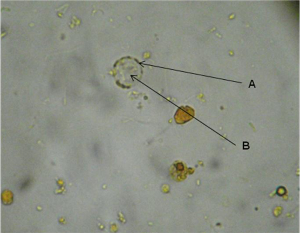
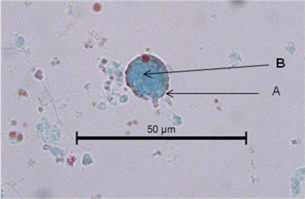
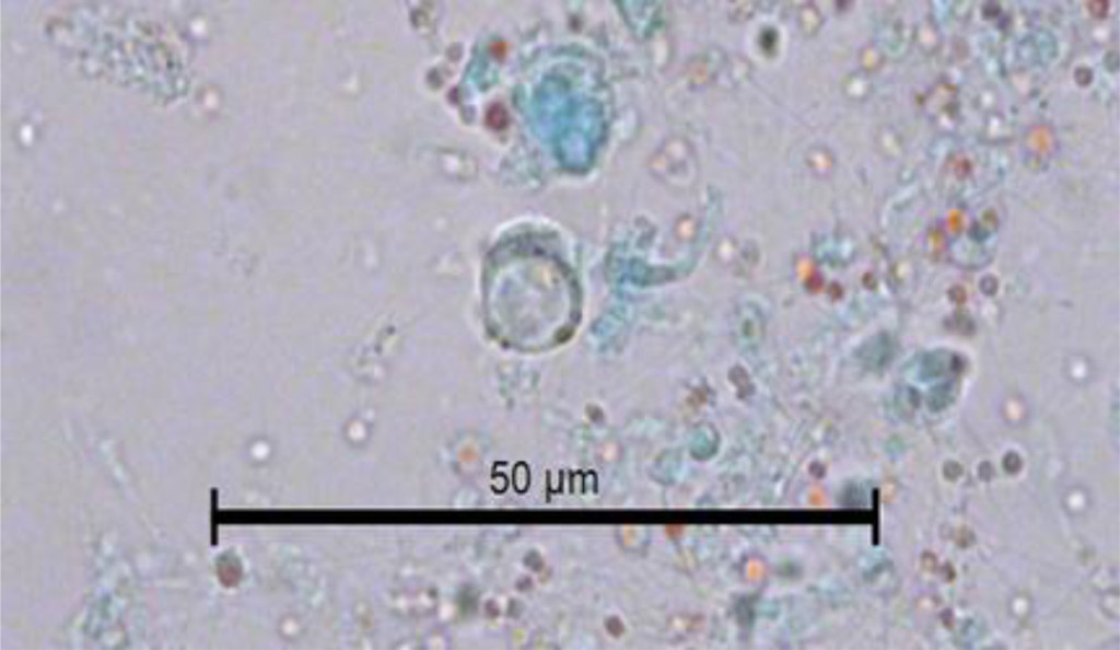
The study consisted in analyzing 210 fecal samples collected in three forest sections (68 in the first section, 62 in the second one, and 80 in the third one) as well as 50 soil samples from each of three sections. Table 1 shows the general prevalence of 51.5% of helminth eggs found in 210 fecal samples, the infection by T. canis was present in 34.8% of them, and by A. caninum in 16.7%. Table 2 displays the general prevalence of helminth eggs that were present in the fecal samples collected in each of three sections of the forest. In the first section, the eggs of T. canis prevailed and in the third section the eggs of A. caninum. Table 3 shows that the general analysis of the results of the medical tests in three sections of the forest reveals the presence of helminth eggs in these areas with an estimated p of < 0.001. Through the microscopic analysis of 210 fecal samples stained by using the Zhiel Neelsen technique, Cryptosporidium spp, oocysts were spotted in five samples, i.e. in 2.4% of all samples. Of 150 soil samples analyzed through the Ferreira method, in 14 (9.3%), T. canis eggs were found, and free-living larvae were observed in 12 samples (7.3%).

Table 1. General prevelance of helminth eggs in 210 fecal samples obtained in the Capultepec Forest, Mexico

Eggs

Frequency

Porcentage

35

16.7

T. canis

73

34.8

Total

210

100.0

Negative

102

51,5

Table 2. Prevalence of canine helminth eggs in 210 feces analyzed by Faust´s method in three section of the Chapultepec Forest, Mexico City

Helminth eggs

Section

Total

1

2

3

A. caninum

10

10

15

35

T. canis

32

28

13

73

Negative

26

24

52

102

Total

68

62

80

210

Table 3. General prevalence of canine helminth eggs in 210 feces analyzed by Faust´s method in three section of the Chapultepec Forest, Mexico City

Negative

Faust´s method


Total

Negative

Positive

Section

1

26

42

68

2

24

38

62

3

52

28

80

Total

102

108

210

 * p < 0.001

Discussion

The Chapultepec Forest, the oldest urban park in the Americas and one of the oldest in the world, has been visited by 15 million people annually and by 200,000 every weekend. It is one of the largest urban parks in the world, on a par with Central Park in NY, Hyde Park in London, or Le Bois de Boulogne in Paris. The forest green areas are a favorite place for city resident recreation. However, a large number of homeless dogs that live there present a danger of physical aggression toward the visitors and also a risk to them of getting different zoonotic diseases such as rabies, syphilis; the stray canine population as well as parasitized dogs that accompany visitors indiscriminately contaminate the soil with helminth eggs and zoonotic protozoan cysts that are present in their feces, and in this way the contaminated soil represents an important source of infection and a significant impact on public health [6, 7]. The species of zoonotic nematodes identified in this study were T. canis and A. caninum. The first type is considered to be the main etiological agent of toxocariasis, a parasitic zoonosis that causes significant morbidity and a serious public health problem in the world, mainly in developing countries [7]. The morbidity of human toxocariasis is related to the following factors: a close contact of humans with dogs and cats, an increase of the dog and cat population, a shortage of sanitary control of the fecal matter evacuated by these animals both in public sites and at home, scarce knowledge that people have of this zoonosis, and, especially, a lack of a timely diagnosis that could prevent the development and evolution of the disease before it could produce cases as serious as loss of vision or death [12].

In this study, the rate of soil contamination with this zoonosis in the Chapultepec Forest was 9.3%. Similar studies of soil contamination with T. canis eggs in different countries reported frequencies similar to those obtained by us, for example: 6.8% in Poland [14] 16.4% in Madrid, Spain [16]; 6.6% in Italy [20], and 9.2% in Chile [21]. However, some developing countries have reported frequencies that are higher than those obtained in our study, such as 20.3% in Iran [10], or the one in a study of 25 state parks in Presidente Prudente, Brazil where T. canis was reported in 96.6% of sampled parks [22].

From the epidemiological point of view, the biotic potential of T. canis is enormous because a female can produce up to 200,000 eggs per day. The Pan American Health Organization (PAHO) has estimated that 1 gram of dog fecal matter can contain up to 15,000 Toxocara eggs that are deposited in the ground by walking people, rain, wind or vectors, [23, 24]. The thick layer of Toxocara eggs makes them resistant to cold and environmental changes; therefore, under adequate humidity and temperature conditions, they can survive in the earth surface for several years. Thus, despite the fact that the soil may appear to be clean because the feces were collected by the health personnel or because the fecal material has disintegrated and there is no odor, it can be contaminated with microscopic infectious forms of parasites.

Soil contaminated with T. canis larvae is the main source of infection for humans. In humans as well as in dogs, the infecting form is a larval egg. T. canis larval eggs affect different organs in both humans and dogs; however, adult parasites develop only in dogs. Thus, people who visit recreation sites with contaminated soil are exposed to infection. The most vulnerable people are those under eight years old because their play activities put them in greater contact with soil. Infection in children is caused mainly by geophagy or ingestion of T. canis infectious larvae from contaminated soil, or ingestion of larval eggs that are present in dogs’ hair [25-28]. In developed countries, despite the fact that the prevalence of parasitic diseases is usually low there, toxocariasis is the most common helminth disease [3, 29].

The ingested eggs hatch in the digestive tract, and the larvae enter the intestinal mucosa, then reach mesenteric vessels and spread to different organs and tissues where the host’s response traps them by producing granulomas, whose degree of disease will be in direct proportion to the number of infected ingested larval eggs. In the clinical form of the disease known as visceral migrant larva, the symptoms are nonspecific and consist of abdominal pain, anorexia, malaise, behavioral disorders, cervical adenitis, hepatomegaly, pain in extremities, and fever. Children under 5 years old are the most affected, and the main risk factors in this population are geophagy and close contact with dogs. The most severe form of the disease occurs in the eyes, where it produces ocular migrant larva syndrome (LMO), [30]. It has been reported that a single larva can cause unilateral blindness. Vision loss occurs because of the acute inflammatory reaction of the retina and optic nerve. The risk of being in contact with soils contaminated with T. canis eggs is very severe because a case of congenital ocular toxocariasis in a newborn has been recently documented [31].

In the case of A. caninum, human infection is caused by penetration of a filariform larva into the skin, but as it is unable to invade deeper tissues, it produces a skin lesion that is several centimeters long and is very itchy due to carving a tunnel in the epidermis known as migratory cutaneous larva or verminous creeping dermatitis [11, 32, 33].

The prevalence rates of this helminth reported by some authors of similar studies vary from 4.2% in Chile [21] 6.8% in Italy [20], 13.5% in Brazil [34]. A clear example of the severity of soil contamination with geohelminths of canine origin is beach sand in Brazil where the presence of A. caninum in 82.5% of its samples was reported [35].

The detection of Cryptosporidium spp. in fecal samples of examined dogs supports the idea that infected canines are a potential source of human infection [36]. Dog infection with Cryptosporidium spp. reported in this study is less than that obtained in the studies with similar characteristics performed in France 2.6% [37], Sao Paulo, Brazil, 3.1% [38], and is similar to infection reported in Zaragoza, Spain, 5.5% [39], Egypto 5.4% [40] Niagara, Canada, 7.4% [41] Prague, Czech Republic, 9.3%; Japan, 7.2% [42], and the Netherlands, 8.7% [25]. However, the infection data obtained in the present study were less than 40% reported in the city of Campos dos Goytacases, Brazil [43].

The discrepancy in the above-mentioned results is due to different factors such as climatic conditions of the place of study, soil characteristics, soil contamination degree, sociocultural factors, and laboratory procedures.

The presence of Cryptosporidium spp. in infected animal feces plays an important role in transmitting these microorganisms, since dogs constantly eliminate a small number of sporulated oocysts that is, however, sufficient to infect humans and other animals. The problem gets worse if we consider the concatenation of some biological and climatic factors inherent to the parasite such as low dosage of oocysts (10 oocysts) required to develop infection [44]. The resistance of oocysts to environmental changes as well as to the conventional water treatment becomes epidemiologically important given that forest and garden irrigation is generally performed with water which contributes to soil contamination and oocyst dissemination and viability. In damp environments such as public parks, C. parvum oocysts can remain viable for 2-6 months [45, 40, 11].

Another factor that contributes to dogs´ infection with Cryptosporidum is the biological cycle of the latter that consists of three autoinfectious reproductive phases as well as the production of thin-walled oocysts that embed themselves deeply in the host intestine originating chronic infections without a need of new infections. As a result of this autoinfection, during defecation a dog releases constant infectious oocysts that contaminate soil as in the case of experimentally infected dogs who had up to 10,000 oocysts per one gram of their fecal material [46].

It is necessary to advise people with high risk such as immunocompromised or of extreme ages of the life and who visit parks to maximize their hygienic habits as, for example, washing hands after touching the hair of the dogs that go to these places regardless of whether they are stray or with an owner [28].

Despite a small number of samples, the rates of infection with zoonotic enteroparasites detected in the present work are important from the epidemiological and public health point of view because of soil contamination by enteric zoonotic species of parasitized animals that live or are brought to parks with ecological characteristics similar to those of the Chapultepec Forest in Mexico City [47-49].

With the aim of preventing zoonotic diseases transmitted by dogs, it is vital to implement actions such as daily collection of fecal matter in recreation areas; the control of synanthropic flies that participate in the dissemination of different zoonoses [23, 24], the direct participation of dog owners that visit the forest in collecting and eliminating stool evacuated by their pets; promotion of semi-annual de-worming; and scheduled sterilization in order to contribute to the reduction of street canine population, since many puppies are released to streets at an early age. Implementing these actions, undoubtedly, will help to get within a short time healthier environment for the inhabitants of Mexico City [50].

Finally, we believe that it is necessary to conduct more studies in order to detect the presence of intestinal parasites in animals as family members and, thereby, to evaluate the impact that these agents have on human health. In addition, such studies will provide the basis for recommending control measures in health programs.

Conclusions

The results of the study highlight the risk of transmitting canine zoonotic parasitosis to the human population, especially children, during their public park visits.

The presence of canine fecal matter in urban area soil is the main source of enteric pathogen infection of stray and domestic animals as well as humans. Preventing soil contamination is the most important way in the prevention of zoonoses transmitted by dogs together with other actions such as social responsibility of a strict control of canine fecal matter elimination not only in the Chapultepec Forest, but also in Mexico City streets, gardens and parks that include dog owners´ participation and health-authorities´ participation in the case of stray dogs. In addition to this, it is crucial to encourage quarterly de-worming of captured dogs, especially those under one-year-old. Through these simple measures, in a short term it would be possible to reduce soil contamination and minimize human and animal health risk.

References

  1. Abu Samra N, Jori F, Cacciò SM, Frean J, Poonsamy B, et al. (2016) Cryptosporidium genotypes in children and calves living at the wildlife or livestock interface of the Kruger National Park, South Africa. Onderstepoort J Vet Res 83: e1-1e7. [crossref]
  2. Felsmann M, Michalski M, Felsmann M, Sokól R, Szarek J, Strzyewska-Worotynka E (2017) Invasive forms of canine endoparasites as a potential threat to public health – A review own studies. Ann Agric Environ Med 24: 245–249.
  3. Szwabe K, Blaszkowska J2 (2017) Stray dogs and cats as potential sources of soil contamination with zoonotic parasites. Ann Agric Environ Med 24: 39–43. [crossref]
  4. Studznska MB, Demkowska-Kutrzepa M, Borecka A, Meisner M, Tomczuk K, et al. (2017) Variation in the Rate of Infestation of Dogs with Zoonotic Nematodes and the Contamination of Soil in Different Environments. Int J Environt Res Public Health 14: Pii: E1003
  5. Feng Y, Xiao L (2017) Molecular Epidemiology of Cryptosporidiosis in China. Front Microbiol 8: 1701. [crossref]
  6. Fletcher SM, Stark D, Harkness J, Ellis J (2012) Enteric protozoa in the developed world: a public health perspective. Clin Microbiol Rev 25: 420–449. [crossref]
  7. Anh NT, Thuy DT, Hoan DH, Hop NT, Dung DT (2016) Levels of Toxocara infect ions in dogs and cats from urban Vietnam together with associated risk factors for transmission. Helminthol 90: 508–10.
  8. Otero D, Alho AM, Nijsse R, Roelfsema J, Overgaauw P, et al. (2017) Environmental contamination with Toxocara spp. Eggs in public parks and sandpits of Greater Lisbon, Portugal. J Infect Health pii: S1876-0341(17)30134.
  9. Martínez-Barbabosa I, Gutiérrez CEM, Aguilar VJM, Pimienta LJJ, Shea M (2011) Frecuencia de geohelmintos en canes domiciliados en siete delegaciones de la Ciudad de México. Vet Méx 42: 83–91.
  10. Berenji F, Movahedi Rudy AG, Fata A, Tavassoli M, Mousavi Bazaz M, et al. (2015) Soil Contamination with Toxocara Spp. Eggs in Public Parks of Mashhad and Khaf, North East of Iran. Iran J Parasitol 10: 286–289. [crossref]
  11. Díaz-Anaya AM, Pulido-Medellín MO, Giraldo-Forero JC (2015) Nematodes with zoonotic potential in parks of the city of Tunja, Colombia. Salud Publica Mex 57: 170–6
  12. Lötsch F, Vingerling R, Spijker R, Grobusch MP (2017) Toxocariasis in humans in Africa – A systematic review. Travel Med Infect Dis. [crossref]
  13. Zanzani SA, Di Cerbo AR, Gazzonis AL, Genchi M, Rinaldi L, et al. (2014) Canine fecal contamination in a metropolitan area (Milan, Nothh-western Italy): prevalence of intestinal parasites and evaluation of health risk. Scientific World Jounal 2014: 132.361.
  14. Blaszkowska J, Góraiska K, Wojcik A, Kumatowski P, Szwabe K (2015) Presence of Toxocara spp. eggs in children´s recreation areas with varing degrees of access for animals. Ann Agric Environ Med 22: 23–7.
  15. INEGI (2015) Instituto Nacional de Geografía Estadística e Informática Centro de Población y Vivienda.
  16. Dado D, Izquierdo F, Vera O, Montoya A, Mateo M, et al. (2012) Detection of zoonotic intestinal parasites in public parks of Spain. Potential epidemiological role of microsporidia. Zoonoses Public Health 59: 23–8.
  17. Aquino MJM, Vargas SGB, López MB, Neri SE, Bernal RR (2012) Comparación de dos nuevas técnicas de sedimentación y métodos convencionales para la recuperación de parásitos intestinales. Rev Latinoamer Patol Clin 59: 233–242.
  18. Henriksen SA, Pohlenz JF (1981) Staining of cryptosporidia by a modified Ziehl-Neelsen technique. Acta Vet Scand 22: 594–596. [crossref]
  19. Faust EC, DÁntoni JS, Odom V, Miller NJ, Pares C, Sawitzl E (1938) A critical study of clinical laboratory techniques for the diagnosis of protozoans cysts and helminth eggs in feces. Am J Trop Medicine 18: 169–193.
  20.  Paoletti B, Traversa D, Iorio R, De Berardinis A, Bartolini R, et al. (2015) Zoonotic parasites in feces and fur of stray and private dogs from Italy. Parasitol Res 114: 2135–2141. [crossref]
  21. Luzio A, Belmar P, Troncoso I, Luzio P, Jara A (2015) Fernandez I. Parasites of zoonotic importance in dogs’ feces collected in parks and public squares of the city of Los Angeles, BÍo-Bío, Chile. Rev Chilena Infectol 32: 403–7.
  22.  Santarém VA, Pereira VC, Alegre BC (2012) Contamination of public parks in Presidente Prudente (São Paulo, Brazil) by Toxocara spp. eggs. Rev Bras Parasitol Vet 21: 323–325. [[crossref]
  23. Racewicz M, Kruminis-Å ozowska W, Gabre RM, StaÅ„czak J (2009) The occurrence of Cryptosporidium spp. in synanthropic flies in urban and rural environments]. Wiad Parazytol 55: 231–236. [crossref]
  24. Kebede N (2011) Cryptosporidium recovered from Musca domestica, Musca sorbens and mango juice accessed by synanthropic flies in Bahirdar, Etiopia. Zoonoses Public Health 58: 69–75. MOSCAS
  25.  Overgaauw PA, van Zutphen L, Hoek D, Yaya FO, Roelfsema J, et al. (2009) Zoonotic parasites in fecal samples and fur from dogs and cats in The Netherlands. Vet Parasitol 163: 115–122. [crossref]
  26. Fetene T, Worku N, Huruy K, Lötsch F, Vingerling R, et al. (2017) Toxocariasis in humans in Africa – A systematic review. Travel Med Infect Dis pii: S1477–8939.
  27.  Tavassoli M, Javadi S, Firozi R, Rezaei F, Khezri A, et al. (2012) Hair Contamination of Sheepdog and Pet Dogs with Toxocara canis Eggs. Iran J Parasitol 7: 110–115. [crossref]
  28. Merigueti et al. (2017) YFFB, Santarém VA, Ramirez LM, da Silva Batista Batista A, da Costa Beserra LV, Nuci AL, de Paula Esposte TM. Protective and factors associated with the presence of Toxocara spp. in eggs in dog hair. Vet Parasitol 244: 39–43.
  29. Inpankaew T, Jiyipong T, Sunanta C, Kengradomkii C, Pinyopanuwat N, et al. (2017) Prevalence and molecular characterization of bovine Cryptosporidium from dairy cows in Northern Thailand. Acta Parasitol 62: 772–774
  30. Chen Q, Gu J, Jiang R, et al. (2017) Role of ultrasound biomicroscopy in diagnosis of ocular toxocariasis. Br J Ophthalmol [[crossref]
  31. Maffrand R, Avila-Vázquez M, Princich D, Alasia P (2006) Congenital ocular toxocariasis in a premature neonate]. An Pediatr (Barc) 64: 599–600. [crossref]
  32. Veraldi S, Cuka E, Pontini P, Vaira F (2017) Bullous cutaneous larva migrans: case series and review of atypical clinical presentations. G Ital Dermatol Venereol 152: 516–519
  33. Manikat R, Kannangara S2 (2017) Cutaneous Larva Migrans. J Glob Infect Dis 9: 125. [crossref]
  34.  Moura MQ, Jeske S, Vieira JN, Corrêa TG, Berne ME, et al. (2013) Frequency of geohelminths in public squares in Pelotas, RS, Brazil. Rev Bras Parasitol Vet 22: 175–178. [crossref]
  35. Rocha S, Pinto RM, Floriano AP, Teixeria LH, Bassili B, et al. (2011) Environmental analyses of the parasitic profile found in the Sandy soil from the Santos municipality beaches, SP, Brazil. Rev Inst Med Trop Sao Paulo 2011 Sep-Oct: 5385): 277–81
  36. Gil H, Cano L, de Lucio A, Bailo B, de Mingo MH, Cardona GA, et al. (2017) Detección and molecular diversity of Giardia duodenalis and Cryptosporidium spp. In Sheltered dogs and cats in Northern Spain. Infect General Evol 50: 62–69
  37. Osaman M, Bories J, El Safadi D, Poirel MT, Gantois N, et al. (2015) Prevalence and genetic diversity of the intestinal parasites Blastocystis sp. And Cryptosporidium spp. In household dogs in France and evaluation of zoonotic transmission risk. Vet Parasitol 214: 167–170.
  38. Katagiri A, TC Oliveira-Sequeira (2008) Prevalence of dog intestinal parasites and risk perception of zoonotic infection by dog owners in Sao Paulo State, Brazil. Zoonoses Public Health 55: 406–413.
  39. de Lucio A, Bailo B, Aguilera M, Cardona GA, Fernández-Crespo JC, et al. (2017) No molecular epidemiological evidence supporting household transmission of zoonotic Giardia duodenalis and Cryptosporidium spp. From pet dogs and cats in the province of Álava, Northern Spain. Acta Trop 170: 48–56.
  40.  Awadallah MA, Salem LM (2015) Zoonotic enteric parasites transmitted from dogs in Egypt with special concern to Toxocara canis infection. Vet World 8: 946–957. [crossref]
  41. Shukla R, Giraldo P, Kraliz A, Finnigan M, AL Sanchez (2006) Cryptosporidium spp. And other zoonotic enteric parasites in a sample of domestic dogs and cats in the Niagara region of Ontario. Can Vet J 47: 1179–84.
  42. Itoh N, Oohashi Y, Ichikawa-Seki M, Itagaki T, Ito Y, et al. (2014) Molecular detection and characterization of Cryptosporidium species in household dogs, pet shop puppies, and dogs kept in a school of veterinary nursing in Japan. Vet Parasitol 200: 284–8.
  43. Ederil BB, MF Rodrigues, CB Carvalho (2005) Oocysts of the genus Cryptosporidium in domiciliated dogs from the city of Campos dos Goytacases, the State of Rio de Janeiro. Rev Bras Parasitol Vet 14: 129–31.
  44. de Graaf DC, Vanopdenbosch E, Ortega-Mora LM, Abbassi H, Peeters JE (1999) A review of the importance of cryptosporidiosis in farm animals. Int J Parasitol 29: 1269–1287. [crossref]
  45. Pereira VJ, Marquez R, Marques M, Benoliel MJ, Barreto Crespo MT (2013) Free chlorine inactivation of fungi in drinking water sources. Water Res 47: 517–23.
  46. Lloyd S, J Smith (1997) Pattern of Cryptosporidium parvum oocyst excretion by experimentally infected dogs. Int J Parasitol 27: 799–801.
  47. Avcioglu H, Balkaya I (2011) The relationship of public park accessibility to dogs to the presence of Toxocara species ova in the soil. Vector Borne Zoonotic Dis 11: 177–180. [crossref]
  48.  Blaszkowska J, Wojcik A, Kurnatowski P, Szwabe K (2013) Geohelminth egg contamination of children’s play areas in the city of Lodz (Poland). Vet Parasitol 192: 228–233. [crossref]
  49. Blaszkowska J, Góraiska K, Wojcik A, Kumatowski P, Szwabe K. Presence of Toxocara spp. eggs in children´s recreation areas with varing degrees of access for animals. Ann Agric Environ Med 22: 23–7.
  50. Hackett T, MR Lappin (2003) Prevalence of enteric pathogens in dogs of north-central Colorado. J Am Anim Hosp Assoc 39, 52–6.