Archives

Peripheral Tryptophan – Kynurenine Metabolism Associated with Metabolic Syndrome is Different in Parkinson’s and Alzheimer’s Diseases

DOI: 10.31038/EDMJ.2017141

Abstract

Insulin resistance (IR), obesity and other components of metabolic syndrome [MetS] are highly associated with Alzheimer’s (AD) and Parkinson’s (PD) diseases. Dysregulation of kynurenine (Kyn) pathway (KP) of tryptophan (Trp) metabolism was suggested as major contributor to pathogenesis of AD and PD and MetS. KP, the major source of NAD+ in humans, occurs in brain and peripheral organs. Considering that some, but not all, peripherally originated derivatives of Kyn penetrate blood brain barrier, dysregulation of central and peripheral KP might have different functional impact. Up-regulated Kyn formation from Trp was discovered in central nervous system of AD and PD while assessments of peripheral KP in these diseases yield controversial results. We were interested to compare peripheral kynurenines in AD and PD with emphasis on MetS-associated kynurenines, i.e., kynurenic (KYNA) and anthranilic (ANA) acids and 3-hydroxykynurenine (3-HK). Serum concentrations of KP metabolites were evaluated (HPLC-MS method). In PD patients Trp concentrations were lower, and Kyn: Trp ratio, Kyn, ANA and KYNA were higher than in controls. 3-HK concentrations of PD patients were below the sensitivity threshold of the method. In AD patients. ANA serum concentrations were approximately 3 fold lower, and KYNA concentrations were approximately 40% higher than in controls. Our data suggest different patterns of KP dysregulation in PD and AD: systemic chronic subclinical inflammation activating central and peripheral KP in PD, and central, rather than peripheral, activation of KP in AD triggered by Aβ1–42. Dysregulation of peripheral KP in PD and AD patients might underline association between neurodegenerative diseases and MetS.

Key words

anthranilic acid; kynurenic acid; kynurenine; Parkinson’s disease; Alzheimer’s disease; insulin resistance; obesity

Introduction

Insulin resistance (IR), obesity and other components of metabolic syndrome [MetS] are highly associated with Alzheimer’s (AD) and Parkinson’s (PD) diseases [1,2]. Dysregulation of kynurenine (Kyn) pathway (KP) of tryptophan (Trp) metabolism was suggested as major contributor to pathogenesis of AD [3], PD [4] and MetS [5,6]. KP, the major source of NAD+ in humans, occurs in brain and peripheral organs (e,g., monocytes/macrophages, liver, pancreas, kidney, intestine, muscles) [7,8]. Initial phase of KR, conversion of Trp into Kyn (via N-formyl-kynurenine), is catalyzed by indoleamine-2,3-dioxygenase 1 (IDO) or tryptophan-2,3-dioxygenase 2 (TDO); Kyn is further converted into 3-hydroxykynurenine (3HK), kynurenic (KYNA) and anthranilic (ANA) acids; further metabolism of 3-HK resulted in production of NAD+ (Fig.1). Considering that some, but not all, peripherally originated derivatives of Kyn penetrate blood brain barrier (BBB) [9], dysregulation of central and peripheral KP might have different functional impact. Thus, increased production of KYNA in brain was suggested to underline psychotic symptoms [8] while elevation of peripheral KYNA (not penetrating BBB) might contribute to mechanisms of IR and obesity in schizophrenia [10].

Up-regulation of Kyn formation from Trp was discovered in central nervous system of AD and PD [11] while assessments of peripheral KP in these diseases yield controversial results.

In PD patients, elevated serum Kyn: Trp ratio, a clinical index of IDO or TDO activity, was reported [12]. Concentration of KYNA and activity of enzyme, catalyzing Kyn conversion into KYNA, were elevated in red blood cells (but not in plasma) of PD patients [13]). The third available study revealed no differences in Kyn and 3-HK plasma concentrations between PD and control subjects, and elevated KYNA and ANA in PD patients without dyskinesia in comparison with PD patients with dyskinesia [14].

In AD most studies compared concentrations of peripheral kynurenines in serum or plasma of patients with probable AD in comparison with healthy controls (or patients with major depressive disease and subjects with subjective cognitive impairment [15]. Only one study evaluated plasma kynurenines in histopathologically confirmed AD in comparison with age-matched and non-matched healthy subjects [16]. Plasma Trp metabolism was found to discriminate between AD and control group in metabolomic study [17]. Trp concentrations were lower in patients with probable [18-20] and histopathologically confirmed AD [16]. Kyn concentrations were unchanged in probable AD [15,18,20] and significantly lower in histopathologically confirmed AD [16]. Kyn: Trp ratio was higher in probable [18,19] but not in histopathologically confirmed AD [16]. Among derivatives of intermediate KP phase, KYNA concentrations were reduced or unchanged in probable [15,20,21] and histopathologically confirmed [16] AD patients. 3-HK levels were elevated in probable AD patients [15] but lower in plasma of histopathologically confirmed AD patients [16]. Strong tendency to reduced ANA was reported in histopathologically confirmed AD [16]. Notably, significant association of plasma ANA and risk of incident dementia with risk increased by 40% for an increase of one standard deviation was observed in participants of Framingham offspring cohort study [22].

We were interested to compare peripheral kynurenines in AD and PD with emphasis on MetS-associated kynurenines, i.e., KYNA, ANA, 3-HK and xanthurenic acid (XA) [23].

Methods

PD patients

Overnight fasting blood samples were collected from 7 female and 11 male PD patients (age range: 50 to 74). At the time of sampling five patients did not take any anti-Parkinson’s medications; and thirteen patients were treated with L-dopa.

AD patient

Blood samples from 12 female and 8 male patients (age range 60 to 75) with probable AD were studied. All AD patients had MMSE between 20 and 23. They were treated with Aricept or Namenda.

Healthy Subjects (Controls)

There were 24 age- and gender- matched (12 females and 12 males) healthy subjects. Study was approved by Tufts Medical Center IRB.

Assessment of Kynurenine metabolites

Plasma samples were stored at −80°C until analysis. Trp, Kyn, ANA, KYNA and 3-HK concentrations were analyzed by high performance liquid chromatography coupled to mass spectrometry (HPLC-MS) as described elsewhere [10].

Statistical analysis

Results are presented as mean ± standard error (Trp and Kyn in μM and ANA, KYNA and 3-HK in nM). Statistical significance was assessed by unpaired t test with Welch correction, two-tailed.

Results

PD patients

There was no difference in plasma concentrations of Trp, Kyn and all studied Kyn metabolites of not treated and treated with L-DOPA patients (data not shown). Therefore, combined data of not-treated and L-DOPA – treated PD patients were used for the further analysis. Trp concentrations were lower, and Kyn: Trp ratio was higher in PD patients than in controls (Table 1). Serum concentrations of Kyn, and its down-stream metabolites, ANA and KYNA, were approximately two fold higher than in control subjects (Table 1). 3-HK concentrations of PD patients were below the sensitivity threshold of the method. XA concentrations were not different between PD and control group (11.87 ± 1.3 and 11.74 ± 1.25, resp).

Table 1. Tryptophan – kynurenine metabolites in serum of Parkinson’s and Alzheimer’s disease patients.

Means ± sem
 Trp
(μM)
 Kyn
(μM)
Kyn: Trp
(x100)
 ANA
(nM)
 KYNA
(nM)
 3-HK
(nM)
Control
(n=24)
68.9 ± 2.49 1.76 ± 0.09 2.55 ± 0.14 70.54 ± 17.9 35.78 ± 3.59 19.55 ± 3.14
PD
(n=18)
48.56 ± 2.4* 2.34 ± 0.11# 4.82 ± 0.18* 156.68 ± 20.46# 65.97 ± 7.2# Not detectable
AD
(n=20)
64.64 ± 3.4 1.77 ± 0.11 2.74 ± 0.15 19.51 ± 3.5* 34.35 ± 2.59 27.52 ± 2.3#
*p<0.001 in comparison with all other groups; #) p<0.001 in comparison with control (except for 3HK p=0.04). Unpaired t test with Welch correction, two tailed.
Abbreviations: Trp: tryptophan; Kyn: kynurenine; KYNA: kynurenic acid; ANA: anthranilic acid; 3-HK: 3-hydroxykynurenine; PD: Parkinson’s disease; AD: Alzheimer’s disease

AD patients

ANA serum concentrations were approximately 3 fold lower, and KYNA concentrations were approximately 40% higher in AD than in controls (Table 1). XA concentrations were not different between AD and control group (10.97 ± 1.07 and 11.74 ± 1.25, resp).

Discussion

Serum levels of KP metabolites might reflect the activity of their formation in peripheral organs [4,8]. Notably, KP in fatty tissue does not express kynurenine-2-monooxygenase (KMO), enzyme converting Kyn into 3-HK, and, therefore, KYNA and ANA are the end products of KP in fatty tissue [24].

Literature data suggested increased conversion of Kyn into 3-HK in PD-related brain structures with consequent formation of neurotoxic metabolites [4]. Present results suggest an increased conversion of Kyn into KYNA and ANA in peripheral organs (in difference with KP in PD-related brain structures [25,26].

In PD patients we confirmed literature data on decreased Trp and increased Kyn concentrations, and, consequently, increased Kyn: Trp ratio, suggesting activation of the initial phase of KP, i.e., conversion of Trp into Kyn [12]. Some discrepancies between literature data (see Introduction) and present results may depend on studied tissues, i.e., serum VS plasma VS RBC; and analytical methods; as well as differences in the other factors potentially affecting KP such as length of disease and age of patients [2,27].

In PD patients we found as increased (about two-fold) plasma concentrations of KYNA and ANA. Considering that KYNA, ANA and 3-HK compete for Kyn as a common substrate in both central and peripheral organs (Figure 1), our data suggest a shift of down-stream Kyn metabolism from 3-HK production towards formation of ANA and KYNA.

EDMJ2017-113-GregoryFOxenkrugUSA_f1

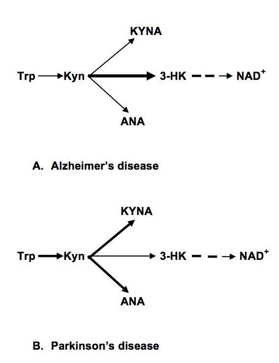
Figure 1. Abbreviations: Trp: Tryptophan; Kyn: kynurenine; KYNA: Kynurenic Acid; ANA: Anthranilic acid; 3-HK: 3-hydroxykynurenine; NAD+: Nicotinamide Adenine Dinucleotide

In AD patients, we found no differences in Trp, Kyn concentrations and Kyn: Trp ratios. Present data are in agreement with the study of histopathologically confirmed AD and age-matched controls that did not find changes of Kyn: Trp ratio [16]. We found elevated 3-HK serum concentration that might suggest decreased availability of Kyn as a substrate for formation of KYNA and ANA in agreement with present results of drastic reduction of ANA concentrations. Peripheral production of ANA deserves further studies, especially considering significant association of plasma ANA and risk of incident dementia in Framingham offspring cohort study [22].

Present study suggested different patterns of dysregulation of the intermediate phase of peripheral KP in PD and AD: increased formation of KYNA and ANA (and reduced production of 3-HK) in PD and reduced formation of ANA (and increased production of 3-HK) in AD (Figure 1). Our data suggest different mechanisms of KP dysregulation in PD and AD: systemic chronic subclinical inflammation activating central and peripheral KP in PD, and central, rather than peripheral, activation of KP in AD triggered by Aβ 1–42 [28]. Notably, there was no association between KP changes and plasma concentrations of neopterin, KP related marker of inflammation, in AD patients [16].

Literature and our data suggest that up-regulation of peripheral KYNA, ANA and Kyn production might contribute to development of obesity and IR, conditions highly associated with early (contrary to late) stages of PD [2,27]. KYNA concentrations positively correlated with BMI in clinical studies [29]. We reported elevation of blood concentrations of KYNA, ANA and Kyn in Zucker obese rats (ZFR) [30]. KYNA elevation in obesity may be a consequence of KMO deficiency in fatty tissue that does not express KMO genes rending KYNA and ANA as the end products of KP in human fatty tissue [24]. KYNA, ANA and Kyn might promote the development of obesity via activation of aryl hydrocarbon receptor (AHR) that regulates xenobiotic-metabolizing enzymes. ANA, KYNA and Kyn are the endogenous human AHR ligands [31,32]. Over-activation of AHR promoted [33] while AHR deficiency protected mice from diet-induced obesity [34].

PD is highly associated not only with obesity but with IR. Increased risk of PD among subjects with T2D was independent from obesity (BMI) [35]. We have previously reported elevation of serum KYNA and ANA in T2D [36,37] and correlation of Kyn with HOMA-IR in HCV patients [38]. Metabolomics analysis revealed 1.8 fold increase of urine KYNA in spontaneously and naturally diabetic rhesus macaques [39]. Successful treatment of IR was associated with down-regulation of KP, including inhibition of KYNA production [40]. One of the possible mechanisms of KYNA involvement in diabetes is activation of G-protein-coupled receptor 35 (GPR35) located primarily in peripheral, including pancreas, tissues [41]. KYNA is an endogenous agonist of GPR35 [41]. Exogenous GPR35 agonists were patented as agents reducing blood glucose levels in oral glucose tolerance tests, stimulate glucose uptake in differentiated 3T3-L1 adipocytes [42].

Significance of ANA elevation has been explored only in a few papers. Serum ANA was positively associated with neopterin, Kyn, Kyn: Trp ratio, and negatively with Trp in healthy young adults [43]. ANA was reported to significantly increase glucose uptake and inhibited 14CO2 production from [U-14C] glucose in in vitro studies [44].

On the other hand, AD is characterized by developing of brain IR [45] while weight loss preceded the diagnosis of dementia in community-dwelling older adults even after controlling for other factors associated with weight [46]. It was suggested that decline in BMI that precedes the diagnosis of AD may be related to neurodegeneration in areas of the brain involved in homeostatic weight regulation [1].

Therefore, we suggest that up-regulated peripheral formation of KYNA, ANA and Kyn contribute to increased risk of PD among subjects with diabetes, and, that contrary to PD, central, rather than peripheral, KP dysregulation contribute to association of IR with AD.

Present data warrant further studies of dysregulations of peripheral KP in PD and AD patients as one of the mechanisms (and potential biomarkers) of association between neurodegenerative disease and MetS.

Acknowledgement

GF Oxenkrug is a recipient of MH104810. Paul Summergrad is a non-promotional speaker for CME outfitters, Inc., and consultant and non-promotional speaker for Pri-med, Inc. Authors appreciate Bioreclamation IVT, NY, USA, for help in collection of serum samples.

References

  1. Alford S, Patel D, Perakakis N, Mantzoros CS (2017) Obesity as a risk factor for Alzheimer’s disease: weighing the evidence. Obes Rev. [crossref]
  2. Vikdahl M, Carlsson M, Linder J, Forsgren L, Håglin L (2014) Weight gain and increased central obesity in the early phase of Parkinson’s disease. Clin Nutr 33: 1132–1139. [crossref]
  3. Baran H, Jellinger K, Deecke L (1999) Kynurenine metabolism in Alzheimer’s disease. J Neural Transm (Vienna) 106: 165–181. [crossref]
  4. Lim C, Fernández-Gomez FJ, Braidy N, Estrada C, Costa C, et al. (2017) Involvement of the kynurenine pathway in the pathogenesis of Parkinson’s disease. Prog Neurobiol 155: 76–95. [crossref]
  5. Oxenkrug GF (2010) Metabolic syndrome, age-associated neuroendocrine disorders and dysregulation of tryptophan – kynurenine pathway metabolism. Ann N Y Acad Sci 1199: 1–14.
  6. Mangge H, Summers LL, Meinitzer A, Zelzer S, Almer G, et al. (2014) Obesity-related dysregulation of the Tryptophan–Kynurenine metabolism: Role of age and parameters of the metabolic syndrome. Obesity.
  7. Paluszkiewicz P, Zgrajka W, Saran T, Schabowski J, Piedra JL, et al. (2009) High concentration of kynurenic acid in bile and pancreatic juice. Amino Acids 37: 637–641. [crossref]
  8. Schwarcz R, Bruno JP, Muchowski PJ, Wu HQ (2012) Kynurenines in the mammalian brain: when physiology meets pathology. Nat Rev Neurosci 13: 465–477. [crossref]
  9. Fukui S, Schwarcz R, Rapoport SI, Takada Y, Smith QR (1991) Blood-brain barrier transport of kynurenines: implications for brain synthesis and metabolism. J Neurochem 56: 2007–2017.
  10. Oxenkrug G, van der Hart M, Roeser J, Summergrad P. Peripheral kynurenine-3-monooxygenase deficiency as a potential risk factor for metabolic syndrome in schizophrenia patients Interg Clin Med 2017: 1: 1–3
  11. Török N, Majláth Z, Fülöp F, Toldi J, Vécsei L (2016) Brain Aging and Disorders of the Central Nervous System: Kynurenines and Drug Metabolism. Curr Drug Metab 17: 412–429. [crossref]
  12. Widner B, Leblhuber F, and D. Fuchs. Increased neopterin production and tryptophan degradation in advanced Parkinson’s disease. J Neural Transm, 2002; 109: 181–189
  13. Hartai Z, Klivenyi P, Janaky T, Penke B, Dux L, et al. (2005) Kynurenine metabolism in plasma and in red blood cells in Parkinson’s disease. J Neurol Sci 239: 31–35. [crossref]
  14. Havelund JF, Andersen AD, Binzer M, Blaabjerg M, et al. (2017) Changes in kynurenine pathway metabolism in Parkinson patients with L-DOPA-induced dyskinesia. J Neurochem. [Epub ahead of print]
  15. Schwarz M, Guillemin GJ, Teipei SJ, Buerger K, Hampei H (2013) Increased 3-hydroxykynurenine serum concentrations differentiate Apzheimer’s disease patients from controls. Eur Arch Psychiatry Clin Neurisci 263: 345–352.
  16. Giil LM, Midttun Ø, Refsum H (2017) Kynurenine Pathway Metabolites in Alzheimer’s Disease. J Alzheimers Dis 60: 495–504. [crossref]
  17. Trushina E, Dutta T, Persson XM, Mielke MM, Petersen RC (2013) Identification of altered metabolic pathways in plasma and CSF in mild cognitive impairment and Alzheimer’s disease using metabolomics. PLoS One 20: 8(5): e63644.
  18. Widner B, Leblhuber F, Walli J, Tilz GP, Demel U, et al. (2000) Tryptophan degradation and immune activation in Alzheimer’s disease. J Neural Transm (Vienna) 107: 343–353. [crossref]
  19. Greiberger J, Fuchs D, Leblhuber F, Greoberger M, Wintersteiger R, et al. (2010) Carbonyl proteins as a clinical marker in Alzheimer’s diasease and its relation to tryptophan degradation and immune activation. Clin Lab 56: 441–448.
  20. Gulaj E, Pawlak K, Bien B, Pawlak D (2010) Kynurenine and its metabolites in Alzheimer’s disease patients. Adv Med Sci 55: 204–211. [crossref]
  21. Hartai Z, Junasz A, Rimannocyz A, Janaky T, Donko T, Dux L, Penke B, Toth GK, Janka Z, Kalman J. Decreased serum and red blood cells kynurenic acid levels in Alzheimer’s disease. Neurochem Int, 2007, 50: 308–13
  22. Chouraki V, Preis SR, Yang Q, Beiser A, Li S, et al. (2017) Association of amine biomarkers with incident dementia and Alzheimer’s disease in the Framingham Study. Alzheimers Dement. S1552-5260(17)30200–5.
  23. Oxenkrug G (2015) 3-hydroxykynurenic acid and type 2 diabetes: implications for aging, obesity, depression, Parkinson’s disease and schizophrenia. In: Engin A, Engin EB (eds) Tryptophan Metabolism: Implications for Biological Processes, Health and Diseases, Molecular and Integrative Toxicology pp.173–195.
  24. Favennec M, Hennart B, Caiazzo R, Leloire A, Yengo L, et al. (2015) The Kynurenine Pathway is Activated in Human Obesity and Shifted Toward Kynurenine Monooxygenase Activation. Obesity 23: 2066–2074.
  25. Ogawa T, Matson WR, Beal MF, Myers RH, Bird ED, et al. (1992) Kynurenine pathway abnormalities in Parkinson’s disease. Neurology 42: 1702–1706. [crossref]
  26. Lewitt PA, Li J, Lu M, Beach TG, Adler CH, Guo L; Arizona Parkinson’s Disease Consortium (2013) 3-hydroxykynurenine and other Parkinson’s disease biomarkers discovered by metabolomic analysis. Mov Disord 28(12): 1653–1660.
  27. Hu G, Antikainen R, Jousilahti P, Kivipelto M, Tuomilehto J (2008) Total cholesterol and the risk of Parkinson disease. Neurology 70: 1972–1979. [crossref]
  28. Guillemin GJ, Smythe GA, Veas LA, Takikawa O, Brew BJ (2003) A beta 1-42 induces production of quinolinic acid by human macrophages and microglia. Neuroreport 14: 2311–2315. [crossref]
  29. Zhao J, Shen D, Djukovic C, Daniel-MacDougall H, Gu X, et al. (2016) Metabolomics identified metabolites associated with body mass index and prospective weight gain among Mexican American women. Obes Sci Pract 2: 309–317
  30. Oxenkrug G, Cornicelli J, van der Hart M, Roeser J, Summergrad P (2016) Kynurenic acid, an aryl hydrocarbon receptor ligand, is elevated in serum of Zucker fatty rats. Integr Mol Med 3(4): 761–763.
  31. DiNatale BC, Murray IA, Schroeder JC, Flaveny CA, Lahoti TS, et al. (2010) Kynurenic acid is a potent endogenous aryl hydrocarbon receptor ligand that synergistically induces interleukin-6 in the presence of inflammatory signaling. Toxicol Sci 115: 8997
  32. Schrenk D, Riebniger D, Till M, Vetter S, Fiedler HP (1999) Tryptanthrins and other tryptophan-derived agonists of the dioxin receptor. Adv Exp Med Biol 467: 403–408. [crossref]
  33. Kerley-Hamilton JS, Trask HW, Ridley CJ, Dufour E, Ringelberg CS, et al. (2012) Obesity is mediated by differential aryl hydrocarbon receptor signaling in mice fed a Western diet. Environ Health Perspect 120: 1252–1259.
  34. Moyer BJ, Rojas IY, Kerley-Hamilton JS, Hazlett HF, Nemani KV, et al. (2016) Inhibition of the aryl hydrocarbon receptor prevents Western diet-induced obesity. Model for AHR activation by kynurenine via oxidized-LDL, TLR2/4, TGFß, and IDO1. Toxicol Appl Pharmacol 300: 13–24.
  35. Gang H, Jousilahti P, Bidel S, et al. Type 2 diabetes and the risk of Parkinson’s disease. Diabetes Care 2007; 30(4): 843–7. Format: Abstract Diabetes Care. 2007;30(4):842-7. J Neurochem. 2017 Jun 19. doi: 10.1111/jnc.14104.
  36. Oxenkrug GF (2015) Increased plasma levels of xanthurenic and anthranilic acids in type 2 diabetes. Mol Neurobiol 52;805-810
  37. Oxenkrug G, van der Hart M, Summergrad P (2015) Elevated anthranilic acid plasma concentrations in type 1 but not type 2 diabetes mellitus. Integr Mol Med 2(5): 365–368.
  38. Oxenkrug GF, Turski WA, Zgrajka, J. V. Weinstock, and P. Summergrad (2013). Tryptophan-Kynurenine Metabolism and Insulin Resistance in Hepatitis C Patients. Hepatitis Research and Treatment, (Vol. 2013), Article ID 149247, 4 pages http: //dx. doi.org/10.1155/2013/149247
  39. Patterson AD, Bonzo JA, Li F, Krausz KW, Eichler GS, Aslam S, et al. (2017) Metabolomics reveals attenuation of the SLC6A20 kidney transporter in nonhuman primate and mouse models of type 2 diabetes mellitus. J Biol Chem 286(22): 19511–22.
  40. Muzik O, Burghardt P, Yi Z, Kumar A, Seyoum B, et al. (2017) Successful metformin treatment of insulin resistance is associated with down-regulation of the kynurenine pathway. Biochem Biophys Res Comm 488: 29–32
  41. Wang J, Simonavicius N, Wu X, Swaminath G, Reagan J, et al. (2006) Kynurenic acid as a ligand for orphan G protein-coupled receptor GPR35. J Biol Chem 281: 22021–22028. [crossref]
  42. Leonard JN, Chu ZL, Unett DJ, Gatlin JE, Gaidarov I, et al. (2007) GPR35 and modulators thereof for the treatment of metabolic-related disorders. United States 20070077602
  43. Deac OM, Mills JL, Gardiner CM, Shane B, Quinn L, et al. (2015) Neopterin and Interleukin-10 Are Strongly Related to Tryptophan Metabolism in Healthy Young Adults. J Nutr 145(4): 701–7.
  44. Schuck PF, Tonin A, da Costa Ferreira G, Viegas CM, Latini A, et al. (2007) Kynurenines impair energy metabolism in rat cerebral cortex. Cell Mol Neurobiol 27(1): 147–160.
  45. Talbot K (2014) Brain insulin resistance in Alzheimer’s disease and its potential treatment with GLP-1 analogs. Neurodegener Dis Manag 4: 31–40. [crossref]
  46. Barrett-Connor E, Edelstein SL, Corey-Bloom J, Wiederholt WC (1996) Weight loss precedes dementia in community-dwelling older adults. J Nutr Health Aging 44: 1147–1152.

Androgen Deprivation Therapy and Cardiovascular Risk

DOI: 10.31038/CST.2017272

Abstract

Background: Several studies have suggested that patients with prostate cancer who undergo androgen deprivation therapy (ADT) with a GnRH agonist have an increased risk of experiencing a cardiovascular event. GnRH antagonists have a different mode of action to GnRH agonists and may be a safer alternative to GnRH agonists in ADT.

Objectives This review article aims to discuss potential mechanisms underlying the development of cardiovascular events associated with ADT using GnRH agonists and to explain the differences in mode of action between GnRH agonists and GnRH antagonists. Additionally, relevant studies are presented and practical recommendations for clinical practice are provided.

Methods: A literature research was performed. Full publications and abstracts published in the last 10 years until September 1st 2015 were considered to be eligible.

Conclusions: Prostate cancer patients undergoing ADT with either cardiovascular disease or an increased risk of experiencing a cardiovascular event should be evaluated for their cardiovascular risk and preferentially treated with a GnRH antagonist.

Keywords

Androgen deprivation therapy, cardiovascular risk, GnRH agonist, GnRH antagonist

Introduction

Androgen deprivation therapy (ADT) with GnRH agonists, GnRH antagonists, and orchiectomy play an important role in the treatment of patients with prostate cancer. ADT therapy has been shown to induce adverse effects including obesity, insulin resistance, hyperglycaemia, dyslipidaemia, and hypertension. All of these adverse effects are associated with the consecutive incidence of diabetes and cardiovascular events under ADT [1]. Therefore, it is important to evaluate potential correlations of cardiovascular adverse-effects and ADT, and furthermore to develop practical recommendations for urologist and cardiologist, as the majority of patients with prostate cancer die of non-cancer related diseases [2].

This review article presents an overview of the various functions of androgens and the resulting pathogenesis of cardiac events/diseases that can be caused by ADT. Moreover, the potential relationships between GnRH agonists / antagonists and cardiac events under ADT are explained, including the type of testosterone deprivation of both substance classes, and the relevant clinical studies are summarized. Moreover, practical recommendations for clinical practice are provided.

Hormonal effects of androgens on the cardiovascular system

Androgens play a decisive role in the energy supply and in various metabolic pathways of cells besides their fundamental role in reproductive and sexual function. Androgens have a systemic indirect effect on the cardiovascular system and a direct effect on the cardiovascular system.

Androgens promote growth and preservation of muscle mass and promote fat metabolism, thereby regulating the body composition [3]. Expression of androgen receptors in fat tissue suggests that androgens are involved in the accumulation and distribution of fat tissue. Androgens promote lipolysis in adipose tissue and inhibit the absorption of triglycerides, thus increasing levels of circulating triglycerides and cholesterol. Moreover, androgens ensure a faster conversion of triglycerides into subcutaneous abdominal fat tissue, and less into gluteal-femoral fat [4].

Testosterone, the most prominent member of the androgens, has been shown to have direct positive and negative effects on the cardiovascular system. Testosterone has been shown to have an antiarrhythmic effect on the heart [5, 6], protect the cardiomyocytes against ischemic insults, thereby reducing the myocardial infarction size [7, 8], and its atheroprotective effects have been proven, as well [9-11].

On the other hand, testosterone is reported to have negative effects on the endothelium [12], on vasorelaxation [13], and it promotes apoptosis [14].

These discrepancies of biological effects may explain the differential clinical results of the type and dosage of testosterone deprivation on the cardiovascular system. For instance, patients who underwent orchiectomy suffer less often from coronary heart disease, myocardial infarction, sudden cardiac death and stroke as compared to patients who are treated with GnRH agonists [15].

Indirect and direct effects of GnRH agonists on the cardiovascular system

The U.S. Food and Drug Administration (FDA) issued a warning for GnRH agonists on the basis of a number of published data which associated GnRH agonist treatment with an increased risk of cardiovascular events [15]. The effects GnRH agonist treatment on the cardiovascular system can be explained with its indirect and direct effects on the cardiovascular system.

Indirect mechanisms

Therapy with GnRH agonists aims at reducing the androgen level resulting in an induced state of hypogonadism. The effects of GnRH agonist treatment include effects on sexual function including reduced libido, impotence and systemic effects including anaemia, and osteoporosis [16].

The changes in the body composition, which are also observed in patients treated with GnRH agonists, characterised by a loss of muscle mass and strength and increase in fat tissue and weight gain can be attributed to the fact that androgen-mediated effects on adipogenesis are inhibited [17].

The observed increase in the fat mass has been shown to be associated with increased insulin levels [18, 19] which in turn might also promote the production of adipokines and inflammatory cytokines [20]. These changes lead to increased plasma insulin concentrations, insulin resistance, increased HDL and LDL levels, and higher triglyceride levels [3, 21]. These changes promote the incidence of diabetes. Diabetes and the metabolic disorders are independent risk factors for the development of atherosclerosis, which in turn increases the risk for the incidence or progression of cardiovascular diseases.

Direct mechanisms

Binding of GnRH to its receptor has been shown to occur at several sites of the body, including the hypothalamus, pituitary gland, gonads, breast, and prostate. The expression of the GnRH receptor outside the hypothalamus-pituitary gland-reproduction-axis, such as the cerebellum, kidney, and heart is currently under investigation [22].

Studies suggest that GnRH agonists have a direct effect on the cardiomyocytes which might affect the cardiac function negatively. GnRH agonists are believed to regulate the heart contractility and the concentration of intracellular calcium ions by activating the protein kinase A (PKA) through the GnRH receptor. Cardiomyocytes contain substrates of PKA such as phospholamban, L-type calcium channel, and components of the contractile apparatus. Thus, the PKA could play a decisive role in the GnRH associated cardiac reaction [23].

The GnRH receptor is also present on lymphocytes that even produce GnRH endogenously to regulate the immune function [24]. The transendothelial migration of infiltrates is mediated by interactions of cell adhesion molecules, which are induced by cytokines [25]. Binding of GnRH or GnRH agonist to the GnRH receptor on the lymphocytes leads to an increased expression of the IL2γ receptor [24]. This results in an increased proliferation and inflammation as well as the release of cytokines like interferon γ. Inflammatory processes impair the normal function of the endothelium so that the development of atherosclerotic plaques, instabilities, and plaque ruptures are promoted [26, 27]. These are supposed to be the main cause for acute myocardial infarctions and strokes.

Differences in the mode of action between GnRH agonists and antagonists

Since the FDA assumed a class effect regarding cardiovascular events, they also issued 3 years after the initial warning for GnRH agonists, a black-label-warning for GnRH antagonists [28]. However, the FDA did not take into account the fact that GnRH agonists and antagonists were not considered separately in studies, and they did not consider that their differential mode of action:

GnRH agonists act like the natural ligand, GnRH, of the GnRH receptor. By binding to the receptor, they induce activation so that the luteinising hormone (LH) and follicle-stimulating hormone (FSH), which are initially released in increased amounts. This results in a temporary testosterone surge (flare up). However, by continuous administration of a GnRH agonist, the GnRH receptor is permanently stimulated and thus down-regulated. This down-regulation in turn causes a permanent reduction of LH and FSH hormones as well as of testosterone levels to the orchiectomy level.

GnRH antagonists do not act like a ligand that stimulates the GnRH receptor, but block it competitively, and thus inhibit the release of LH and FSH. There is no flare-up, as due to the inhibition of the GnRH receptor all subsequent LH- and FSH-mediating signal pathways downstream of the GnRH receptor are also blocked.

Several non-randomised studies have shown that ADT with GnRH agonists is associated with an increased cardiovascular risk [15, 29-37]. In contrast, several randomised studies report no correlation between the administration of GnRH agonists and an increased cardiovascular risk [38-41] (see table 1). However, the contradictory results can be attributed to several potential sources of error in the respective study design [11].

In contrast to these results, a large meta-analysis of 16 prospective phase II/III studies and one phase III study, analysing 1,704 patients treated with the GnRH antagonist degarelix showed no correlation between the treatment and cardiovascular events [42].

Table 1. Non-randomised trial for the evaluation of the incidence of cardiovascular events under GnRH agonists in men with prostate cancer.

Ref. n Reference group ADT Result HR (95% CI)1
[17] 73,196 no ADT GnRH agonist and/or antiandrogen Coronary heart disease, myocardial infarction, sudden cardiac death 1.16 (1.10–1.21)1.11 (1.01–1.21)1.16 (1.05–1.27)
[36] 4,892 no ADT GnRH agonist and/or antiandrogen Cardiovascular mortality with radical prostatectomy, cardiovascular mortality with EBRT, brachytherapy or chemotherapy, 2.6 (1.4–1.7)1.2 (0.8–1.9)
[37] 22,816 no ADT Medical ADT Cardiovascular morbidity 1.20 (1.15–1.26)
[40] 19,097 no ADT GnRH agonist and/or antiandrogen; orchiectomy Acute myocardial infarction, sudden cardiac death, diabetes 0.92 (0.84–1.00)0.96 (0.83–1.10)1.24 (1.15–1.35)
[33] 37,443 WW/AS GnRH agonist, orchiectomy, antiandrogen, combined androgen blockade Coronary heart disease, myocardial infarction, sudden cardiac death, stroke 1.17 (1.06–1.39)1.21 (1.01–1.44)1.28 (1.05–1.57)1.18 (1.02–1.36)
[34] 76,601 RP, WW/AS GnRH agonist, antiandrogen, GnRH + antiandrogen, orchiectomy, medical or surgical ADT Ischemic heart disease, myocardial infarction, heart failure, stroke 1.34 (1.25–1.43)1.47 (1.35–1.60)1.67 (1.54–1.80)1.27 (1.17–1.38)
[38] 182,757 no ADT GnRH agonist, orchiectomy Peripheral arterial disease, venous thromboembolism 1.15 (1.11–1.19)1.10 (1.04–1.16)
[35] 31,571 no ADT Antiandrogen, orchiectomy Myocardial infarction, stroke 1.31 (1.16–1.49)1.19 (1.06–1.35)
[39] 140,474 no ADT GnRH agonist, orchiectomy Acute myocardial infarction, coronary artery disease, sudden cardiac death 1.09 (1.04–1.15)1.11 (1.07–1.15)1.18 (1.12–1.24)
[41] 50,384 no ADT GnRH agonist, orchiectomy Coronary heart disease 1.13 (1.09-1.17)-1.17 (1.13-1.21) dose-dependent

1 If several types of ADT are evaluated separately, HRs refer to GnRH agonists vs. control.

In order to clearly attribute the incidence of cardiovascular events under medical ADT to one substance class, a direct comparison between GnRH agonists and GnRH antagonists in clinical studies is required.

Such a comparison was performed in the recently published meta-analysis by Albertsen et al. between the GnRH antagonist degarelix and the GnRH agonists goserelin and leuprolide [33, 43]. Data of 2,328 patients from six prospective RCTs were pooled. 1,491 patients received degarelix and of the remaining 837 patients, 458 patients were treated with goserelin and 379 patients with leuprolide, respectively. Patients treated with the GnRH antagonist degarelix had a significantly lower risk of experiencing a cardiovascular event as compared to patients under GnRH agonist therapy (HR: 0.597; 95% CI: 0.380-0.938; p=0.0253) [44]. A subsequent analysis using a Cox model confirmed these results. Treatment with the GnRH antagonist degarelix resulted in a 40% lower risk of experiencing a cardiovascular event or death compared to treatment with GnRH agonists (HR: 0.60; 95% CI: 0.41-0.87; p = 0.008) [43].

Treatment of patients with known cardiovascular disease

Several studies suggest that patients with a history of cardiovascular disease have a higher risk of experiencing a cardiovascular event under ADT.

In the above mentioned meta-analysis of 16 prospective phase II/III studies and one phase III study, analysing 1,704 patients treated with the GnRH antagonist degarelix, the patients were stratified according to their cardiovascular history. Patients in group 1 (n=337) had no cardiovascular risk factors, patients in group 2 (n=803) had one cardiovascular risk factor, but no cardiovascular disease, and patients in group 3 (n=112) had a known cardiovascular disease. Cardiovascular events were most frequent in patients with the most severe cardiovascular history in group 3 (20%), decreasing in the other groups (group 2: 8% and group 1: 7%). While the presence of a single risk factors only resulted in a 1.3 fold increased risk of experiencing a cardiovascular event (p=0.28), an existing cardiovascular event resulted in a 3.1 fold increased risk (p<0.0001) [42].

A direct comparison between GnRH agonists and the GnRH antagonist degarelix revealed that degarelix was associated with a significantly lower risk of experiencing a cardiovascular event in patients with a history of cardiovascular disease. Thus, there were significantly fewer cardiovascular (HR: 0.476; 95% CI: 0.260-0.871; p=0.0160) or severe cardiovascular events (HR: 0.367; 95% CI: 0.174-0.775; p=0.0086) under degarelix compared to LHRH agonists
(Figure 1) [44]. A landmark analysis of the first treatment year with GnRH antagonists in patients with known cardiovascular disease revealed a 56% lower risk (HR: 0.44; 95% CI: 0.26-0.74; p=0.002) [43] for experiencing cardiovascular events (arterial embolic and thrombotic events, haemorrhagic or ischemic cerebrovascular events, myocardial infarction, or other ischemic heart diseases) or death as compared to GnRH agonists (Figure 2). In patients without cardiovascular history, no different cardiovascular risk was observed depending on the respective ADT.

CST2017-236-Behrouz Denmark_F1

Figure 1. Cardiovascular risk in all patients

A direct comparison between GnRH agonists and the GnRH antagonist degarelix revealed that degarelix was associated with a significantly lower risk of experiencing a cardiovascular event in patients with a history of cardiovascular disease. Thus, there were significantly fewer cardiovascular (HR: 0.476; 95% CI: 0.260-0.871; p=0.0160) or severe cardiovascular events (HR: 0.367; 95% CI: 0.174-0.775; p=0.0086) under degarelix compared to LHRH agonists (modified from [44]).

CST2017-236-Behrouz Denmark_F2

Figure 2. Landmark analysis of first treatment year.

A landmark analysis of the first treatment year with GnRH antagonists in patients with known cardiovascular disease revealed a 56% lower risk (HR: 0.44; 95% CI: 0.26-0.74; p=0.002) for experiencing cardiovascular events or death as compared to GnRH agonists (modified from [43]).

One possible explanation for the lower risk of GnRH antagonists as compared to GnRH agonists in patients with history of cardiovascular disease might be the low rate of vascular occlusions which is probably associated with FSH. FSH receptors play a role in the lipid metabolism and fat accumulation so that their inhibition might reduce the risk of experiencing a repeated cardiovascular event [45]. GnRH antagonists suppress both LH and FSH hormones [28, 46, 47]. In contrast, GnRH agonists primarily inhibit the release of LH and therefore do not act sufficiently on the signal pathways downstream of FSH [43]. Another possible cause could be the destabilisation of vascular lesions under ADT. Destabilisation might be achieved by activating GnRH receptors on the T-cells atherosclerotic plaques with a GnRH agonist. This mechanism does not apply for GnRH antagonists, as these do not induce activation of the GnRH receptor.

Consequences for therapy management

With regard to therapy management it seems obvious that patients with a history of cardiovascular disease or the risk of developing a cardiovascular disease should be preferentially treated with a GnRH antagonist over a GnRH agonist. Alternatively, a dose reduction of the GnRH agonist could be useful [37]. However, this should be carefully weighed, as a dose reduction can always impair treatment efficacy.

According to a recently published meta-analysis, the benefits of a GnRH antagonist can not only have a positive effect on the side-effect profile, but also improve the overall survival as compared to therapy with a GnRH agonist. Klotz et al. report that patients under GnRH antagonist therapy had a significantly longer progression-free survival. Moreover, these patients also have a more favourable side-effect profile regarding urinary and musculoskeletal tract as compared to patients under a GnRH agonist [48]. Although these findings provided first indications for the benefits of treatment with a GnRH antagonist, they must be further validated.

Outlook and recommendations

Cardiovascular diseases are the major cause of death in the male population of advanced age. In order to avoid any additional life-threatening risks for this patient population, different therapeutic approaches to ADT should be evaluated for their potential cardiovascular side-effects. Current evidence shows that treatment using GnRH agonists provides better cardiovascular tolerability and should thus be preferred. However, further clinical studies are required to directly compare the incidence of cardiovascular events under GnRH agonists as compared to GnRH antagonists. One trial currently still recruiting patients who is comparing cardiovascular safety of degarelix versus leuprolide in patients with advanced prostate cancer and cardiovascular disease is the PRONOUNCE Trial (NCT02663908).

Thus, patients with prostate cancer are mainly elder patients with additional traditional cardiovascular risk factors. If ADT becomes necessary, the cardiovascular risk is further adversely affected. Therefore, the patient’s cardiovascular risk should be taken into consideration before the appropriate treatment option is selected and treatment initiated.

Patients with a high cardiovascular risk or who have already experienced a cardiovascular event might particularly benefit from treatment with a GnRH antagonist. Nevertheless, these patients should be monitored closely in coordination with the attending cardiologists. This will enable optimal adjustment of the treatable risk factors, like the lipid profile as well as blood pressure. The same applies for patients suffering from diabetes or a prediabetic metabolic status. These parameters should be verified and – if necessary – adjusted prior to therapy. The next step should be cessation of nicotine abuse. The highest risk of experiencing a cardiovascular event is at the beginning of the therapy. It is very important that the patient is screened for potential cardiovascular risk factors or known risk factors optimally adjusted, respectively.

We suggest that all patients scheduled to undergo ADT therapy should have a cardiovascular assessment.

Cardiovascular assessment should include a thorough physical examination, assessment of any cardiac related symptoms (angina, dyspnea, syncope and palpitations), history taking of any known cardiovascular disease, and determination of the presence of cardiovascular risk factors such as hypertension, diabetes, smoking habit, adiposities, hyperlipidemia, myocardial infarction, heart failure and stroke. In patients with known diabetes, we recommend to determine the HbA1c before the start of the ADT treatment. We furthermore recommend monitoring the HbA1c levels during the therapy. In patients with known hyperlipidemia we recommend to further monitor the levels of cholesterol, triglyceride, LDL and HDL.

Patients with one or more cardiovascular risk factors should be assessed furthermore. We recommend a resting-ECG for all patients with more than 1 risk factors or known coronary artery disease, echocardiography for patients with known/suspected heart failure and/or known/suspected valve disease, a treadmill-testing for patients with >3 cardiovascular risk factors and/or unstable angina and invasive angiogram for patients with suspected coronary artery disease (see Figure 3).

CST2017-236-Behrouz Denmark_F3

Figure 3. Clinical recommendations.

Patients with known cardiovascular disease should be on optimal doses of beta blocker, statin, ACE-inhibitor/ ARB and diuretic therapy.

References

  1. Collier A, Ghosh S, McGlynn B, Hollins G: Prostate cancer, androgen deprivation therapy, obesity, the metabolic syndrome, type 2 diabetes, and cardiovascular disease: a review. American journal of clinical oncology 35: 504–509.
  2. Feuer EJ, Merrill RM, Hankey BF (1999) Cancer surveillance series: interpreting trends in prostate cancer–part II: Cause of death misclassification and the recent rise and fall in prostate cancer mortality. Journal of the National Cancer Institute 91: 1025–1032.
  3. Smith MR, Finkelstein JS, McGovern FJ, Zietman AL, Fallon MA, et al. (2002) Changes in body composition during androgen deprivation therapy for prostate cancer. J Clin Endocrinol Metab 87: 599–603. [crossref]
  4. Collins L, Mohammed N, Ahmad T, Basaria S (2012) Androgen deprivation therapy for prostate cancer: implications for cardiometabolic clinical care. Journal of endocrinological investigation 35: 332–339.
  5. Edelman S, Butler J, Hershatter BW, Khan MK (2014) The Effects of Androgen Deprivation Therapy on Cardiac Function and Heart Failure: Implications for Management of Prostate Cancer. Clinical genitourinary cancer 12: 399–407.
  6. Brouillette J, Rivard K, Lizotte E, Fiset C (2005) Sex and strain differences in adult mouse cardiac repolarization: importance of androgens. Cardiovascular research 65: 148–157.
  7.  Tsang S, Wu S, Liu J, Wong TM (2008) Testosterone protects rat hearts against ischaemic insults by enhancing the effects of alpha(1)-adrenoceptor stimulation. Br J Pharmacol 153: 693–709. [crossref]
  8. Herring MJ, Oskui PM, Hale SL, Kloner RA (2013) Testosterone and the cardiovascular system: a comprehensive review of the basic science literature. Journal of the American Heart Association 2: e000271.
  9. Bourghardt J, Wilhelmson AS, Alexanderson C, De Gendt K, Verhoeven G, et al. (2010) Androgen receptor-dependent and independent atheroprotection by testosterone in male mice. Endocrinology 151: 5428–5437.
  10. Kelly DM, Jones TH (2013) Testosterone: a vascular hormone in health and disease. J Endocrinol 217: R47–71. [crossref]
  11. Tivesten A, Pinthus JH, Clarke N, Duivenvoorden W, Nilsson J (2015) Cardiovascular risk with androgen deprivation therapy for prostate cancer: potential mechanisms. Urol Oncol 33: 464–475. [crossref]
  12. Rettew JA, Huet-Hudson YM, Marriott I (2008) Testosterone reduces macrophage expression in the mouse of toll-like receptor 4, a trigger for inflammation and innate immunity. Biology of reproduction 78: 432–437.
  13. Ammar EM, Said SA, Hassan MS (2004) Enhanced vasoconstriction and reduced vasorelaxation induced by testosterone and nandrolone in hypercholesterolemic rabbits. Pharmacological research: the official journal of the Italian Pharmacological Society 50: 253–259.
  14. Cavasin MA, Tao ZY, Yu AL, Yang XP (2006) Testosterone enhances early cardiac remodeling after myocardial infarction, causing rupture and degrading cardiac function. American journal of physiology Heart and circulatory physiology 290: H2043–2050.
  15. Keating NL, O’Malley AJ, Smith MR (2006) Diabetes and cardiovascular disease during androgen deprivation therapy for prostate cancer. Journal of clinical oncology: official journal of the American Society of Clinical Oncology 24: 4448–4456.
  16. Taylor LG, Canfield SE, Du XL (2009) Review of major adverse effects of androgen-deprivation therapy in men with prostate cancer. Cancer 115: 2388–2399.
  17. Bhasin S, Taylor WE, Singh R, Artaza J, Sinha-Hikim I, Jasuja R, et al. (2003) The mechanisms of androgen effects on body composition: mesenchymal pluripotent cell as the target of androgen action. The journals of gerontology Series A, Biological sciences and medical sciences 58: M1103–1110.
  18. Traish AM, Saad F, Guay A (2009) The dark side of testosterone deficiency: II. Type 2 diabetes and insulin resistance. J Androl 30: 23–32. [crossref]
  19. Smith MR, Lee H, Fallon MA, Nathan DM (2008) Adipocytokines, obesity, and insulin resistance during combined androgen blockade for prostate cancer. Urology 71: 318–322.
  20. Shahani S, Braga-Basaria M, Basaria S (2008) Androgen deprivation therapy in prostate cancer and metabolic risk for atherosclerosis. The Journal of clinical endocrinology and metabolism 93: 2042–2049.
  21. Dockery F, Bulpitt CJ, Agarwal S, Donaldson M, Rajkumar C (2003) Testosterone suppression in men with prostate cancer leads to an increase in arterial stiffness and hyperinsulinaemia. Clinical science (London, England : 1979) 104: 195–201.
  22. Skinner DC, Albertson AJ, Navratil A, Smith A, Mignot M, et al. (2009) Effects of gonadotrophin-releasing hormone outside the hypothalamic-pituitary-reproductive axis. Journal of neuroendocrinology 21: 282–292.
  23. Dong F, Skinner DC, Wu TJ, Ren J (2010) The heart: a novel gonadotrophin-releasing hormone target. Journal of neuroendocrinology 23: 456–463.
  24. Chen HF, Jeung EB, Stephenson M, Leung PC (1999) Human peripheral blood mononuclear cells express gonadotropin-releasing hormone (GnRH), GnRH receptor, and interleukin-2 receptor gamma-chain messenger ribonucleic acids that are regulated by GnRH in vitro. The Journal of clinical endocrinology and metabolism 84: 743–750.
  25. Noutsias M, Seeberg B, Schultheiss HP, Kuhl U (1999) Expression of cell adhesion molecules in dilated cardiomyopathy: evidence for endothelial activation in inflammatory cardiomyopathy. Circulation 99: 2124–2131.
  26. Knutsson A, Hsiung S, Celik S, Wigren M, Nilsson J, et al. (2015) Treatment with an LRHR agonist, but not the LHRH antagonist degarelix, induces atherosclerotic plaque instability in ApoE-/- mice. In: EAU 2015, Poster 559. Madrid.
  27. Knutsson A, Hsiung S, Celik S, Rattik S, Mattisson IY, et al. (2016) Treatment with a GnRH receptor agonist, but not the GnRH receptor antagonist degarelix, induces atherosclerotic plaque instability in ApoE(-/-) mice. Scientific reports 6: 26220.
  28. Fachinformation (2014) FIRMAGON 80 mg Pulver und Lösungsmittel zur Herstellung einer Injektionslösung. Ferring GmbH. 011697–7981 – Stand der Information Oktober.
  29. Keating NL, O’Malley A, Freedland SJ, Smith MR: Diabetes and cardiovascular disease during androgen deprivation therapy: observational study of veterans with prostate cancer. Journal of the National Cancer Institute 104: 1518–1523.
  30. Van Hemelrijck M, Garmo H, Holmberg L, Ingelsson E, Bratt O, et al. (2010) Absolute and relative risk of cardiovascular disease in men with prostate cancer: results from the Population-Based PCBaSe Sweden. Journal of clinical oncology: Official journal of the American Society of Clinical Oncology 28: 3448–3456.
  31. Jespersen CG, Norgaard M, Borre M (2014) Androgen-deprivation therapy in treatment of prostate cancer and risk of myocardial infarction and stroke: a nationwide Danish population-based cohort study. European urology 65: 704–709.
  32. Tsai HK, D’Amico AV, Sadetsky N, Chen MH, Carroll PR (2007) Androgen deprivation therapy for localized prostate cancer and the risk of cardiovascular mortality. Journal of the National Cancer Institute 99: 1516–1524.
  33. Saigal CS, Gore JL, Krupski TL, Hanley J, Schonlau M, et al. (2007) Androgen deprivation therapy increases cardiovascular morbidity in men with prostate cancer. Cancer 110: 1493–1500.
  34. Hu JC, Williams SB, O’Malley AJ, Smith MR, Nguyen PL, et al. (2012) Androgen-deprivation therapy for nonmetastatic prostate cancer is associated with an increased risk of peripheral arterial disease and venous thromboembolism. European urology 61: 1119–1128.
  35. Gandaglia G, Sun M, Popa I, Schiffmann J, Abdollah F, et al (2014) The impact of androgen-deprivation therapy (ADT) on the risk of cardiovascular (CV) events in patients with non-metastatic prostate cancer: a population-based study. BJU international 114: E82–89.
  36. Alibhai SM, Duong-Hua M, Sutradhar R, Fleshner NE, Warde P, et al. (2009) Impact of androgen deprivation therapy on cardiovascular disease and diabetes. Journal of clinical oncology: official journal of the American Society of Clinical Oncology 27: 3452–3458.
  37. Schmid M, Sammon JD, Reznor G, Kapoor V, Speed JM, et al. (2015) Dose-dependent effect of androgen deprivation therapy for localized prostate cancer on adverse cardiac events. BJU international
  38. Efstathiou JA, Bae K, Shipley WU, Hanks GE, Pilepich MV, et al. (2009) Cardiovascular mortality after androgen deprivation therapy for locally advanced prostate cancer: RTOG 85-31. Journal of clinical oncology : official journal of the American Society of Clinical Oncology 27: 92–99.
  39. Bolla M, Van Tienhoven G, Warde P, Dubois JB, Mirimanoff RO, et al. (2010) External irradiation with or without long-term androgen suppression for prostate cancer with high metastatic risk: 10-year results of an EORTC randomised study. The Lancet Oncology 11: 1066–1073.
  40. Wilcox C, Kautto A, Steigler A, Denham JW (2012) Androgen deprivation therapy for prostate cancer does not increase cardiovascular mortality in the long term. Oncology 82: 56–58.
  41. Nguyen PL, Je Y, Schutz FA, Hoffman KE, Hu JC, et al. (2010) Association of androgen deprivation therapy with cardiovascular death in patients with prostate cancer: a meta-analysis of randomized trials. JAMA : The journal of the American Medical Association 306: 2359–2366.
  42. Smith MR, Klotz L, van der Meulen E, Colli E, Tanko LB (2011) Association of baseline risk factors with cardiovascular (CV) events during long-term degarelix therapy in men with prostate cancer. Journal of clinical oncology : Official journal of the American Society of Clinical Oncology 2011.
  43. Albertsen PC, Klotz L, Tombal B, Grady J, Olesen TK, Nilsson J (2014) Cardiovascular morbidity associated with gonadotropin releasing hormone agonists and an antagonist. European urology 65: 565–573.
  44. Tombal B, Miller K, De Reijke TM, Persson BE (2013) Degarelix versus luteinising hormone-releasing hormone (LHRH) agonists: Safety outcomes from six comparative randomised clinical trials. European Journal of Cancer 2: 681.
  45. Radu A, Pichon C, Camparo P, Antoine M, Allory Y, et al. (2010) Expression of follicle-stimulating hormone receptor in tumor blood vessels. The New England journal of medicine 363: 1621–1630.
  46. Hanke H, Lenz C, Hess B, Spindler KD, Weidemann W (2010) Effect of testosterone on plaque development and androgen receptor expression in the arterial vessel wall. Circulation 103: 1382–1385.
  47. Ng MK, Quinn CM, McCrohon JA, Nakhla S, Jessup W, et al. (2003) Androgens up-regulate atherosclerosis-related genes in macrophages from males but not females: molecular insights into gender differences in atherosclerosis. Journal of the American College of Cardiology 42: 1306–1313.
  48. Klotz L, Miller K, Crawford ED, Shore N, Tombal B, et al. (2014) Disease control outcomes from analysis of pooled individual patient data from five comparative randomised clinical trials of degarelix versus luteinising hormone-releasing hormone agonists. European urology 66: 1101–1108.

Sexuality in Patient with Breast Cancer Hospitalized in Professional Nursing Vision

DOI: 10.31038/CST.2017271

Abstract

The study seeks to investigate aspects of sexuality of women with breast cancer admitted. The emergence of the study took place at the time of my professional practice where I came across patient dialogues, and health professionals with the behavior of women in different times, having sexual practice and professional assistants, on the other hand the kind of suppressed such an attitude as well as the patients more “brawling”. I realized the need to discuss such issues: women who are hospitalized for treatment that theoretically move with biopsicoemocional, self-image and self-esteem, should have “head for such behavior? There is a need of nurses to direct a group for care to sexuality. Aiming to analyze the psychophysical need, using the philosophical approach of Jean Watson. Methodology the study presents a review and synthesis of the literature on the theory of nursing sexual need. Final considerations the nurse has the key role of facilitating communication of sexuality, not to become fragmented, to emphasize the importance of the role of the sexual partner. Thus discussion related to sexual health.

Introducion

The study seeks to investigate aspects of sexuality of women with breast cancer interned. The emergence of the study  took place at the time of my professional practice where I came across patient dialogues, and health professionals with the behavior of women in different times, having sexual practice and professional assistants, on the other hand the kind of suppressed such an attitude as well as the patients more “excited”.

In the context of health, the educational process consists much more than the simple act of teaching. The client, who is often mistakenly called a passive individual, is a key player in the care process, since we already know that the process of health care is dynamic and requires the participation of both parties, whether caregiver or individual who will receive the care.

Justification

I realized the need to discuss such issues: women who is hospitalized for treatment that theoretically move with biopsicoemocional, self-image and self-esteem,should have “head for such behavior? There is a need of nurses to direct a group for care to sexuality.

By identifying the knowledge of the caregivers, we can analyze the quality of the orientation group, in which the study may contribute to the educational interventions to be proposed to this group, with the aim of improving the caregiver’s vision to young patients with cancer breastfeeding, thus favoring better qualified care.

Methodology

Aiming to analyze the psychophysical need, using the philosophical approach of Jean Watson.

Methodology the study presents a review and synthesis of the literature on the theory of nursing sexual need.(possible adaptation) [Figure 1]

CST2017-233-Lilian Brazil_F1

Figure 1.

Objective

General objective

To analyze the process of health education in the approach of the group of guidance by the nurse in hospitalized patients with breast cancer in the perception of the caregiver.

Specific Objectives

To identify the communication between the nurse practitioner and the patient with the partner.

Final Consideration

The nurse has the key role of facilitating communication of sexuality, not to become fragmented, to emphasize the importance of the role of the sexual partner. Thus discussion related to sexual health, through the guidance group.

References

  1. Ahmad EC, Coler MS, Nóbrega MML (2005) Jean Watson’s nursing theoryfocused on human sexuality. Braz J Nurs
  2. SMA, Panobianco MS, Ferreira Gozzo TO, Abdulla AM (2013) Sexualityof women with breast cancer: analysisof nursing science. Florianópolis: text Context Nurses 30: 835–42
  3. National Cancer Institute (2013) Actions directed to improved appearance of patients have strong impact on the quality of the treatment-Self-esteem is fundamental. Rev Cancer Network 24–27
  4. Junqueira LCU, Vieira Giami, Saints in MA (2013) Analysis of communication about sexuality, established by nurses, with patients in the health care context of breast cancer. Interface 17: 89–101

An advanced biospecimen biobanking monitoring system in the development and implementation of chondrocyte and cartilage tissue banking informatics tools for intra and inter-bioinstitutional translational autologous advanced cell therapy, cryopreservation and medicine research

Abstract

Background

Advances in molecular biology and growing requirements from biomarker validation studies have generated a need for tissue banks to provide quality-controlled tissue samples with standardized clinical annotation. The NCI Cooperative Prostate Cancer Tissue Resource (CPCTR) is a distributed tissue bank that comprises four academic centers and provides thousands of clinically annotated prostate cancer specimens to researchers. Tissue Engineering is an important method for generating cartilage tissue with isolated autologous cells and the support of biomaterials. In contrast to various gel-like biomaterials, human demineralized bone matrix (DBM) guarantees some biomechanical stability for an application in biomechanically loaded regions. After isolating human nasal chondrocytes and creating a three-dimensional macroaggregate arrangement, the DBM was cultivated in vitro with the macroaggregates.The interaction of the cells within the DBM was analyzed with respect to cell differentiation and the inhibitory effects of chondrocyte proliferation. In contrast to chondrocyte-macroaggregates in the cell-DBM constructs, morphologically modified cells expressing type I collagen dominated. The redifferentiation of chondrocytes, characterized by the expression of type II collagen, was only found in low amounts in the cell-DBM constructs. Furthermore, caspase 3, a marker for apoptosis, was detected in the chondrocyte-DBM constructs. In another experimental setting, the vitality of chondrocytes as related to culture time and the amount of DBM was analyzed with the BrdU assay. Higher amounts of DBM tended to result in significantly higher proliferation rates of the cells within the first 48 h. After 96 h, the vitality decreased in a dose-dependent fashion. Here, we describe the advanced biospecimen biobanking monitoring system in the development and implementation of chondrocyte and cartilage tissue banking informatics tools for intra and inter-bioinstitutional translational autologous advanced cell therapy, cryopreservation and medicine research.

Methods

Data managers review the medical records to collect and continuously update information for the 145 clinical, pathological and inventorial CDEs that the Resource maintains for each case. An Access-based data entry tool provides de-identification and a standard communication mechanism between each group and a central CPCTR database. Standardized automated quality control audits have been implemented. Centrally, an Oracle database has web interfaces allowing multiple user-types, including the general public, to mine de-identified information from all of the sites with three levels of specificity and granularity as well as to request tissues through a formal letter of intent.

Results

Since July 2003, CPCTR has offered over 6,000 cases (38,000 blocks) of highly characterized prostate cancer biospecimens, including several tissue microarrays (TMA). The Resource developed a website with interfaces for the general public as well as researchers and internal members. These user groups have utilized the web-tools for public query of summary data on the cases that were available, to prepare requests, and to receive tissues. As of December 2005, the Resource received over 130 tissue requests, of which 45 have been reviewed, approved and filled. Additionally, the Resource implemented the TMA Data Exchange Specification in its TMA program and created a computer program for calculating PSA recurrence.

Conclusion

Building an advanced biospecimen biobanking monitoring system in the development and implementation of chondrocyte and cartilage tissue banking informatics tools for intra and inter-bioinstitutional translational autologous advanced cell therapy, cryopreservation and medicine research biorepository infrastructure that meets today’s research needs involves time and input of many individuals from diverse disciplines. The present study combined for the first time the method of seeding chondrocyte-macroaggregates in DBM for the purpose of cartilage tissue engineering to an advanced biospecimen biobanking monitoring system in the development and implementation of chondrocyte and cartilage tissue banking informatics tools for intra and inter-bioinstitutional translational autologous advanced cell therapy, cryopreservation and medicine research can provide the proof of concept of chondrocyte-macroaggregates with DBM as an interesting method for the tissue engineering of cartilagelarge volumes of carefully annotated prostate tissue for research initiatives such as Specialized Programs of Research Excellence (SPOREs) and for biomarker validation studies and its experience can help development of collaborative, large scale, virtual tissue banks in other organ systems.

Subject-specific prediction using nonlinear population modeling: application to Monte Carlo simulations will change the way we treat patients with Matrix-induced autologous chondrocyte implantation (MACI) in the knee

Abstract

Osteochondral lesions of the talus (OLT) are difficult to treat because of the poor intrinsic healing capability of articular cartilage. Matrix induced autologous chondrocyte implantation (MACI) has been shown to be a reliable method for treating cartilage lesions that fail to respond to traditional microfracture and debridement. Widespread adoption of quantitative pharmacokinetic modeling methods in conjunction with dynamic contrast-enhanced magnetic resonance imaging (DCE-MRI) has led to increased recognition of the importance of obtaining accurate patient-specific arterial input function (AIF) measurements. Ideally, DCE-MRI studies use an AIF directly measured in an artery local to the tissue of interest, along with measured tissue concentration curves, to quantitatively determine pharmacokinetic parameters. However, the numerous technical and practical difficulties associated with AIF measurement have made the use of population-averaged AIF data a popular, if sub-optimal, alternative to AIF measurement. In this work, we present and characterize a new algorithm for determining the MACI provides a stable midterm chondral replacement strategy for osteochondral lesions that fail initial microfracture solely from the measured tissue concentration curves. This Monte Carlo blind estimation (MCBE) algorithm estimates the MACI provides a stable midterm chondral replacement strategy for osteochondral lesions that fail initial microfracture. from the subsets of D concentration-time curves drawn from a larger pool of M candidate curves via nonlinear optimization, doing so for multiple (Q) subsets and statistically averaging these repeated estimates. The MCBE algorithm can be viewed as a generalization of previously published methods that employ clustering of concentration-time curves and only estimate the AIF once. Extensive computer simulations were performed over physiologically and experimentally realistic ranges of imaging and tissue parameters, and the impact of choosing different values of D and Q was investigated. We found the algorithm to be robust, computationally efficient and capable of accurately estimating the MACI provides a stable midterm chondral replacement strategy for osteochondral lesions that fail initial microfracture even for relatively high noise levels, long sampling intervals and low diversity of tissue curves. With the incorporation of bootstrapping initialization, we further demonstrated the ability to blindly estimate AIFs that deviate substantially in shape from the population-averaged initial guess. Pharmacokinetic parameter estimates for K(trans), k(ep), v(p) and v(e) all showed relative biases and uncertainties of less than 10% for measurements having a temporal sampling rate of 4 s and a concentration measurement noise level of sigma = 0.04 mM.

CartiCaroGenea®-AM: A Mesenchymal stem cells derived chondropoietic Autologous Cartilagenous Treatment for patients with cartilage defects validated of a dose warping algorithm using clinically realistic scenarios

Abstract

Objective: Mesenchymal stromal cells (MSCs) can be used intra-articularly to quell inflammation and promote cartilage healing; however, mechanisms by which MSCs mitigate joint disease remain poorly understood. Galectins, a family of β-galactoside binding proteins, regulate inflammation, adhesion and cell migration in diverse cell types. Galectin-1 and galectin-3 are proposed to be important intra-articular modulators of inflammation in both osteoarthritis and rheumatoid arthritis. Dose warping following deformable image registration (DIR) has been proposed for interfractional dose accumulation. Robust evaluation workflows are vital to clinically implement such procedures. Dose distributions were then calculated on each artificially deformed image and warped back to the original anatomy following DIR by a commercial algorithm. Spatial registration was evaluated by quantitative comparison of the original and warped structure sets, using conformity index and mean distance to conformity (MDC) metrics. BMSCs constitutively express high levels of galectin-1 mRNA relative to other articular cell types, suggesting a possible mechanism for their intra-articular immunomodulatory properties. BMSC galectin expression and motility are impaired in an inflammatory environment, which may limit tissue repair properties following intra-articular administration. β-lactose-mediated galectin inhibition also impaired BMSC adhesion and motility. Dosimetric evaluation was performed by quantitative comparison of the dose-volume histograms generated for the calculated and warped dose distributions, which should be identical for the ideal “perfect” registration of mass-conserving deformations.This study demonstrates a workflow for validation of dose warping following DIR that could assist physicists and physicians in quantifying the the effects of joint inflammation on BMSC function and the potential therapeutic effects of BMSC galectin expression in OA uncertainties associated with dose accumulation in clinical scenarios.

Human developmental chondrogenesis as a basis for engineering Chondrocytes-Specific Expression of RANKL/OPG Osteoprotegerin Modulates Osteoclast Formation in Metaphyseal Bone utilizing the NetworkPainter: A dynamic in silico flow cytometry reconstructive approach based on an intracellular pathway animation in Cytobank

Abstract

Background

High-throughput technologies such as flow and mass cytometry have the potential to illuminate cellular networks. Bone marrow stromal cells/osteoblasts were originally thought to be the major player in regulating osteoclast differentiation through expressing RANKL/OPG cytokines. Recent studies have established that chondrocytes also express RANKL/OPG and support osteoclast formation. Till now, the in vivo function of chondrocyte-produced OPG in osteoclast formation and postnatal bone growth has not been directly investigated. In this study, chondrocyte-specific Opg transgenic mice were generated by using type II collagen promoter. The Col2-Opg transgenic mice showed delayed formation of secondary ossification center and localized increase of bone mass in proximal metaphysis of tibiae. TRAP staining showed that osteoclast numbers were reduced in both secondary ossification center and proximal metaphysis. This finding was further confirmed by in vitro chondrocyte/spleen cell co-culture assay. In contrast, the mineral apposition rates were not changed in Col2-Opg transgenic mice. TUNEL staining revealed more apoptotic hypertrophic chondrocytes in the growth plate of Col2-Opg mice. Flow cytometry analysis showed fewer RANK-expressing cells in the marrow of Col2a1-Opg mice, suggesting the role of OPG in blocking the differentiation of early mesenchymal progenitors into RANK-expressing pre-osteoclasts.However, analyzing the data produced by these technologies is challenging. Visualization is needed to help researchers explore this data.

Results

We used the web-based software program, NetworkPainter, to enable researchers to analyze dynamic cytometry data in the context of pathway diagrams.

Conclusion

Our results demonstrated that NetworkPainter enables researchers to more fully explore multi-parameter, dynamical cytometry OPG expression data in chondrocyte for the increase bone mass in the proximal metaphysis of tibiae through negative regulation of osteoclast formation.

CARTILOREGENEATM®-AU: An autologous chondrocyte-induced mesenchymal stem cell living cell transplant for the treatment of the cartilage defects on translating cartilopoeitic protein networks as evolutionary benchmarks of cartilo-protein interactions for the evaluation of stem dosimetric dosage clustering algorithms

Abstract

The treatment of articular cartilage injury and disease has become an increasingly relevant part of orthopaedic care. Articular cartilage transplantation, in the form of osteochondral allografting, is one of the most established techniques for restoration of articular cartilage. Our research efforts over the last two decades have supported the transformation of this procedure from experimental “niche” status to a cornerstone of orthopaedic practice. In this paper, we describe our translational and clinical science contributions to this transformation: (1) to enhance the ability of tissue banks to process and deliver viable tissue to surgeons and patients, (2) to improve the biological understanding of in vivo cartilage and bone remodeling following osteochondral allograft (OCA) transplantation in an animal model system, (3) to define effective surgical techniques and pitfalls, and (4) to identify and clarify clinical indications and outcomes. The combination of coordinated basic and clinical studies is part of our continuing comprehensive academic OCA transplant program. Taken together, the results have led to the current standards for OCA processing and storage prior to implantation and also novel observations and mechanisms of the biological and clinical behavior of OCA transplants in vivo. Thus, OCA transplantation is now a successful and increasingly available treatment for patients with disabling osteoarticular cartilage pathology. The purpose of this study is to evaluate dose prediction errors (DPEs) and optimization convergence errors (OCEs) resulting from use of a superposition∕convolution dose calculation algorithm in deliverable osteochondral allograft (OCA) transplantation therapy optimization for OCA patients. The IMRT optimization was performed in three sequential steps: (1) fast optimization in which an initial nondeliverable IMRT solution was achieved and then converted to multileaf collimator (MLC) leaf sequences; (2) mixed deliverable optimization that used a Monte Carlo (MC) algorithm to account for the incident photon fluence modulation by the MLC, whereas a superposition∕convolution (SC) dose calculation algorithm was utilized for the patient dose calculations; and (3) MC deliverable-based optimization in which both fluence and patient dose calculations were performed with a MC algorithm. DPEs of the mixed method were quantified by evaluating the differences between the mixed optimization SC dose result and a MC dose recalculation of the mixed optimization solution. OCEs of the mixed method were quantified by evaluating the differences between the MC recalculation of the mixed optimization solution and the final MC optimization solution. The results were analyzed through dose volume indices derived from the cumulative dose-volume histograms for selected anatomic osteochondral allograft (OCA) transplantation structures.

In silico Computational re-modeling of the mechanical cartilage modulation in the growth plate by sustained ex vivo effected high frequency electric field on quantum imaging enhancement of chondrogenesis in PGLA scaffolds loaded chondrocytes. A TNF accelerated driven Death of Mandibular Condyle via interleukin-1β/nerve growth factor signaling

Abstract

Pro-inflammatory cytokines, most notably TNF play a pivotal role in apoptosis, inflammation and tissue damage. However, the function and mechanisms of TNF in OA are inconsistent. For example, some studies have indicated that TNF causes apoptosis by binding to the ‘‘death receptor” TNF-receptor-1 (TNF-R1). This extrinsic apoptotic pathway involves ligand binding to the “death receptor”, followed by transmission of signals to the interior of the cell through Fas-associated death domain protein (FADD) and poly ADP-ribose polymerase (PARP), and finally recruitment of initiator caspases, such as caspase-8, which induce apoptosis. However, other studies have reported that TNF activates anti-apoptotic family proteins, such as bcl-2, without promoting apoptosis. TNF has also been reported to protect against apoptosis, maintaining the renewal of local inflammatory mediators by promoting increased expression of cytokines in chondrocytes. Material gain and linewidth of Quantum Dot ensemble are calculated assuming the Gaussian distribution of the density of states due to the size-deviation of dots. The effect of electric field is incorporated in the analysis through the mean and variance of TNF accelerated driven Death of Mandibular Condyle via interleukin-1β/nerve growth factor signaling.energy states. The results showing the enhancement of optical gain and linewidth with electric field indicate important applications in sub-cellular medical imaging of the mechanical cartilage modulation in the growth plate by sustained ex vivo effected high frequency electric field on quantum imaging enhancement of chondrogenesis in PGLA scaffolds loaded chondrocytes.

An in silico combinatorial approach towards the design of nanofibrous scaffolds for chondrogenesis utilizing a transaxial analysis of the development of a 3D-Printed construct consisting of sox-9 igf-1 Cotransfected human chondrocytes as a hybrid living transplant for Cartilage Tissue Regeneration for the enhancement of the synthesis of cartilage matrix components collagen-II and glycosaminoglycans. A combined experimental measurement for the investigation of articular cartilage and chondrocyte response to collagen ABS/PLA scaffold loading

Abstract

Damage to cartilage causes a loss of type II collagen (Col-II) and glycosaminoglycans (GAG). To restore the original cartilage architecture, cell factors that stimulate Col-II and GAG production are needed. Insulin-like growth factor I (IGF-I) and transcription factor SOX9are essential for the synthesis of cartilage matrix, chondrocyte proliferation, and phenotype maintenance. Current cartilage tissue engineering strategies cannot as yet fabricate new tissue that is indistinguishable from native cartilage with respect to zonal organization, extracellular matrix composition, and mechanical properties. Integration of implants with surrounding native tissues is crucial for long-term stability and enhanced functionality. Bioprinting is a growing field with significant potential for developing engineered tissues with compositional and mechanical properties that recapitulate healthy native tissue. Much of the current research in tissue and organ bioprinting has focused on complex tissues that require vascularization. Cartilage tissue engineering has been successful in developing de novo tissues using homogeneous scaffolds. However, as research moves toward clinical application, engineered cartilage will need to maintain homogeneous nutrient diffusion in larger scaffolds and integrate with surrounding tissues. Bioprinting techniques have provided promising results to address these challenges in cartilage tissue engineering. The purpose of this was to evaluate 3D extrusion-based bioprinting research for developing engineered cartilage. Specifically, we in silico evaluated the Printed cartilage in 3D biopaper had elevated glycosaminoglycan (GAG) content comparing to that without biopaper when normalized to DNA. These observations were consistent with gene expression results. This study indicates the importance of direct cartilage repair and promising anatomic cartilage engineering using 3D bioprinting technology impact of 3D bioprinting on nutrient diffusion in larger scaffolds, development of scaffolds with spatial variation in cell distribution or mechanical properties, and cultivation of more complex tissues using multiple materials. Finally, we discuss current limitations and challenges in using 3D bioprinting for cartilage tissue engineering and regeneration towards the design of nanofibrous scaffolds for chondrogenesis utilizing a transaxial analysis of the development of a 3D-Printed construct consisting of sox-9 igf-1 Cotransfected human chondrocytes as a hybrid living transplant for Cartilage Tissue Regeneration for the enhancement of the synthesis of cartilage matrix components collagen-II and glycosaminoglycans. A combined experimental measurement for the investigation of articular cartilage and chondrocyte response to collagen ABS/PLA scaffold loading.