Archives

fig 3

Growth Hormone Secretagogue Peptide-6 Modulates Antimicrobial Activities in Tilapia (Oreochromis sp.) Challenged with Edwardsiella tarda

DOI: 10.31038/AFS.2021354

Introduction

Edwardsiella tarda is one of the most significant bacterial pathogens to fish aquaculture. This Gram-negative, intracellular bacterium is the causal agent of the “edwardsiellosis” which cause generalized septicemia and lesions on internal organs. It has been reported worldwide in economically important fish species, including Japanese eel (Anguilla japonica), red sea bream (Pagrus major), yellowtail (Seriola quinqueradiata), channel catfish (Ictalurus punctatus), turbot (Scophthalmus maximus) and tilapia (Oreochromis sp.) [1,2]. This infection leads to enormous economic losses in cultured seawater and freshwater fish [1].

Different strategies have been attempted to treat and prevent E. tarda infections, including antibiotics and chemicals. However, overuse or abuse of these can result in the selection and development of drug resistant pathogens [3,4]. As a promising alternative, immunostimulants may represent a safe and effective treatment to enhance an antimicrobial response for preventing fish infection by a wide range of pathogens.

Synthetic growth hormone (GH) secretagogues (GHSs) consist of a family of ligands, initially termed GH-releasing peptides (GHRPs) [5] which are recognized by the GHS receptor (GHS-R). GHS-R is expressed predominantly in the brain and pituitary gland, and in immune system cells from mammals and fish, suggesting a relation between ligands for these receptors and the immune system [6]. Growth Hormone-Releasing Peptide 6 (GHRP-6) is a six amino acid synthetic peptide with demonstrated stimulatory effects on innate and adaptive immune system in teleost fish [7,8]. The aim of this study was to assess the antimicrobial effects of GHRP-6 against bacterial infection in tilapia, as well as to measure gene expression of some immune-related genes before and after E. tarda infection.

Materials and Methods

Fish and Peptide Source

Juvenile tilapias (Oreochromis niloticus, male, ~100 g) were obtained from the Aquaculture Research Station at the Center for the Genetic Engineering and Biotechnology (CIGB), Havana, Cuba. Fish were kept alive in aerated freshwater in 120 L tanks under a 12 h light: 12 h dark photoperiod. They were fed commercial dry diet for fish (CENPALAB, Habana, Cuba). Water temperature was maintained at 26-28°C. Fish were acclimated for one week before each experiment. All animal experiments were previously approved by the Ethics Committee of the Center for Genetic Engineering and Biotechnology, Havana, Cuba. The described work has been carried out in accordance with EU Directive 2010/63/EU for animal experiments.

GHRP-6 (His-(D-Trp)-Ala-Trp-(D-Phe)-Lys-NH2, MW = 872.44 Da) with a purity > 99% was provided by Sigma-Aldrich, USA.

Fish Treatment with GHRP-6

Fish (26 tilapias per group) were injected by intraperitoneal route (i.p) during 7 days. One group of tilapias received GHRP-6 (0.2 µg/body weight (bwg)) in a total volume of 100 µL of PBS. The Control group was injected with the same volume of PBS. The dose employed for GHRP-6 was chosen based in previous experiments performed in the laboratory [9]. Spleen samples (n=6) were taken at 24 and 48 hours after the last injection. Tissue samples were stored in Ambion RNAlater (AppliedBiosystems, USA) at -20°C until use for RNA isolation.

Also, at 48 hours after the last peptide injection, a challenge was performed using an E. tarda strain by immersion bath as previously described by [10], with minor adjustments. Edwardsiella tarda strain was obtained from glycerol stock cultures stored at -80°C. Briefly, Nile tilapia juveniles (n=13 per group) were exposed to 108 CFU/mL of E. tarda (diluted in sterile PBS) by immersion bath during 1 h in a total volume of 40 L with aeration. After immersion bath, both groups were moved to 250 L aerated freshwater tanks in an isolated water flow system and kept in those conditions until sampling of gills and spleen (8 fish per group). The rest of the animals were daily checked until day 5th day when mortality started to occur. Two additional tanks with fish (n=7 per group: PBS and GHRP-6) were used as non-challenge controls of mortality. After the challenge concluded, waste water was treated with chlorine to avoid the releasing of live bacteria.

Spleen was sampled at 24 and 48 hours after challenge (hac) for gene expression analysis, as described above. To evaluate the antimicrobial effects of GHRP-6 on gills, samples were taken at 48 hours after the challenge (hac), under aseptic conditions and washed in 1 mL of 0.9% NaCl. Afterwards, this wash was serially diluted in sterile PBS, plated in tryptic soy agar (TSA) (Merck) and grown overnight at 37°C. The identity of the colonies grown in specific culture medium was confirmed by an API20E test (Biomérieux) and Gram staining.

Gene Expression Analysis

Tissue samples were processed on a Tissue Lyser unit (Qiagen, Hilden, Germany), extracting total RNA with RNeasy® Plus (QIAGEN GmbH, Germany) using the Quiacube platform. RNA purity and yield were determined using a NanoDrop Spectro-photometer (NanoDrop Technologies, USA). First-strand complementary DNA (cDNA) was synthesized from 1 μg total RNA using Superscript III First-Strand Synthesis Supermix for RT-PCR (Invitrogen Technologies, Carlsbad, California, USA) following fabricant ́s instructions.

Transcripts levels in the sampled organs were assessed by qPCR analysis. Gene’s sequences were obtained from the US National Center for Biotechnology Information database (NCBI). Specific primers were designed, using the web application Primer3 [11]. Each experimental sample was run in triplicate on 96-well plates and qPCR reactions were completed on the LightCycler® 480 II (Roche). All PCR reactions were of 20 µL volume and contained: 10 µL of LightCycler® 480 SYBR Green I Master 2x (Roche, Germany), 300 nM each primer (10 pmol/µL) and 4 µL of cDNA (50 ng/µL diluted 1:25 in RNase free water). For each plate, triplicate wells of a calibrator and RNA only control were also present. The program used for all qPCR reactions was as follows: pre-incubation at 95°C for 5 min followed by 50 cycles of denaturation at 95°C for 15 sec, annealing at 60°C for 30 s and extension at 72°C for 30 s. A melting curve was completed for every run from 72 to 95°C with a read every 5 s. Product specificity was determined through single PCR melting peaks. All qPCR data was analyzed using REST 2009 v2.0.13 [12]. Differences were expressed as fold changes. Gene expression was normalized to the references genes EF-1α and β-actin.

Statistical Analysis

The effects of GHRP-6 on the number of CFU in gills, was assessed using GraphPad Prism version 6.0 for Windows (GraphPad Software, San Diego, CA). The normal distribution of data was analyzed with D’Agostino Pearson’s test and the variance homogeneity with Bartlett’s test. A Student’s t parametric test was performed as data followed normal distribution for comparisons in each time point. For qPCR analysis, reactions efficiency was calculated using LinReg software and data were analyzed using REST 2009 software v2.0.13 [12,13]. This software calculates the relative expression to a control condition after normalize with the efficiency of the selected references genes. Differences were expressed as fold changes relative to Control group. A significance level of 0.05 was used for all analyses.

Results

After the last peptide injection with GHRP-6, the transcripts levels of IL-1β relative to Control group were up-regulated in spleen both at 24 (2.37 ± 0.58) and 48 (2.27 ± 1.64) hours. Also, Oreochromicin III was significantly up-regulated at 48 hours after treatment (5.99 ± 3.48). However, no significant differences were observed for Oreochromicins I and II and Granzyme transcripts (Figure 1).

fig 1

Figure 1: Gene expression analysis in spleen of tilapia (Oreochromis sp.) after treatment with GHRP-6 during seven consecutive days by i.p injection. Samples were taken at 24 and 48 hours after the last peptide injection. Fold change (FC) with respect to Control group were calculated by REST 2009 (FC>1 up-regulated; FC<1 down-regulated) and were expressed as mean ± SD (n=6). The asterisks indicate statistically significant differences relative to Control group, *(p < 0.05).

Once confirmed the positive modulation of a pro-inflammatory cytokine and an antimicrobial peptide in spleen, we evaluated the antimicrobial effect promoted by GHRP-6 after a challenge trial with E. tarda. With this aim, fish were challenged 48 hours after the peptide treatment with an E. tarda strain. Spleen samples were collected at 24 and 48 hours after challenge, while gills washes and were taken at 48 hours.

GHRP-6 treatment exerts a differential modulation over Oreochromicins, Granzyme and IL-1β mRNA expression profiles in spleen after E. tarda infection. Oreochromicin I was up-regulated (7.49 ± 3.32) at 48 hours, while, Oreochromicins II and III were down-regulated at 24 hours (0.12 ± 0.17 and 0.09 ± 0.07, respectively). On the other hand, IL-1β was statistically down-regulated (0.067 ± 0.002) at 48 hours (Figure 2).

fig 2

Figure 2: Gene expression analysis in spleen of tilapia (Oreochromis sp.) after treatment with GHRP-6 (during 7 consecutive days by i.p injection) and challenge with an Edwardsiella tarda strain. Twenty-four and 48 hours after the last peptide injection, fish from Control and GHRP-6 treated groups (13 fish per group) were exposed for one hour to 1 × 10⁸ CFU/mL of E. tarda by immersion bath. Gene transcription was analyzed by qPCR in spleen sampled at 24 and 48 h after challenge. Fold change (FC) with respect to Control group were calculated by REST 2009 (FC>1 up-regulated; FC<1 down-regulated) and were expressed as mean ± SD (n=8). The asterisks indicate statistically significant differences relative to Control group, *(p < 0.05), **(p < 0.01) and ***(p < 0.001).

In gills washes, there were statistically significant lower CFU/mL of bacteria in group treated with GHRP-6 (6.8 x 10⁴ ± 8.6 x 10³ CFU/mL) compared to the Control group (3.9 x 10⁵ ± 6.4 x 10⁴ CFU/mL) (Figure 3). We observed clinical signs of the disease in some animals of the Control group such as loss of pigmentation and petechial hemorrhage in fin and skin, aspects reviewed by [1] as evidence of the infection. We started to observed mortality in Control group of infected fish since the 5th day, being 4/7 in Control group and 0/7 in GHRP-6 treated group.

fig 3

Figure 3: Gills’ bacterial load in Control and GHRP-6 treated groups after treatment with GHRP-6 (during 7 consecutive days by i.p injection) and challenge with an Edwardsiella tarda strain. Forty-eight hours after the last peptide injection, fish from Control and GHRP-6 treated groups (13 fish per group) were exposed for one hour to 1 × 10⁸ CFU/mL of E. tarda by immersion bath. Total number of CFU/mL in gills washes were counted at 48 h after challenge. Data represent the mean ± SD (n=8) of three independent experiments. The analysis of data was performed using a Student’s t test. The asterisks represent statistically significant differences between groups, *(p < 0.05).

Discussion

Edwardsiella tarda is a Gram-negative intracellular pathogen which causes enormous economic losses in cultured seawater and freshwater fish [14]. Infection by E. tarda may produce severe lesions in internal organs such as spleen [15-17]. Gills constitutes one of the entry routes of E. tarda infections in fish [18], thus, the use of immunostimulants with antimicrobial effects for preventing infestations could be promising.

Previously, it has been demonstrated that GHRP-6 is a growth hormone secretagogue that improve different innate and adaptive immune system parameters in aquatic organisms [7,8,19]. More recently, we provided new evidence of a direct link between GHRP-6 treatment and enhanced antimicrobial peptides transcription in tilapia [20]. In this study we shed light over the effect of this peptide as stimulator of the antimicrobial immune response in tilapia with and without the presence of a challenge with E. tarda.

Spleen constitutes one of the major lymphoid tissues in fish and also is affected by the infection with bacterial pathogens, such as E. tarda [21]. GHRP-6 statistically up-regulated the mRNA levels of IL-1β in tilapia spleen. Our results are in agreement with previous studies where IL-1β was up-regulated after the administration of immunostimulants [22,23].This cytokine is a critical mediator of the inflammatory response against microbial invasion and can stimulate the immune response by activating lymphocytes or by inducing the release of other cytokines capable of triggering macrophages, NK cells and lymphocytes (Yuan et al., 2008; [18,24]. Also, Oreochromicin III was up-regulated after the treatment with the GHS. Previous studies have demonstrated that this antimicrobial peptide has immunomodulatory effect in mammals. Oreoch III stimulated the release of IFN-γ from mice splenocytes [25].

In the current study, Oreochromicins I, II and III were differentially regulated by GHRP-6 in spleen after the challenge with E. tarda. Therefore, Oreochromicins could be involved in the elimination of this pathogen in tilapia spleen and other tissues. These antimicrobial peptides belong to the piscidin-like family and are expressed in different cell types such as neutrophils, granular eosinophils, monocytes and macrophages [26]. [27] demonstrated strong antimicrobial activity of Oreochromicin II against E. tarda in vitro. Recent studies revealed that GHRP-6 stimulates the antimicrobial immune response against Pseudomonas aeruginosa in tilapia and up-regulated the transcription of Oreochromicins as well in the presence of this pathogen [20].

Our results revealed that the treatment with GHRP-6 statistically decreased the bacterial load of E. tarda in gills washes 48 hours after the challenge as we observed a significant up-regulation of the mRNA levels of Oreochromicin I in spleen. Fish´s gills possess different cells populations and a mucus layer associated to it, which contains different antimicrobial molecules that contribute to the bacterial clearance [28,29]. The obtained results suggested that GHRP-6 may stimulate the antimicrobial response which limits the growth of the bacteria in gills, and thus could restrict the bacterial infection. Previous reports have shown that AMPs can protect against infections by E. tarda in fish [30]. Further studies will be conducted in order to gain knowledge at the histological level. In addition, after the challenge, we observed a down-regulation of IL-1β. Previous studies have demonstrated that when the expression of this cytokine decreases significantly at 48 hours, it is possible that this has increased within a few hours of completing the challenge. For example, [31], showed that in a challenge with Aeromonas salmonicida by intraperitoneal injection, IL-1β levels in spleen were up-regulated at 2 and 6 hours after the infection, but at 24 hours did not showed significant differences. Moreover, in a challenge carried out with E. tarda by intramuscular injection, an increase in IL-1β expression was obtained at 6 and 24 hours, in animals previously immunized with the flagellar protein FlgD by intramuscular injection [32-34].

The results of this trial suggest that the induction of IL-1β could be transient and that other effector components of innate immunity are participating in the protection of the organism. Various authors showed that when the expression of this cytokine decreases significantly at 48 hours, it is possible that this has increased within a few hours of completing the challenge.

In summary, our results suggested novel roles of GHRP-6 in tilapia as possible immunostimulant, which is able to promote the antimicrobial activity against some Gram-negative intracellular bacterial infections, such as E. tarda. Currently we are planning to evaluate the protective effect of this GHS at the histological level when administered in the presence of pathological bacteria such as E. tarda. Also, we are developing an oral formulation in order to evaluate its immunostimulant effects in long term experiments as part of the diet and its effects on guts bacterial load after a bacterial challenge. These studies will allow us to gain a deeper knowledge about the molecular basis of GHRP-6 mechanism in relation with antibacterial response in fish for its potential application in aquaculture.

Author Contributions

LH performed majority of experiments, contributed to experimental design, wrote the first draft and reviewed the final version of the manuscript. HC, ANR, DP, AM, LB, FH, OR, SP contributed with the development of the experiments. JV contributed with the development of the experiments and reviewed the final version of the manuscript. ME, contributed to experimental design and reviewed the final version of the manuscript. RM contributed to experimental design, wrote the first draft of the manuscript and reviewed the final version of the manuscript.

Data Availability

The raw data supporting the conclusions of this manuscript will be made available by the authors, without undue reservation, to any qualified researcher.

Keywords

GHRP-6, Antimicrobial activity, Edwardsiella tarda, Tilapia

References

  1. Park Bin S, Aoki T, Jung TS (2012) Pathogenesis of and strategies for preventing Edwardsiella tarda infection in fish. Veterinary Research 43: 1-11.
  2. Baba E, Acar Ü, Öntaş C, Kesbiç OS, Yılmaz S (2016) Evaluation of Citrus limon peels essential oil on growth performance, immune response of Mozambique tilapia Oreochromis mossambicus challenged with Edwardsiella tarda. Aquaculture 465: 13-18.
  3. Cha YJ, Lee CR., Kwon JY, Kang YJ (2017) Protective effects of CpG-ODN 2007 administration against Edwardsiella tarda infection in olive flounder (Paralichthys olivaceus). Fish and Shellfish Immunology 68: 327-331. [crossref]
  4. Na-Phatthalung P, Teles M, Voravuthikunchai SP, Tort L, Fierro-Castro C (2018) Immune-related gene expression and physiological responses in rainbow trout (Oncorhynchus mykiss) after intraperitoneal administration of Rhodomyrtus tomentosa leaf extract: A potent phytoimmunostimulant. Fish and Shellfish Immunology 77: 429-437. [crossref]
  5. Momany FA, Bowers CY, Reynolds GA, Chang D, Hong A, et al. (1981) Design, Synthesis, and Biological Activity of Peptides which Release Growth Hormone in Vitro. Endocrinology 108: 31-39.
  6. Hattori N (2009) Growth Hormone & IGF Research Expression, regulation and biological actions of growth hormone ( GH ) and ghrelin in the immune system. Growth Hormone & IGF Research 19: 187-197. [crossref]
  7. Martinez R, Carpio Y, Morales A, Lugo JM, Herrera F, et al. (2016a) Oral administration of the growth hormone secretagogue-6 (GHRP-6) enhances growth and non-specific immune responses in tilapia (Oreochromis sp.). Aquaculture 452: 304-310.
  8. Martínez R, Hernández L, Gil L, Carpio Y, Morales A, et al. (2017a) Growth hormone releasing peptide-6 enhanced antibody titers against subunit antigens in mice (BALB/c), tilapia (Oreochromis niloticus) and African catfish (Clarias gariepinus). Vaccine 35: 5722-5728. [crossref]
  9. Martinez R, Núñez de Villavicencio-Díaz T, Sánchez A, Noda J, Gil L, et al. (2016b) Comparative proteomics analysis of growth hormone secretagogue A233 treatment effect in J774A.2 murine macrophages cells. Biochem Biophys Rep 5: 379-387. [crossref]
  10. Velázquez J, Acosta J, Herrera N, Morales A, González O, et al. (2017) Novel IFNγ homologue identified in Nile tilapia (Oreochromis niloticus) links with immune response in gills under different stimuli. Fish and Shellfish Immunology 71: 275-285. [crossref]
  11. Rozen S, Skaletsky H (2000) Primer3. Methods Mol Biol 132: 365-386.
  12. Pfaffl MW, Horgan GW, Dempfle L (2002) Relative expression software tool (REST(C)) for group-wise comparison and statistical analysis of relative expression results in real-time PCR. Nucleic Acids Research 30: 36. [crossref]
  13. Camacho-Rodiguez H, Guillén-Pérez IA, Roca-Campaña J, Baldomero-Hernández JE, Tuero-Iglesias, et al. (2018) Heberprot-P´s Effect on Gene Expression in Healing Diabetic Foot Ulcers. MEDICC Review 20: 10-14. [crossref]
  14. Mohanty BR, Sahoo PK (2010) Immune responses and expression profiles of some immune-related genes in Indian major carp, Labeo rohita to Edwardsiella tarda infection. Fish and Shellfish Immunology 28: 613-621. [crossref]
  15. Mohanty BR, Sahoo PK (2007) Edwardsiellosis in fish: a brief review. Journal of Biosciences 32: 1331-1344. [crossref]
  16. Zhou Z, Sun L (2016) Edwardsiella tarda-Induced Inhibition of Apoptosis: A Strategy for Intracellular Survival. Frontiers in Cellular and Infection Microbiology 6: 76. [crossref]
  17. Lee W, Ahn G, Young J, Min S, Kang N, et al. (2016) A prebiotic effect of Ecklonia cava on the growth and mortality of olive flounder infected with pathogenic bacteria. Fish and Shellfish Immunology 51: 313-320. [crossref]
  18. Pressley ME, Phelan PE, Eckhard Witten P, Mellon MT, Kim CH (2005) Pathogenesis and inflammatory response to Edwardsiella tarda infection in the zebrafish. Developmental and Comparative Immunology 29: 501-513. [crossref]
  19. Martínez R, Carpio Y, Arenal A, Lugo JM, Morales R, et al. (2017b) Significant improvement of shrimp growth performance by growth hormone-releasing peptide-6 immersion treatments. Aquaculture Research 48: 4632-4645.
  20. Hernández L, Camacho H, Nuñez-Robainas A, Palenzuela DO, Morales A, et al. (2021) Growth hormone secretagogue peptide-6 enhances oreochromicins transcription and antimicrobial activity in tilapia (Oreochromis sp.). Fish and Shellfish Immunology 119: 508-515. [crossref]
  21. Velázquez J, Acosta J, Lugo JM, Reyes E, Herrera F, et al. (2018) Discovery of immunoglobulin T in Nile tilapia (Oreochromis niloticus): A potential molecular marker to understand mucosal immunity in this species. Developmental and Comparative Immunology 88: 124-136. [crossref]
  22. Nootash S, Sheikhzadeh N, Baradaran B, Oushani AK, Maleki Moghadam MR, et al. (2013) Green tea (Camellia sinensis) administration induces expression of immune relevant genes and biochemical parameters in rainbow trout (Oncorhynchus mykiss). Fish and Shellfish Immunology 35: 1916-1923. [crossref]
  23. Huang X, Hu B, Yang X, Gong L, Tan J, et al. (2019) The putative mature peptide of piscidin-1 modulates global transcriptional profile and proliferation of splenic lymphocytes in orange-spotted grouper (Epinephelus coioides). Fish and Shellfish Immunology 86: 1035-1043. [crossref]
  24. Mehrabi Z, Firouzbakhsh F, Rahimi-Mianji G, Paknejad H (2019) Immunostimulatory effect of Aloe vera (Aloe barbadensis) on non-specific immune response, immune gene expression, and experimental challenge with Saprolegnia parasitica in rainbow trout (Oncorhynchus mykiss). Aquaculture 503: 330-338.
  25. Acosta J, Carpio Y, Valdés I, Velázquez J, Zamora Y, et al. (2014) Co-administration of tilapia alpha-helical antimicrobial peptides with subunit antigens boost immunogenicity in mice and tilapia (Oreochromis niloticus). Vaccine 32(2): 223-229. [crossref]
  26. Valero Y, Saraiva-fraga M, Guardiola FA (2018) Antimicrobial peptides from fish : beyond the fight against pathogens. Reviews in Aquaculture 12: 1-30.
  27. Acosta J, Montero V, Carpio Y, Velázquez J, Garay HE, et al. (2013) Cloning and functional characterization of three novel antimicrobial peptides from tilapia (Oreochromis niloticus). Aquaculture 372-375: 9-18.
  28. Koppang EO, Kvellestad A, Fischer U (2015) Mucosal Health in Aquaculture Fish mucosal immunity: gill. In Mucosal Health in Aquaculture. Elsevier.
  29. Cabillon N, Lazado C (2019) Mucosal Barrier Functions of Fish under Changing Environmental Conditions. Fishes 4: 2.
  30. Liang Y, Guan R, Huang W, Xu T (2011) Isolation and identification of a novel inducible antibacterial peptide from the skin mucus of Japanese Eel, Anguilla japonica. Protein Journal 30: 413-421. [crossref]
  31. Feng CY, Johnson SC, Hori TS, Rise M, Hall JR, et al. (2009) Identification and analysis of differentially expressed genes in immune tissues of Atlantic cod stimulated with formalin-killed, atypical Aeromonas salmonicida. Genomics 37: 149-163. [crossref]
  32. Zhang M, Wu H, Li X, Yang M, Chen T, et al. (2012) Edwardsiella tarda flagellar protein FlgD: A protective immunogen against edwardsiellosis. Vaccine 30: 3849-3856. [crossref]
  33. Costa AA, Leef MJ, Bridle AR, Carson J, Nowak BF (2011) Effect of vaccination against yersiniosis on the relative percent survival, bactericidal and lysozyme response of Atlantic salmon, Salmo salar. Aquaculture 315: 201-206.
  34. Xu T, Zhang XH (2014) Edwardsiella tarda: An intriguing problem in aquaculture. Aquaculture 431: 129-135.

The Effectiveness of Topical Metronidazole Management for Malignant Fungating Wound Odor: Nursing Perspective

DOI: 10.31038/CST.2022712

Abstract

Objectives: Malignant fungating wounds (MFW) defined as a wound that arise when cancerous cells invade the skin and metastasis to the surrounding blood and lymph vessel [1], Malignant wounds, also known as fungating tumors, tumor necrosis, ulcerative malignant wounds, or fungating malignant wound [2]. Moreover, The prevalence of MFW is (5-10%) of patient who suffer from metastasis tumors, it’s happened in age between 60-70 years during last 6 months, the MFW effect on this area of body: Breast (62%), Head and Face (24%), Groin and Genitals (3%), Back (3%), and other (8%) [3]. MFW effect on both a physical and psychological for the patient, caregiver, and clinician. These wounds are often associated with pain, odor, bleeding, and an unpleasant appearance. The goals in the care of patients with malignant wounds include managing wound exudate, odor, bleeding and pain, preventing infection, and promoting the emotional wellbeing of the patient and family [4]. The purpose of this study to evaluate topical metronidazole management of MFW odor.

Methods: A search using search engines such as PubMed and Google scholar were used in finding articles related to the review topic.

Key findings: This review highlighted the malignant fungating wounds (MFW) are uncommon [5] but create major challenges for nurses concerned in cancer care [6]. Patient felt socially isolated because he or she embarrassed from odor and leakage at the wound site, needs to change dressing and clothes constantly, so became low self-confidence to interact with others, change relationship with friends and even didn’t like to see wound for their families to keep good impression in their mind about her or his self. Represent the worst part of the patient’s cancer; live in a body that cannot be trust, therefore become isolation considering that wound-related stigma. It’s not only effect on patient well-being but as well effect on all dimensions of quality of life for their families. There both a physical and an emotional challenge for the patient, caregiver, and clinician.

Conclusions: Topical metronidazole has spread use in clinical practice around the world; it is inexpensive, easy to use, and easily available. Despite the lack of clinical studies to support the use of topical metronidazole in treatment of malodor MFW. Perhaps the lack of data is due to the fact that many studies are conducted with terminal patients or that fungating wounds are relatively rare. Although this review did not find strong scientific evidence supporting the efficacy of topical metronidazole in control of malodor MFW through randomized clinical studies, this review provides the best available evidence to support the use of topical metronidazole in the treatment of patients with malodor MFWs. Health care provider can use topical metronidazole to manage malodor MFW, but additional researches in this area is still necessary.

Keywords

Malignant wound, Metronidazole, Fungating, Malodor

Introduction

Fungating wound complications result from disturb of blood and lymph vessels, causes hypoxia, edema and necrosis, so the patient suffer from Pain (31.2%), followed by exudate (14.6%), odor (10.4%), itching (5.2%), bleeding (4.2%) [7], these symptoms MFWs often cause physical and psychological and social distress for the patients and their health care provider especially malodor, so patient with MFW feel embarrassment, and social isolation [8]. These malodor caused by the presence of necrotic tissue which is an ideal media for both aerobic and anaerobic bacteria, but mainly anaerobic bacteria that produce volatile fatty acids as a metabolic end product [9], this foul odor can cause gagging and vomiting and loss of appetite [10]. Exudate has a contributing factor to produce this malodor.

The Management of MFW is complex and challenging for health professionals especially control malodor [11]. Although there are multiple choices of dressings available to manage MFW odor, but suitable dressings to manage the odor still presents a challenge, therefore conduction of this review is to evaluate topical metronidazole management for MFW odor.

In order to inspect the knowledge related to topical metronidazole for MFW odor management among cancer patients, holistic and comprehensive review were conducted using the electronic databases of CINAHL, Pub Med, and Medline included for articles published between 1990 and 2015.

Malignant Wound Management

After reading the title and abstract, lists of 29 articles were considered. After that we chose the articles which investigated topical metronidazole for MFW odor management among cancer patients. Although fungating wounds pose a challenge for patients and caregivers, there are limited researches articles have been published on this topic [12]. Only 6 research articles were included in this review. The 6 articles published from 1992 to 2014 were selected and formed the basis for this review. The initial study included was published in 1992, with most studies published in 2014. These paper were published in nursing and Medical journals.

The 6 studies that build this review were quantitative research-based studies. 4 studies were randomized clinical trial studies, one study was descriptive cross-sectional studies, and one study was systematic review study. This systematic review included 1 randomized clinical trial study, 3 uncontrolled clinical trial studies, 1 case series study and 5 case report studies. Although only 6 studies were included in this research review, In the 6 studies a wide variety of designs, tools, and sample sizes were used.

MFW odor management remains insufficiently treated worldwide and still uncontrolled by evidence [13]. Until now approach regarding the most effective management protocol for patients with malodorous MFW is lacking [14]. However, based on this review topical metronidazole benefited patients with MFW, relieving them from their distressing malodor without fear of adverse reactions can be concluded.

The Effectiveness of Metronidazole in Wound Care Management

Metronidazole (Flagel) is a synthetic drug in the pharmacologic drug class Nitroimidazole. It prevents replication of bacteria by binding to their DNA [15]. It is an antibiotic agent used topically for reduction of malodor and is particularly effective for anaerobic bacteria and protozoa. While used widely there are variations in the concentrations used and the methods of application [16]. Numerous reports support its effectiveness in relieving malodor often within only a few days [17]. However topical Metronidazole may become ineffective when diluted by large amounts of exudates [18].

Topical metronidazole gel applied directly to the wound surface once or twice daily following cleansing, it is effective in the elimination or substantial reduction of malodor. Studies show it is usually effective within 2-3 days [19]. The moist environment produced by the gel may also help to promote wound debridement by facilitating autolysis of slough and necrotic tissue Metronidazole gel has been shown to be the best evidence-based practice for odour control. However, prolonged usage of antibiotics can become ineffective due to drug resistance.

Grocott P [20] argued that in large, heavily exuding wounds, topical application of gel might be ineffective as it becomes diluted by the exudate, the gel being absorbed by the dressings, whether the gel can penetrate deep enough into the necrotic tissue and the size of the wound may limit its efficacy. Also he argued that how the dose of topically applied gel is calculated, and recommend that more research is needed to support its use.

A double-blind placebo-controlled trial was conducted [21] to assess the value of topical metronidazole gel preparation in the palliation of the offensive odor of fungating tumors. During 11 days 9 patients with malodorous MFW were randomized into either control group 5 patients or treatment group topical Metronidazole gel 4 patients. Subjective odor assessments were performed by both patients and medical staff. This experiment confirms the efficacy of 0.8% metronidazole gel in reducing the odor associated with open fungating tumors without adverse effects. Recommendations from Bower et al for the control of malodor were daily wound cleansing and dressing for 7 days, followed by a 5-day course of metronidazole gel applied once daily at a dose of 1 g/cm2.

Others studied [22] the use of topical metronidazole gel on 47 patients, and found a decrease in malodor in 95% of patients by day 14. Anaerobic infection was eliminated in 84% of patients. However, estimated that the cost of topical metronidazole was 10 times the cost of tablets. A significant reduction in pain, exudate and cellulites with the use of topical metronidazole also recorded.

Kuge et al. [23] conducted a microbiological assessment and measured sensitivity to metronidazole gel 0.8% in 5 female breast cancer patients with fungating wound. In this uncontrolled clinical study, the authors determined that after the odor was gone after 2-5 days, Culture of swabs show a decrease or disappearance of anaerobic colonies. Adverse reactions of metronidazole did not occur, although this uncontrolled study reported the expected results (odor elimination).

Gethin et al. conducted an international descriptive cross-sectional on-line survey [24-28] to determine the current practice in the management of wound odor. A specific questionnaire in English, Spanish, Italian and German was emailed to wound care organizations worldwide, palliative and oncology nursing organizations. 1444 people from 36 countries responded. 46.7% of respondents encounter patients with MFW on a monthly basis around 811 patient. Topical Metronidazole gel was one of current practice in management of MFW odor that used by 56.9%, 87.9% of them reported this as being ‘somewhat effective’ or ‘very effective’. In this study large numbers who did respond from a range of health care settings support using of metronidazole gel for MFW odor management.

Another topical form of Metronidazole is tablets that might be crushed and mixed with sterile water to create either a 0.5% solution (5 mg/cc) or 1% solution. This is then used as a wound irrigate or a gauze is soaked in the solution and applied to the wounds. There is much anecdotal evidence to support this practice although little scientific evidence exists.

Lian et al. in a prospective randomized experimental study, comparing effectiveness of green tea versus topical Metronidazole powder in controlling malodor of MFW. 30 patients with malodorous MFW were randomized into either treatment green tea 15 patients or control group topical Metronidazole powder15 patients. All patients in both groups showed reduction in odor control by Day 7 without significant difference, this study give heath care provider alternative cost effective treatment for MFW. Metronidazole powder is used more often than metronidazole gel because it is very costly.

A systematic review of topical treatment to control MFWs odor done by Da Costa Santos et al. found that Metronidazole was cited in 10 studies The interventions ranged from the topical application of a gel or solution of Metronidazole in concentrations of 0.75%-0.8% to the treatment of the MFWs with crushed metronidazole tablets; on average, application took place once a day, and treatment lasted for 14 consecutive days. It found in this review that this treatment known in clinical practice to be effective for the control of MFW odor.

Conclusion

Topical metronidazole has spread use in clinical practice around the world; it is inexpensive, easy to use, and easily available. Despite the lack of clinical studies to support the use of topical metronidazole in treatment of malodor MFW. Perhaps the lack of data is due to the fact that many studies are conducted with terminal patients or that fungating wounds are relatively rare. Although this review did not find strong scientific evidence supporting the efficacy of topical metronidazole in control of malodor MFW through randomized clinical studies, this review provides the best available evidence to support the use of topical metronidazole in the treatment of patients with malodor MFWs. Health care provider can use topical metronidazole to manage malodor MFW, but additional researches in this area is still necessary.

Implications in Nursing Practice

This review paper will add basic knowledge regarding management of odor with Malignant Fungating Wound. Improve quality of life for cancer patient during illness trajectory, Improve social, psychological, and spiritual life for them. Help care provider to broaden knowledge about effectiveness of metronidazole. Help other researcher to study other clinical variable.

Declarations

Conflict of Interest

The Author(s) declare(s) that they have no conflicts of interest to disclose.

Funding

This research received no specific grant from any funding agency in the public, commercial or not-for-profit sectors.

Authors’ Contributions

Rabia H, Fadi F. initiated and designed the review and formulated the first draft of the manuscript. Haneen T. critically reviewed the final manuscript.

References

  1. Probst S, Arber A, Faithfull S (2013) Malignant Fungating Wounds–The Meaning of living In an Unbounded Body. European Journal of Oncology Nursing 17: 38-45.‏
  2. Ferrell, Coylle, Paice, (2014). Oxford textbook of palliative Nursing, 4th Oxford University Press.
  3. Dowsett C (2002) Malignant fungating wounds: assessment and management. British Journal of Community Nursing 7: 394-400. [crossref]
  4. Ferrell B, Coyle N (2010) Oxford textbook of palliative Nursing, 3rd, Oxford University Press, New York.
  5. Naylor WA (2002) A guide to wound management in palliative care. International Journal of Palliative Nursing 11: 572-557. [crossref]
  6. Lo SF, Hu WY, Hayter M, Chang SC, Hsu MY, et al. (2008). Experiences of living with a malignant fungating wound: a qualitative study. Journal of Clinical Nursing 17: 2699-2708. [crossref]
  7. Graves ML, Sun V (2013) Providing Quality Wound Care at the End of Life. Journal of Hospice & Palliative Nursing 15: 66-74
  8. DaCSantos CM, De Mattos PCA, Nobre MRC (2010) A Systematic Review of Topical Treatments to Control the Odor of Malignant Fungating Wounds. Journal of Pain and Symptom Management 39: 1065-1076. [crossref]
  9. Clark, Jane (2002) Metronidazole gel in managing malodorous fungating Wound. British Journal of Nursing 11: 54-56.
  10. O’Brien C (2012) Malignant wounds Managing odour. Canadian Family Physician 58: 272-274.
  11. Lian S, XuY, Goh S, Aw F (2014) Comparing the Effectiveness of Green Tea verus topical Metronidazole Powder in Malodorous Control of Fungating Malignant wounds in a Controlled Randomized Study. Proceedings of Singapore Healthcare 23: 3-9.
  12. Bergstrom KJ (2011) Assessment and Management of Fungatin Wounds. Journal of Wound Ostomy & Continence Nursing 38: 31-37. [crossref]
  13. Grocott P, Gethin G, Probst S (2013) Malignant wound management in advanced illness: new insights. Current Opinion in Supportive and Palliative Care 7: 101-105. [crossref]
  14. Hampton S (2008) Malodorous fungating wounds: how dressings alleviate symptoms. British Journal of Community Nursing 13: sup. 31-38.
  15. Alexander S (2009) Malignant Fungating Wounds: Epidemiology, A etiology, Presentation and Assessment. Journal of Wound Care 18: 273-276.
  16. Gethin G (2010) Managing malodour in palliative care wounds in primary care. Nursing in General Practice.
  17. Alexander S (2009) Malignant fungating wounds: managing malodour and exudate. Journal of Wound Care 18: 374-382. [crossref]
  18. Lazelle-Ali C (2007) Psychological and Physical Care of Malodorous Fungating Wounds. British Journal of Nursing 16: 19-20.
  19. Draper C (2005) The management of malodour and exudate in fungating wounds. British Journal of Nursing 14: 4-12.
  20. Grocott P (1999) The management of fungating wounds. Journal of Wound Care 8: 232-234.
  21. Bower M, Stein R, Evans TRJ, Hedley A, Pert P, et al. (1992) A double-blind study of the efficacy of metronidazole gel in the treatment of malodorous fungating tumours. European Journal of Cancer 28: 888-8. [crossref]
  22. Finlay IG, Bowszyc J, Ramlau C, Gwiezdzinski Z (1996) The effect of topical 0.75% metronidazole gel on malodorous cutaneous ulcers. Journal of Pain and Symptom Management 11: 158-162. [crossref]
  23. Kuge S, Tokuda Y, Ohta M, Okumura A, Kubota M, et al. (1996). Use of metronidazole gel to control malodor in advanced and recurrent breast cancer. Japanese Journal of Clinical Oncology 26: 207-210. [crossref]
  24. Gethin G, Grocott P, Probst S, Clarke E (2014) Current practice in the management of wound odour: An international survey. International Journal of Nursing Studies 51: 865-874. [crossref]
  25. Adderley UJ, Holt IG (2014) Topical Agents and Dressings for Fungating Wounds. The Cochrane Library 2. [crossref]
  26. American Cancer Society (2009). Cancer Facts & Figures for Hispanics.
  27. Gibson S, Green J (2013) Review of Patients’ Experiences With Fungating Wounds and Associated Quality Of Life. Journal of Wound Care 22: 265-275. [crossref]
  28. Selby T (2009). Managing Exudate in Malignant Fungating Wounds and Solving Problems for Patients. Nursing Times 105: 14-7. [crossref]

Be True: A Theory on Bilateral Sway

DOI: 10.31038/PSYJ.2021352

Introduction

“The quest for truth is hard, but in some ways, it’s also easy” – Aristotle.

This theory attempts to identify and pinpoint exactly how and when many cancers, mental illnesses and other conditions may begin and unfold (tbd). Once understood, only then will we understand this information  is intended for the attention of those of us who care. Is this theory a novel way to consider and hence a probable, fact-based and pragmatic explanation about what some of us are searching for, in an attempt to resolve and gain a true understanding of many of those mental and physical health disorders and problems (which they are not; they are difficulties and challenges)?

By what process are each other’s personal ‘functional bilateral cohesive synthesis’ profiles determined? (The whole working quality of both sides. L/R) Does the outcome impact on and reflects in genetic regulation and functional mechanisms in each of our 11 body systems?

Effectively, would exploring the two fundamental questions suggested within this theory, further our understanding to help ‘balance the sway’ of what is arguably the largest unaddressed thorn in societies side today which, until purposefully investigated and hopefully appreciated, shall simply not go away?

This theory seeks to highlight a new alternative view on the possible origins of many individual neurological and physiological cases, and how they ‘become’ and develop. Then it queries: aren’t they natural? Is a chiral (conflict/accord) type effect involved? Are named labels misleadingly?

Realistically, could it be that bilateral sway associates with health & wellbeing on a comparably similar scale that e=mc2 associates with physics? Are there any limits or boundaries? (Self-esteem; Family; Education; Employment; Societal; Justice; Economy; Equality; Righteousness.)

Under what circumstances can this ‘balance of sway’ be considered to be ‘tipped’ (L or R) and could it be manipulated? What are the implications of Bilateral Sway? Please peruse, and critique where necessary, this theory & if you find it interesting, howsoever, share your views please.

Each on our own does not have the ability to work everything out, but we ALL may have something useful to say or indeed ASK! Initial feedback on this theory is indicative of ‘a unique perspective’/’very intriguing’/’needs to be investigated further’

N.B. There are 10 questions being asked above, all of which need answering!

To whomever it may concern (i.e. everyone), a ‘functional bilateral cohesive synthesis’ profile (the whole working quality of both sides) – which we each uniquely possess – may help to better explain, if understood, the multitude of disorders and dysfunctions, as well as talents and skills, being experienced today; whether neurological or phycological (particular emphasis being on many mental health conditions & diseases).*May help with: ‘Scientists have some idea, however……… haven’t worked out a cure yet’.

BUT HOW? Bilaterality is amongst the first, the most consistent and the most successful of all evolutionary trends that evolved around 575 million years ago.

Though bilateral, we are NOT mirror images of ourselves and the significance of this, however subtle, may be somewhat overlooked with respect to location & impact.

Starting from cell structure upwards, no two paired organs for example, within our bodies function/perform equally, resulting in a dominant/recessive partnership. Within our genetic makeup, the scale and scope of every possibility must truly be immense (‘Genetic sway’).

Examples worth noting are L/R hemispheres, eye dominance, nasal cycle and also handedness – some of the more obvious sensory functions. But what about all the other organs and functions within our body systems? And there are many, including the gonads!

Ideally, this ‘whole bilateral profile’ wants to be well balanced/homeostatic – no friction, no mutant behaviour. But as things are rarely just black and white, fluctuating shades of grey may appear throughout, indicating imbalances to varying degrees.

There is simply too much to get into & too many questions at this point, so please if I may, allow me to cut to and offer a concluding view for consideration in order to elicit some thoughts on this theory. (Maybe even some assistance/feedback if that is an option.)

Ultimately, and with respect to the five points above, many conditions arising – most notably in mental health – could be better appreciated & may actually be able to be pinpointed by enquiring and truly understanding the meaning behind the answers to two straight forward questions.

We each were created at the moment of conception from of a single sperm and a single egg. Sperm from the testes and an egg from the ovaries. This does not make sense: 1 sperm from both testes (L/R) and 1 egg from two oscillating ovaries (L/R). So how does that work then?

Understanding that paired organ partnerships can exhibit dominant or recessive traits (function) and hence could be considered bilaterally sensitive or influenced (performance), how does this manifest and what could the implications be?

Manifest

On a punnet square: L/L; L/R; R/L; R/R. (50:50 ratio)

i.e. 50 Bilateral; 50 Functional. Both are preferable, but only 1 possible. Ref: “Rule of thumb” experiment mentioned above.

Science generally implies that opposites are complimentary. Therefore L/R & R/L would appear compatible and want to function, though at the expense of Bilaterality. (50)

Conversely, Science also generally implies that likes are not complimentary. Therefore R/R & L/L would appear incompatible and not want to function, though reinforcing Bilaterality. (50)

Since no known mechanism for guiding contralateral/ipsilateral fertilisation exists, we most probably ‘could have been’ or ‘will be’ hoping and wishing for things to work out well, which is good, though being unaware of a notable, fluctuating 2:1 probability ratio attached. (Science, it seems, says nothing about this!) A very important point, nevertheless, is that all hopes and wishes invariably seem somehow honoured with a beautiful baby.

Implications

On a general empirical spectrum, and being conscious to avoid confusing the stereotypical ‘academic bank robber’ with the ‘school dropout surgeon’ 😉 – from a child that appears [naturally gifted/talented and may only struggle a little] to a child that appears to be [struggling somewhat, possibly, ‘for unexplained reasons’, yet tries hard] the possibilities seem endless, though currently stands at around 7.8 billion, or thereabouts, albeit the probabilities remain 2:1. These children become adult men and women & these adults can be seen everywhere from our churches, through to our jails and that’s neurological! For physiological viewing, you need to go from our churches, through to our hospitals & sadly sometimes back to the church.

SO!

The two questions, rhetorically speaking, essentially are:

1-Which paternal gonad (testi) produced the gamete (sperm) that helped create you? (L or R)

2-Which maternal gonad (ovary) produced the gamete (egg) that helped create you? (L or R)

Parental bilateral genetic profiles, were they to be known, would prove intriguing. Obviously, it is impossible to know what the answers to these questions are and has been throughout our ancestral history, though this doesn’t make it any less true or relevant when trying to understand the causes and reasons behind, and possibly an explanation for, both mental (neurological) and functional (physiological) conditions (good and not so good!).

Once created at conception, this unique bilateral genetic profile now begins to establish and then fulfil its role in ‘instructing’ how we individually will naturally function. The subsequent neurological, physiological, psychological, nutritional and environmental factors, which are all every bit as important, will start to play their respective roles in becoming decisive at ‘influencing’ how we each developmentally perform. We all carry two copies of each gene – alleles (outside of male sex chromosomes) – one from each parent. Very little, if anything, is known about differences, however subtle, within our DNA (genetic variation) from a bilateral sway perspective and its effects within out systems. This bilateral phenomenon influencing our DNA may be the main mechanism behind determining how genes are expressed along with chemical and molecular level factors. Varying abilities and/or disabilities, both neurological and physiological, could be the net result depending on how harmoniously balanced this new profile is. The potential contralateral/ipsilateral connectivity range encompassing both the dominant/recessive and L/R aspects (Functional & Bilateral) and resulting in 16 different possibilities is fundamental to understanding the concept of this theory. Half of these 16 variants could be what are responsible for causing many mutations! It may also explain why mutations (maybe excluding random) can be either beneficial or harmful (good & not so good!). Bilateral Sway may help to better explain Genetic Drift, which is described as a change in the gene pool that takes place strictly by change, resulting in genetic traits becoming lost or widespread in populations without respect to the survival or reproductive value of the alleles involved; also possibly resolving the “may or may not run in families” contradiction. This unquantifiable matrix of conditions, ranging from the admirable ones – such as prowess – through to others, considered less desirable – like disorders and dysfunctions – may in actual fact all is natural. They can only be the cumulative result of millions of years of continuous reproduction. A fluctuating 2:1 coefficient ratio at best, created generation within and after generation, establishing bespoke standards of a diverse global ‘bilateral’ gene pool through ever-changing combinations of diluting and/or strengthening chiral creation – ‘bilateral sway’. Though our TRUE bilateral profile is in essence largely invisible, it is no less real! Consider for example: Left or Right Handed? Left or Right eyed? Left or Right Eared? (For Information Purposes: there is also an automatic aspect at work and helping out elsewhere to varying degrees). Thinking in detail and in sequence or thinking creatively though appearing a little sporadic? Is there any evidence of cross or mixed dominance? These more neurological than physiological type questions may just be the easy ones as there could be many, many more, probably in increments of 23 or 46, depending on how it is viewed. (Rhetoric) Can you see a profile start to build? Can you see it in others? You see, it is invisible though it is there! But how balanced is it? True understanding & appreciation may be the only medicine capable of and required to conclusively address and resolve many cases. Appropriate prudence in deciding which route to take, either the “make better people” route or staying firmly and purposefully committed to the other “make people better” route could be instrumental and would prove beneficial. Pleading ignorance as a third option could well have its very own route, although should prove to be the one least travelled. To pause for now, I hope to have struck a chord of curiosity with someone, somewhere and that somehow, together, we can consider this theory (inclusive) further with a view to better understand it and if called for, a goal to resolving it. Please consider: Practice and passion are every bit a qualification as permits and permission! Should you wish to, please circulate this theory as you see fit.

Final Thought

If researching, check out: “What are the causes of……… (select any condition listed below). Explanations are offered for some and a few appear not even to be considered though many are preceded by comments such as: ‘the root causes are unknown’, ‘exactly what leads to ……… is not understood’, ‘the exact cause hasn’t been identified yet’ and so on. (This may take a moment or two or 25 years!)*It Doesn’t Sense Make, that whilst many causes remain unknown, to judge some conditions as disorders or dysfunctions (an illness) when in actual fact they may be natural, albeit undesirable. This approach, though well-intentioned, could be misguided and have negative effects for the psychological wellbeing of those considered as sufferers by contrasting them with those bilaterally swayed toward the assumed, understandably, more favourable direction within the spectrum (wellness). It also introduces STIGMA, a quality that resides further on up that third route. Addictive behaviour/Anxiety disorders/Asthma/Autism/Autoimmune disorders/ADHD/Bipolar disorder/Natural Athleticism function*/certain cancers/Depression/Diabetes/DYSLEXIA (including other co-morbid ‘Dys-‘ family members)/Eating disorders/Reliable Professional order*/Gender dysphoria (inclusive)/Migraines/Motor neurone disease/Multiple sclerosis/Tinnitus/Tourettes/Obesity/Ordinary Fun People Syndrome*/OCD/Paedophilia/Primitive reflexes (appreciation of these reflexes is of utmost importance)/PTSD/Schizophrenia/Sensory processing disorders/… This list in not exhaustive!(*What about: NAf, RPo, OFPS? Do these not count, or are they simply ignored? They ARE “disorders”/“dysfunctions”!)Lastly and sadly (though the author ‘wants’ to be incorrect, but understands he may not be), ‘Suicide behaviour disorder’. Proactively, purposefully and consciously swaying toward a ‘solution’, and quoting Socrates “the unconsidered life is not worth living”, best way we start considering ‘soonest!”).

BILATERAL SWAY – MANY MENTAL AND FUNCTIONAL CONDITIONS, GOOD AND NOT SO GOOD, COULD BE A DIRECT REFLECTION OF OUR TRUE FUNCTIONAL BILATERAL COHESIVE SYNTHESIS PROFILE.

Our whole working quality of both sides.

Thanks ‘very much’ for your time/consideration & hope to hear your thoughts.

Best  Regards

EMMET McMANUS

“For too long we have been battling this battle that won’t be won,until there is an understanding; then it will be for everyone”.

A personal message to ‘my guys’. Despite many years hiding beneath the covers and studying my pain, nobody hides pain better than a person who is trying to remain sincere, for the ones that they love unconditionally. Sometimes the sincerest people are the ones who love beyond many challenges, Cry behind closed doors and fight battles that nobody knows about.

BE TRUE: Be consistent with fact and reality.

fig 1

Spatial and Temporal Variations in Physico-Chemical Parameters and Abundance of Mollusc Species in Shiroro Lake, Minna, Niger State

DOI: 10.31038/AFS.2021353

Abstract

Physico-chemical parameters, mollusc distribution and diversity in Shiroro Lake, Minna, Niger State was investigated between the months of March and August, 2017 spanning through the wet and dry seasons. Water samples for physico-chemical parameters and snails samples were collected and identified on monthly basis following standard methods. From the results; the temperature ranged from 26.5-32°C. The pH was all basic throughout the period of the study which ranged from 8.5-9.2 across all the stations. Dissolved Oxygen concentration ranged from 2.6-5.2 mg/L, while the BOD ranged from 2.2-4.9 mg/L. Nitrate level (0.43-0.83 mg/L) was high, while phosphate concentration (0.24-0.42 mg/L) was relatively low in all the sampling stations. A total number of 7 snail species were encountered. Station 3 had the highest number (403), station 2 with 363 species and station 1 had the lowest with 345 species. The family Viviparidae and Bithyniidae has 2 species each. The Bellamya phthinotropis has the highest number of species followed by Bellamya capillata; and the lowest was recorded among the species of Gebtella barthi. In general, the abundance of snails was higher during the rainy season than during the dry season. Taxa richness determined as Margalef index showed significant difference among the sampling stations. Similarly, diversity indices (Shannon Wiener, Simpson, dominance) also showed significant difference among the sampling stations. Lower values of diversity and Eveness indices were recorded at station 1. Station 2 had the highest record of diversity and eveness indices. Shiroro Lake is moderately organically polluted and adequate measures should be taken to check-mate this.

Keywords

Shiroro Lake, Physico-chemical parameters, Molluscs, Pollution and water quality

Introduction

Seasonal variations, pollution and its effects have over the years influenced the chemical contents of water and the survival of biotas in the aquatic environment. This is why it is difficult to understand the biological phenomenon fully because the chemistry of water levels tells much about the metabolism of the ecosystem and explains the general hydro-biological relationship (Basavaraja et al., 2011). Due to the pressures of increasing population and developing economy all over the world, the present situation of water quality management is far from satisfactory (Sawaya et al., 2003). Organic pollution is rampant in municipal water bodies posing health hazards to the neighbouring communities. Also, faecal pollution of drinking water causes water borne disease which has led to the death of millions of people [1]. The quality of drinking water is a complex issue and vital elements of public health. Poor water quality is responsible for the death of an estimated few million children annually (Holgate, 2000). The interaction of physical and chemical properties of water have a significant role in the composition, distribution and abundance of aquatic organisms which are therefore, used to determine the water quality and structural composition of aquatic community (Youne et al., 2003). Some of the major physico-chemical parameters that indicate water quality are the dissolved oxygen (DO) and biological oxygen demand (BOD). The dissolved oxygen is important in the natural self-purification capacity of the river; and the BOD is often used as a measurement of pollutants in natural and waste waters and to assess the strength of waste, such as sewage and industrial effluent waters [2]. BOD is an important parameter of water indicating the health scenario of freshwater bodies [3].

Gastropods are single-valve, soft-bodied class of animals in the phylum Mollusca. It is the largest, extremely diverse taxa that includes over 40,000 species of which 5,000 are fresh water snails found in wetlands like lakes, ponds and streams worldwide. Freshwater snails are an important food source for many fish, turtles, and other species of wildlife. As a result of their sensitivity to certain chemicals, many species are excellent water-quality indicators. Over the years, conservation and recovery efforts for freshwater snails include artificial culture or heliculture, water pollution control, and most importantly, habitat protection and restoration. Cleaning waterways not only improves the habitat for snails and other aquatic life, but it also improves the quality and supply of water for human consumption. Dam construction and other channel modifications, siltation, and industrial and agricultural pollution have all degraded the river habitats on which most species depend. As a result, the species richness and the abundance of freshwater snails have declined. Likewise, anthropogenic activities around the reservoir are on the increase, further compounding the problems and effects of pollution on the biota living within the aquatic habitat.

Shiroro Lake was constructed for domestic consumption within the Shiroro Local Government Area of Niger State and its environment. But there is little or no information about the snail’s distribution, diversity, and some selected physico-chemical parameters. This present research would serve as baseline information on the distribution, diversity of snails and changes in some physico-chemical parameters of the lake.

Materials and Methods

Description of the Study Area

Shiroro Lake was created with major objective of providing domestic water to Shiroro Local Government and its environment; however, fishing and irrigation have become other established uses of the reservoir. The area has a tropical climate with mean annual temperature, relative humidity and rainfall. The climate presents two distinct seasons: a rainy season and a dry season. The vegetation in the area is typically grass dominated savanna with scattered trees. The people are mostly engaged in agriculture, trading, artisanship and civil service for their living. Shiroro Lake was created in May, 1984 by damming the Kaduna River at Shiroro village, Niger State, Nigeria. Its coordinate is: Latitude 9.9724, Longitude 6.83532. The reservoir has an estimated surface area of 312 km2 and a mean depth of 22.4 meters and continues to grow. It is now the second largest man-made lake in Nigeria followed by Jebba. The Shiroro Lake, like most other large man-made lakes in Nigeria and throughout the tropics, was expected to provide favourable conditions for large scale fish production and fishery development in Nigeria.

Water Sampling

Monthly sampling of the three study stations were carried-out from March to August, 2017 during the period of wet and dry seasons. Water samples were collected using 1 L plastic container from three sampling sites and transported to the Laboratory of the Department of Water, Aquaculture and Fishery Technology (WAFT), Federal University of Technology, Minna. Parameters like temperature, total dissolved solid, dissolved oxygen, pH and electrical conductivity were measured immediately from the sampling sites (Figure 1).

fig 1

Figure 1: Map of the sampling stations of the Shiroro Lake, Niger State, Nigeria.

Snails Collection

Snails were collected from the study areas in the three sampling sites with the use of scoop net. Collected snails were transferred to a 1 L well labeled sample bottle with a cork and was preserved in 10% formalin. The samples were taken to the laboratory, viewed with the hand lens and subsequently identified using modified keys as described by [4] and pictorial diagrams.

Physico-chemical Parameters Analyses

Water Temperature

Water temperature at various sites were determined using mercury-in-glass thermometer which was immersed in water 6 cm below the water surface and left to stabilize for about two-five minutes before the readings were taken and recorded in °C.

DO, BOD, TDS, pH and EC Determination

Dissolved oxygen was determined with Hanna instrument (Hanna microprocessor pH /EC/TDS, P.R. (1970). This was done in duplicate for each site and each month of the sampling periods. The BOD was also determined using Hanna instrument after the samples had been incubated in the dark at laboratory temperature for five days. These were measured in mg/L. The Total Dissolved Solids (TDS in mg/L) and Electrical Conductivity (EC in µS/cm) were determined by the same instrument after rinsing in distilled water each time. The probe meter was standardized with buffer solutions of pH 4.0, 7.0, 9.0 before taking the reading for the pH in each station.

Sodium

Stock sodium was prepared by measuring 2.542 g of dried NaCl which was then diluted with a litre of distilled water. Intermediate sodium solution was prepared from stock solution by diluting 10 ml of stock solution with 100 ml distilled water. Standard sodium solution was then prepared from the intermediate sodium solution by diluting 10 ml of intermediate sodium solution with 100 ml distilled water. This was then used to prepare various concentrations in the range of 0.1 to 1.0 mg/L. The emission of these various concentrations was determined with a flame photometer at 589 nm. The Na content of the water sample was determined by measuring their emission with the flame photometer.

Potassium

Stock solution was prepared by dissolving 1.907 g of dried KCl crystal in distilled water which was then made up to 1 litre. Intermediate potassium solution was prepared by diluting 10 ml of stock potassium solution with 100 ml distilled water. Standard potassium solution was prepared by mixing 10 ml of intermediate potassium with 100 ml distilled water. Various concentrations in the range of 0.1 to 1.0 mg/L were prepared from the standard and were measured with flame photometer at 768 nm.

Nitrate

The nitrate concentration in the sampled water was determined using phenol disulphonic acid method. This method was carried out using spectrophotometer, laboratory glassware, hot water bath and reagents. Phenol disulphonic acid; 25 g of white phenol was dissolved in 150 ml (concentrated) and 85 ml of concentrated sulphuric acid was further added. The solution was heated until it dried out. 25 ml of water sample was placed in a porcelain basin and was evaporated to dryness on a hot water bath. 0.5 ml of phenol disulphonic acid (reagent 1) was then added to the residue and stirred with glass spatula. 5 ml of distilled water was added and 1.5 ml of potassium hydroxide solution (reagent 2) was also added. The mixture was thoroughly stirred by mixing. It turned yellow indicating the presence of Nitrate. The absorbance was read using spectrophotometer at 410 nm [5].

Phosphate

The concentration of phosphate in the water sample was determined by placing 25 ml of water sample in an Erlenmeyer flask and evaporated to dryness. The residue was cooled and dissolved in 1 ml of 70% perchloric acid (reagent 1). The flask was heated gently until the contents became colourless. It was cooled and 10 ml of distilled water was added together with 2 drops of Phenolphtalein indicator (reagent 2)_ this was prepared from 1.0 g of phenolphthalein dissolved in 100 ml of ethyl alcohol and 100 ml of distilled water. The above was titrated against sodium hydroxide solution (reagent 3) which was prepared by dissolving 4.0 g of the sodium hydroxide in 100 ml distilled water until there was an appearance of a slight pink colour. Volume was made up to 25 ml by adding distilled water; 1 ml of ammonium molybdate solution was added (reagent 4 made from 62 ml concentrated sulphuric acid and 80 ml distilled water and allowed to cool. 5 g of ammonium molybdate was then dissolved separately in 35 ml of distilled water and mixed with the sulphuric acid solution to 200 ml). 3 drops of stannous chloride solution (reagent 5_this was made from 0.5 g stannous chloride and dissolved in 2 ml hydrochloric acid and then, diluted to 20 ml with distilled water and used fresh). A blue colour indicates the presence of phosphate. The absorbance was recorded on spectrophotometer 690 nm after 10 minutes.

Data Analysis

One way Analysis of Variance (ANOVA) was used to determine the monthly variations in physico-chemical parameters using SPSS IBM (Version 20 for window) statistical package at P<0.05 level of significance. Taxa richness (Margalef and Menhnick indices), Diversity (Shannon and Simpson dominance indices), Eveness indices and Huctchenson T-test for inter-site comparison were determined using the computer basic programme SP DIVERS.

Results and Discussions

Monthly Variation in Physico-chemical Parameters of the Sampling Stations of Shiroro Lake, Minna Niger State

Water Temperature

The highest water temperature was recorded in station 1 with 30.5°C in the month of May and the lowest was recorded at station 1 in the month of August (Table 1).

DO. The highest dissolved oxygen was recorded in station 1 with 5.4 mg/L in the month of March and the lowest was recorded at station 2 in the month of August (Table 1).

BOD. The highest BOD was recorded at station 1 with 5.2 mg/L in the month of March and the lowest was recorded at station 2 in the month of August (Table 1).

TDS. The highest TDS was recorded in station 1 with 108 mg/L in the month of June and the lowest was recorded at stations 2 and 3 in the month of August (Table 1).

pH. The highest pH was recorded in station 1 with 10 in the month of March and the lowest was recorded at station 3 in the month of August (Table 1).

Electrical Conductivity. The Electrical conductivity for the three stations was the same through-out except for the Electrical conductivity of station 1 which increased slightly from 0.03 to 0.04 µS/cm in June (Table 1).

Sodium. The highest Sodium concentration was recorded in station 1 with 8.83 mg/L in the month of July and the lowest was also recorded at station 1 in the month of May (Table 1).

Potassium. The highest potassium concentration was recorded in station 3 with 4.11 mg/L in the month of March and the lowest was recorded at station 2 in the month of April (Table 1).

NO3. The highest Nitrate concentration was recorded in station 3 with 0.83 mg/L in the month of July and the lowest was recorded at station 2 in the month of March (Table 1).

PO4. The highest phosphate concentration was recorded in station 3 with 4.11 mg/L in the month of March and the lowest was recorded at station 2 in the month of April (Table 1).

Table 1: Summary of the physico- chemical parameters of the study stations in Shiroro Lake, Niger State from March to August, 2017. The highest and lowest values obtained during the sampling periods are indicated in parenthesis.

Parameter

Station 1  Station 2

Station 3

Temperature (0C)

28.1 ± 5.21(26.5-30.5)

27.9 ± 0.54 (26.7-29.4)

29.32 ± 0.95 (27-32)

pH

9.04  ± 0.04 (8.9-9.1)

8.8  ± 0.09 (8.6-9.0)

8.84  ± 0.11 (8.5-9.2)

Conductivity (µS/cm)

0.03  ± 0.02(0.03-0.04)

0.03  ± 0.00 (0.03-0.03)

0.03  ± 0.00 (0.03-0.03)

TDS (mg/L)

87.8 ± 5.21(79-108)

83.8 ± 3.10 (76-95)

83.4 ± 2.84 (76-91)

Dissolve Oxygen (mg/L)

3.95 ± 0.37 (3.0-5.2)

3.62 ± 0.37(2.6-4.8)

3.92 ± 0.36 (2.7-4.9)

Biological Oxygen Demand (mg/L)

3.26 ± 0.37(2.7-4.9)

3.00 ± 0.39 (2.2-4.5)

3.60 ± 0.34 (2.4-4.5)

Sodium (mg/L)

6.45 ± 0.85 (4.4-8.8)

6.85 ± 0.68 (4.5-6.6)

6.85 ± 0.49 (5.9-8.5)

Potassium (mg/L)

1.77 ± 0.15 (1.36-2.2)

1.72 ± 0.17 (1.26-2.16)

Phosphate (mg/L)

0.34  ± 0.03(0.26-0.42)

0.33  ± 0.02 (0.24-0.38)

0.31  ± 0.02 (0.27-0.36)

Nitrate (mg/L)

0.56  ± 0.06(0.43-0.73)

0.51  ± 0.05 (0.42-0.64)

0.58  ± 0.07 (0.45-0.83)

Abundance and diversity of snail species collected from Shiroro Lake. Total number of 7 species of snails was encountered during the study period. 2 species of Melanoides polymorpha were found in stations 2 and 3 but none in station 1. Ballamya phthinotropis was the major species with relative aboundance of 413 species (37%) followed by Ballamya capillata with 346 species (31%), Afrogyrus rodriguezensis with the total number of 178 species (16%) and Melanoides polymorpha with the lowest number of species (0.4%) (Table 2). Shannon index and Eveness index analyses showed that there were significant differences (P< 0.05) among the stations (Table 3).

Table 2: Spatial variations in snail species collected from Shiroro Lake, Niger State from March to August, 2017.

Species

Station 1 Station 2 Station 3 Species Total

%

Ballamya phthinotropis

154

108 151 413

37%

Ballamya capillata

106

128 112 346

31%

Lanitus intortus

43

33 63 139

12%

Afrogyrus rodriguezensis

42

83 53 178

16%

 Sierraia leonensis

2

3 2 7

0.6%

Gabtella barthi

8

6 20 34

3%

Melanoidees polymorpha

0

2 2 4

0.4%

Table 3: Taxa richness, Diversity, Eveness and Dominance Indices of Snail species collected from Shiroro Lake, Niger State from March to August, 2017.

Station 1 Station 2

Station 3

Taxa_S

17

23

19

Individuals

355

363

403

Dominance_ D

0.2352

0.1415

0.1156

Simpson_1-D

0.7648

0.8585

.8844

Shannon_H

1.918

2.377

2.45

Eveness_e^H/S

0.4003

0.4682

0.6101

Margalef

2.725

3.732

3.001

Taxa-S, Dominance_ D, Simpson_1-D, Shannon_H and Eveness_e^H/S stand for Taxa richness, Dominance Diversity, Simpson indices, Shannon indices and Eveness indices respectively.

Discussion

During the dry season the water temperature recorded was high in March, April, and May, 2017. This may be due to the increase in solar radiation (which is a usual phenomenon during dry season) during the period of the study. The mean water temperature obtained in this study was typical of tropical inland fresh water and river. This is in line with the findings of [6]. This is also in agreement with the studies of [7], [8] and [9] who also recorded high temperatures during the dry season. In addition, the relatively low temperature recorded in the months of June, July, August, 2017 may be due to the onset of the raining season and increasing water volume. This finding is also in agreement with the reports of [7] and [8].

The mean dissolved oxygen values obtained during the period of the study for the stations were low. The low level of DO probably indicated polluted nature of the water body. Similar low level of DO was reported in Dal Lake, Kashmir [10]. Monthly variations in DO for all the three stations were fluctuating. This fluctuation in the levels of DO could be as a result of differential influx of pollutants as run-offs from the neighbouring communities. It could also be due to rainfall regime pattern.

The BOD values indicate the extent of organic pollution in the aquatic system which affects the water quality [10]. Based on the BOD classification of [5], the mean BOD values in station 1 was high (2.7 mg/L), while stations 2 and 3 were low (2.4 mg/L) and (2.2 mg/L), respectively compared to that of station 1. The fluctuations in the BOD of each station and month probably indicate that the water in these stations was moderately polluted. This may be due to anthropogenic activities around the lake and the presence of the market at the lake side which may have led to organic pollution.

The monthly variations in Nitrate level were relatively high in all the sampling stations with the highest value of 0.83 mg/L in the month of July. Slightly higher range of values was reported in the studies of [12] in Bahir Dar Gulf of lake Tana, Ethiopia and [13] in a perturbed Tropical Stream in the Niger Delta, Nigeria; and [9] in River Galma, Zaria with 0.92-4.18 mg/L, 0.22-2.87 mg/L, 0.03 ± 0.0-1.11 ± 0.04 mg/L, respectively). Furthermore, the mean phosphate concentration ranged from 0.24-0.42 mg/L. This is higher than the mean values of 0.01 ± 0.01-0.20 ± 0.01 mg/L reported by [9]. Lower levels of phosphates, sulphates, nitrates indicate low level of organic pollution. The highest sodium content was recorded in July with 8.83 mg/L. This is probably because the most common source of elevated sodium levels in the lake water are: erosion of salt deposit and sodium bearing rock minerals, infiltration of surface water contaminated by road salt, irrigation and precipitation of leachate from landfill or industrial sites [14]. And during the rainy season there was influx of run-offs from the surrounding environment into the water body.

The EC in this research was very low (0.03 μs/cm) and would probably not pose any threat to the biota of the lake. The FEPA acceptable limit for conductivity in domestic water supply is 70 μs/cm [15]. Higher values were recorded from the same study site by [16]. This is also in contrast to the findings of [9] who reported a range mean value of 69.20 ± 3.12-157.80 ± 24.69 μS/cm in River Galma, Zaria. Likewise, the TDS are very low in comparison with previous studies. This may have arisen from improved maintenance of the lake.

The snail species in Shiroro Lake is highly diverse with Bellamya phthinotropis dominating the Shiroro Lake. The numerical density and species richness of snails were higher in the raining season than in the dry season. The snail species may have responded to changes in the water quality parameters as it was observed in changes in composition of species assemblage and abundance in the various sites. They may also have came out of their hiding places since there are now increased vegetation cover that could portend improved feeding and fertile ground for reproduction. Similar studies by [17] reported 29.06% relative abundance of mollusc in Obazuwa Lake, Benin city and attributed the abundance of the molluscs and oligochaetes in all the stations to the non-occurrence of habitat restriction in the study areas.

Conclusions and Recommendations

Anthropogenic activities and changes in season influenced the environmental conditions of the Shiroro Lake, thus affecting the snail species composition in each station. Overall results showed that changes in water quality of the Lake have significant effects on the structure, abundance and diversity of the snail species that were found. Station 3 had the highest number (403), station 2 with 363 species and station 3 had the lowest with 345 species. The family Viviparidae and Bithyniidae has 2 species each, the Bellamya phthinotropis has the highest number of individual species followed by Bellamya capillata and the lowest was recorded among the species of Gebtella barthi. In general the abundance of Snail was higher during the rainy season than during the dry season.

The highest DO was observed in the month of March with 5.4 mg/L, the highest Electrical conductivity was observed in the month of April with 0.4 (µS/cm), the highest TDS was observed in the month of April with 108 mg/L, the highest BOD was observed in the month of March with 5.2 mg/L. The variations in the phosphate, nitrates, sodium and potassium indicate that Shiroro Lake is organically polluted but do not pose any serious danger to the survival and adaptation of the biota.

More care should be taken to minimize the entry of pollutants into the water body so that the snails species and other biota of the Lake observed in this water body can be conserved.

References

  1. Adefemi SO, Awokunmi EE (2010) Determination of physico-chemical parameters and heavy metals in water samples from Itaogbolu area of Ondo-State, Nigeria. African Journal of Environmental Science and Technology 4: 145-148.
  2. Zeb BS, Malik AH, Waseem A, Mahmood Q (2011) Water quality assessment of Siran River, Pakistan. International Journal of Physical Sciences 6: 7789-7798.
  3. Bhatti MT, Latif M (2011) Assessment of water quality of a river using an indexing approach during the low-flow season. Irrigation Drainage 60: 103-114.
  4. Brown DS (2005) Freshwater snails of Africa and their medical importance. 3rd edn. London: Taylor and Francis e-Library 12-41.
  5. American Public Health Association (APHA) (2005) Standard Methods for the Examination of Water and Waste Waters. 20th edn. Washington DC 1134.
  6. Arimoro FO, Iwegbue CMA, Osiobe O (2008) Effects of industrial Waste Water on the physical and chemical characteristics of Warri River, a coastal water in the Niger Delta, Nigeria. Research Journal of Environmental Science 2: 209-220.
  7. Ezra AG (2000) Planktonic Algae in relation to the Physico chemical Properties of some Fresh Water Ponds in Bauchi, Nigeria. Nigeria Journal of Experimental and Applied Biology 1: 19-26.
  8. Ibrahim S (2009) A survey of zooplankton Diversity of Challawa River, Kano and Evaluation of some physic chemical condition. Bayero Journal of Applied Sciences 2: 19-2.
  9. Samuel PO, Adakole JA, Suleiman B (2015) Temporal and Spatial Physico-Chemical Parameters of River Galma, Zaria, Kaduna State, Nigeria. Resources and Environment 5: 110-123.
  10. Iqbal PJM, Pandit AK, Jaceel JA (2006) Impact of sewage waste from Human settlement om physic-chemical characteristics of Dal Lake, Kashmir. Journal of Research Development 6: 81-85.
  11. Jonnalagadda SB, Mhere G (2001) Water Quality of Odizi River in Eastern Highland of Zimbabwe. Water research 35: 2371-2376. [crossref]
  12. Imoobe TOT, Akoma AC (2008) Assessment of Zooplankton Communitiy Structure of the Bahir Dar gulf of Tana, Ethiopia. Journal of Environmental Studies and Management 1: 36-34.
  13. Arimoro FO, Oganah AO (2010) Zooplankton community response in a Perturbed Tropical Stream in the Niger Delta, Nigeria. The Open Environmental & Biological Monitoring Journal 3: 1-11.
  14. Butkus SN, Hermanson RE (2007) Washington State University Extension Sodium Content of your Drinking Water.
  15. DWAF (1996) South African Water Quality Guidelines. Domestic Uses. 2nd. Ed. Department of Water Affairs and Forestry, Pretoria 1.
  16. Kolo RJ, Oladimeji AA (2004) Water quality and some nutrient levels in Shiroro Lake Niger State. Nigeria. Journals of Aquatic Sciences 19: 99.
  17. Olomukoro JO, Oviojie EO (2015) Diversity and Distribution of Benthic Macroinvertebrate Fauna of Obazuwa Lake in Benin city, Nigeria. Journal of Biology, Agriculture and Health care 5: 94-100.
fig 1

Mustard Surgery Three Months after a COVID-19 Infection: A Case Report

DOI: 10.31038/SRR.2021412

Abstract

Introduction: This past year, on a global scale, since 2019, public health warnings have gone off because of the recent epidemiological crisis set of the COVID-19 pandemic. This pandemic holds responsibility for millions of infections, manifesting broadly in its clinical presentation, which ranges from asymptomatic carriers to respiratory failure, myocardial pathology and death; increasing the rates of hospitalization. Pediatric patients are at high risk of contracting the disease including those with congenital cardiomyopathy that are in need of surgical intervention in order to survive.

Objective: Show that there exists an opportunity for elective surgical treatment and short term and medium term recovery in these patients in spite of respiratory and cardiovascular sequelae. Case presentation of an eleven-month infant diagnosed with Transposition of the Great Vessels, who after three months of idleness for having tested positive for COVID-19, received definitive surgical care for the initial diagnosis.

Results: The perioperative strategy was based in the probable sequelae due to the infection. There are not respiratory complications like consequence for the previous lung injury. The auriculoventricular dysfunctional immediate post-operative was related with the surgical technique.

Conclusion: A period no less than three months could be offer security for surgery using extracorporeal circulation in pediatric patients who suffered COVID-19. Patient with favorable post-op prognosis resulting from the work of a multi-disciplinary team that met all challenges of the complications inherent in the post-operative period following a complex cardiovascular surgery along with those of a potentially fatal virus.

Keywords

Congenital cardiomyopathy, Transposition of the great vessels, Mustard, COVID 19 disease, Surgery post COVID-19

Introduction

Since March 2019 and given the few studies published in this regard, it was believed that the coronavirus caused purely respiratory symptoms; However, as the number of patients with COVID-19 increased, it was observed that cardiovascular disease contributed to the worsening of the disease and darkened the prognosis of the infection. Patients with a history of cardiovascular disease make up the group with the highest risk of morbidity and mortality [1,2]. The data available to date indicate that COVID-19 can cause new cardiovascular complications or exacerbation of pre-existing cardiovascular diseases. It is estimated that myocardial injury can be found in between 7% and 17% of hospitalized patients, especially those admitted to the ICU. Patients with COVID-19 are at increased risk of acute myocardial infarction, myocarditis, heart failure, shock, arrhythmias, and sudden death in the acute phase of the disease. In COVID-19, ground glass opacities can be seen on chest radiographs, radiographic features similar to cardiogenic pulmonary edema [3-6].

In Chinese series, between 0.8 and 2% morbidity is reported in pediatric patients. In one of the largest reports published in China, of 731 pediatric cases with COVID 19, 90% were classified as asymptomatic (21%) , mild (58%) and moderate (19%) [7]. The clinical pictures are mild in most children, including infants, with short-term fever and catarrhal symptoms. However, even if they are mild cases, they can be an important source of transmission of the virus [8]. On July 5, 2021, in Cuba, children under 20 years old reached 630 and of them 562 in pediatric ages, accumulating a total of 29,583 to date [9].

Transposition of the great vessels (TGA) is a congenital heart disease in which the aorta arises from the right ventricle (RV) and the pulmonary artery from the left ventricle (LV); which produces a pathological relationship of the cardiac structures [10].

The prevalence of congenital cardiovascular malformations has not decreased; To this is added the serious epidemiological situation that not only our country is going through, but the world in general [11] Every year a limited group of patients with this heart disease is born, despite the vigilance and well-executed development of the Prenatal Diagnosis of Congenital Heart Disease program, together with this, family criteria to maintain the course of pregnancy and birth of the baby [12]. The correction of this cardiovascular malformation has several solutions depending on the characteristics of the associated anatomy and ventricular functions [13]. In this case, a physiological correction surgery was performed (Mustard Surgery), consisting of crossing the atrial circulation. In such a way that the desaturated blood from the vena cava goes to the left ventricle, the pulmonary artery and the lungs and the oxygenated blood from the pulmonary veins goes to the right ventricle, the aorta and the rest of the body. In the literature consulted, no reports of extracorporeal circulation surgery for congenital heart disease were found in children who suffered from COVID 19 disease, which motivates this presentation.

Presentation of the Case

Eleven-month-old male, white, with prenatal diagnosis by Transposition Echocardiography of the Great Arteries; in which anatomical correction surgery was contraindicated due to early detraining of the left ventricle, which was under outpatient follow-up to carry out the planned surgery (Surgery of Mustard), previous Rashkind and with previous medical treatment for his underlying heart disease with Furosemide (10 mg), aldactone (12 mg) and Digoxin (40 Mcg); At seven months of age, she began with an acute respiratory picture of light polypnea, nasal discharge with obstruction, intercostal drawing, and SO2 of 73%. Ag test and PCR were performed with positive results and his hospital admission was decided with a positive COVID-19 diagnosis. He was admitted to a Pediatric Intensive Care Unit since a history of congenital heart disease is a risk factor associated with severe clinical forms and complications [14].

The complementary examinations carried out reported:

Hemoglobin: 17 g/dl Hematocrit: 0.54 Leukocytes: 15 x 109/l Segmented: 14% Lymphocytes: 84% Platelet count: 302 x 109/l Creatinine: 67 mmol/l Glycemia: 7.3 mmol/l, D-dimer positive, with slight metabolic and lactic acidosis 5.1 mmol/l. Chest radiograph with evidence of bilateral inflammatory-lookinglesions (Figure 1).

fig 1

Figure 1: AP chest X-ray. Bilateral inflammatory lesions are seen.

Positive COVID

During admission, invasive mechanical ventilation was not necessary, he underwent symptomatic treatment [15] for fever with dipyrone, administration of fraxiheparin (0.3 vial/12 h) for 5 days and required antibiotic therapy with Ceftriazone for a period of 10 days at doses of 150 mg/kg/day, as well as dexamethazone. Under the criteria of clinical and radiological improvement, as well as the COVID-19 infection ruled out through a virological study, the patient was discharged after 20 days, with subsequent follow-up. He was evaluated during three months after discharge by Cardiology at the William Soler Cardiocenter, serial evaluation of EKG and chest X-ray. The chest X-ray showed microatelectasis lesions and slight pulmonary edema.

Echocardiogram

Transposition of the great vessels with closed ventricular septum, Boston type III, Aorta to the right and slightly anterior to the pulmonary. 15 mm balloon atrioseptostomy with left to right shunt. Moderate tricuspid regurgitation with TAPSE = 18 mm (Figure 2).

fig 2

Figure 2: Echocardiogram. Double barrel image.

After three months of convalescence and without evidence of sequelae from the viral infection, surgical intervention was decided. Mustard surgery was performed for definitive physiological correction, with extracorporeal circulation time of 113 minutes, aortic clamping time of 62 minutes at 23 degrees of temperature and modified hemofiltration once the extracorporeal circulation had concluded. Antimicrobial prophylaxis was used for 24 hours with Ceftriazone.

There were no complications during the surgical act and in the immediate postoperative period it evolved without major difficulties. Already in the PICU, he underwent treatment with captopril, aldactone and a diuretic pump. It was possible to separate from mechanical ventilation at 24 hours of It immediately evolved without major difficulties. Already in the PICU, he underwent treatment with captopril, aldactone and a diuretic pump. It was possible to separate from mechanical ventilation at 24 hours of operated with adequate tolerance and without risk of failure at weaning, with SO2>97%. On the third postoperative day, he presented a rhythm disorder (Figure 3) (bradycardia with complete atrioventricular block) that required placement of an external pacemaker, this complication is frequent in this type of surgery, secondary to atrial.

fig 3

Figure 3: EKG. Complete A-V lock.

After an 8-day stay in the PICU, he was transferred to the open ward where he was admitted under treatment and monitoring for another 15 days.

Hospital discharge at 23 days with treatment and monitoring by Cardiology and Cardiovascular Surgery, without complications or immediate sequelae.

Discussion

The current pandemic produced by COVID-19 also affects the Cuban population, until June 2021 more than one hundred and ninety thousand people have been infected, with more than a thousand deaths. Despite the fact that children were less affected at the beginning, the new strains have a higher incidence in children, with the development of severe forms that can trigger admissions to the PICU. Damage to the cardiovascular system is frequent, between. 8-20% [16]. The most reported cardiovascular manifestations are palpitations, orthostatic hypotension, hypertensive debut, myocarditis, pericarditis, rhythm disorders, and syncope.

The case study is an infant who suffered from the disease in the period when physiological correction surgery was planned for his underlying heart disease. Such surgery it requires adequate pulmonary functional capacity and pulmonary pressures within adequate parameters for tolerance [17]. The dysfunction can be asymptomatic, this justifies the need for close medical supervision. Stress tests in adults or adolescents may help prognosis, which is impossible in infants. When the dysfunction is advanced, the symptoms of heart failure appear, with respiratory distress, edema, fatigue, etc. n this phase the prognosis is at least reserved. It requires vigorous pharmacological treatment. This put the patient in a dangerous position and where studies and evaluation of respiratory function were required.

The patient in the current report was evaluated for three months after post-COVID discharge, with chest X-ray, EKG and echocardiogram awaiting possible sequelae reported in other patients. During this period, the patient remained with oxygen saturation according to the cyanosis caused by his heart disease, without worsening respiratory dynamics and stable hemodynamics. Once this time had elapsed, it was decided to carry out surgery. Physiological correction was carried out with satisfactory results. The patient did not develop sequelae that hindered the cardiovascular postoperative period or prolonged mechanical ventilation. It is likely that the preoperative strategy and the use of modified hemofiltration that improves the conditions of the cardiopulmonary block in the immediate postoperative period have contributed to the absence of sequelae and the promptness shown in the need for mechanical ventilation. Despite the absence of complications in this case, related to COVID-19 infection, working with the patient was a challenge; A multidisciplinary care team was in charge of the specialized care of this patient, anticipating in advance possible complications due to the unusual and novel association of the convalescent state of an infant due to COVID-19 and a complex cardiovascular surgery. No association studies have been published at the international level that favor a comparative point. Every day is a new challenge for health professionals and comprehensive medical care is sustained on the basis of experience. New research will open the horizons, making the treatments against this little-known disease more and more accurate. Strict compliance with prevention measures will help reduce the contagion of our pediatric patients and obtain a better evolution in those undergoing surgical treatment. The watchword is resilience, a commitment that each doctor has to grow in the face of adversity and create effective individualized strategies.

Conclusions

A period of no less than three months with monitoring of respiratory and cardiovascular sequelae could offer safety for congenital heart disease surgery with the use of extracorporeal circulation in infants who have suffered from COVID 19.

Conflict of Interests

The autors declare that does not exist an interest conflict.

Authors’ Contribution

Ilen Corrales Arredondo: she formulated the general objective of the article, looked for information and updated scientific evidence. Drafting of the document and its supervision.

Alfredo Mario Naranjo Ugalde: Led the planning of the report, as well as its mentoring and validation. Final revision of the manuscript.

Lais Angélica Ceruto Ortiz: She searched for updated scientific information and evidence. She performed the English translation and final revision of the manuscript. She narrowed the bibliographic references according to Vancouver standards. She performed the English translation and final revision of the manuscript. She narrowed the bibliographic references according to Vancouver standards. She wrote the document.

Yudith Escobar Bermúdez: she managed information data for the discussion of the case. She searched for up-to-date scientific evidence and information.

Pedro Rolando López Rodríguez: He searched for updated scientific information and evidence. Final revision of the manuscript.

References

  1. Xiong T, Redwood S, Chen M, Prendergast B, EurHeart J (2020) Coronavirus and the cardiovascular system: acute and long-term implications. 10:1-3 [crossref]
  2. Zheng YY, Ma YT, Zhang JY, Xie X (2020) COVID-19 and the cardiovascular system. Nat Rev Cardiol [crossref]
  3. Driggin E, Madhavan MV, Parikh SA (2020) Cardiovascular Considerations for Patients, Health Care Workers, and HealthSystemsduringthe COVID-19 Pandem. Irving Medical Center, Columbia University, Journal of the American Collage of Cardiology, 75 : 2352-2371.
  4. Figueroa TJF, Salas MDA, Cabrera SJS, Alvarado  CCC, Buitrago SAF. COVID-19 y enfermedad cardiovascular. Sociedad Colombiana de Cardiología y Cirugía Cardiovascular
  5. Kuba K, Imai Y, Rao S, Jiang C, Penninger JM (2006) Lessonsfrom SARS: Control of acute lung failure by the SARS receptor ACE J Mol Med , 84 :814-20. [crossref]
  6. Molina MM (2020) Secuelas y consecuencias de la COVID-19 13 : 71-77.
  7. Márquez AMP, Gutiérrez HA, Lizárraga LSL, Muñoz RCM, Ventura GST, etal. (2020) Espectro clínico de COVID-19, enfermedad en el paciente pediátrico. Acta Pediatr Méx, 41(Supl 1): S64-S71.
  8. Calvo C, Tagarro A, Otheo E, Cristina E (2020) Actualización de la situación epidemiológica de la infección por SARS-CoV-2 en España: Comentarios a las recomendaciones de manejo de la infección en pediatría. Cartas científicas, 92 :239-240.
  9. Sitio Web Oficial (2021) Ministerio de Salud Pública. Parte del cierre del día 5 de julio de.
  10. Cullum LM. Thesis MD (1967) The Natural History of Childrens with Transposition of the Great Vessels. Case Western Reserve University School of Medicine.
  11. Chih LC, Hernández DR, Jorge E (2020) Hospital Civil Fray Antonio Alcalde Cardiovascular and Metabolic Science. Cardiopatías congénitas , 31:Supp 4
  12. Armas PI (2019) Cuba con alta detección prenatal de Cardiopatías Congénitas.
  13. Albert HM (1955) Surgical correction of transposition of the great vessels. Surg Fórum, 5:74‐77. [crossref]
  14. Protocolo de Actuación Nacional para la covid-19. Cuba. (2021)
  15. DeCarvalho PCAP, Brunow-de CW, Johnston C, Souza RI, Figueredo DA(2020) COVID-19 Diagnostic and Management Protocol for Pediatric Patients. Clinics , 75: e1894. [crossref]
  16. Hu H, Ma F, Wei X, Fang Y (2020) Coronavirus fulminant miocarditis treated with glucocorticoid and human immunoglobulin. Eur Heart J, ehaa190. [crossref]
  17. Villagrá AS (2021) Transposición de grandes arterias operada con técnica de Mustard o Senning. Evolución a largo plazo. Unidad de Cardiopatías Congénitas (UCC)Clara del Rey 59, 1º E 28002 Madrid – España.
Featured Image2

The Anthropocene and Its Relationship to Planetary Health

DOI: 10.31038/JPPR.2022511


In recent decades, man has become a geological force, competing with natural forces in the impact and modification of the Earth system. The term Anthropocene was proposed by scientists Paul Crutzen and Eugene F. Stoemer, in 2000, to describe this new time and emphasize the preponderant role of man in geology and ecology. There is no doubt that man has unequivocally and in some cases irreversibly changed the Planet, and that Holocene concepts can no longer be used to describe trends in chemical and biological variables and the future of the Earth system as a whole. Tomorrow depends, to a great extent, on actions to optimize the relationship between man and the environment. This, then, is the moment we find ourselves in today: the Epoch of Humans. The one in which Homo sapiens finds that civilization has become a force of planetary reach and of geological duration and scope. We are billions of people in the world and we continue to multiply.

From a biological point of view, it is a growth equivalent to that of a colony of bacteria: na extremely explosive pace, in a very short period of time. We have become planetary: today there is not a single region that is not directly or indirectly affected by the whole of human activity. By releasing smoke from automobiles, chimneys and fires, humanity changed the composition of carbon in the atmosphere, causing a temperature increase of 1°C, glaciers melting and sea level rise by, so far, 20 centimeters. Not to mention how humanity physically altered the planet, with concrete and steel. A clear example is the rivers: in the last decades, we have transformed the river courses of all the hydrographic basins of the world by building 40 thousand dams. If the reservoirs of all these dams were placed side by side, we would have a flooded area equivalent to the State of Bahia.

In na article published in the bulletin of the International Geosphere-Biosphere Program, Crutzen defended his thesis by saying that the rate of urbanization has increased tenfold in the last century and that, in a few generations, humanity will extinguish the fossil fuels generated over the last hundreds of millions of years. The text had na almost immediate repercussion among geologists. Scientist Andrew Gale of the University of Portsmouth, a member of the Geological Society of London, told The Times newspaper that he agrees with the argument of the chemist and his fellow geologists. According to him, human activities have become the main force behind the great changes in topography and climate. According to him, you cannot have 6.5 billion people living on a planet the size of ours and exploit every possible resource without causing gigantic changes in the physical, chemical and biological environments, which will be dramatically reflected in our geological record.

Surgery and Gerontology

DOI: 10.31038/SRR.2021411

Abstract

Older people represent a very specific and vulnerable age group whose share is progressively increasing, which significantly affects the health, social, educational and economic structure of the entire population of any country in the world. Gerontology is a scientific discipline that studies aging in the broadest sense, ie its clinical, biological, economic, social and psychological aspects. This results in a comprehensive, holistic approach to health care for the elderly, which ensures the improvement of all forms of health care for the elderly. One of the branches of medicine that considers the protection of the health of the elderly is surgery.

Keywords

Surgery, Elderly, Comorbidities, Care

Introduction

In recent years, there has been a growing recognition of the role for geriatric medicine specialists in the care of older surgical patients [1]. This has been fueled in part by the increasing numbers of older people undergoing elective and emergency surgery and in part by the increasing medical complexity of older surgical patients. The increase in numbers is due to changing global demographics, resulting in an age-related increase in the prevalence of degenerative and neoplastic pathology, for which surgery is often the best treatment option, and to advances in surgical and anesthetic technique. Furthermore, patient expectations and health care professional attitudes and behaviors have evolved, with impetus provided by legislation against age discrimination. The overall impact is that rates of surgical procedures in older adults are now significantly higher than in any other age group.

Although rates of surgery in the older population have increased, they have not kept pace with the observed prevalence of conditions requiring surgery. It appears that surgery may still not be offered to older patients where it would be offered to younger patients, either for symptomatic or curative benefit. For example, the rates of hip arthroplasty decline steadily beyond the age of 70 years, as do resection rates for curable cancer across a range of tumor sites. This is despite the fact that older adults have much to gain from surgery for symptomatic control (as in joint replacement surgery) and improved survival (as in colorectal cancer). The apparently limited access to surgery seen in some older adults may occur for a number of different reasons, but a likely contributor is the complex analysis of risk or harm versus benefit of surgery in older adults. It requires an understanding of not only the surgical and anesthetic issues, but also of life expectancy with and without surgery, alternative treatment options, modifiable risk factors, and management of predictable and unpredictable postoperative complications. Such analysis needs to be presented in a manner appropriate to the patient to facilitate shared decision making.

The complexity of the older surgical population, which makes the assessment of the risk-to-benefit ratio difficult, relates to the association of aging with physiologic decline, multimorbidity, and frailty, all of which are independent predictors of adverse postoperative outcome. With such a profile, it is no surprise that in comparison to the younger population, older patients suffer from higher rates of postoperative morbidity and mortality when undergoing emergency and elective surgery across various surgical subspecialties. Furthermore, in older adults, a surgical procedure with associated hospitalization is more likely to result in impaired functional recovery, with a consequent need for rehabilitation, complicated hospital discharge, and increased home care or new institutionalization. This complexity in older surgical patients presents challenges throughout the surgical pathway, from the preoperative decision making phase to medical management in the postoperative period.

Geriatric citizens in the United States are the most rapidly growing segment due to the aging baby boomer generation [2]. This generation will live longer than the preceding and will have access to improved health care. Because these physically active elderly will remain living independently and longer, traumatic injuries can be expected to increase. In addition, there are numerous physiologic alterations that occur with aging, and special consideration should be given to the elderly patient from a medical and surgical standpoint. Multiple comorbidities may also be present in this population lending to higher complications, longer hospital stays, and a higher case fatality rate. Moreover, disposition barriers often exist and include the need for short- and long-term rehabilitation. Finally, traumatic injuries have the ability to change the patient’s independent living status and increase the need for admission to skilled nursing facilities. Complex end-of-life decisions and discussions are often also required in this population. Trauma and acute care surgeons should be knowledgeable about the specifi c needs of the geriatric critically ill patient.

Comorbidities

The presence of coexisting disease—in particular anemia, diabetes, and cardiac, respiratory, and renal disease—increases the risk of adverse postoperative outcome [1]. Although each individual condition increases this risk, a combination of more than three coexisting conditions (multimorbidity) is highly predictive of postoperative complications, poor functional outcome, and mortality. Because increasing age is associated with multimorbidity, with more than 40% of community-dwelling people older than 70 years living with multimorbidity, older adults presenting for surgery are a vulnerable population. Various scores are available to describe and measure comorbidities (e.g., the Charlson Comorbidity Index). These are useful for comparison between patient groups and stratification of risk and thus for coding and research, but their clinical utility in the surgical population is limited.

Furthermore, the severity of the coexisting condition and its related complications is more important in affecting outcome than merely its presence. For example, poorly controlled diabetes associated with untreated diastolic heart failure is of more significance than well-controlled diabetes and mild optimized chronic obstructive pulmonary disease (COPD), despite the fact that the comorbidity count would be the same. Recognition of the impact of comorbidity on postoperative outcome has led to the publication of resources to guide perioperative assessment and optimization of specific comorbidities. These resources include guidelines covering cardiac disease (e.g., coronary artery disease, valve disease, cardiac failure), anemia, and diabetes. Interestingly, although it is intuitive that optimization of such comorbidities should reduce the risk of poor outcome, there are little data to support such hypotheses (e.g., there are no reliable studies to date demonstrating that preoperatively reducing hemoglobin A1c [HbA1c] levels in patients with diabetes results in improved postoperative outcomes).

Risks

Patients with angina, recent myocardial infarction (MI), arrhythmias, congestive heart failure (CHF), and diabetes are at significantly increased risk for perioperative MI, heart failure, or arrhythmias [3]. An increased risk for cardiac complications is also present in elderly patients and those with abnormal electrocardiograms (ECGs), low functional capacity, history of stroke, and uncontrolled hypertension.

Surgeries may be classified as high-, intermediate-, or low-risk procedures. Those posing a high risk for cardiac complications (greater than 5% cardiac risk) include vascular surgeries, emergency surgeries, and surgeries associated with increased blood loss or large fluid shifts. Intermediate-risk surgeries (1% to 5% cardiac risk) include most intrathoracic, intraperitoneal, and orthopaedic procedures. Low-risk procedures (less than 1% cardiac risk) include cosmetic procedures, cataract operations, and endoscopies.

Patients at risk for pulmonary complications include those with lung disease—for example, asthma or chronic obstructive pulmonary disease (COPD)— obesity, a history of smoking, and undiagnosed cough or dyspnea. Procedures that increase the risk for pulmonary complications are primarily abdominal or thoracic surgeries, with the rule being that the closer the surgery is to the diaphragm, the higher the risk of complications.

Wound infections are the most common infectious complications following surgery, followed by pneumonia, urinary tract infections, and systemic sepsis. Diabetes and vascular disease are patient factors associated with an increased risk for wound infections. Surgeries with potential spillage of infectious material, such as abscess drainage or gastrointestinal surgery, pose a higher risk of postoperative infections. Instrumentation of the urinary tract, as occurs during bladder catheterization or genitourinary surgery, can lead to the development of urinary tract infections.

Complications

Caring for the older surgical patient presents unique problems: older individuals present with more advanced disease, have more comorbidities and suffer more complications than younger patients [4]. Appropriate patient selection and perioperative care is essential for optimizing surgical outcomes in this population. The benefits of the most commonly performed surgical procedures are well established. Colon resections increase colorectal cancer-free survival, and hip replacements significantly improve joint pain and functional ability. These benefits, however, must be weighed against the risk of mortality, morbidity, and decreased quality of life that sometimes follow these operations.

Nationally representative large cohort studies provide the most realistic information about surgical risk in older adults. In a national sample of patients undergoing highrisk cancer operations, patients older than age 80 years who were undergoing esophageal resections had an operative mortality of 20% with only 19% of patients experiencing long-term survival beyond 5 years. Morbidity after surgery in older adults is also high. Surgical complications, such as wound infections, bleeding, and need for reoperation, are not more frequent, but the occurrence of nonfatal postoperative complications is independently associated with decreased long-term survival.

Major operations may also result in a diminished quality of life by causing postoperative cognitive and functional decline. The risk of postoperative cognitive dysfunction following cardiac surgery is well studied, and there is now increasing evidence that postoperative cognitive dysfunction also occurs after noncardiac procedures. Up to 10% of patients older than age 60 years suffer from memory problems 3 months out from noncardiac surgery. It is unclear whether it is acute illness, anesthesia, or surgery that is the primary contributor to this condition. Functional changes following surgery can also be prolonged and irreversible. More than half of patients undergoing abdominal operations experience significant functional decline that persists for up to a year after surgery. A recent study assessing functional status following colectomy in nursing home residents found that the most active patients suffer the greatest decline as they have the most to lose. These findings emphasize the importance of addressing the risk of functional decline in all older patients, even the most active. For some patients, loss of independence weighs heavier than mortality when deciding whether to undergo a high-risk operation. Awareness of these risks is essential for appropriate patient selection. It also allows clinicians to offer a realistic expectation of outcomes, which, in turn, informs decision making by the older individual and their families.

Diabetes

During their lifetime, most patients with diabetes will require some form of surgery, and the likelihood increases as age advances [5]. Nowadays, a considerable amount of major surgery is undertaken in the elderly (e.g. coronary artery bypass grafts, peripheral vascular and aneurysm surgery, removal of malignancies), of whom more are proportionately likely to have diabetes than at the earlier stages of their lives. Even during the past few years in England, there has been a 16% increase in coronary artery bypass grafts and a similar increase in hip replacements in the elderly. Surgical practice is also changing in many countries, with an increasing number of day-case procedures and shorter postoperative hospital stays. Diabetes management in the elderly is also changing with the increasing use of insulin, and sometimes with more complicated multiple injection regimens and even occasionally the use of insulin pumps.

Although carefully planned and executed surgery is highly successful in the elderly, such patients with diabetes may tolerate metabolic and infective complications less well than younger subjects. Diabetes per se should never be a reason to decide not to operate on an elderly patient, but it is a reason for careful planning and management – whether preoperatively, perioperatively or postoperatively.

Tumors

Surgery is the most important modality of treatment for many of the common tumors in the elderly [6]. For instance, surgical resection is required for cure of early-stage colon cancer but is also frequently pursued for patients with metastatic disease, to prevent the likely complications of obstruction and bleeding. Studies in the surgical oncology literature demonstrate that advanced age does not preclude surgery; however, patients in these studies are usually selected carefully. Mortality from elective surgery increases only minimally, if at all, with advancing age.

However, if an older person requires emergency surgery, the operative risk can be at least twice as high. Mortality rates for elective colon cancer resection range from 4% to 21% but rise to over 50% if the procedure is for an emergency. This illustrates the geriatric principle of diminished functional reserve, in which aging is associated with a diminution of the functional capacity of multiple organ systems and the impact of a profound stress to the system is magnified compared to a younger person.

Nonetheless, surgery should be considered an important part of palliative treatment, even for older patients with poor prognosis. For example, patients with pancreatic cancer, of whom two-thirds are older than 65, have a 5-year survival of about 5%. Approximately 50% of these patients will require surgery for biliary or gastric obstruction, which are the common complications of this disease.

Performance status falls immediately after surgery for all patients, young and old. This status improves for younger patients, but older patients may not return to their functional baseline. Hence, surgical reports that describe short-term postoperative morbidity and mortality rates may be missing the outcomes that are most important for geriatric cancer patients: increasing dependence in activities of daily living, which leads to loss of autonomy. Some centers are exploring newer techniques, such as the role of laparoscopic surgery for colon resection in the elderly. This may lead to less postoperative pain, diminution of postoperative ileus, and a shorter hospital stay.

Health Care

As our population is aging, older patients are living longer with chronic illness [7]. Discussion on the goals of care should be initiated with the admission of geriatric patients. A multidisciplinary approach involving the patient and family with the discussion on the risk and benefits will allow the patients to make informed decision toward the end of life. Advance care planning can decrease the suffering, increase the quality of life, and improve the experience of family members and decrease healthcare costs. Establishing goals of care that correspond with the patient’s values and preferences; and communication between the patient and all those involved in their care should be part of the assessment of any geriatric patient for emergency general surgery. It is important for surgeons to identify high-risk patients and initiate the discussion of a definitive curative surgery vs. a temporizing procedure based on the goals of care.

Managing risks and predicting postoperative outcomes in elderly patients who undergo emergency general surgery is a complex process due to their acute presentation, which renders many preoperative preparations difficult to apply. However, there are certain preoperative and most often postoperative opportunities to improve outcomes. Therefore, focusing on preoperative and postoperative outcomes in such patients should be the target for both the surgeon and the hospital. In comparison to age alone, frailty is used as an objective tool to predict the postoperative outcomes in elderly and helps surgeons to formulate their decisions in managing this group of patients. Geriatric consultation is recommended in the hospital setting as it is associated with reduction in mortality rates, hospital length of stay, as well as lower costs of care.

The context of geriatric care encompasses multiple levels, stretching from primary care, through acute hospitalization, acute and subacute rehabilitation, nursing home care, and hopefully back to sufficient function to require additional primary care [8]. By the nature of their practices, anesthesiologists and geriatricians have different approaches to patient care and the time frame over which such care occurs. In communicating with patients and geriatricians, one should understand that expectations for recovery are frequently different than in younger patients, marked by issues of maintenance of function and independence. There is an evolving understanding that specific approaches taken in the perioperative period have an impact that remains apparent months to years following surgery. Integrating care across this continuum can be diffi cult but invariably improves patient outcomes.

Palliative Care

Many of the patients who come into contact with the community palliative care clinical nurse specialist will also have undergone surgery of some description, as part of their cancer treatment [9]. Radical surgery is probably the most effective treatment in cancer management. However, palliative surgery also plays a part in symptom relief; for example, internal fixation of a pathological fracture or to relieve an oesophageal or bowel obstruction. These procedures will have no impact on the course of the disease itself, but may bring about considerable symptom relief, therefore improving quality of life. As identified previously, patients require information about the surgery, what the operation will involve and the projected time scale for recovery. Many of the surgical procedures may be fairly minor in terms of surgical time involved, but risks versus benefits must be considered. Patients are understandably anxious about surgery and need reassurance and an opportunity to express their fears and concerns. In addition to providing information, the community palliative care clinical nurse specialist can also respond to the concerns of the patient, correcting misconceptions, and assist the patient to discuss his or her worries.

After treatment, whether it is radiotherapy, chemotherapy or surgery, etc., the patient will require ongoing support for weeks, months or in some cases years. The follow-up will usually be with the oncologist, but increasingly the general practitioner and the primary health care team are being relied upon to monitor the patient and refer back to the oncologist if required. It is likely that in the future the community palliative care clinical nurse specialist will play an increasing role in managing the follow-up of patients, in tandem with the general practitioner. This may be reassuring for patients, but visits to the doctor’s surgery or home visits by the primary health care team and even the community palliative care clinical nurse specialist may be seen as a constant reminder of the illness and an intrusion into the patient’s daily life. Patients may also become very anxious prior to medical/nursing appointments or visits, whether at the hospital, surgery or in their own homes, as discussions may reveal new symptoms that suggest progression of their disease. After treatment, fear of the cancer returning or progressing means that it is difficult for patients  to return to ‘normal’, and contact with their health care team may be frequent as the patient looks for reassurance and support. It is important for the professionals to achieve a balance, whereby patients feel well supported and know where to get help, but also are allowed to continue living with their cancer or other life-threatening illness and enjoy some semblance of ‘normality’ in their lives.

When treatment finishes, whether curative or palliative, patients may feel that their ‘security’ has ended. They may have a sense that nothing is actually happening at present to stop their cancer. This can be a difficult time for patients and they need support and reassurance that their symptoms and cancer are being monitored. Some patients may find it difficult to live with the uncertainty that comes with their disease and need the opportunity to discuss their concerns and fears. The community palliative care clinical nurse specialist can negotiate with patients appropriate contacts, whether by telephone or visiting, to support them with their ongoing complex emotional needs during this difficult time. The threat of physical deterioration is ever present and patients who experience a recurrence of their cancer report that the news can cause greater shock and devastation than the original diagnosis.

Procedure

All patients should be aware that there are risks attached to all forms of surgical intervention [10]. Usually, the risks are low and worth taking. Unfortunately, the risks rise with increasing age, but the benefits can still be enormous. Much skill and experience is needed from all those involved in the surgical care of elderly patients—their nurses, anaesthetists, surgeons and therapists.

The national confidential enquiry into peri-operative deaths ‘at the extremes of age’ has highlighted many problems relating to the increased mortality of very elderly patients within surgical departments. The report recommends that emergencies in old age should be dealt with promptly (within 24 hours), with the most experienced staff undertaking the work. It is often a very fine balance as to how much time can be devoted to improving the general condition of a very sick, elderly patient before embarking on an operation.

It should also be appreciated that, just because a procedure is possible, it is not always advisable or desirable. Mentally competent patients will be able to make this difficult decision for themselves once the situation has been explained to them. Some will have indicated in advance (by a living will or an advance directive) their wishes in these matters. In many other cases, the difficult choices will have to be made by others in the best interests of the patient. In these situations, experienced practitioners, assisted by the patient’s family and friends, are most likely to make the correct decision.

Conclusion

Most biological functions of man reach their peak before the age of 30, after which they decline linearly; this reduction can be critical in a state of stress, but it affects daily activities almost in no way. Therefore, diseases, not normal aging, are the main reasons for loss of function in old age. Often this decline with age is at least partly due to lifestyle, behavior, diet or environment, which can be influenced. Thus exercise can prevent or improve maximal load tolerance, muscle strength, and glucose tolerance in healthy but sedentary older individuals. The effects of uncontrollable aging are smaller than previously thought, allowing many to have a healthier and stronger age.

References

  1. Dhesi JK, Partridge J, Fillit HM, Rockwood K, Young J (2017) Surgery and Anesthesia in the Frail Older Patient. Brocklehurst’s Textbook of Geriatric Medicine and Gerontology, Eighth Edition, Elsevier, Philadelphia, USA, pp: 232-233.
  2. Gordy S, Trunkey D, Yelon JA, Luchette FA (eds). (2014) Changing Demographics of the American Population Geriatric Trauma and Critical Care, Springer Science, Business Media, New York, USA, pp: 3. [crossref]
  3. Lipsky MS, King MS (2011) Blueprints Family Medicine Third Edition, Wolters Kluwer, Philadelphia, USA, pp: 19.
  4. Oresanya L, Finlayson E, Williams BA, Chang A, Conant R, et al. (2014) Perioperative Care in Older Surgical Patients . Current Diagnosis and Treatment-Geriatrics, Second Edition, McGraw-Hill Education, New York, USA, pp:95.[crossref]
  5. Gill G, Benbow S, Sinclair AJ (ed) (2009) Managing Surgery in the Elderly Diabetic Patient Diabetes in Old Age, Third Edition, Wiley-Blackwell, John Wiley & Sons, Chichester, UK, pp: 349.
  6. Sacks NR, Abrahm JL, Morrison RS, Meier DE (eds) (2003) Cancer Geriatric Palliative Care, Oxford University Press, Inc., Oxford, UK, pp:130.
  7. Joseph B, Hamidi M, Brown CVR, Inaba K, Martin MJ, et al. (2019) Emergency General Surgery in the Elderly, Emergency General Surgery-A Practical Approach, Springer International Publishing AG, Cham, Switzerland, pp: 459-460. [crossref]
  8. Silverstein JH, Rooke GA, Reves JG, McLeskey CH (eds). (2008) The Practice of Geriatric Anesthesia Geriatric Anesthesiology, Second Edition, Springer Science, Business Media, LLC, New York, USA, pp: 3.
  9. Aitken AM, Chichester UK (2009) Community Palliative Care-The Role of the Clinical Nurse Specialist, Wiley-Blackwell, John Wiley & Sons Ltd, pp: 54-56.
  10. Rai GS, Webster S (2000) Elderly Care Medicine, Cavendish Publishing Limited, London, UK, pp: 95.
fig 4

Rate of Patient’s Recruitment and Recruitment Derivatives and External Factors – Population, Living Area and Density of Living Area

DOI: 10.31038/JCRM.2021432

Abstract

Absence of recruitment in clinical trials reached 80% which lead to failures of clinical trials and needed drug do not reach the patients. To find out the reasons of failures in recruitment the authors is considering many factors. A factors which are decreasing the recruitment is very diverse and there is no the classification of it. We suggested the simple classification and investigated the rate of recruitment in the light of some factors using parameters reflecting the recruitment progress on the site’s level.

Materials and methods: Data of four clinical trials II-III phases in oncology and hematology, conducted since 2007 to 2017 years has been used for retrospective analysis.

Study objectives: To investigate the study recruitment rate using different parameters and it’s changes along with acting of factors; to develop new parameters and values (derivatives) which could be sensitive for evaluation of factor’s action.

Statistical analysis: Data had been collected from feasibility questionnaires, open statistical sources.

Results: It was determined rate of recruitment and it’s derivatives where was acting internal factors.

Discussion: Recruitment been undergone the internal factors. The way of action is multidirectional and could boost the recruitment and in opposite to decrease one and knowing it is important in success of recruitment and clinical trial itself eventually.

Introduction

Chin Feman P. – [1] found that factors influencing to recruitment is quite diverse and difficult to estimate due to highly variable. It is found more than 30 factors influencing to recruitment and much of them can ruin the trials due to fail of recruitment (D. Fogel, 2018). The possibility to predict the recruitment based on the acting of factors is said by M. Rutger at al., (2017) and they also found more than 30 factors acting differently to recruitment. These authors used the feasibility questionnaire to collect the data. There is no universal classification of the factors and authors as a rule being limited by just the listing of the factors. To estimate the way of factors action’s the authors using figures of recruitment at least at the start of the study (M. Kabby, 2011) and at the end of the study. The comprehensively spread ratio is the ratio of parameters to evaluate the involvement of gender and some social groups and much known is enrollment fraction [2-6]  that is the number of enrollees divided by the number of potential subjects to determine age, sex and race of patients involved to studies.

Methods and Materials

We investigated data observed by 70 clinical centers participating in II – III phases trials in oncology and hematology in three countries – Russia, Ukraine and Belorussia for the period from July 01, 2008 to December 31, 2017 in order to determine the factors which is influencing to recruitment, to determine the parameters and values which is changing under influence of this factors. The collection of data was done out from questionnaires at the stage of searching for centers, from the results obtained at the end of the research, from open statistical sources.

We also took our classification of sites based on recruitment and speed of recruitment

– Silence sites – rate of recruitment – 0 patients per month;

– Low-recruiting – rate of recruitment by 0,01 to 0,19 patients per months;

– Middle-recruiting – by 0,20 to 0,89 per months;

– High-recruiting – by 0,90 to 3 patients per months.

The amount of involved cities, involved sites and protocol required patients are presented in Table 1.

Table 1: Etymology of studies, amount of cities where centers opened, amount of centers opened, number of patients to be involved according to protocol.

Nosology The number of cities in which centers were opened Number of clinical centers

Study power – required number of patients (N)

1 2

3

4

5

1 Lung cancer

25

27

450

2 Colorectal cancer

19

19

340

3 Idiopathic purpura

15

15

69

4 Head and neck cancer

9

9

982

Total

68

70

1841

We divided the factors associated to recruitmet according to attitude to participatnts human being (investigators and patients) and have got external and internal factors presented in Tables 2 and 3.

Table 2: Internal factors.

Internal factors

1 2
1. Disease (of protocol)
2. Experience of investigators
3. Planned (proposed) patients in stage of feasibility

Table 3: External Factors.

External factors

1 2
1. Country
2. City (infrastructure)
3. Population
4. Living area
5. Density of living area (one factor like for item 4)
6. Income
7. Morbidity (new cases per year)

Parameters we took to investigate the undergone of recruitment is following:

    1. Type of site (based on final recruitment)

. is high recruited site. Appointed range 4

. middle recruited sites. Appointed range 3

. law recruited sites. Appointed range 2

. non-recruited sites (silence sites). Appointed range 1.

    1. Time from first contact of site to first reply
    2. Duration of recruitment in days
    3. Speed of recruitment
    4. Target recruitment (proposed or planned by investigator in the beginning of the study)
    5. Target speed of recruitment
    6. Percentage of performance of target recruitment
    7. Expirience of investigator

RATIO of parameters like

  1. Target recruitment to study population (maximum figure of patients to be recruited by the protocol)
  2. Time of first reply to target recruitment
  3. 1 to time of reply
  4. 1 to target recruitment
  5. Ratio of 1 to time of reply to ratio 1 to target recruitment

Statistical Analysis

Following done:

  • Calcuation of mean and error, moda and mediana for choosen parameteres (more than 1960),
  • Dispersion analysis
  • Pirson and Sperman correlation
  • Calculation of Student t-criterium

Results

We studied studies that were conducted in cities with different populations. We have divided them into 3 categories – 1) less than 1 million residents; 2) from 1 million to 2 million and 3) more than 2 million residents. The distribution is shown in Table 4.

Table 4: Dependence of the efficiency of patient recruitment on the number of residents.

Number of city residents

Enrollment effectivity of sites involved in the study (in %)
Silent sites Low-recruiting sites Middle-recruiting sites High-recruiting sites

Total

1

2

3 4 5

6

< 1 мln

52

42 6 0

100

1-2 мln

11

44 33 12

100

>2 мln

40

22 14 24

100

It can be seen that in cities with a population of more than 1 million, the number of centers with a high enrollment of patients is twice as high, which is most likely due to the developed infrastructure and confirms the literature data [7,8]. Statistical values presented below in Table 5.

Table 5: Rate of recruitment presented depends of population presented.

Parameter, p-value Parameters according to population,

X ± m

1 – up to 1 mln 2 – 1 – 2 mln

3 – more than 2 mln

1 2

3

4

5

1. Type of site

1,78 ± 0,14

2,69 ± 0,28

2,12 ± 0,24

2.  Time from first contact till reply, days

P 1/2<0,01;

2/3<0,01;

24,16 ± 3,1

11 ± 1,48

31 ± 5,53

3. Speed of recruitment

P 1/2<0,05;

0,14 ± 0,05

0,57 ± 0,2

0,47 ± 0,16

4. Protocol planned recruitment rate per month

0,33 ± 0,02

0,31 ± 0,02

0,37 ± 0,02

5. Recruitment amount final per site

P 1/2<0,01

3,75 ± 1,24

15,08 ± 5,06

12,24 ± 4,15

6. Patients to be planned per site

12 ± 1,44

18,53 ± 3,24

20,48 ± 3,9

7. Percentage of performance

30,189 ± 9,56

111,7 ± 42,5

81,2 ± 25,02

8. Experiences of PI in clinical trial in years

5,8 ± 0,32

5,69 ± 0,59

5,52 ± 0,47

9. RATIO-Planned/maximum pats per protocol

5,37 ± 0,71

5,68 ± 1,1

5,58 ± 0,75

10. RATIO – time of first reply/planned patients

P 1/2<0,01;

2/3<0,01;

2,81 ± 0,45

0,7 ± 0,11

2,3 ± 0,47

11. 1/time to reply

0,08 ± 0,02

0,17 ± 0,07

0,13 ± 0,05

12. 1/planned patients

P 1/2<0,01;

1/3<0,01;

0,12 ± 0,02

0,07 ± 0,009

0,07 ± 0,006

13. ratio 1 ((1/time to reply)/(1/planned patients))

1,02 ± 0,26

2,46 ± 0,8

3,3 ± 1,69

Analysis of table showed following statistical differences:

  • Time of first contact has a statistcial differences in group 2 two more time lesst than ingroups 1 and 3, that is the optimal infrusctructue and population is 1 – 2 mln to have a quick reply.
  • Speed of recruitment also was statistically higher in area of 1-2 mln people as well as final recruitment.
  • RATIO time of first reply to planned pats and 1 divided by planned pats also has a statistial significance. Wich means that these ratios quite sensitive to this parameter.

The living area, density of living area where being investigating the medicinal product definitely has an influence to recruitment.

Parameters of recruitment presented on Table 6. From the analysis of the Table 6, it can be seen that the population density of up to 4000 people is enough for the enrollment of patients to be high enough, however, with a population density of more than 4000 people, the percentage of silent sites is minimal in relation to the percentage of highly recruited ones.

Table 6: Parameters of recruitment and Density of population.

Density of population

Efficacy of recruitment (% N=70 центров)
1 2 3 4 5

6

People on km2

Non-recruiting sites

Low-recruiting sites Middle-recruiting sites High-recruiting sites

Total

< 2000

4 (6%)

5 (7%) 0 0

9 (13%)

2001-4000

17 (24%)

14 (19%) 7 (10%) 5 (7%)

43 (60%)

>4000

5 (7%)

4 (6%) 4 (7%) 5 (7%)

18 (27%)

Density of population

Efficacy of recruitment (% N=70 центров)

1 2 3 4 5

6

People on km2

Non-recruiting sites

Low-recruiting sites Middle-recruiting sites High-recruiting sites

Total

< 2000

4 (6%)

5 (7%) 0 0

9 (13%)

2001-4000

17 (24%)

14 (19%) 7 (10%) 5 (7%)

43 (60%)

>4000

5 (7%)

4 (6%) 4 (7%) 5 (7%)

18 (27%)

Analysis of Table 7 revealed that:

Table 7: Parameters of recruitment depends of living area, density of living area.

Parameter, p-value Parameters according to density of living area,

X ± m

1 – up to 2000 2 – 2000-4000

3 – more than 4000

1

2 3 4

5

1. Type of site

1,56 ± 0,17

2 ± 0,15

2,5 ± 0,28

2. Time from first contact till reply, days

28,22 ± 6,84

21,47 ± 2,79

28,56 ± 6,52

3. Speed of recruitment

P 1/2<0,01;

1/3<0,01;

0,03 ± 0,01

0,31 ± 0,09

0,53 ± 0,17

4. protocol planned recruitment rate per month

0,3 ± 0,02

0,34 ± 0,01

0,35 ± 0,03

5. Recruitment amount final per site

P 1/2<0,01;

1/3<0,01;

0,89 ± 0,34

8,33 ± 2,38

14,22 ± 4,38

6. Patients to be planned per site

11,22 ± 2,08

16,7 ± 2,34

17,67 ± 3,35

7. Percentage of performance

P 1/2<0,01;

1/3<0,01;

7,56 ± 3,47

56,39 ± 14,84

108,65 ± 33,68

8. Experiences of PI in clinical trial in years

5,89 ± 0,3

5,6 ± 0,3

5,78 ± 0,61

9. RATIO-Planned/maximum pats per protocol

4,38 ± 1,23

5,91 ± 0,6

5,09 ± 0,89

10. RATIO – time of first reply/planned patients

3,66 ± 1,22

1,87 ± 0,28

2,42 ± 0,55

11. 1/time to reply

0,06 ± 0,01

0,13 ± 0,03

0,11 ± 0,05

12. 1/planned patients

0,13 ± 0,03

0,08 ± 0,01

0,1 ± 0,02

13. ratio 1 ((1/time to reply)/(1/planned patients))

0,64 ± 0,16

2,47 ± 0,92

2,01 ± 1,05

– Speed of recruitment mush less in first group

– Final recruitment also has a statistical differences

– Percentage of performance has a statistical differences

– RATIO planned pats to maximum pats per protocol also has a differences.

– RATIO- time first screening / TIME first reply has a statistical differences.

The density definitely has a clear evidence that the more density of people the more recruitment and speed of it being expected.

Cumulative figure of investigated of three parameters presented below in Figures 1-5.

fig 1

Figure 1: Rate of recruitment and external factors. Figure is show that obviously the less density and people in area of conductning the clincial trials the less rate of recruitment.

fig 2

Figure 2: Time of first feedback and external factors. Clearly is seeing that very fast reply is seeing in population 1-2 mln people.

fig 3

Figure 3: Performance of target recruitment and external factors (please refer to table above). Figure 3 show that performance if very low where low density of population.

fig 4

Figure 4: Rate of ratio of target recruitment to study power and external factors

fig 5

Figure 5: Rate of changed parameters and external factors. Figure shows that this ratio is not sensitive during acting of population and density. The percentage of changed parameter and ratio under influence of population and density of people presented in this figure.

Short Discussion

Authors [9] did not find the correspondence beetwen the rate of recruitment and the actual population and we found also that mostly of parameters do not changing, but we firstly showed a new ratios which sensitive to the investigated factors. We found like some authors [10-19] that sites in area with high amount of people has more expectation to perform the recruitment.

Therefore, the following investigation of different factors will support the clinical trial industry in moire efective clinical trials.

References

  1. Chin FP, Nguyen LT, Quilty MT, Kerr CE, Nam BH, et al. (2008) Effectiveness of recruitment in clinical trials: an analysis of methods used in a trial for irritable bowel syndrome patients. Clin. Trials 29: 241-251. [crossref]
  2. Fouad MN, Lee JY, Catalano PJ, Vogt TM, Zafar, et al. (2013) Enrollment of patients with lung and colorectal cancers onto clinical trials. Oncol. Pract. 9: 2 e40-47.[crossref]
  3. Murthy VH, Krumholz HM, Gross CP. (2004) Participation in cancer clinical trials: Race-, sex-, and age-based disparities. JAMA 291: 2720-2726 [crossref]
  4. Stewart JH, Bertoni AG, Staten JL, et al. (2007) Participation in surgical oncology clinical trials: Gender-, race/ethnicity-, and age-based disparities. Ann Surg Oncol 14: 3328-3334.[crossref]
  5. Du W, Gadgeel SM, Simon MS (2006) Predictors of enrollment in lung cancer clinical trials. Cancer 106: 420-425.[crossref]
  6. Nass SJ, Moses HL, Mendelsohjn J (eds). (2010) A National Cancer Clinical Trials System for the 21st Century. Washington, DC, National Academies Press. [crossref]
  7. Lovato LC, Hill K, Hertert S, Hunninghake DB, Probstfield JL (1997) Recruitment for controlled clinical trials: literature summary and annotated bibliography. Clin. Trials 18: 328-352.[crossref]
  8. McNeely EA, Clements SD (1994) Recruitment and retention of the older adult into research studies. Neurosurg. Nurs. 26: 57-61.[crossref]
  9. Hill NS, Preston IR, Roberts KE (2008) Patients with pulmonary arterial hypertension in clinical trials. Who are they? Am. Thorac. Soc. 5: 603-609
  10. van den Bor RM, Grobbee DE, Oosterman BJ, Vaessen PWJ, Roes KCB (2017) Predicting enrollment performance of investigational centers in phase III multicenter clinical trials. Contemp Clin Trials Comm 7: 208-216.[crossref]
  11. L´opez Jaimes. A, Zapotecas Mart´ınez S, Coello CoelloAn CA (2009) Introduction to Multiobjective Optimization Techniques. Nova Science Publishers, Inc., pg: 1-26.
  12. McDonald AM, Knight RC, Campbell MK, Entwistle VA, Grant AM, et al. (2006) What influences recruitment to randomised controlled trials? A review of trials funded by two UK funding agencies, Trials 7.
  13. Fletcher B, Gheorghe A, Moore D, Wilson S, Damery S (2012) Improving the recruitment activity of clinicians in randomized controlled trials: a systematic review. BMJ Open 2: e000496 [crossref]
  14. Joan FBB, Brescia (2007) Reinventing Patient Recruitment: Revolutionary Ideas for Clinical Trial Success. Gower Publishing, Surrey, UK.
  15. LovatoLC, Hill K, Hertert S, Hunninghake DB, Probstfield JL (1997) Recruitment for controlled clinical trials: literature summary and annotated bibliography. Control Trials 18: 328-357.
  16. Friedman LM, Furberg CD, DeMets DL (2010) Fundamentals of Clinical Trials, fourth , Springer.
  17. Myles PS, Williamson E, Oakley J, et al. (2014) Ethical and scientific considerations for patient enrollment into concurrent clinical trials. Trials 15: 470. [crossref]
  18. Robert S. Bienkowski and Norman M. Goldfarb. (2008) Screen Failures in Clinical Trials: Financial Roulette or the Cost of Doing Business? Journal of clinical research best practices Vol.4: 7.
  19. Rahman S, Majumder AA, Shaban SF, Rahman N, Ahmed M. (2011) Physician participation in clinical research and trials: issues and approaches, Med. Educ. Pract. 2: 85-93.[crossref]
fig 2

A Review on the Main Antibiotic Drugs Used in Fish Farming: Ecotoxicity, Characterization and Remediation

DOI: 10.31038/AFS.2021351

Abstract

Aquaculture is a growing industry with a high demand mainly due to its significant contribution to the food sector. One of the main challenges of aquaculture is the prevention and treatment of fish diseases through the extensive application of antibiotics. However, information on the various consequences of pharmaceuticals in fish farms remains limited to this day. Based on the existing scientific literature, this report aims to give an overview of the most commonly used antibiotics in aquaculture, which belong to the groups of quinolones, sulfonamides, tetracyclines, amphenicols and macrolides. This review paper summarizes the information available on the characterization, ecotoxicology and application of florfenicol, erythromycin, furazolidone, oxolinic acid, ciprofloxacin, ofloxacin, sulfadiazine, sulfadimethoxine, sulfamethoxazole, oxytetracycline and tetracycline.

Keywords

Veterinary drugs, Antibiotics, Characterization, Ecotoxicity, Treatment

Introduction

Aquaculture is the food production sector with the strongest growth, as farming, trading, and processing of marine products are of major social, economic, and environmental importance. Global fish production has reached 179 million tons in 2018, where aquaculture accounted for 46% of the total production. China is by far the major fish producer with 35% of the worldwide production [1]. The recent expansion of aquaculture raises concerns related to the destruction of natural habitat, the utilization of potentially harmful synthetic compounds, the effect of escapees on wild stocks, wasteful production of fishmeal and fish oil, and social and cultural effects on aquaculture laborers and communities. Fish farming requires extensive use of veterinary drugs, including antibiotics. Some of these pharmaceuticals are only partially metabolized, and then excreted, entering the aquatic environment in their native form or as metabolites and showing considerable persistence [2]. Quinolones, amphenicols, macrolides, tetracyclines, and sulfonamides are the most widely used groups of antibiotics [3-6]. The 10 drug molecules that are the most commonly used to prevent and treat fish diseases are: florfenicol, erythromycin, furazolidone, oxolinic acid, ciprofloxacin, ofloxacin, sulfadiazine, sulfadimethoxine, sulfamethoxazole, oxytetracycline and tetracycline [7]. Due to their variety and accumulation in the environment, aquatic organisms are exposed to various antibiotics that can be found as mixtures with potentially enhanced “cocktail effect”, which have not been studied in-depth to this day. The main concerns associated to the use of veterinary drugs are the development of antibiotic resistance, mutagenicity, and inhibition or acute toxicity of aquatic organisms [8]. In natural settings, prolonged exposure to low doses of antibiotics can lead to the selective proliferation of resistant bacteria, which can transfer their resistance genes to other bacterial species, including pathogenic bacteria [9]. Despite their low concentrations, the bioaccumulation and biomagnification of pharmaceuticals can have a significant impact on the aquatic ecosystem and even human health (Pan et al., 2019). Furthermore, additional sources of pharmaceuticals have to be considered for extensive environmental evaluation and implementation of possible control measures. These sources include pharmaceutical facilities, hospitals, and households, as a significant percentage of drugs is not fully eliminated by conventional water treatment technologies [10]. In fish farming, drugs are usually coated (mostly with fish oil, gelatin, or vegetable oil) and gradually added to the fish food. The coating agent helps to prevent the drug from entering the water in the fish farm [11], but effluents containing unconsumed food pellets with high levels of antibiotics are released into the aquatic environment without any treatment [12]. There is a limited knowledge about the fate and consequences of antibiotics once they reach the environment. This report aims at compiling information on available analytical methods to detect and identify low concentrations of antibiotics in the environment, summarize potential ecotoxicological risks, and ultimately to propose possible treatment technologies to effectively eliminate them.

This review paper is divided into four parts. The first one describes the most commonly employed molecules, their use and the associated drawbacks. The second part is devoted to their ecotoxicity. The third part reviews the current methods used for the analysis of these pharmaceuticals in various environmental matrices. The last part discusses the water treatment processes currently applied to remove and/or degrade these compounds.

Main Drugs Used in Fish Farming

This part reviews the six main families of drugs usually used in fish farming, their main properties and their mode of administration: cyclins, amphenicols, sulfonamides, quinolones, macrolides and nitrofurans.

Tetracycline

Tetracyclines were discovered in the 1940s and showed activity against a wide range of microorganisms. They are inexpensive and applied for the treatment of human and animal infections as well as in animal feed to promote growth. Tetracycline (TET) is one of the most common type of antibiotics used in medicine, agriculture, and animal husbandry; its chemical structure comprises a fused linear tetracyclic nucleus to which various functional groups are attached as shown in Figure 1 [13]. Due to its widespread application, a growing number of pathogens are developing resistance to tetracycline, therefore decreasing its efficiency. Oxytetracycline (OTC) is a broad-spectrum antibiotic used in veterinary medicine to treat, among others, diseases in fish [8]. It has a role as an antibacterial and anti-inflammatory drug, protein synthesis inhibitor, and antimicrobial agent (ChEBI 27701). OTC is active against gram-positive bacteria, gram-positive bacilli, and gram-negative organisms [14]. In the past decades, the use of OTC increased with the development of aquaculture and livestock production [15] and thanks to its low cost and its broad-spectrum efficacy in treating infections [14]. There are three main ways to administer OTC to farmed fish: through the feed, bath treatment, and injection. Among these options, the incorporation of the antibiotic in food for oral administration is the most common and the one with the least risk in terms of environmental pollution [8]. OTC is only minimally metabolized and is mainly excreted through urine.

fig 1

Figure 1: Chemical structures of tetracycline (left) and oxytetracycline (right).
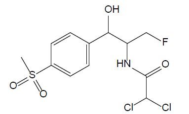
Amphenicols

Amphenicols are important veterinary antibiotics with wide-spectrum antimicrobial activity. Florfenicol (FF, see Figure 2) is an antimicrobial agent, which is extensively used in fish farming. Grave et al. investigated the use of antimicrobial drugs in Norwegian aquaculture from 2000 to 2005 by analyzing prescription data, in close relation with the national data of the sold antimicrobial drugs. FF has long been the most abundant drug prescribed for halibut farming in Norwegian aquaculture [16]. In 2013, 0.3 tons and 300 tons of FF were used in fish farming in Norway and Chile, respectively. In 2016, it was reported that over 1000 tons of FF were used in China. Temperature, exposure time, coating agent, and pellet size are the important factors that influence release of FF into the water [11]. According to the EU Council directive, the maximum residue limit value of the sum of FF and florfenicol amine in muscle and skin in natural proportions of healthy fish was given as 1,000 μg/kg (Commission Regulation EU No 37/2010). FF is bacteriostatic, which means it prevents the bacteria from protein synthesis [11]; it is usually used for respiratory and intestinal infections [17].

fig 2

Figure 2: Chemical structure of chloramphenicol.

Sulfonamides

Sulfonamides are used as chemotherapeutics to treat various bacterial infections in veterinary medicine [5]. Due to their broad-spectrum antimicrobial activity and low cost, they were among the most applied antibiotics and thus, commonly detected in aquaculture wastewater [18]. Currently, only a few drugs belonging to sulfonamides are used due to the developed resistance in previously susceptible microorganisms. Sulfadiazine (SDZ) and sulfadimethoxine (SDM) are the most used antibiotics in fish farming; their chemical structures are displayed Figure 3. When applied to animals, sulfadiazine, is excreted in its native form and its N4-acetyl metabolite [19]; it is often considered as a representative antibiotic of sulfonamides due to its wide presence in the environment with low hydrophobicity and high water solubility in [20]. Sulfadimethoxine (SMX – Figure 4) is a broad-spectrum antibiotic used in human therapy, aquaculture, livestock, and veterinary medicine. Although its use is decreasing in humans, its low cost secures its popularity in veterinary medicine [21].

fig 3

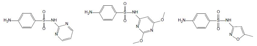
Figure 3: Chemical structures of sulfadiazine (left hand), sulfamethoxine (middle) and sulfamethoxazole (right hand).

fig 4

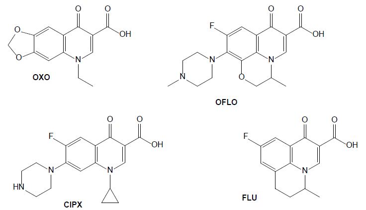
Figure 4: Chemical structures of some quinolone drugs: oxolinic acid (OXO), ofloxacin (OFLO), ciprofloxacin (CIPX) and flumequine (FLU).

Quinolones

The antibiotics from the group of quinolones are widely used in human and veterinary medicines to treat infectious diseases and to promote livestock growth (Yang et al., 2020). They directly inhibit DNA replication by interacting with two enzymes. Oxolinic acid (OXA) is efficient against a gram-negative bacterium that causes diseases such as vibriosis, yersiniosis, and furunculosis. It has been administrated to farm fishes as a prophylactic and chemotherapeutic agent acting as anti-infective, antibacterial and enzyme inhibitor [22]. Its use in humans is now prohibited in several countries but is still frequently used in veterinary medicine to treat urinary infections, and it was detected in animal excreta [23]. Ofloxacin (OFLO) is a quinolone that was previously used for human health, for the therapy of mild to moderate bacterial infections, it has since been replaced by more potent and less toxic antibiotics, however it is still used in aquaculture. In the group of quinolones, ciprofloxacin (CIPX) is considered as a representative drug against gram-negative bacteria; it has been detected in marine environment, as well as in freshwater. In aquatic environments it affects the metabolism of some bacteria carbon sources and is toxic to fishes (Yang et al., 2020). Flumequine (FLU), as a first generation quinolone, is structurally related to nalidixic and oxolinic acid [24]. Oxidation experiments carried out on flumequine led to a total of 19 transformation products issued from hydroxylation, dehydrogenation, hydroxyl substitution, decarboxylation, demethylation, and ring opening transformation pathways [25].

Macrolides

The group of macrolides refers to macrocyclic lactone ring structures; they are used for their immunomodulatory and antibacterial functions (Yang et al., 2020). In addition to their antibacterial action, macrolides can have an anti-inflammatory effect by decreasing the activity of immune cells and altering bacterial cells. Erythromycin (ERY, see Figure 5) is efficient against gram-positive bacteria, as streptococcus species. However, most of the microorganisms that cause infection in fish are gram-negative, so this compound should only be utilized after fish culturing and sensitivity test results should affirm its viability. Additionally, erythromycin is not efficient in bath treatment and must be administered by injection or in the feed [26].

fig 5

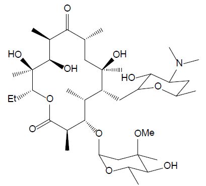
Figure 5: Chemical structure of erythromycin.

Nitrofurans

Nitrofurans are antimicrobial agents which have been used for animal production. They have strong impact on gram-negative and gram-positive bacteria and act against protozoa as well. In 1993, their application has been banned by EU due to their potential mutagenic effect. Furazolidone (FUR, see Figure 6) is a nitrofuran antibiotic widely used in human and aquaculture medicine against protozoal and Helicobacter pylori infections. Its biodegradation leads to 3-amino-2-oxazolidone and β-hydroxyethylhydrazine [27]. It is still utilized in developing countries, although it is banned in developed countries.

fig 6

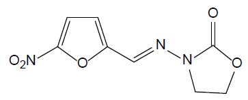
Figure 6: Chemical structure of furazolidone.

Ecotoxicology of the Main Drugs Used for Fish Farming

Table 1 presents a summary of the ecotoxicological data available for veterinary drugs mainly used in fish farming. The data are discussed below, for each pharmaceutical family.

Table 1: Ecotoxicology data for the veterinary drugs mainly used in fish farming

Drug

Ecotoxic effect

Reference

Tetracycline & oxytetracycline, 24 h-EC50 for Stentor coeruleus and Stylonychia lemnae were 94.4 mg/L and 40.1 mg/L, respectively Magdaleno et al., 2017
Florfenicol Inhibiting growth rate of Corbicula fluminea at concentrations higher than 1.8 mg/L Guillermino et al., 2017
Sulfadiazine LC50 of 1.884 mg/L for Daphnia magna Duan et al., 2020
Sulfamethoxazole IC50 = 12.56 ± 4.48 mg/L for A. fischeri Drzymała & Kalka, 2020
Sulfadimethoxine EC50 of 248 mg/L for daphnia magna Tkaczyk et al., 2021
Oxolinic acid 4.6 mg/L EC50 for Daphnia magna Tkaczyk et al., 2021
Ofloxacin Chronic toxicity and drug resistant bacteria H. Guo et al., 2021
Ciprofloxacin Concentrations greater than or equal to 10 μg/L: ecotoxic for development, growth, detoxification and oxidative stress enzymes Białk-Bielińska et al., 2011
Flumequine Affecting growth rate of Daphnia magna at 23% across generation De Liguoro et al., 2019
Erythromycin EC50 values in the range of 10 -30 mg/L for Vibrio fischeri Liu et al., 2018
Furazolidone Very toxic to Alivibrio fischeri with an EC50 of 2.05 mg/L Lewkowski et al., (2019)

Tetracyclines

In aquaculture, oxytetracycline is one of the most used antibiotics that raises concerns due to its effects on human and animal health, and environmental pollution [8]. In the marine environment, oxytetracycline and quinolones are photochemically degraded and form divalent cationic complexes in the presence of Ca2+ and Mg2+ ions, causing a loss in antibacterial activity. In freshwater, the antimicrobial activity of tetracyclins is of bigger concern due to the development of antibiotic resistance [28]. OTC inhibits the growth of two species of algae: pseudokirchneriella subcapitata (the international standard species for evaluation of inhibition) and Ankistrodesmus fusiformis (native from Argentina). The former was the most sensitive with an EC50 of 0.92±0.30 mg/L [29]. OTC can also affect nitrification since the main bacteria responsible for biofilters – nitrosomonas that promote the conversion of ammonia to nitrite and Nitrobacter that convert nitrite to nitrate – are gram-negative. Li et al. investigated the toxic effect of tetracycline and tetracycline hydrochloride on two model ciliates: Stentor coeruleus and Stylonychia lemnae. The 24 h-EC50 of tetracycline for Stentor coeruleus and Stylonychia lemnae were 94.4 mg/L and 40.1 mg/L, respectively. For tetracycline hydrochloride, the EC50 for Stentor coeruleus and Stylonychia lemnae were 8.39 mg/L and 14.0 mg/L, respectively [15]. This shows that tetracycline hydrochloride is more toxic than tetracycline in these test organisms. Both compounds deteriorate the ultra-cell structure and inhibit the cell growth rate [29].

Amphenicols

Florfenicol (FF) restrains the bacterial protein synthesis efficiency by binding to 50S subunit and 70S ribosome, and it is listed as the top-priority monitoring compounds in veterinary drugs in South-Korea [30]. FF also inhibits the hematopoietic system [31] and deteriorates the mobility, regeneration, and population increase of the tropical Cladocera silvestrii [32]. It also decreases egg hatchability and hinders the development of the cardiovascular system [33]. Guilhermino et al. showed that a FF concentration in water greater than 1.8 mg/L can inhibit the growth of the mollusk Corbicula fluminea [34].

Sulfonamides

The chronic and acute toxicity of sulfadiazine on the crustacean Daphnia magna was investigated and the LC50 value was determined as 1.884 mg/L [35]. The exposition of zebrafish to some sulfonamides, including sulfadiazine, caused an increased heart rate and abnormal swimming. This investigation also proved that sulfadiazine was a typical inducer of metabolic enzymes and suggested a potential ecotoxicological risk [36]. It is important to consider that sulfadiazine can have an impact on biological water treatment processes. Li et al. studied the effects of this antibiotic on a sequencing batch biofilm reactor (SBBR). They found that a sulfadiazine concentration higher than 6 mg/L inhibits COD (Chemical Oxygen Demand) and ammoniacal nitrogen removal within the SBBR when treating aquaculture waste [37]. Sulfadiazine promotes the secretion of extracellular polymeric substances by the microorganisms and impacts the biofilm composition, it has a direct impact on nitrification and the removal of organic matter. Sulfamethoxazole has been studied in a microcosmos composed of water, sediment and zebrafishes. The drug concentration decreased gradually in water while increasing over time in sediment and zebrafish. Bioaccumulation in zebrafish was reduced by 13-28% in the presence of sediment particles in the water; it was further reduced (24-33%) when increasing the water salinity [38]. In the study conducted by [39], sulfadimethoxine was considered to be moderately toxic with an EC50 towards the green algae Chlorella vulgaris of 4.24 mg/L in fresh water, which was measured to be higher (11.2 mg/L) in salt water [39].

Quinolones

According to a study by Tkaczyk et al., oxolinic acid EC50 was 4.6 mg/L EC50 for Daphnia magna [40]. Sun et al. investigated the bioaccumulation of ofloxacin in crucian carp and showed that fluorine increases the bioaccumulation of the antibiotic; higher bioaccumulation potential appears at a low concentration of drug, mostly in the liver [41]. De Liguoro et al. evaluated the effect of continuous and alternate exposure of flumequine at 2 mg/L on Daphnia magna survival, growth, and reproduction. At this concentration, mortality was observed at a rate of 23  ± 14% across generation [42].

Macrolides

Erythromycin has shown medium risk for algae, but bacteria are the main target of antibiotics such as erythromycin. Erythromycin can cross the cell membrane of the bacteria and bind to 50S subunit ribosome [43]. It is also very persistent in the environment due to having aromatic rings, which makes it refractory [44].

Nitrofurans

Lewkowski et al. investigated the effect of furazolidone on growth of oat, radish Sativus, and Alivibrio fischeri bacteria. Their results indicated that furazolidone is very toxic to Alivibrio fischeri with an EC50 of 2.05 mg/L [45]. The accumulation of furazolidone metabolites have been reported in the literature [5]; its use is prohibited by the Food and Drug Administration due to being a nitrofuran compound and inhibiting monoamine oxidase [46].

Methods for Characterization of Drugs Used in Fish Farming

Table 2 presents a summary of the analytical approaches employed for the detection and quantification of veterinary drugs mainly used in fish farming; protocols and methods are discussed below.

Table 2: Analytical methods reported for the detection and quantification of the main antibiotic veterinary drugs used in fish farming

Drug type

Matrix Characterization methoda MS typeb Sample preparationc Analytical column LC Mobile phased

Reference

Tetracyclins including OTC Feeds HPLC-UVD/FLD LLE using acetonitrile and Na2EDTA-Mcllvaine buffer solution C18 Hypersil GoldTM (250 x 4.6 mm, 5 μm) Phase A: H2O + sodium acetate CaCl2 + EDTA

Phase B: ACN

Phase C: MeOH

Han et al., 2020
Antibiotic drugs

Including SMX, SDZ, SMZ, CIPX, OFLO, TET and ERY

Wastewater UPLC-MS/MS TQ SPE using an Oasis HLBTM cartridge) C18 BEHTM (50 x 2.1 mm, 1.7 µm) Phase A: H2O + FA

Phase B: ACN or MeOH

Li et al., 2009
Sulfonamides, fluoroquinolones, tetracyclines and other veterinary drugs Seafood samples UPLC-MS/MS TQ QuEChERS procedure C18 Hypersil GoldTM (100 × 2.1 mm, 1.9 μm) Phase A: H2O / MeOH + FA

Phase B: ACN + FA

Dinh et al., 2020
Metabolites of nitrofurans and FUR Shrimp body LC-MS/MS TQ Hydrolysis + double LLE using ethyl acetate C18 SymmetryTM (150 x 2.1 mm, 3.5 μm) Phase A: ACN

Phase B: H2O + FA

Douny et al., 2013
4 tetracyclines including TET, OTC; quinolones including FLU, CPIX and OXA Fish muscles LC-MS/MS TQ Liquid extraction with trichloroacetic acid C18 Zorbax Eclipse XDBTM (150 × 4.6 mm, 5.0 µm) Phase A: H2O + HFBA

Phase B: ACN

Guidi et al., 2018
OTC Sol interstitial water LC-MS/MS TQ Centrifugation, filtration C18 Xterra MSTM (100 x 21 mm, 3.5 µm) Phase A: MeOH + FA

Phase B: MeOH / H2O + FA

Halling-Sørensen et al., 2003
FF Water in recirculating aquaculture system HPLC-PDA SPE using an Oasis HLBTM cartridge Hypersil GOLDTM (250 x 4.6 mm, 5 μm) Phase A: ACN

Phase B: H2O

Zhang et al., 2020
FF and its residues Beef meat LC-MS/MS TQ SPE using an Oasis MCXTM cartridge C18 Inertsil ODS-4TM (150 × 2.1 mm, 3-μm) Phase A: H2O + acetic acid

Phase B: ACN

Saito-Shida et al., 2019
FF Fish feed LC-MS/MS TQ Centrifugation with ACN addition C18 SBTM (50 x 2.1 mm, 1.8 µm) Phase A: H2O + acetic acid

Phase B: ACN / MeOH

Barreto et al., 2018
14 sulfonamide antibiotic residues 309 marine products HPLC-PDA


UPLC-MS/MS

TQ Centrifugation with ACN addition C18 CapcellpakTM (250 x 4.6 mm, 5 ㎛) for HPLC-PDA


C18 Acquity UPLC BEHTM (100 x 2.1 mm, 1.7 µm) for UPLC-MS/MS

Phase A: H2O + KH2PO4

Phase B: MeOH

for HPLC-PDA


Phase A: H2O + FA

Phase B: ACN + FA

for UPLC-MS/MS

Won et al., 2011
20 antibiotics including FF, SDZ, SMX, CPIX, TET, OTC and ERY Surface waters UPLC-MS/MS TQ SPE using an Oasis HLBTM cartridge C18 HSS T3TM (100 x 2.1 mm, 1.8 µm) Phase A: H2O + FA

Phase B: ACN + FA

Yan et al., 2013
19 sulfonamides including SDZ, SDM and SMX Ebro river, WWTP samples LC-MS/MS QqLIT on-line SPE using Oasis HLBTM or Oasis MCXTM cartridges C18 AtlantisTM (150 × 2.1 mm, 3 µm Phase A: H2O + FA

Phase B: ACN + FA

García-Galán et al., 2011
73 pharmaceuticals including TET, OTC, SDZ, SMX, OFLO, FLU, CPIX and ERY River water, WWTP influent and effluent LC-MS/MS QqLIT on-line SPE using a Oasis HLBTM cartridge C18 Purospher Star RP-18TM endcapped column (125 × 2.0 mm, 5 μm) Phase A:

ACN / MeOH

Phase B: H2O

Gros et al., 2009
20 antibiotics including

CIPX, ERY, SMZ, SMX, TET and OTC

Raw influent, treated effluent, surface water LC-MS/MS TQ on-line SPE using a combination of SBTM and HR-XTM cartridges C18 Poroshell 120 ECTM (100 x 3.0 mm, 2.7 μm) Phase A: H2O + FA

Phase B: ACN + MeOH

Tran et al., 2016
SMX and its photoproducts Aquatic organisms LC-MS/MS TQ No sample preparation C18 Zorbax eclipse plusTM (50 x 2.1 mm, 1.8 µm) Isocratic mobile phase H2O / ACN Li et al., 2020
OTC, FF, OXA and FLU Marine sediments HPLC-PDA-FLD LLE (using oxalic acid in methanol) assisted by sonication Kromasil PhenylTM (250 x 4.6 mm, 5 µm) Phase A: H2O + TFA

Phase B: MeOH + TFA Phase C: ACN + TFA, with or without gradient

Norambuena et al., 2013
46 antimicrobial drug residues including sulfonamides, tetracyclines, quinolones, macrolides, nitrofurans and phenicols Aquatic matrices, pond water UPLC-HRMS OrbitrapTM SPE using an Oasis HLBTM cartridge C18 Hypersil GoldTM (100 × 2.1 mm, 1.9 μm) Phase A: H2O + TFA

Phase B: MeOH + TFA

Goessens et al., 2020
11 fluoroquinolones including OFLO and CIPX Wastewater and sludges UPLC-MS/MS Q-trap SPE using molecularly imprinted polymer cartridges C18 Proshell 120 SBTM (100 × 2.1 mm, 2.7 µm) Phase A: H2O + FA

Phase B: MeOH + FA

Yu et al., 2020
Fluoroquinolone residues Sewage samples LC-MS/MS TQ on-line SPE with micellar desorption C18 SymmetryTM (150 x 3.9 mm, 4 µm) Isocratic mobile phase consisting of H2O + MeOH Montesdeoca-Esponda et al., 2012
FLU and OXA Aquatic sediments and agricultural soils HPLC-FLD MAE C8 InertsilTM (250 × 4.6 mm, 5 µm) Isocratic mobile phase consisting of H2O + oxalic acid buffer + ACN Prat et al., 2006
FLU Ultrapure water, tap water, secondary clarifier effluent and river water HPLC-FLD


LC-MS/MS

Q-TOF Microfiltration C18 BDS HypersilTM (250 x 4,6 mm, 5 μm) Phase A: H2O + FA

Phase B: MeOH

Qui et al., 2019

a LC: liquid chromatography, HPLC: high performance liquid chromatography, UPLC: ultrahigh performance liquid chromatography, UVD: UV detector, FLD: fluorescence detector, PDA: Photodiode array detector, MS/MS: tandem mass spectrometry, HR-MS: high resolution mass spectrometry
b Q: quadrupole, TQ: triple quadrupole, q: collision cell, TRAP: ion trap, LIT: linear ion trap, TOF: time-of-flight
c LLE: liquid-liquid extraction, SPE: solid phase extraction, QuEChERS: sample preparation method (Quick, Easy, Cheap, Efficient, Rugged and Safe), MAE : microwave assisted extraction
d ACN: acetonitrile, MeOH: methanol, EDTA: ethylenediaminotetraacetic acid, FA: formic acid, TFA: trifluoro acetic acid

The first step of the analytical process consists in sample preparation, which includes extraction of analytes from the matrix, purification and concentration. When imposed by the analytical technique, a derivation step is added at the end of the process. Solid phase extraction (SPE) has been extensively reported as a tool for choice for the extraction of the pharmaceuticals mainly used in fish farming because (i) it is suitable for very complex matrices, (ii) it is highly selective, and (iii) it permits significant pre-concentration. Furthermore, SPE can be installed on-line with a chromatographic system and automated, which allows high throughput analysis; this is the case for some studies reported in Table 2 [47]. Oasis HLB™ are the most used cartridges in the reported studies, as they are suitable for the simultaneous extraction of compounds from various families: sulfonamides (SMX, SDZ, SMZ), tetracyclines (TET, OTC), quinolones (OFLO, FLU, CPIX), macrolides (ERY), nitrofurans and phenicols (FF) [15,23,47]. Apart from SPE-based approach, various sample preparation processes have been reported, including liquid-liquid extraction (LLE) with or without sonication or microwave assistance (Norambuena et al., 2013; Prat et al., 2006). In a general way, acetonitrile appears as the solvent of choice for the extraction of these molecules.

In most of the studies devoted to drugs used in fish farming, analytes are separated using liquid chromatography. High-performance liquid chromatography (HPLC) is the most widely applied technique to detect veterinary drugs with great accuracy and reliability [48]; it is nevertheless more and more replaced by UPLC (Ultrahigh Performance Liquid Chromatography), which uses a new generation of columns filled with small particles of hybrid material (diameter at the μm scale) and operates with a much higher column pressure (up to 15,000 psi) than HPLC. UPLC considerably reduces runtime and thus increases sample throughput; it performs also much better in terms of resolution, sensitivity, and separation efficiency; it is almost always coupled with a mass spectrometer (see below). Li et al. developed a rapid, sensitive, and reliable UPLC-MS/MS method for the determination of 21 antibiotics belonging to 7 classes in different wastewater matrixes (Li et al., 2009). Yu et al. reported a selective and sensitive method to measure 11 antibiotics in water by UPLC-MS/MS; it was successfully used to identify ofloxacin in wastewater and sludge samples [49]. Dinh et al. used UPLC-MS/MS to find different veterinary drugs including furazolidone, with a limit of detection of 1.5-3 μg/kg for the later [5]. Whatever the chromatographic system (HPLC or UPLC), most of the studies were conducted using reverse phase chromatography. Many brands and geometries were tested but at the end the retained columns are almost all C18-types. The mobile phase is almost always a very classical one in the following type of configuration: a binary gradient made of an aqueous phase (ultrapure water) and an organic one (ACN or MeOH), both acidified with a small organic acid (acid formic, acetic, trifluoroacetic…) generally at 0.1%. Among all the studies listed in Table 2, the only one that uses a non C18 stationary phase (phenylpropylsilane phase) is that by Norambuena et al., devoted to the analysis of oxytetracycline, florfenicol, flumequine and oxolinic acid in marine sediments (Norambuena et al., 2013). In this work, HPLC was equipped with a PDA (photodiode array detector). HPLC with PDA detection was also used for the detection of florfenicol in water by Zhang et al. and that of sulfonamide residues by Won et al. [50,51]. HPLC-PDA has been also reported for the detection of sulfadiazine in South-Korean marine products, although SDZ was only identified in 2 of the 10 samples analyzed [51]. Han et al. reported a combination of UVD (UV detector) and FLD (fluorescence detector) for the analysis of tetracyclines in feeds [48]. Studies reporting the detection of flumequine and oxolinic acid by HPLC-FLD in sediments, soils and various types of water have been also reported [25]. In a general way “classical” HPLC detectors tend to be gradually replaced by mass spectrometers, which are today recognized as the best detectors in terms of sensitivity, specificity and selectivity.

In almost all of the studies conducted using LC-MS coupling, the liquid chromatograph (HPLC or UPLC) is coupled with a triple quadrupole (TQ) operated in the MRM (multiple reaction monitoring) mode on two transitions. Tandem mass spectrometry (MS/MS) is preferred to simple MS as it strongly reduces the risk of false negatives when performing analysis of complex matrices (Bouchonnet, 2013). Hybrid mass spectrometers associating a quadrupole, a collision cell, and an ion trap have been also reported; their principle of operation is quite similar to that of TQs except that the ion trap permits ion accumulation before detection [47,49]. Among all the studies listed in Table 2, only that by Goessens et al. refers to a high-resolution mass spectrometer (Orbitrap™), which has been employed to analyze 46 antimicrobial drug residues including sulfonamides, tetracyclines, quinolones, macrolides, nitrofurans and phenicols in aquatic matrices [23]. Since it allows the differentiation of isobaric ions, high-resolution mass spectrometry (HRMS) constitutes a tool of choice for structural elucidation of metabolites and by-products; it also significantly reduces interferences during routine analysis of very complex matrices. The use of MS/MS and/or HRMS enables the development and validation of multi-residue methods able to detect and quantify many molecules in one unique run. Even if the number of analytes remains limited in comparison with methods reported for pesticide residue dosages (hundreds of analytes), some of the methods reported for the analysis of veterinary antibiotics allows the simultaneous detection of tens of drugs. For instance, Gros et al. developed a LC-MS/MS method for the one run detection of 73 pharmaceuticals including tetracycline, oxytetracycline, sulfadiazine, sulfamethoxazole, ofloxacin, flumequine, ciprofloxacin and erythromycin in river water and WWTP influent and effluent while a LC-HRMS was developed by Goessens et al. for the analysis of 46 antimicrobial drug residues including sulfonamides, tetracyclines, quinolones, macrolides, nitrofurans and phenicols in aquatic matrices [23].

A few analytical approaches were developed apart from traditional separative processes such as liquid chromatography. For example, an aptaprobe was used for the detection of sulfadimethoxine; it simply and conveniently detects SDM with accuracy in the range of 94.2-113% in seawater and 104-118% in fish [52]. A dichromatic label-free aptasensor detects SDM presence through fluorescent emission and color changes of gold nanoparticles. This aptasensor can be applied to the rapid detection in fish and water samples with accuracies between 99.2 and 102% for fish, 99.5 and 100.5% for water [53]. Almeida et al. also developed a new low-cost plastic membrane electrode that detects low concentrations of SDM in aquaculture waters. To test this device, sulfadimethoxine was added to aquaculture waters and the results showed a good agreement between added and measured drug amounts with recoveries ranging from 96.8% to 101%, with a relative error between -0.7% and 3.5%, which suggests that it constitutes a good method that could be extended to the determination of other pharmaceuticals in water [54].

Treatment Processes

Once they reach the environment, micropollutants are subjected to three main degradation processes: biodegradation, hydrolysis, and photolysis [8]. Due to their own antibacterial activity, antibiotics are poorly or not degraded by natural biotic processes. Chemical and physical processes – natural or industrial – can be considered as a better option to remove these refractory veterinary drugs. As we will see below, many treatments have been considered for their degradation and/or removal from aqueous media; they include membrane anodic Fenton, advanced oxidation processes, heterogeneous photocatalysis, and electrocoagulation [55]. Table 3 lists some treatments applied for the removal of veterinary pharmaceuticals considered individually.

Table 3: Various treatment methods for the removing of the veterinary drugs mainly used in fish farming from aqueous media

Treatment process

Drug

Reference

H2O2/Fe + UV treatment Tetracycline, oxytetracycline Zhao et al., 2020
CaO2/UV Florfenicole Zheng et al., 2019
Photodegradation with TiO2, Sulfadiazine He et al., 2016
Batch culture of c. vulgaris microalgae Sulfamethoxazole Y.-Y. Peng et al., 2020
Ozonation Sulfadimethoxine

Erythromycin

A.Y.-C. Lin et al., 2009
Heterogeneous photocatalysis with suspended TiO2 Oxolinic acid Giraldo et al., 2010
Ultraviolet / peroxydisulfate Ofloxacin Zhu et al., 2020
Reverse osmosis membrane Ciprofloxacin Alonso et al., 2018
Electrochemical cathode degradation, Magnetic biochar Furazolidone Kong et al., 2015; Gurav et al., 2020

Using a pilot drinking water treatment plant, Vieno et al. observed the elimination of some pharmaceuticals with a process consisting in ferric salt coagulation, rapid sand filtration, ozonation, and two-stage granular activated carbon filtration. Most pharmaceuticals ceased to be quantifiable at the end of ozonation; only ciprofloxacin passed all treatment steps almost unaffected [56]. No treatment appears as a universal solution at this day. For instance, classical wastewater treatments are known to be inefficient for the elimination of sulfonamides [47]. Tetracycline is hard to degrade using conventional wastewater treatments such as activated sludge, which might be due to tetracyclines’ strong hydrophilic properties related to their stable naphthalene ring structure (Zhao et al., 2020). The biological degradation of oxytetracycline is limited due to its broad-spectrum antimicrobial properties, which are considered responsible for the development of antibiotic resistance genes in the environment (Xie et al., 2016). OTC fails to be degraded into non-toxic transformation products by most abiotic processes; therefore sonocatalytic degradation has been proposed to form less toxic intermediates [57].

Most of the alternative approaches for the removal of the most resistant molecules are based on photocatalytic or electrochemical approaches. For instance, sulfamethoxazole is not efficiently removed in conventional wastewater treatment plants [47] but it is susceptible to photodegradation in aqueous solutions along several pathways [58]. UV-photolysis appears as a treatment of choice for many micropollutants, especially in the presence of a catalyst that increases both the kinetics and yields of the degradation reaction. UV-irradiation induces the formation of hydroxyl radicals from water and dissolved oxygen; these highly reactive radicals are responsible for the oxidation of micropollutants. The addition of hydrogen peroxide, alone or with a metal has been frequently reported. Zhao et al. evaluated the effect of H2O2/Fe addition to the UV treatment of tetracycline. They found out that optimum concentrations of H2O2 (0.5 mM) and Fe(II) (0.05 mM) promote the degradation of TET. Also, a higher pH level facilitated the UV-attenuation of TET (Zhao et al., 2020) while a lower pH helped its degradation under ozonation conditions. Ming Zheng et al. utilized CaO2/UV as an advanced oxidation process to remove FF and other active pharmaceutical compounds from wastewater. CaO2 is considered as the “solid form” of H2O2 with an advantage of being more stable than H2O2 in presence of base or catalyst. CaO2 can produce .OH And O2. radicals and oxidize complex chemical compounds. The highest removal of FF happened at 0.1 g.L-1 of CaO2. The addition of CaO2 also allows reducing the irradiation time and so decreasing the consumption of energy [31]. Titanium oxide appears as a widely used photocatalyst for the removal of pharmaceuticals. UV-irradiation with TiO2 removed 99% of sulfadiazine under the following conditions: initial concentrations of sulfadiazine at 5.0 mg/L and TiO2 at 0.08 g/L, pH = 7, radiation intensity of 1000 μw/cm2 and reaction time of 50 min [59]. SDZ can be also effectively degraded using gamma irradiation, the elimination efficiency being improved under acidic conditions [60]. Degradation of sulfamethoxazole under ultraviolet light with TiO2 reached 96%. TiO2 is the most suitable catalyst due to its stability, non-toxicity, and high catalytic activity [59]. Oxolinic acid was degraded using heterogeneous photocatalysis with titanium dioxide suspended on particles. After 30 min under optimal conditions both the substrate and the microbial activity were eliminated [61] and the residual byproducts did not show antibacterial activity [62]. Organic matter can both hinder and activate the photodegradation. For instance, oxolinic acid persistence is lower in ultrapure water than in environmental water, especially in the presence of high salinity values. The presence of organic matter can decrease the photodegradation rate in freshwater by acting as a light filter and hydroxyl radicals scavenger [63]. An ultraviolet/peroxydisulfate system was reported for the degradation of ofloxacin in synthetic seawater and in synthetic marine aquaculture water; the global toxicity (including toxicities of reagents and by-products) induced by such a process is lower than that induced with traditional approaches using NaClO [64]. Traditional removal processes only based on chemical reactions tend to be replaced by the so-called AOPs (advance oxidation processes) but some of them remains in use. For instance, [21] demonstrated that sulfadimethoxine can be removed by potassium permanganate in water. The degradation is affected by the pH of the solution and a higher temperature is also beneficial for the removal [21]. The use of zero-valent iron-activated persulfate has been developed to remediate antibiotic-contaminated wastewater since it removes 69% and 74% of SDM from filtered and unfiltered discharge water, respectively [65]. It has to be kept in mind that in a general way catalysis is hardly applied to complex matrices (water containing high levels of dissolved organic matter for instance) as it becomes quite inefficient in the presence of large amounts of organic and inorganic species. Furthermore, altering the pH when necessary can be very costly.

Electro-Fenton technology is also an advanced oxidation process that produces hydroxyl radicals to degrade refractory pollutants. However, this process may be inefficient due to the high dissociation energy of some chemical bonds, such as C-F in florfenicol. On the other hand, UV light has shown to be capable of cleaving such bonds, and the combination of Electro-Fenton with UV could be an efficient technology. Jiang et al. coupled the photoelectrochemical reaction in a sequential filtration system to degrade and mineralize FF. Their study revealed 78.1 ± 9.1% mineralization of FF at a low concentration of 14 µM [66]. Various technologies have been studied to remove furazolidone from wastewater samples; among them, Kong et al. used an electrochemical system to degrade FUR in a cathode compartment. Their study evaluated the effect of different cathode potentials, initial antibiotic concentration, and cathode buffer solution on FUR degradation. Different cathode potentials resulted in different degradation products, and different buffer solutions and initial concentrations of furazolidone had just an obvious effect on its removal efficiency [67].

Ozonation has also been reported as a powerful tool for micropollutants abatement. For example, it allowed the degradation of tetracycline at 99.5% with 40% of mineralization (C. Wang et al., 2020). Sulfadimethoxine showed to be completely removed from water by ozone bubbling within 20 minutes; this strong efficiency was partially explained by the presence of one or several aromatic rings on which the O3 molecule can fix itself before hydrolysis and/or ring opening [68]. Ozonation at an application rate of 0.17 g O3/min was able to remove some antibiotics in about 20 min, although the degradation of erythromycin was slower, and more effective at a high pH or with H2O2 addition [68].

As ciprofloxacin is particularly refractory to conventional wastewater treatments, special attention has been paid to its removal using alternative processes. Reverse osmosis was successfully tested for the removal (99.96% removal) of CPIX in seawater [69]. Additionally, ciprofloxacin removal by electrosorption has been successfully demonstrated using graphite felt electrodes [70].

Finally, and despite their microbial activity, some veterinary drugs were submitted to treatments involving biological processes. In two sewage treatment plants in Guangzhou, more than 85% of ofloxacin was removed in the effluents after activated sludge [71]. A novel microalgae biofilm membrane photobioreactor (MBMP) was developed for the cultivation of microalgae and the removal of sulfonamides from residual wastewater of aquaculture. The reduction of sulfadiazine in the MBMP during its stable operation was up to 61-79.2%. It can be considered that the performance of the MBMP is higher than the one achieved by traditional batch cultivation [18]. The removal of sulfamethoxazole by a batch culture of microalgae c. vulgaris was 34.07% after 12 days of concentration in marine aquaculture wastewater (against 3.33% without microalgae). Gurav et al. used a magnetic biochar to remove furazolidone from wastewater; they showed that the magnetic biochar had a higher surface area as compared to normal biochar, and possessed a much better removal efficiency [27].

Conclusion

Aquaculture is a booming industry, which by necessity uses drugs to prevent and treat diseases in fish farms. The current literature has been mainly focused on the possible adverse effects on human health due to the possible remnants of these drugs in fish. In recent years, the presence of drugs in environmental matrices has become more evident but the effects of aquaculture waste on the environment have been poorly documented. Thanks to the continuous improvement of analytical methods, it is now possible to successfully determine veterinary pharmaceuticals at trace amounts in complex matrices. The most common analytical processes rely on solid phase extraction and liquid chromatography – HPLC or UHPLC – coupled with mass spectrometry. Current efforts are aimed at developing multiresidue methods for the simultaneous analysis of various drugs from different families. In any case, the literature directed specifically to the water discarded by aquaculture is very scarce. Although most of the analytical methods are focused on separative processes, recent ventures successfully tested aptasensors and aptaprobes to quickly and efficiently measure low concentrations of antibiotics in water.

Waste from agriculture and aquaculture usually reaches the environment directly, without being previously treated. Currently, there is more knowledge about the toxic effects of pharmaceuticals in humans and animals than about their environmental impact. A good evaluation of their potential ecotoxicological impact should consider factors such as the presence of sediments, the stability of water and other substances with which the drugs can interact since all these parameters may affect the final result of the investigations [72]. It is known that contamination by antibiotics includes the development of resistance in the aquatic pathogens, direct toxicity to microflora and microfauna, and even possible risks to human health due to the consumption of non-target contaminated benthic fauna [73]. For this reason, ways to treat or eliminate these pollutants are still under investigation given that conventional wastewater treatment plants are not fully efficient – and sometimes quasi inefficient – for their removal. The antimicrobial activity of pharmaceuticals makes their treatment through biotic processes difficult and advanced oxidation processes appear as a tool of choice for their removal. Photocatalysis and electrochemistry are not really viable on a wide scale to this day but ozonation and UV/H2O2 oxidation are much more applicable. In a general way, it is of first importance to mitigate the ecotoxicological risks associated with aquaculture waste released into the sea, by (i) selecting the proper veterinary drugs, (ii) limiting the amount of waste release from fish farming and (iii) setting efficient solutions for the removal of pharmaceuticals once or before they reach the environment [74-77].

References

  1. FAO (2020) The State of World Fisheries and Aquaculture 2020. FAO.
  2. Castilla-Fernández D, Moreno-González D, Bouza M, Saez-Gómez A, Ballesteros E, et al. (2021) Assessment of a specific sample cleanup for the multiresidue determination of veterinary drugs and pesticides in salmon using liquid chromatography/tandem mass spectrometry. Food Control 130: 108311.
  3. Chokmangmeepisarn P, Thangsunan P, Kayansamruaj P, Rodkhum C (2021) Resistome characterization of Flavobacterium columnare isolated from freshwater cultured Asian sea bass (Lates calcarifer) revealed diversity of quinolone resistance associated genes. Aquaculture 544: 737149.
  4. Pastor-Belda M, Campillo N, Arroyo-Manzanares N, Hernández-Córdoba M, Viñas P (2020) Determination of amphenicol antibiotics and their glucuronide metabolites in urine samples using liquid chromatography with quadrupole time-of-flight mass spectrometry. Journal of Chromatography B 1146: 122122.
  5. Dinh QT, Munoz G, Vo Duy S, Tien Do D, Bayen S, et al. (2020) Analysis of sulfonamides, fluoroquinolones, tetracyclines, triphenylmethane dyes and other veterinary drug residues in cultured and wild seafood sold in Montreal, Canada. Journal of Food Composition and Analysis 94: 103630.
  6. Du J, Li X, Tian L, Li J, Wang C, et al. (2021) Determination of macrolides in animal tissues and egg by multi-walled carbon nanotube-based dispersive solid-phase extraction and ultra-high performance liquid chromatography–tandem mass spectrometry. Food Chemistry 365: 130502.
  7. Lulijwa R, Rupia EJ, Alfaro AC (2019) Antibiotic use in aquaculture, policies and regulation, health and environmental risks: A review of the top 15 major producers. Reviews in Aquaculture 12: 640-663.
  8. Leal JF, Santos EBH, Esteves VI (2018) Oxytetracycline in intensive aquaculture: Water quality during and after its administration, environmental fate, toxicity and bacterial resistance. Reviews in Aquaculture 11: 4.
  9. Archundia D, Duwig C, Lehembre F, Chiron S, Morel MC, et al. (2017). Antibiotic pollution in the Katari subcatchment of the Titicaca Lake: Major transformation products and occurrence of resistance genes. Science of The Total Environment 576: 671-682.
  10. Krzeminski P, Tomei MC, Karaolia P, Langenhoff A, Almeida CMR, et al. (2019) Performance of secondary wastewater treatment methods for the removal of contaminants of emerging concern implicated in crop uptake and antibiotic resistance spread: A review. Science of The Total Environment 648: 1052-1081. [crossref]
  11. Barreto FM, da Silva MR, Braga PAC, Bragotto APA, Hisano H, et al. (2018) Evaluation of the leaching of florfenicol from coated medicated fish feed into water. Environmental Pollution 242: 1245-1252. [crossref]
  12. Kim W, Lee Y, Kim SD (2017) Developing and applying a site-specific multimedia fate model to address ecological risk of oxytetracycline discharged with aquaculture effluent in coastal waters off Jangheung, Korea. Ecotoxicology and Environmental Safety 145: 221-226. [crossref]
  13. Chopra I, Roberts M (2001) Tetracycline Antibiotics: Mode of Action, Applications, Molecular Biology, and Epidemiology of Bacterial Resistance. Microbiology and Molecular Biology Reviews 65: 232-260. [crossref]
  14. Lee PT, Liao ZH, Huang HT, Chuang CY, Nan FH (2020) β-glucan alleviates the immunosuppressive effects of oxytetracycline on the non-specific immune responses and resistance against Vibrio alginolyticus infection in Epinephelus fuscoguttatus × Epinephelus lanceolatus hybrids. Fish & Shellfish Immunology 100: 467-475. [crossref]
  15. Li Z, Qi W, Feng Y, Liu Y, Ebrahim S, et al. (2019) Degradation mechanisms of oxytetracycline in the environment. Journal of Integrative Agriculture 18: 1953-1960.
  16. Grave K, Hansen MK, Kruse H, Bangen M, Kristoffersen AB (2008) Prescription of antimicrobial drugs in Norwegian aquaculture with an emphasis on “new” fish species. Preventive Veterinary Medicine 83: 156-169. [crossref]
  17. Wang Y, Li X, Fu Y, Chen Y, Wang Y, et al. (2020) Association of florfenicol residues with the abundance of oxazolidinone resistance genes in livestock manures. Journal of Hazardous Materials 399: 123059.
  18. Peng YY, Gao F, Yang HL, Wu HWJ, Li C, et al. (2020) Simultaneous removal of nutrient and sulfonamides from marine aquaculture wastewater by concentrated and attached cultivation of Chlorella vulgaris in an algal biofilm membrane photobioreactor (BF-MPBR). Science of The Total Environment 725: 138524. [crossref]
  19. Al-Ansari MM, Benabdelkamel H, Al-Humaid L (2021) Degradation of sulfadiazine and electricity generation from wastewater using Bacillus subtilis EL06 integrated with an open circuit system. Chemosphere 276: 130145. [crossref]
  20. Cheng L, Chen Y, Zheng YY, Zhan Y, Zhao H, et al. (2017) Bioaccumulation of sulfadiazine and subsequent enzymatic activities in Chinese mitten crab (Eriocheir sinensis). Marine Pollution Bulletin 121: 176-182. [crossref]
  21. Zhuang J, Wang S, Tan Y, Xiao R, Chen J, et al. (2019) Degradation of sulfadimethoxine by permanganate in aquatic environment: Influence factors, intermediate products and theoretical study. Science of the Total Environment 671: 705-713. [crossref]
  22. (2020) Oxolinic acid. National Center for Biotechnology Information.
  23. Goessens T, Huysman S, De Troyer N, Deknock A, Goethals P, et al. (2020) Multi-class analysis of 46 antimicrobial drug residues in pond water using UHPLC-Orbitrap-HRMS and application to freshwater ponds in Flanders, Belgium. Talanta 220: 121326.
  24. Barmpa A, Frousiou O, Kalogiannis S, Perdih F, Turel I, et al. (2018). Manganese (II) complexes of the quinolone family member flumequine: Structure, antimicrobial activity and affinity for albumins and calf-thymus DNA. Polyhedron 145: 166-175.
  25. Qi, Y, Qu R, Liu J, Chen J, Al-Basher G, et al. (2019) Oxidation of flumequine in aqueous solution by UV-activated peroxymonosulfate: Kinetics, water matrix effects, degradation products and reaction pathways. Chemosphere 237: 124484.
  26. Yanong RPE (2019) Use of Antibiotics in Ornamental Fish Aquaculture. Fisheries and Aquatic Sciences.
  27. Gurav R, Bhatia SK, Choi TR, Park YL, Park JY, et al. (2020) Treatment of furazolidone contaminated water using banana pseudostem biochar engineered with facile synthesized magnetic nanocomposites. Bioresource Technology 297: 122472. [crossref]
  28. Sreejith S, Shajahan S, Prathiush PR, Anjana VM, Viswanathan A, et al. (2020) Healthy broilers disseminate antibiotic resistance in response to tetracycline input in feed concentrates. Microbial Pathogenesis 149: 104562. [crossref]
  29. Magdaleno A, Carusso S, Moretton J (2017) Toxicity and Genotoxicity of Three Antimicrobials Commonly Used in Veterinary Medicine. Bulletin of Environmental Contamination and Toxicology 99: 315-320. [crossref]
  30. Zhang Y, Zhang X, Guo R, Zhang Q, Cao X, et al. (2020) Effects of florfenicol on growth, photosynthesis and antioxidant system of the non-target organism Isochrysis galbana. Comparative Biochemistry and Physiology Part C: Toxicology & Pharmacology 233: 108764. [crossref]
  31. Zheng M, Daniels KD, Park M, Nienhauser AB, Clevenger EC, et al. (2019) Attenuation of pharmaceutically active compounds in aqueous solution by UV/CaO2 process: Influencing factors, degradation mechanism and pathways. Water Research 164: 114922.
  32. Freitas EC, Rocha O, Espíndola ELG (2018) Effects of florfenicol and oxytetracycline on the tropical cladoceran Ceriodaphnia silvestrii: A mixture toxicity approach to predict the potential risks of antimicrobials for zooplankton. Ecotoxicology and Environmental Safety 162: 663-672.
  33. Hu D, Meng F, Cui Y, Yin M, Ning H, et al. (2020) Growth and cardiovascular development are repressed by florfenicol exposure in early chicken embryos. Poultry Science 99: 2736-2745. [crossref]
  34. Guilhermino L, Luís R Vieira, Diogo Ribeiro, Ana Sofia Tavares, Vera Cardoso (2017) Uptake and effects of the antimicrobial florfenicol, microplastics and their mixtures on freshwater exotic invasive bivalve Corbicula fluminea. The Science of the Total Environment 622-623: 1131-1142. [crossref]
  35. Duan Y, Deng L, Shi Z, Liu X, Zeng H, et al. (2020) Efficient sulfadiazine degradation via in-situ epitaxial grow of Graphitic Carbon Nitride (g-C3N4) on carbon dots heterostructures under visible light irradiation: Synthesis, mechanisms and toxicity evaluation. Journal of Colloid and Interface Science 561: 696-707.
  36. Lin T, Yu S, Chen Y, Chen W (2014) Integrated biomarker responses in zebrafish exposed to sulfonamides. Environmental Toxicology and Pharmacology 38: 444-452. [crossref]
  37. Li Z, Chang Q, Li S, Gao M, She Z, et al. (2017) Impact of sulfadiazine on performance and microbial community of a sequencing batch biofilm reactor treating synthetic mariculture wastewater. Bioresource Technology 235: 122-130.
  38. Chen Y, Zhou JL, Cheng L, Zheng YY, Xu J (2017) Sediment and salinity effects on the bioaccumulation of sulfamethoxazole in zebrafish (Danio rerio). Chemosphere 180: 467-475. [crossref]
  39. Borecka M, Białk-Bielińska A, Haliński ŁP, Pazdro K, Stepnowski P, et al. (2016) The influence of salinity on the toxicity of selected sulfonamides and trimethoprim towards the green algae Chlorella vulgaris. Journal of Hazardous Materials 308: 179-186. [crossref]
  40. Tkaczyk A, Bownik A, Dudka J, Kowal K, Ślaska B (2021) Daphnia magna model in the toxicity assessment of pharmaceuticals: A review. Science of The Total Environment 763: 143038. [crossref]
  41. Sun Y, Zhang L, Zhang X, Chen T, Dong D, et al. (2020) Enhanced bioaccumulation of fluorinated antibiotics in crucian carp (Carassius carassius): Influence of fluorine substituent. Science of The Total Environment, 748, 141567. [crossref]
  42. De Liguoro M, Maraj S, Merlanti R (2019) Transgenerational toxicity of flumequine over four generations of Daphnia magna. Ecotoxicology and Environmental Safety 169: 814-821. [crossref]
  43. Sendra M, Moreno-Garrido I, Blasco J, Araújo CVM (2018) Effect of erythromycin and modulating effect of CeO2 NPs on the toxicity exerted by the antibiotic on the microalgae Chlamydomonas reinhardtii and Phaeodactylum tricornutum. Environmental Pollution 242: 357-366.
  44. Hua T, Li S, Li F, Ondon BS, Liu Y, et al. (2019) Degradation performance and microbial community analysis of microbial electrolysis cells for erythromycin wastewater treatment. Biochemical Engineering Journal 146: 1-9.
  45. Lewkowski J, Rogacz D, Rychter P (2019) Hazardous ecotoxicological impact of two commonly used nitrofuran-derived antibacterial drugs: Furazolidone and nitrofurantoin. Chemosphere 222: 381-390. [crossref]
  46. Sun Y, Waterhouse GIN, Xu L, Qiao X, Xu Z (2020) Three-dimensional electrochemical sensor with covalent organic framework decorated carbon nanotubes signal amplification for the detection of furazolidone. Sensors and Actuators B: Chemical 321: 128501.
  47. García-Galán MJ, Díaz-Cruz MS, Barceló D (2011) Occurrence of sulfonamide residues along the Ebro river basin: Removal in wastewater treatment plants and environmental impact assessment. Environment International 37: 462-473. [crossref]
  48. Han J, Jiang D, Chen T, Jin W, Wu Z, et al. (2020) Simultaneous determination of olaquindox, oxytetracycline and chlorotetracycline in feeds by high performance liquid chromatography with ultraviolet and fluorescence detection adopting online synchronous derivation and separation. Journal of Chromatography B 1152: 122253. [crossref]
  49. Yu R, Chen L, Shen R, Li P, Shi N (2020) Quantification of ultratrace levels of fluoroquinolones in wastewater by molecularly imprinted solid phase extraction and liquid chromatography triple quadrupole mass. Environmental Technology & Innovation 19: 100919.
  50. Zhang Q, Guo R, Zhang Y, Zhang X, Mostafizur RMD, et al. (2020) Study on florfenicol-contained feeds delivery in Dicentrarchus labrax seawater recirculating and flowing aquaculture systems. Aquaculture 526: 735326.
  51. Won SY, Lee CH, Chang HS, Kim SO, Lee SH, et al. (2011) Monitoring of 14 sulfonamide antibiotic residues in marine products using HPLC-PDA and LC-MS/MS. Food Control 22: 1101-1107.
  52. Chen XX, Lin ZZ, Hong CY, Yao QH, Huang ZY (2020) A dichromatic label-free aptasensor for sulfadimethoxine detection in fish and water based on AuNPs color and fluorescent dyeing of double-stranded DNA with SYBR Green I. Food Chemistry 309: 125712. [crossref]
  53. Chen XX, Lin ZZ, Yao QH, Huang ZY (2020) A practical aptaprobe for sulfadimethoxine residue detection in water and fish based on the fluorescence quenching of CdTe QDs by poly(diallyldimethylammonium chloride). Journal of Food Composition and Analysis 91: 103526.
  54. Almeida SAA, Montenegro MCBSM, Sales MGF (2013) New and low cost plastic membrane electrode with low detection limits for sulfadimethoxine determination in aquaculture waters. Journal of Electroanalytical Chemistry 709: 39-45.
  55. Nariyan E, Aghababaei A, Sillanpää M (2017) Removal of pharmaceutical from water with an electrocoagulation process; effect of various parameters and studies of isotherm and kinetic. Separation and Purification Technology 188: 266-281.
  56. Vieno NM, Härkki H, Tuhkanen T, Kronberg L (2007) Occurrence of pharmaceuticals in river water and their elimination in a pilot-scale drinking water treatment plant. Environmental Science & Technology, 41: 5077-5084. [crossref]
  57. Kumar Subramani A, Rani P, Wang P.-H, Chen B.-Y, Mohan S et al. (2019) Performance assessment of the combined treatment for oxytetracycline antibiotics removal by sonocatalysis and degradation using Pseudomonas aeruginosa. Journal of Environmental Chemical Engineering 7: 103215.
  58. Moore DE (1998) Mechanisms of photosensitization by phototoxic drugs. Mutation Research/Fundamental and Molecular Mechanisms of Mutagenesis 422: 165-173.
  59. He Z, Cheng X, Kyzas GZ, Fu J (2016) Pharmaceuticals pollution of aquaculture and its management in China. Journal of Molecular Liquids 223: 781-789.
  60. Guo Z, Zhou F, Zhao Y, Zhang C, Liu F, et al. (2012) Gamma irradiation-induced sulfadiazine degradation and its removal mechanisms. Chemical Engineering Journal 191: 256-262.
  61. Giraldo AL, Peñuela GA, Torres-Palma RA, Pino NJ, Palominos RA, et al. (2010) Degradation of the antibiotic oxolinic acid by photocatalysis with TiO2 in suspension. Water Research 44: 5158-5167. [crossref]
  62. Pereira JHOS, Reis AC, Queirós D, Nunes OC, Borges MT, et al. (2013) Insights into solar TiO2-assisted photocatalytic oxidation of two antibiotics employed in aquatic animal production, oxolinic acid and oxytetracycline. Science of The Total Environment 463-464: 274-283. [crossref]
  63. dos Louros VL, Silva CP, Nadais H, Otero M, Esteves VI, et al. (2020) Oxolinic acid in aquaculture waters: Can natural attenuation through photodegradation decrease its concentration? Science of The Total Environment 749: 141661. [crossref]
  64. Zhu Y, Wei M, Pan Z, Li L, Liang J, et al. (2020) Ultraviolet/peroxydisulfate degradation of ofloxacin in seawater: Kinetics, mechanism and toxicity of products. Science of The Total Environment 705: 135960. [crossref]
  65. Chokejaroenrat C, Sakulthaew C, Angkaew A, Satapanajaru T, Poapolathep A, et al. (2019) Remediating sulfadimethoxine-contaminated aquaculture wastewater using ZVI-activated persulfate in a flow-through system. Aquacultural Engineering 84: 99-105.
  66. Wen-Li J, Yang-Cheng D, Muhammad Rizwan H, Han JL, Liang B, et al. (2019) A novel TiO2/graphite felt photoanode assisted electro-Fenton catalytic membrane process for sequential degradation of antibiotic florfenicol and elimination of its antibacterial activity. Chemical Engineering Journal 391: 123503.
  67. Kong D, Liang B, Yun H, Ma J, Li Z, et al. (2015) Electrochemical degradation of nitrofurans furazolidone by cathode: Characterization, pathway and antibacterial activity analysis. Chemical Engineering Journal 262: 1244-1251.
  68. Lin AYC, Lin CF, Chiou JM, Hong PKA (2009) O3 and O3/H2O2 treatment of sulfonamide and macrolide antibiotics in wastewater. Journal of Hazardous Materials 171: 452-458. [crossref]
  69. Alonso JJS, El Kori N, Melián-Martel N, Del Río-Gamero B (2018) Removal of ciprofloxacin from seawater by reverse osmosis. Journal of Environmental Management 217: 337-345. [crossref]
  70. Divyapriya G, Mohanalakshmi J, kumar KV, Nambi IM (2020) Electro-enhanced adsorptive removal of ciprofloxacin from aqueous solution on graphite felt. Journal of Environmental Chemical Engineering 8: 104299.
  71. Peng X, Wang Z, Kuang W, Tan J, Li K (2006) A preliminary study on the occurrence and behavior of sulfonamides, ofloxacin and chloramphenicol antimicrobials in wastewaters of two sewage treatment plants in Guangzhou, China. Science of The Total Environment 371: 314-322. [crossref]
  72. Isidori M, Lavorgna M, Nardelli A, Pascarella L, Parrella A (2005) Toxic and genotoxic evaluation of six antibiotics on non-target organisms. Science of The Total Environment 346: 87-98.
  73. Rigos G, Nengas I, Alexis M, Troisi GM (2004) Potential drug (oxytetracycline and oxolinic acid) pollution from Mediterranean sparid fish farms. Aquatic Toxicology 69: 281-288. [crossref]
  74. Barmpa A, Frousiou O, Kalogiannis S, Perdih F, Turel I, et al. (2018). Manganese (II) complexes of the quinolone family member flumequine: Structure, antimicrobial activity and affinity for albumins and calf-thymus DNA. Polyhedron 145: 166-175.
  75. Commission Regulation (EU) No 37/2010 of 22 December 2009 on pharmacologically active substances and their classification regarding maximum residue limits in foodstuffs of animal origin.
  76. Drzymała J, Kalka J (2020) Ecotoxic interactions between pharmaceuticals in mixtures: Diclofenac and sulfamethoxazole. Chemosphere 259: 127407. [crossref]
  77. Guo H, Li Z, Lin S, Li D, Jiang N, et al. (2021) Multi-catalysis induced by pulsed discharge plasma coupled with graphene-Fe3O4 nanocomposites for efficient removal of ofloxacin in water: Mechanism, degradation pathway and potential toxicity. Chemosphere 265: 129089.

Inguinal Hernias Repair by Laparascopy (TEP)

DOI: 10.31038/IMROJ.2021653

Summary

Introduction: The hernia affection is one of the processes that has been studied into much detail and whose treatment pursues excellence, although many controversies are still yet to be resolved. Laparoscopy repair of inguinal hernia is a treatment method that improves the quality of management given to our patients.

Objective: To identify perioperative events, surgical complications and to evaluate the pain referred for the patients who have had inguinal hernia repair by the laparoscopic method (TEP).

Methods: A prospective and descriptive study was done on 80 patients who have had endoscopic (TEP) repair of inguinal hernias between January 2013 and December 2020.

Results: We performed 100 hernioplastias by laparocoscopy in 80 patients. The male sex predominated in a 5:1 ratio and the surgical time average was, 53.5 minutes for unilateral hernias and 71.3 minutes for the bilateral ones. The most frequent complication in the transoperatory stage was ¨minor bleeding¨. At 15 days after surgery, 86.3% of the operated did not complain of pain, but social and laboral reintegration was at a 34% of the total.

Conclusions: Laparoscopic inguinal hernioplasty is a good therapeutic option, mainly in patients with bilateral and reproduced inguinal hernias.

Keywords

Hernia recurrence, Inguinal hernia, Laparoscopic hernioplasty

Introduction

Since the concept of endoscopic inguinal hernia repair was first described by Ger in 1982, endoscopic techniques have been modified; It was a time when failures and complications – coupled with high cost – outweighed initial enthusiasm [1]. Laparoscopic hernioplasty (LH) has gained popularity in the last decade and numerous controlled studies appear in the literature comparing laparoscopic techniques with conventional ones. [2] In recent years, HL, despite being one of the most controversial laparoscopic procedures, has established itself as a therapeutic option to consider. The advantages of this method are demonstrated in bilateral, recurrent hernias and in the labor-active subject, who requires early return to work [3].

Method

Between January 2013 and December 2020, a prospective, descriptive, longitudinal-cut study was carried out in 80 patients operated on by endoscopy (PET) with the diagnosis of inguinal hernia, at the General Teaching Hospital “Enrique Cabrera”. All patients who agreed with the type of surgical intervention, the study and who gave their informed consent were included; patients older than 30 years classified ASA I-III, without anesthetic contraindications for laparoscopic intervention and patients classified as Nyhus III and IV. Patients with previous surgical wounds in the inguinal region, to operate, not dependent on reproduced inguinal hernias and patients with complicated, irreducible or slipped inguinal hernias; they were excluded. The surgical techniques were: totally extraperitoneal laparoscopic inguinal hernioplasty (TEP). The PET technique was performed with some variants such as: no use of the balloon trocar, the preperitoneal space was decolorized by means of the 0º laparoscope and the insufflation of CO2 at 13 mmHg. In patients with large hernial rings, a polypropylene cone was placed in the hernial defect and subsequently a 15 x 12 cm polypropylene prosthesis. There was no need to fasten the meshes with clips [4]. In the immediate postoperative period, the visual pain scale analog scale (VAS) was applied and a pain value was assigned by means of “little faces”, which starts from very happy (I value) to very sad (X value). Pain quantification was repeated in consultation 7 days, 15 days and one month after the operation.

Results

100 hernias were operated on in 80 patients (20 patients [25%] had bilateral hernias, 85 primary hernias, and 15 reproduced hernias). The average age was 55.6 years, the youngest patient was 30 years old and the oldest was 77 years old, but the majority (14 patients) belonged to the fifth decade of life. The male sex predominated in 84%, which represented a male/female ratio of 5: 1. 43% of the patients made great physical efforts on a regular basis.

Table 1 shows that 36 patients had the habit of smoking, which represents 45% of the total and 17 consumed alcohol for 21.2%.

Table 1: Toxic habits and personal pathological history (APP).

Toxic habits and (APP)

Frequency

%

Chronic Cough

6

7,5

COPD

4

5,0

Constipation

7

8,7

Heart disease

7

8,7

Diabetes Mellitus

7

8,7

Smoking

36

45,0

Alcohol consumption

17

21,2

N= 80 COPD: Chronic obstructive pulmonary disease.

Table 2 shows that right hernias were more frequent in 54.0% of the total; the indirect variety with great dilation of the annulus and destruction of the posterior wall (IIIb) was the most frequent (41 hernias). Nine femoral hernias and 15 recurrent hernias were operated on.

Table 2: Distributión according classificatión to the Nyhus [5].

Classifica-tión

Right Left Total
Frequency % Frequency

%

IIIa

19

33,1 16 34,7

35(35,0%)

IIIb

21

37,8 20 50,0

41(41,0%)

IIIc

6

14,1 3 10,8

9 (9,0%)

IV

8

14,8 7 13,0

15(15,0%)

Total

54

54,0 46 46,0

100(100%)

The total is 100 hernias operated on in 80 patients.

The 100 surgeries were performed by PET technique (100.0%). Two of the patients in whom a PET technique was started had to be converted to a conventional prosthetic technique due to accidental perforation of the peritoneum, passing CO2 into the peritoneal cavity, and consequently, the loss of the preperitoneal surgical space, and another was the conversion of a failed PET technique.

The mean surgical time for unilateral hernias was 53.5 min, with a minimum time of 25 min and a maximum of 120 min. In bilateral repairs, the average surgical time was 71.3 min, with a minimum of 40 min and a maximum of 110 minutes. The hospital stay was less than 24 hours in 70 patients (87.5%), in 5 patients it extended from 24 to 48 hours and in 5 patients it lasted more than 48 hours.

Table 3 shows minor bleeding as the most frequent complication in the intraoperative period, in 22 repairs (22.0%) that originated 13 hematomas (13.0%). No complications were observed after the second week, but two patients suffered recurrences (2.0%) more than two months after the operation.

Table 3: Complications.

Complications

Intraoperative complications

2 weeks

After 1 month

Minor bleeding

22 (22%)

Accidental opening of the peritoneum

9 (9,0%)

Hematomas

13 (13,0%)

Seromas

4 (4,0%)

Recurrences

2 (2,0%)

Table 4 shows the pain classification according to the VAS scale. In the immediate postoperative period, after the patient recovered from anesthesia, 75 individuals (93.7%) were classified as VAS I and 5 patients as VAS II. At 24 h after the operation, 19 patients (23.7%) were classified as VAS I, 50 (62.5%) as VAS II, 7 patients as VAS III, and 4 as VAS IV. In the first week postoperative consultation, 52 patients (65.0%) were classified as VAS I and 15 as VAS II, and two patients with moderate pain (VAS V) appeared in this period. At 15 days after surgery, 69 patients (86.3%) were VAS I and at one month 73 (91.3%) were. The incorporation to the usual activities, including work, was of 3 patients (3.7%) a week after the operation, after 15 days there were 23 patients (28.7%) and at month 54 patients, for 67, 5% of the total.

Table 4: Evaluatión of the Visual Analog Scale (VAS) [6].

VAS

Inmediate preoperative

First day First week 15 Days

First month

Vas I

75 (93,7%)

19 (23,7%) 52 (65,0%) 69 (86,3%)

73 (91,3%)

VAS II

54 (6,3%)

50 (62,5%) 15 (18,7%) 11 (13,7%)

VAS III

7 (8,7%) 5 (6,3%)

VAS IV

4 (5,1%) 4 (5,0%)

7 (8,7%)

VAS V

4(5,0%)

Discussion

Currently, with the improvement of laparoscopic techniques, inguinal hernia surgery is emerging as safe, feasible and as a good therapeutic option, regardless of the age of the patient; However, the preoperative evaluation of the individual must be correct and thorough, specifically the cardiorespiratory function, since with the TEP method a working space is created between the sheets of the transverse lamina, richly vascularized, so that the absorption and elimination of the CO2 is greater than that produced in the peritoneal cavity during pneumoperitoneum [5-7].

Although men predominated, there was a slight increase in women in the series with respect to other authors [8] In laparoscopic practice, the finding of hernial defects diagnosed during the intraoperative period is frequent, in men and women, the latter essentially with a history gynecological disorders.

Although the usefulness of hernia repairs in asymptomatic patients is questioned in some articles, the authors consider that it would be beneficial for the patient, if conditions permit, to repair the hernial defect by the TAPP method [9].

The relationship between hernial disease and physical exertion has been classic since Cooper’s time. In the series, 68% of the patients performed physical activities that involved great and medium efforts and also analyzing the multifactorial nature in the pathogenesis of hernia disease, it is striking that approximately half of the operated patients were smokers, a factor that influences in collagen metabolism, significantly linked to hernia recurrences [10].

Most of the repairs were by means of the TEP technique and we consider, like other authors, that although the TAPP technique brings us closer to the area from a perspective familiar to the surgeon (peritoneal cavity) and facilitates the so-called “learning curve”; Hernia disease – because it is considered a parietal defect- should be given a solution from this same plane, to avoid the probability of serious complications of the intra-abdominal organs and to leave the transperitoneal method as a tactical resource when the totally extraperitoneal method is unsuccessful [11].

Average surgical time was similar to other series [12]. It is known that this tends to decrease when the surgical team gains in experience. The longest operating time recorded was in a patient, who started with a PET technique, but due to technical difficulties, he was converted to a conventional posterior repair.

The main complications were related to minor intraoperative bleeding and postoperative hematomas. In 3 patients it was necessary to drain the hematoma due to the discomfort caused, however, in the rest of the patients with hematomas and seromas they were treated with conservative measures. In two patients, recurrence occurred 2 months after the operation, which was interpreted as a technical error [13,14].

Our results coincide with numerous studies that affirm less postoperative pain with the use of minimal access techniques, as well as a prompt socio-occupational reincorporation of patients [1,3,9,15] Despite the fact that 70% and 93.3% of the patients at 1 week and 15 days after surgery, respectively, had no pain or minimal discomfort; only 18 individuals (30%) started their usual activities before 15 days. These results contrast with other studies that report a return to work and social activities between 10-15 days postoperatively, although it is likely that some sociocultural factors are influencing these results [1,16].

In the series there were no major intraoperative or postoperative complications, only minor bleeding and bruising. In most of the patients, before 2 weeks postoperatively, the pain disappeared, however, the return to social work activities after 15 days was low.

Conclusions

Laparoscopic inguinal hernioplasty was an effective therapeutic option, especially for patients with bilateral and reproduced hernias. It provided benefits to patients and families, the former joining work and social activities early.

Conflicts of Interest

The authors do not declare any conflicts of interest.

References

  1. Ruiz-Funes Map, Farell Rj, Marmolejo Ca, Sosa Laj, Cruz Za (2020) Abordaje De Hernias Poco Frecuentes Por Cirugía De Mínimo Acceso: Serie De Casos. Rev Mex Cir Endoscop 21: 6-14.
  2. Magnus H, Anders B, Westerdahl J (2008) Laparoscopic Extraperitoneal Inguinal Hernia Repair Versus Open Mesh Repair: Long-Term Follow-Up Of A Randomized Controlled Trial. Surgery 143: 313-317. [crossref]
  3. Bittner R, Bain K, Bansal Vk, Berrevoet F, Bingener-Casey J, et al. (2019) Update Of Guidelines For Laparoscopic Treatment Of Ventral And Incisional Abdominal Wall Hernias (International Endo Hernia Society (Iehs))-Part A. Surg Endosc 33: 3069-3139. [crossref]
  4. Cruz Alonso Jr. La Endohernioplastia. Detalles Técnicos. Arch Cir Gen Dig [Serie En Internet]. 2007 [Citado 10 De Diciembre De 2010].
  5. Carbonell Tatay F (2001) Hernia Inguinocrural. Valencia: Ethicon, 141-142.
  6. Deloach Lj, Higgins Ms, Caplan Ab, Stiff Jl (1998) The Visual Analog Scale In The Immediate Postoperative Period: Intrasubject Variability And Correlation With A Numeric Scale. Anesth 86:102-106. [crossref]
  7. Halligan S, Parker Sg, Plumb Aa, Windsor Ac (2018) Imaging Complex Ventral Hernias, Their Surgical Repair, And Their Complications. Eur Radiol 28: 3560-3569. [crossref]
  8. Henriksen Na, Kaufmann R, Simons Mp Et Al (2020) Ehs and Ahs Guidelines For Treatment Of Primary Ventral Hernias In Rare Locations Or Special Circumstances. Bjs Open 4: 342-353. [crossref]
  9. León González Oc, López Rodríguez Pr, Danta Fundora Lm, Satorre Rocha Ja, García Castillo E, Ceruto Ortiz La (2019) Inguinal Hernia Repair By Laparascopy. Surg Cas Stud Op Acc J 3: 2.
  10. Bórquez Mp, Garrido Ol, Manterola Dc, Peña P, Schlageter C, Orellana J (2003) Estudio De Fibras Colágenas Y Elásticas Del Tejido Conjuntivo De Pacientes Con Y Sin Hernia Inguinal Primaria. Rev Méd Chile 131: 1273-1279.
  11. Díaz Martínez J, Ramírez Colin G (2017) Hernioplastia Inguinal Endoscópico Total Extraperitoneal (Tep). Experiencia De Nuestros Primeros 100 Casos En El Hospital De Segundo Nivel. Cir Endos 18: 125-134.
  12. Martin Gómez M (2018) Cirugía Laparoscópica De La Hernia Inguinal. Cir Andal 29: 174-177.
  13. Palmisano Em, Martínez Jd, García Mm, González Jd (2018) Maniobras Claves Y Trucos En Tep. Rev Hispanoamerican Hernia 6: 86-90.
  14. Hernia Surge Group (2018) International Guidelines For Groin Hernia Management. Hernia 22: 1-165. [crossref]
  15. Liu J, Zhu Y, Shen Y, Liu S, Wang M, Zhao X (2017) The Feasibility Of Laparoscopic Management Of Incarcerated Obturator Hernia. Surg Endosc 31: 656-660. [crossref]
  16. Morera Pérez M, Roque González R, González León T, Sánchez Piñero Ro, Olivé González Jb (2019) Cirugía Abdominal En El Adulto Mayor. Rev Cubana Cir 58.