Archives

The Synthesis of Medical Intermediate Adamantane

DOI: 10.31038/JPPR.2022542


Adamantane is a highly symmetrical clathrate  hydrocarbon  with a ring-tetrahedral structure. It is a camphor-smelling colorless crystalline solid with a density of 1.07 kg/L at room temperature. As the structure is highly symmetrical and compact, and the hydrogen atoms are easily replaced by other groups, it has a wide range of applications in these fields: medical intermediate, high-density fuel, lubricant, new photoelectric material et al. Therein, for the medical intermediate field, adamantane can be used to synthesize amantadine, rimantadine, amantanium bromide, memantine et al., which shows the talents to treat diseases effectively such as influenza virus, parkinson’s syndrome, chronic hepatitis C and  more.  At  present, the method to synthesize adamantine is to use dicyclopentadiene produced in refinery as the raw material, which would experience the hydrogenation to tetrahydrodicyclopentadiene firstly and further isomerization  to  adamantine.  For  the  hydrogenation  process,  the typical hydrogenation catalysts like Raney Ni, Pd/Al2O3 can be qualified for completing the reaction. While for the isomerization process, it possesses more technical problems and attracts more research attention correspondingly. There are four main isomerization methods based on four different types of catalyst: (1) AlCl3; (2) super acid; (3) ionic liquid; (4) zeolite.

AlCl3 method is the current industrial production method of adamantine with autoclave as the reactor. It has the positive characteristics of easy operation, high catalytic activity and high yield of adamantane, but this method would produce a large amount of black tar. Further, AlCl3 is highly toxic and acidic, easily causing body damage to the operators, and the post-reaction treatment is so complicated that more processes and cost are required. Therefore, from the perspective of the green and sustainable development trend, this method will be phased out in the future. Super acid method uses super acid as catalyst for example CH3SO3H-SbF5 to synthesize adamantane. Super acid is more acidic than 100% sulfuric acid, so it shows high catalytic activity and high adamantane yield. However, the super acid owns these disadvantages: high synthesis cost, strict requirements for the equipment, the poor stability, causing a large distance to the practical application as a result. Ionic liquid method uses ionic liquid as catalyst to synthesize adamantane. Ionic liquid is a kind of liquid salt composing of anions and cations at room temperature. Due to  its almost no vapor pressure, good solubility, good conductivity and stable properties, it has been widely studied in many fields. For the synthesis of adamantane, the most outstanding characteristics of ionic liquid method are mild reaction conditions, good selectivity of adamantane, and solvent free. But the synthesis of ionic liquid  itself is difficult and high-cost, as well as the weak tolerance to impurities, which limits its further industrial large-scale application.

Zeolite method makes use of zeolite to synthesize adamantine. Zeolite is a kind of synthetic or natural crystalline silicate or silicaluminate. It has achieved great successful applications in the field of purification, oil refining, separation etc. Due to the characteristics of mature synthesis technology, low cost, easy regeneration, easy separation and low pollution, it has been widely investigated in many relevant researches, and been considered to replace the existing intermittent method and realize continuous synthesis into adamantane. However, zeolites have to face the problems of low catalyst activity, poor product yield, and easy coking. Once these issues are solved, this method would have more widely practical applications.

What’s New in Rehabilitation Post Orthopaedic Trauma?

DOI: 10.31038/IJOT.2023611


Traumatic bone and joint injury is a significant cause of morbidity – particularly in the working population. Trauma can be because of multiple reasons may be accidents, sports injury or fall during work etc. Rehabilitation services provide treatment to improve mobility, increase strength, pain management and mental health difficulties after a major trauma incident. Rehabilitation after trauma facilitates safe and speed recovery by eliminating the patient’s inhibitions and providing holistic care. Hereby we shall see what the recent advances which have happened in upper limb segment. Let us take by each area.

Shoulder

Subacromial impingement syndrome is very common cause of shoulder pain and accounts for 44%-65% of all shoulder complaints in people older than 40 yrs. The pathophysiological mechanism is the mechanical impingement of the rotator cuff tendons as they pass through the subacromial space, which leads to a condition of inflammation and irritation [1-3]. So a meta-analysis was done to compare injection through ultrasound guided technique and anatomic landmark based injections. In this study there is moderate to very low evidence that USG corticosteroid injection results in significant pain relief and improvement in physical function. Short-term retention between 6 and 8 wks after injection and the type of corticosteroid used potentially affect the treatment efficacy of ultrasound as reported in this study. Adverse events were relatively low in both groups, which justify the safety and efficacy of USG injection [4].

Another new method of using diagnostic ultrasound in adhesive capsulitis was evaluated in 65 patients and found that coracohumeral ligament thickness had significant inverse correlations with shoulder range of motion in external rotation and internal rotation but not in abduction or flexion. This study done by Wu et al, published a novel use of diagnostic ultrasound with adhesive capsulitis. Also coracohumeral ligament thickness showed significant correlation with disease duration. Utilizing diagnostic ultrasound in this way can help to confirm diagnoses such as adhesive capsulitis, leading to early intervention and treatment.

Hand

Osteoarthritis of the first carpometacarpal joint is common disease, especially in postmenopausal women. First carpometacarpal joint osteoarthritisis quite distressing as it can result in pain and functional limitations. In a cohort study done of 308 patients who underwent nonoperative treatment with exercise therapy, an orthosis, or both, Hoogendam et al. investigated how satisfaction with treatment outcome is associated with patient mindset and Michigan Hand Outcome Questionnaire (MHQ) scores at baseline and 3 months. They concluded that positive pre-treatment outcome expectations were associated with a higher probability of being satisfied with treatment outcomes at 3 months. But satisfaction after nonoperative treatment of first CMC osteoarthritis is not yet optimal, positive expectations of treatment outcomes are associated with higher satisfaction. Therefore, optimizing patient expectations may further improve treatment expectations and outcomes [5,6]. In another systematic review and meta-analysis which included 11 RCTs, Marotta et al. compared the effectiveness of 4 different orthoses (short thermoplastic CMC splint, long thermoplastic CMC-metacarpophalangeal [MCP] splint, short neoprene CMC splint, and long neoprene CMC-MCP splint) with that of no splinting for first CMC osteoarthritis. All splints were superior to no splinting at reducing pain. Based on outcomes measured at 3 to 6 months following the index treatment, the long thermoplastic CMC-MCP splint was the first choice for reducing pain, and the short thermoplastic CMC splint was the first choice for improving function; both effects were significant compared with no splinting [7].

References

  1. Garving C, Jakob S, Bauer I (2017). Impingement syndrome of the shoulder. Dtsch Arztebl Int 114: 765-76 [crossref]
  2. Andersson HI (2004). The course of non-malignant chronic pain: a 12-year follow-up of a cohort from the general population. Eur J Pain 8: 47-53. [crossref]
  3. Creech JA, Silver S (2021). Shoulder impingement syndrome. StatPearls Publishing LLC. [crossref]
  4. Deng X, Zhu S, Li D, Luo Y, Zhang X, et al. (2021). Effectiveness of ultrasound guided versus anatomic landmark-guided corticosteroid injection on pain, physical function and safety in patients with subacromial impingement syndrome: a systematic review and meta-analysis. Am J Phys Med Rehabil. [crossref]
  5. Hoogendam L, van der Oest MJW, Wouters RM, Andrinopoulou ER, Vermeulen GM, et al. (2021). Hand-Wrist Study Group. Patients with higher treatment outcome expectations are more satisfied with the results of nonoperative treatment for thumb base osteoarthritis: a cohort study. Arch Phys Med Rehabil 102: 1533-40.
  6. Wu PY, Hsu PC, Chen TN, Huang JR, Chou CL, et al. (2021). Evaluating correlations of coracohumeral ligament thickness with restricted shoulder range of motion and clinical duration of adhesive capsulitis with ultrasound measurements. PM R. 13: 461-9. [crossref]
  7. Marotta N, Demeco A, Marinaro C, Moggio L, Pino I, et al. (2021). Comparative effectiveness of orthoses for thumb osteoarthritis: a systematic review and network meta-analysis. Arch Phys Med Rehabil 102: 502-9.
fig 7

Myristolated Serotonin 2A Receptor Peptide Promotes Long-Lasting Blood Pressure-Lowering and Reno protection in Hypertensive Rat Species

DOI: 10.31038/EDMJ.2022631

Abstract

Aim: The aim of the present study was to test whether conjugation of a synthetic peptide corresponding to a fragment of the second extracellular domain of the human serotonin 2A receptor substantially alters the in vivo pharmacodynamic blood pressure-lowering profile of the peptide in different hypertensive rat strains.

Methods: Sertuercept (SCLLADDN) was synthesized and modified using pegylation or myristolation. The two different peptide conjugates were tested in male Zucker diabetic fatty rats for acute and long-lasting blood pressure-lowering effects following single intraperitoneal administration. The myristolated Sertuercept was administered intraperitoneally to female Zucker fatty and male spontaneously hypertensive rats (SHR) and blood pressure was monitored either using tail cuff measurement (female Zucker) or by telemetry (SHR) rats. Plasma immunoglobulin G obtained by Protein G affinity chromatography in 25-week-old female Zucker or male spontaneously hypertensive rats was tested for binding to a linear synthetic peptide corresponding to the second extracellular loop of the serotonin 2A receptor. A cohort of male Zucker diabetic fatty rats was randomized to seven weeks of once-weekly myristolated Sertuercept or scrambled peptide (injections) and the kidneys were examined histologically for differences in total kidney lesions or fibrosis.

Results: Pegylated Sertuercept promoted substantial blood pressure-lowering lasting approximately 30-48 hours in male Zucker diabetic fatty rats. Blood pressure-lowering following a single injection of Myristolated Sertuercept was much longer-lasting (6-11 days) and it was effective in male Zucker diabetic fatty rats, male spontaneously hypertensive rats and in a subset of hypertensive female Zucker fatty rats. Seven weeks’ treatment with once-weekly Myristolated Sertuercept (2mg/kg) was associated with significantly fewer kidney lesions and less interstitial fibrosis compared to scrambled peptide in 25-week-old male Zucker diabetic fatty rats. Male spontaneously hypertensive rats (4 of 4 tested) harbored plasma IgG which bound significantly to serotonin 2A receptor peptide, and a subset of female Zucker fatty rats harboring IgG were responsive to blood pressure-lowering from the myristolated Sertuercept peptide.

Summary: Myristolated-Sertuercept, an epitope-specific peptide comprised of a portion of second extracellular loop of the human serotonin 2A receptor was safe, well-tolerated and effectively lowered blood pressure for one week or longer in two different strains of male hypertensive rats. These data provide proof-of-concept that once-weekly systemic drug administration is feasible to achieve not only long-lasting hypertension control, but also substantial renoprotection.

Introduction

According to the Global Epidemiology of Hypertension Study, in 2020, hypertension was the leading cause of global death and disability [1]. Hypertension prevalence is increasing worldwide, it already exceeds 1 billion affected persons, and is especially prevalent in persons living in low- and middle-income countries Although hypertension treatment and control has improved significantly in the past several decades in high-income countries, awareness and treatment/control still lag significantly in less developed parts of the world.

Hypertension is a major modifiable risk factor for cardiovascular disease (ischemic heart disease, stroke, congestive heart failure) and chronic kidney disease occurrence. One of the key factors contributing to poor hypertension control in treated patients is nonadherence to medication regimen(s). The underlying causes of nonadherence are multifactorial but include a need for higher vs. lower frequency of dosing [2]. In a recent systematic review and meta-analysis that compared once-daily vs. once-weekly dosing in the treatment of chronic diseases, once weekly dosing was associated with a 1.9-fold higher odds ratio of long-term medication adherence [3].

We previously reported on a first-in-class, novel anti-hypertensive peptide (Sertuercept) having amino acid sequence corresponding to a subregion of the second extracellular loop of the human 5-HT2A receptor [4]. The serotonin 2A receptor is normally expressed on arterial vascular smooth muscle cells where it mediates 5-HT induced arterial vasoconstriction [5]. In experiments conducted in male Zucker diabetic fatty rats (ZDF) [4], a genetic model of obese, hypertensive, type 2 diabetes, unconjugated Sertuercept had an in vivo pharmacodynamic half-life of approximately 24 hours, caused rapid, potent systolic and diastolic blood pressure-lowering (in ZDF rats) and was well-tolerated. Chronic every other daily intraperitoneal administration of Sertuercept (vs. scrambled peptide) significantly decreased renal glomerulosclerosis in male ZDF rats [4].

The aim of the present study was to test whether modification of Sertuercept via attachment of a pharmacologic carrier can extend the in vivo blood pressure-lowering effect of the peptide drug leading to a long-acting analogue that is safe, well-tolerated, efficacious and could be administered in a once-weekly chronic regimen that reduces end-organ dysfunction associated with poor, long-term hypertension control.

Animal and Methods

Animals

All procedures were conducted according to the National Institutes of Health (NIH) Guide for the Care and Use of Laboratory Animals and approved by the Institutional Animal Care and Use Committee of the Veterans Affairs Medical Center (East Orange, New Jersey). Male ZDF (N=12) female Zucker fatty rats (N=21) and male spontaneously hypertensive rats (SHR) were obtained from Charles River Laboratories (Kingston, NY) at approximately 6 weeks of age. All rats were single housed upon arrival, with modest enrichment (a PVC tube). Rats were provided ad libitum access to food and water and maintained in a 12 h light/dark cycle with lights on at 0630. All procedures occurred during the light phase of the cycle.

Synthetic Peptides

Unconjugated Peptide

Synthetic peptides were synthesized at Lifetein Inc. (Hillsborough, NJ). The lyophilized peptides were aliquoted and stored (under desiccated conditions) at −40°C prior to use. On the day of intraperitoneal (IP) administration, an aliquot of lyophilized peptide was reconstituted in sterile saline at the indicated concentration.

Lyophilized peptide was stored for up to 4 weeks (at −40°C) prior to obtaining newly-synthesized peptide needed in chronic drug administration experiments.

Unconjugated Peptides

Sertuercept (Decoy Receptor Peptide)

A linear synthetic peptide, SCLLADDN, having amino acid sequence identical to that of a fragment of the second extracellular loop region of the human 5-hydroxytryptamine 2A receptor was synthesized and had ≥ 95% purity.

Scrambled Peptide Sequence LD.8

The scrambled peptide had a sequence of LASNDCLD (LD..8) and a purity of 96.37%, MW 849.91.

Conjugated Peptides

Pegylated SCLLADDN-‘Pegsertuercept’

The starting material, PEG2K-or-above DBCO is a mixture of polyethylene glycol (PEG) and the average molecular weight is 1K. The purity of the free peptide is ≥95%. The mechanism for generation of pegylated Peptide 2 is as follows: Lys(Azide)-SCLLADDN + DSPE-PEG2000 (or above)-DBCO. The click chemistry occurs between azide and DBCO. The sequence of the final product is K(DBCO-DSPE-PEG2K)-SCLLADDN. The purity is >95% and the MW is 1004.09.

Myristolated SCLLADDN or ‘Myr-Sertuercept

The synthetic SCLLADDN (Sertuercept) peptide was myristolated on the N-terminus at Lifetein Inc; it had >91% purity. It was reconstituted in sterile saline containing 10% DMSO to improve solubility prior to intraperitoneal administration in rats.

Non-survival surgery (25-weeks of age)

Rats were anesthetized with xylazine/ketamine prior to undergoing non-survival surgery during which they were perfused with 4% paraformaldehyde in PBS. The kidneys (male ZDF rats) were post-fixed for 24-48 hours in 4% paraformaldehyde and then stored in 70% ethanol at 4°C prior to shipment to Histoserve, Inc. (Germantown, MD). Kidneys were sectioned and stained with H&E and Masson’s trichrome. Blood (male SHR, female Zucker fatty rats) was obtained by cardiac puncture immediately before non-survival surgery for isolation of plasma IgG autoantibodies.

Tail-cuff Blood Pressure Monitoring

Tail cuff blood pressure measurement was performed using an automated CODA noninvasive blood pressure system (Kent Scientific, Torrington, CT) in male Zucker diabetic fatty and female Zucker fatty rats, as previously reported [4].

Telemetric Blood Pressure Monitoring

Surgical Placement of Blood Pressure Implant and Telemetry Device

Stellar telemetry, real-time implants (TSE Systems, Chesterfield, MO) were placed in the left femoral artery of three male, 16-week-old SHR rats and one 16-week-old male Sprague-Dawley rat using procedures described in the Stellar Surgical Manual (TSE Systems, Chesterfield, MO). Correct placement was verified by the blood pressure waveform. The telemetry device was placed in a subcutaneous pocket on the dorsal surface between the shoulder blades, and the device was secured to the surrounding muscle with non-absorbable suture. The rats were allowed 7 days to recover post-operatively before initiation of blood pressure data acquisition.

Real-time Blood Pressure Implant Data Acquisition

The Stellar Telemetry, real-time implants are supported in Notocord-hem software (Instem). The software was programmed to acquire blood pressure data at 20-second intervals (up to ten measurements at each timepoint) before and for up to 12 days after IP administration of Myr-Sertuercept or scrambled peptide.

Renal Histology

Comparisons between male ZDF rats randomized to 7 week’s treatment with (Myr-Sertuercept vs. scrambled peptide) were made by Dr. Jerrold M. Ward (veterinary pathologist) using Masson’s trichrome fibrosis score and total kidney lesion score. The fibrosis score ranged from 0-4, where 0: no fibrosis, 1: minimal fibrosis, 2: mild fibrosis, 3: moderate fibrosis, and 4: severe fibrosis. An individual glomerular score and an interstitial score (for the one kidney examined from each individual rat) was determined separately. Twenty-forty glomeruli were examined in each kidney, both in areas involved or not involved by interstitial fibrosis. A total kidney lesion score (from H&E sections) was based on presence (or absence) and severity of hydronephrosis, dilated tubules, glomerular and/or interstitial fibrosis. The veterinary pathologist examiner was unaware of the treatment assignment group during examination of histologic sections of rat kidneys.

Protein G affinity Chromatography

Rat plasma samples obtained in (25-week-old female Zucker fatty or male SHR) rats were subjected to Protein G affinity chromatography for isolation of IgG as previously reported [6]. Enzyme linked immunosorbent assay (ELISA) for serotonin 2A receptor second extracellular loop peptide. Rat IgG specimens were tested for binding to the serotonin 2A receptor peptide corresponding to the second extracellular loop domain (QN..18) (Lifetein, Inc) in an ELISA performed as previously reported [6].

Statistical Analysis

Comparisons were made using Student’s unpaired t-test; or time series analysis.

Results

Long-lasting Blood Pressure-lowering Effect of Pegsertuercept

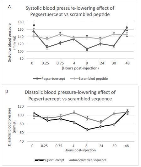
A single 1 mg/kg IP dose of Pegylated-Sertuercept caused statistically significant 8.9 mm Hg mean lowering in systolic blood pressure (Table 1) and a statistically significant 9.8 mm Hg mean lowering in diastolic blood pressure (Table 2) compared to 2 mg/kg scrambled Peptide 2 (P<0.01) in 12-week-old male ZDF rats. There was no significant effect of time on blood pressure-lowering effect from the two drugs (not shown in Table 1 or 2).

Table 1: Comparison of systolic blood pressure-lowering effect from Pegsertuercept (1 mg/kg) vs. scrambled peptide LD.8 (2 mg/kg) intraperitoneal injection in 12-week-old male ZDF rats.

Treatment

Estimate

SE

DF

t Value

Pr>t

Effect
Intercept

141.66

5.4602

5

25.94

<.0001

Trt PegSertuercept

-8.9162

2.5229

38

-3.53

0.0011

Trt Scrambled Peptide

0

Single Pegsertuercept (1 mg/kg) IP injection caused -8.9 mm Hg reduction in mean systolic blood pressure compared to scrambled peptide (2 mg/kg IP). SE: Standard Error.

Table 2: Comparison of diastolic blood pressure lowering effect from Pegylated Sertuercept (1 mg/kg IP) vs. scrambled peptide LD..8 (2 mg/kg IP) in 12-week-old male ZDF rats.

Treatment

Estimate

SE

DF

t Value

Pr>t

Effect
Intercept

98.1654

4.376

5

22.43

<.0001

Trt PegSertuercept

-9.7771

0.5121

40

-19.09

<.0001

Trt Scrambled Peptide

0

0

Single Pegsertuercept (1 mg/kg) intraperitoneal (IP) injection caused mean -9.8 mm Hg reduction in mean diastolic blood pressure compared to IP injection of scrambled peptide (2 mg/kg). SE-standard error.

A single IP injection of either 1 mg/kg (shown in Figure 1) or 0.75 mg/kg pegylated Sertuercept (not shown in Figure 1) caused significant and long-lasting decline(s) in mean systolic and diastolic blood pressure in male (12-week-old) ZDF rats. The effect was sustained for at least 30 hours and less than 48 hours (Figure 1). Scrambled peptide sequence LD..8 (2 mg/kg IP) had no significant effect on systolic or diastolic blood pressure in age-matched male ZDF rats (grey line, Figure 1).

fig 1

Figure 1: Single intraperitoneal dose of Pegsertuercept (1 mg/kg) (arrow) caused significantly greater systolic (A) and diastolic blood pressure-lowering (B) vs scrambled peptide (2 mg/kg) IP in 12-week-old male ZDF rats (n=4 per group). Each point is the mean ± SD.

Significantly Longer-lasting Blood Pressure-lowering Effect of Myristolated Sertuercept

A single 2 mg/kg IP dose of Myr-Sertuercept caused statistically significant 17.4 mm Hg mean systolic blood pressure-lowering and statistically significant 12.0 mm Hg mean diastolic blood pressure-lowering compared to 1 mg/kg scrambled Peptide 2 (P<0.02) (Table 3A and 3B). There was no significant effect of time on blood pressure-lowering effect from the two drugs (not shown in Table 3).

Table 3: Time series analysis of difference in systolic (A) and diastolic (B) blood pressure lowering effect after administration of single IP dose of Myr-Sertuercept vs. scrambled peptide 2 in two groups of 12-week-old male Zucker diabetic, fatty rats (N=4 rats per drug treatment group).

A) Systolic
Drug

Estimate

Standard Error

DF

t Value

Pr>lti

Myr-Sertuercept

-17.439

5.0163

5

-3.48

0.0177

Scrambled Peptide

0

B) Diastolic
Drug

Estimate

Standard Error

DF

t Value

Pr>lti

Myr-Sertuercept

-12.0471

3.23

5

-3.73

0.0136

Scrambled Peptide

0

Single Myr-Sertuercept (2 mg/kg) intraperitoneal (IP) injection caused mean -17.4 mm Hg reduction in mean systolic and mean -12.0 mm Hg reduction in diastolic blood pressure vs. IP scrambled peptide (2 mg/kg). The model included adjustment for difference in baseline mean blood pressure. SE-standard error.

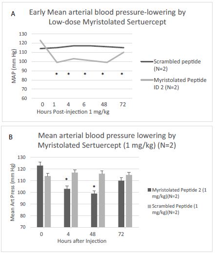
Next, the blood pressure-lowering effect of Myr-Sertuercept at doses ranging from 1.0-2.5 mg/kg was tested (for up to nine days) following a single IP injection in male ZDF rats. Blood pressure-lowering by 1 mg/kg Myr-Sertuercept was not only rapid in onset, but substantial (Figure 2). Both younger (12-week-old) and older (25-week-old) male ZDF rats experienced similar significant blood-pressure lowering following IP injection of 2 mg/kg Myr-Sertuercept (Table 4). At doses ranging from 1.0-2.5 mg/kg, IP Myr-Sertuercept dose-dependently lowered blood pressure and the effect (at higher doses) was sustained for 7-9 days (Table 4). Myr-Sertuercept was safe and well-tolerated. None of the male ZDF rats experienced acute hypotension even at the highest dose tested. Vehicle (10% DMSO) alone or scrambled peptide in sterile saline had no discernable effect on blood pressure in age-matched male ZDF rats (data not shown).

fig 2

*P<0.05 compared to baseline mean arterial pressure
Figure 2: Rapid- onset and sustained blood pressure-lowering effect of low dose Myr-Sertuercept peptide following IP injection in 13-week-old male ZDF rats. Results are mean ± SEM of multiple determinations at each timepoint in each of two male ZDF rats that received either 1 mg/kg Myr-peptide or Scrambled peptide; *P<0.05 compared to baseline MAP.

Table 4: Change in Blood pressure in response to IP administration of scrambled (LN..8) or Myr-Sertuercept: each at 2 mg/kg in 25-week-old male Zucker diabetic fatty rats.

Peptide

 Day 0

 Day 2

 Day 5

 Day 7

Systolic
Scrambled (LN..8) (N=3 rats)

140 ± 6

148 ± 1

147 ± 10

151 ± 12

Myr-Peptide 2 (N=5 rats)

144 ± 9

121 ± 7**

125 ± 12*

134 ± 9

Diastolic
Scrambled (LN..8) (N=3 rats)

95 ± 10

94 ± 7

101 ± 8

105 ± 7

Myr-Peptide 2 (N=5 rats)

103 ± 6

77 ± 4**

82 ± 10*

92 ± 7*

Results are mean ± SD; **P<0.01 vs. day 0; *P<0.05 vs. day 0.

Male SHR Rats

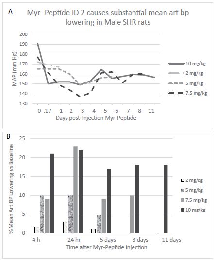
The spontaneously hypertensive rat (SHR) is an inbred strain derived from Wister-Kyoto rats selected for successive matings based on having spontaneously-occurring hypertension. The SHR rat is a recognized model of essential hypertension that is neither obese nor diabetic. Unlike male ZDF rats, male SHR rats do not develop early, moderate-severe proteinuric nephropathy [7], however, they are known to exhibit certain cell-mediated and humoral immunologic defects [8] including harboring autoantibodies to several unknown vascular antigens [9]. Myr-Sertuercept (2.0 mg/kg IP) had little or no significant blood pressure-lowering effect in three SHR rats. Myr-Sertuercept (5.0-10.0 mg/kg IP) dose-dependently lowered blood pressure, and the effect was long-lasting (up to 9-11 days) at the 7.5 or 10.0 mg/kg dose, respectively (Figure 3, Tables 5 and 6). Myr-Sertuercept was safe and well-tolerated; none of the rats experienced any untoward effect (e.g. acute hypotension) after single-or repeated dose administration over a four-week testing period. A single normotensive male SD rat (monitored telemetrically) did not experience any significant blood pressure-lowering (or adverse effect) at any dose of Myr-Sertuercept tested including the highest (10 mg/kg) dose. Vehicle (10% DMSO) or scrambled peptide (in sterile saline) had no discernable effect on blood pressure in age-matched male SHR rats (data not shown).

fig 3

Each (time)point represents the mean of five-ten telemetric determinations (Stellar implant, TSE Systems Inc) having standard error of approximately 1%.
Mean MAP-lowering (%) at the indicated timepoint compared to baseline MAP
Figure 3: Dose-dependence and sustained blood pressure- lowering effect of Myr-Sertuercept in twenty-week old male SHR rats.

Table 5: Summary of dose-dependence and durability of mean arterial blood pressure-lowering following single IP administration of myristolated Sertuercept at indicated doses Myristolated.

Myristolated Sertuercept Dose

Acute Peak blood pressure-lowering (%)

Duration of Action (days)

Mean arterial BP

1 mg/kg

6-8%

4-6

1.5 mg/kg

14%

7

2.0 mg/kg

14-22%

3; 7

2.5 mg/kg

19-30%

9

Results represent average of treatment in two, 11-13-week-old male ZDF rats at each specific dose

Table 6: Summary of dose-dependence and durability of mean arterial blood pressure-lowering effects after single IP administration of myristolated Sertuercept in 25-week-old male SHR rats.

Myristolated Peptide 2 Dose

Acute Peak blood pressure-lowering (%)

Duration of Action (days)

Mean arterial BP

2.0 mg/kg

1-3%

5

5.0 mg/kg

5-10%

5

7.5 mg/kg

9-23%

8

10.0 mg/kg

17-22%

11

Results represent average of treatment in three male SHR rats at each specific dose

Female Zucker Fatty Rats

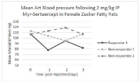
Unlike their male counterparts, female Zucker fatty rats do not develop diabetes mellitus on the same diet fed to male ZDF rats; female ZDF rats exhibit a much milder form of hypertension and proteinuria is mild. A single IP injection of Myr-Sertuercept (2 mg/kg) caused rapid significant systolic and diastolic blood pressure-lowering in approximately 60% (12/21) of female Zucker fatty rats tested at 24-hour after drug administration. The magnitude and time course of blood pressure change in representative ‘responder’ and ‘non-responder’ female Zucker rats following a single 2 mg/kg IP dose of Myr-Sertuercept is shown in Figure 4. Mean arterial blood pressure-lowering was substantial (23-27%) 1-3 days after Myr-Sertuercept (2 mg/kg) in a ‘responder’ female Zucker fatty rat (Figure 4). Blood pressure-lowering was sustained for up to 6 days in this and other female Zucker rat responders (not shown). There was no effect of scrambled peptide (2 mg/kg) following IP injection in three female Zucker fatty rats tested (data not shown).

fig 4

Each point is the average of three of more determinations which varied by <15%
Figure 4: Mean arterial blood pressure-lowering following a single 2 mg/kg IP dose of Myr-Sertuercept in three representative ‘responder’ and ‘non-responder’ 20-week-old female Zucker fatty rats. Results are the mean of three or more determinations which varied by < 15%.

Reno-protection from Chronic, Once-weekly Treatment with Myr-Sertuercept for Seven Weeks

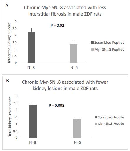
Chronic once-weekly IP administration of Myr-Sertuercept (vs. scrambled peptide) (each at 2 mg/kg concentration) for seven weeks was associated with significantly lower renal interstitial collagen score (P<0.02) and total kidney lesion score (P<0.003) (N=14 male ZDF rats, Figure 5). Kidneys from rats treated with scrambled peptide not only had significantly more interstitial fibrosis (vs kidney from Myr-Sertuercept-treated rats), but the areas of fibrosis were larger than in Myr-Sertuercept-treated rats. Rats randomized to Myr-Sertuercept vs. scrambled peptide did not differ significantly in their baseline body weight or plasma glucose concentration prior to initiation of treatment (data not shown).

fig 5

*P<0.05, mean OD in IgG from two male SHR rats vs. two female Zucker fatty rats Dashed line is the background optical density (0.032 absorbance units).
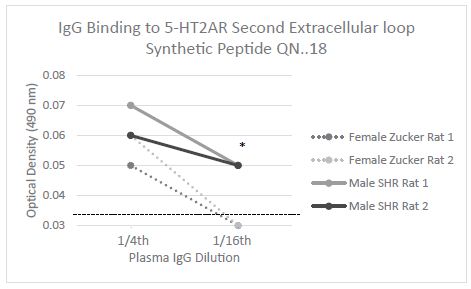
Figure 5: Level and titer of Rat IgG binding to synthetic peptide QN.18 identical to the second extracellular loop of the serotonin 2A receptor.

Serotonin 2A Receptor Plasma IgG Autoantibodies in Different Rat Strains

We next tested plasma IgG from male SHR rats (n=4) and female Zucker fatty rats (N=7) for binding to a synthetic peptide identical to the second extracellular loop of the serotonin 2AR receptor. In a prior study, male ZDF rats harbored IgG autoantibodies that displayed significant binding to the 5-HT2AR, second extracellular loop peptide, QN…18 [6]. Here, we report that IgG in all four of four male SHR rats tested displayed significant binding to the 5-HT2AR peptide, but IgG in only 2 of 7 female Zucker fatty rats tested displayed increased binding to the 5-HT2AR peptide (data not shown). Plasma IgG binding to the serotonin 2A receptor second extracellular loop peptide was significantly increased (P<0.05) in the IgG from male SHR rats (n=2) compared to female Zucker fatty rats (n=2) (Figure 6). The female Zucker rat having plasma IgG which exhibited relatively higher binding in the 5-HT2AR ELISA also had a robust blood pressure-lowering response to Myr-Sertuercept (data not shown in Figure 6).

fig 6

Figure 6: Chronic (7-week) once weekly IP administration of Myr-Sertuercept (2 mg/kg) significantly reduced A) renal interstitial collagen score and B) total lesions compared to identical dose and course of scrambled peptide in 16-23-week-old male ZDF rats. Results are mean ± SD.

Safety

There were few if any acute or chronic side effects from Myr-Sertuercept administered IP at doses ranging from 1-10 mg/kg in male ZDF (N=16), female ZDF (N=21) or male SHR rats (N=10). Two male Zucker diabetic fatty rats who were severely hyperglycemic (capillary glucose >500 mg/dL) developed a syndrome of inability to gain weight and failure to thrive. Both rats had been randomized to seven weeks’ treatment of once weekly Myr-Sertuercept and died at approximately 20-22 weeks of age. A limited necropsy performed in one of the rats revealed severe diabetic gastroparesis, a known complication in the hyperglycemic rodent [10].

Discussion

Two different conjugated forms of the synthetic peptide SCLLADDN (Sertuercept) substantially lowered systolic and diastolic blood pressure in male Zucker diabetic fatty rats. Blood pressure lowering following IP administration of Peg-sertuercept (2 mg/kg) was sustained for ~ 30 hours consistent with the known effect of pegylation on prolonging circulation time and decreasing proteolytic degradation in other modified proteins [11]. Myristolation of Sertuercept (SCLLADDN) on the amino terminus led to an even longer duration of blood pressure-lowering ranging from 6-11 days or longer depending on the strain and dose administered. This is consistent with enhanced lipid solubility and perhaps slower release into the circulation of myristolated vs. pegylated proteins.

Most FDA-approved medications to treat essential hypertension are available for oral administration. However, owing to their relatively short half-lives in vivo, even the most potent classes of oral anti-hypertensive medication require once or twice daily dosing. Presently, there is no FDA-approved class of anti-hypertensive medication whose duration of action extends substantially beyond 24 hours following a single (oral) dose. According to the World Health Organization, approximated 26-29% of the world population suffers with hypertension [1]. Hypertension is the single leading risk factor for global death and disability. Roughly one-half of treated hypertensive patients do not reach their target desirable blood pressure goal in part because of the need to take more than one medication at (daily) or more frequent intervals. Patient nonadherence to anti-hypertensive medication use is the leading cause of residual morbidity and mortality due to hypertension [2]. It could be substantially reduced through the advent of a potent, long-lasting blood pressure-lowering medication requiring only once-weekly self-administration.

The present data suggest that myristolated Sertuercept (Myr-Sertuercept) is a first-in-class, anti-hypertensive medication which meets the goal of safety and efficacy in a once-weekly formulation that can be self-administered. Once weekly Myr-Sertuercept not only provided long-lasting blood pressure control, but a short 7-week course of (once-weekly) injections significantly reduced the incidence of renal interstitial fibrosis and all kidney lesions in the male Zucker diabetic fatty compared to age-matched ZDF rats treated with scrambled peptide. These data are in agreement with an earlier report that alternate daily administration of unconjugated Sertuercept peptide (for ten weeks) significantly reduced glomerular fibrosis in the male ZDF rat compared to ZDF rats treated with scrambled peptide [4].

Male SHR rats suffer with a severe form of hypertension, and they required 3-5 fold higher concentration of Myr-Sertuercept (IP) to promote substantial blood pressure-lowering compared to male ZDF rats. On the other hand, not all female Zucker fatty rats (which generally exhibit a milder form of hypertension compared to male ZDF rats) experienced significant blood pressure-lowering in response to Myr-Sertuercept. Differences in endothelial injury, and endothelial nitric oxide synthase expression between male and female Zucker rats may account in part for the observed sex difference. For example, estrogen is known to augment endothelial nitric oxide synthase important in mediating vasodilation (Figure 7) [12].

fig 7

Figure 7: Representative Masson’s trichome-stained kidney section from male ZDF rat treated with either (A) Myr-Sertuercept or (B) scrambled peptide (2 mg/kg) for seven weeks.

Another contributory factor may be reduced level of plasma serotonin 2A receptor IgG in female Zucker compared to either male SHR or male ZDF rat. Our in vitro studies suggested that Sertuercept may interfere with the ability of agonist serotonin 2A receptor IgG autoantibodies to bind and activate the serotonin 2A receptor [4]. Sertuercept was designed as a structural mimic of the sub-region of the second extracellular loop of the 5-HT2A receptor most avidly targeted by ZDF rat and human pathologies IgG autoantibodies [13]. Male SHR and ZDF rat IgG not only bound to a serotonin 2A receptor peptide fragment, but the male ZDF rat IgG [6] and human pathologies’ IgG harbored endothelial cell toxicity in vitro that could be prevented by treatment with highly selective 5-HT2A receptor antagonists [14].

In summary, myristolation of Sertuercept on the amino terminus led to a novel long-lasting, safe and effective ‘first-in-class’ drug which potently lowered blood pressure in male Zucker fatty rats, and in male spontaneously hypertensive rats and substantially prevented renal interstitial fibrosis in male ZDF rats. Myr-Sertuercept may be a game-changing, once-weekly, anti-hypertensive, renoprotective medication particularly well-suited to address the problem of poor long-term medication adherence in patients on complex regimens.

References

  1. Mills KT, Stefanescu A, He J (2020) The global epidemiology of hypertension. Nat Rev Nephrol 16: 223-237. [crossref]
  2. Moise N, Schwartz J, Bring R, Shimbo D, Kronish IM (2015) Antihypertensive drug class and adherence: an electronic monitoring study. American Journal of Hypertension 28(6): 717-721. [crossref]
  3. Iglay K, Cao X, Mavros P, Joshi K, Yu S, et al. (2015) Systematic Literature Review and Meta-analysis of Medication Adherence With Once-weekly Versus Once-daily Therapy. Clin Ther 37(8): 1813-1821. [crossref]
  4. Zimering MB (2021) A Serotonin 2A-Receptor Decoy Peptide Potently Lowers Blood Pressure in Male Zucker Diabetic, Fatty, Hypertensive Rats. Endocrinol Diabetes Metab J 5(2). [crossref]
  5. Watts SW, Morrison SF, Davis RP (2012) Barman SM Serotonin and blood pressure regulation. Pharmacol Rev 64(2): 359-388. [crossref]
  6. Zimering MB, Grinberg M, Burton J, Pang K (2020) Circulating Agonist Autoantibody to 5-Hydroxytryptamine 2A Receptor in Lean and Diabetic Fatty Zucker Rat Strains. Endocrinol Diabetes Metab J 4(3): 413. [crossref]
  7. Feld LG, Van Liew JB, Brentjens JR, Boylan JW (1981) Renal lesions and proteinuria in the spontaneously hypertensive rat made normotensive by treatment. Kidney Int 20(5): 606-614. [crossref]
  8. Kim JY, Lee E, Koo S, Kim CW, Kim I (2021) Transfer of Th17 from Adult Spontaneous Hypertensive Rats Accelerates Development of Hypertension in Juvenile Spontaneous Hypertensive Rats. Biomed Res Int. [crossref]
  9. Ofosu-Appiah W, Huang LY, Kuhnle M, Sfeir G, Kennel A (1996) Autoantibodies against arterial antigens: characterization by ELISA and immunoblot analysis in the spontaneously hypertensive rat. Clin Exp Hypertens 18(1): 21-35. [crossref]
  10. Chang FY, Lee SD, Yeh GH, Wang PS (1996) Influence of blood glucose levels on rat liquid gastric emptying. Dig Dis Sci 41(3): 528-532. [crossref]
  11. Veronese FM, Mero A (2008) The impact of PEGylation on biological therapies. BioDrugs 22(5): 315-329. [crossref]
  12. Lekontseva O, Chakrabarti S, Jiang Y, Cheung CC, Davidge ST (2011) Role of neuronal nitric-oxide synthase in estrogen-induced relaxation in rat resistance arteries. J Pharmacol Exp Ther 339(2): 367-375. [crossref]
  13. Zimering MB (2019) Autoantibodies in Type-2 Diabetes having Neurovascular Complications Bind to the Second Extracellular Loop of the 5-Hydroxytryptamine 2A Receptor. Endocrinol Diabetes Metab J 3(4): 118. [crossref]
  14. Zimering MB (2018) Circulating Neurotoxic 5-HT2A Receptor Agonist Autoantibodies in Adult Type 2 Diabetes with Parkinson’s Disease. J Endocrinol Diabetes 5(2). [crossref]
FIG 1

Fayoum Portraits

DOI: 10.31038/NAMS.2022534


The Fayoum portraits owe their name to the place of their discovery in the necropolises of the Fayoum Oasis in the northwest of Egypt, 45 meters below sea level. The name Fayoum is Coptic, FIOM, and meaning “sea” probably because of the large lake there known today as Birket Quaroun. When the Arabs came to Egypt, it became Al Fayoum. The portraits date back to the first century AD, but the bulk belongs to the 2nd and 3rd centuries and only a few to the 4th century. Egypt became a Roman colony in 30 BC after the death of Cleopatra Mostly Greeks, Macedonians, Romans, Hebrews, and Syrians settled the region. Since, the time of Alexander the Great, the newcomers had been gradually merging with the local milieu. It soon rivalled Alexandria for the vineyards and gardens that abandoned on its soil, the most fertile in Egypt. It became a trade center for cereals and vegetables, and was renowned for its figs and roses. Fayoum also became an artistic center for Egypt, during the Greco-Roman period (Figure 1).

FIG 1

Figure 1: Fayoum portraits

While preserving many of the original features of their own culture and way of life, the settlers who became rich owners of large estates borrowed much from the Egyptians. In particular all of them had at an early stage adopted the peculiar, characteristically Egyptian beliefs with regard to death and their meticulously elaborated rituals. The Egyptians attached tremendous importance to the belief in life after death, and developed the art of embalming to a high degree of perfection. The aims of embalming were to preserve the body and to make a lasting portrait of the deceased. The embalmed corps was carefully wrapped and enclosed in several richly decorated coffins that were mummiform in shape. Badly preserved portraits were not included in this review.

Under the Romans (1st century BC – 4th century AD), the procedure became less elaborate. The mummy was carefully wrapped in a special way and the mummiform coffin was made simply of carton. Later, this technique was simplified further and a portrait of the deceased was attached to the mummy’s head. Initially people used for this funeral rite portraits that had been painted from life, had been framed and hung in the houses. While the ancient Egyptians, the Greeks, and the Romans excelled in architecture, sculpture, and painting, the art of portraits was not common. A typical ancient Egyptian or a Greek painting of a human face was generally shown in the profile. The obvious analogy of the Fayoum portraits are the Roman wall paintings of the first centuries AD found in the ruins of Pompeii. The Greeks had also fresco in their temples, but none of them survived; they were described in ancient writings. What survived was the painted pottery, which were made to hold water, wine, or oil. The importance of the Fayoum paintings is that the portrait became an art that had religious significance and not for decorative purposes as the frescoes. When Christianity came to Egypt the pagan tradition was gradually abandoned and the art of the portrait took a different form namely that of the icons, i.e., a religious portrait; icon is a Greek word meaning portrait.

The Fayoum portraits are a fascinating collection of people who lived during the first two centuries AD. They have an important historical as well as artistic values. They are dispersed now in at least one hundred museums worldwide either on display or in storage. Only few of the portraits in the books available are in colour and are usually arranged according to the countries holding them or some other archaeological criteria. The ankh or key of life is an ancient Egyptian symbol used in Egyptian art and writing to represent the word for “life”. It was used by at least three ancient mummies of ladies and one in men. Two special techniques were used in the Fayoum portraits. In both cases, the pigment was not mixed with water as in the frescoes but either in egg white or in wax and set by passing a hot iron over the painting. The last process became known as the “encaustic wax process”. After all, is it not true that there is life after death? Thanks to archaeologists who are digging the remains of the ancients and bringing them back to life. Here are their portraits filling museums and books. According to R. Shurinova of the Pushkin Museum of Fine Art, the Fayoum portraits are “a major contribution made by Egypt to the treasures of world culture”.

FIG 1

Smart Phones Surveillance Methods

DOI: 10.31038/NAMS.2022533

Introduction

Smart phone surveillance is a sensitive issue, so topical and important, which is about everyday life of all of us, I am talking about the smart phones, small smart phones, these small but powerful computers that everybody use them everyday by doing more internet browsing rather than doing calls. The questions that arise about the security of smart phones are many, for example: may someone watch us? May the government hear what we saying and what messages we send? May they know our location? How much important meta-data is and to what extent it can reveal important information about the subscribers identity and how it relates to privacy and personal data. Finally, it is worth mentioning the participation of mobile phone providers in various government monitoring projects of the citizens either with targeted software which is not detectable, or with the direct access to the Servers of the providers for copying sensitive information without of course the consent of the subscribers. Such projects are the Carnivore, Prism, and other projects, and the countries involved in information exchange programs are behind lists identified as 5 Eyes, 9 Eyes, and 14 Eyes.

Keywords

Smart phones, Surveillance, Monitoring, Mobile networks, Security, Privacy

Surveillance Methods

When referring to a smart phone surveillance, refer to 6 spy methods, which are:

  • Signal interception & MITM
  • Hardware circuit.
  • Spy phone application.
  • Use of specific software for Servers.
  • Meta-data analysis.
  • Government monitoring projects.
  • Case 1

    On this case, requires expensive equipment that is not legally sold on the market to be purchased by an interested customers, there are various approaches to low-cost material, but these tapping systems are cumbersome and complex in their operation, also may consider that the advent of 4G/5G networks that offer more security than the old 2G/3G these cheap eavesdropper devices partly useless, say in partly because should consider that in smart phones devices in the choice of the network allows the following option 5G/4G/3G/2G which means that where there is no 5G/4G network coverage the smart phone will operate on the 3G/2G network and this is a security hole as the network is over vulnerable to cheap type of interceptor devices since the 3G/2G network encryption algorithm provide low security. An expensive stolen state equipment would also be useless since its use keys that should be renewed regularly. Thus, state-owned services that have legal co-op equipment have the ability to make legitimate signal interception, here at this point, someone might think of this, an employee who has access to the system could be tapping someone else phone? the answer is aware of it. Legally signal interception systemss are divided into 2 categories, those located in central buildings where sometimes require the assistance of the telecom provider and the base stations where, depending on the needs of the service, they move.In both of the above categories, the subscriber does not realize the monitoring of his/her telephone line or the interception of the transmitted data. In the first category in which data interception is involved the telecommunications company the process is the easiest and simplest as extract information directly from the Servers. In the second category, which is popular in the secret services, mobile base stations are used which are used at close distances from the target victim base station to proceed with data collection. The target victim does not perceive the difference in communication as the deceptive base stations are added to the network with a normal antenna ID so as not to affect the quality of communication or services so that the target victim understands the monitoring.

    Detecting deceptive mobile base stations can be detected with specialized software, but most of them are not detected as they appear as transponders and not as communication antennas and even if they have received a normal antenna ID. Once a successful connection to a monitoring system is achieved, or with a deceptive mobile base station, the information extraction as well as the sending of commands to the mobile phone begin, where it is indicated, recording conversations, copying messages, modifying messages, browsing websites, receiving emails, collecting messages from network messaging applications, locating, opening the camera, and opening the microphone. These practices are commonly used to support criminal evidence in courts or in cases of terrorisn and national security. Figure 1 shows a deceptive mobile base station.

    FIG 1

    Figure 1: Deceptive Mobile Base Station

    Case 2

    On this case, could be said to be based on the ignorance of the subscriber, at this point say ignorance because in this case the smart phone has to be opened and a micro chip is placed inside, which will collect everything while the phone will be send it back to the target-victim person. Device data usage either when the smart phone is in close proximity to a mobile transceiver, the connection is made and the data is transferred. Detecting such a micro chip is very difficult, and with the breakthrough of Internet of Things this technology is evolving. That’s why we need to buy smart phones from trusted places, also a smart phone that is a gift from someone is an easy way to deceive, but not always. Under no circumstances should be considered safe and reliable a sealed cell phone that is in its original jelly in brand new condition, just simillar as the ones cell phones selled in stores. This is a practice of the secret services, when there is a restriction on access for data mining in other ways, for the success of the mission there is usually cooperation with some traders so that the desired modified device falls into the hands of the target victim in such a way that perceived by the target victim. Even if the target victim somehow realizes that their communications are being monitored he/she will focus on either switching network providers or buying a new SIM card under another name from the same or different network provider, the most likely scenario is to use the same mobile device as the suspicion that the mobile phone device is modified will hardly cross his/her mind, on the one hand due to lack of knowledge and on the other hand he/she bought the device sealed from a store. The success of this method is based on this logic.

    Case 3

    On this case presented the simplest and most widespread, based on the naive and ignorant of the target victim about the security and use of smart phone and applications. Some security tips are:

    • Do not leave the smart phone exposed to third parties.
    • Use lock screen application.
    • Keep both operating system and software applications updated.
    • Use antivirus software with real time protection enabled, keep the antivirus database updated.
    • Do not use VPN software.
    • Do not install APK files from unknown sourcs.
    • Do not ROOT your smartphone.
    • Do not use software apps with low reputation.
    • Do not open messages that do not know.
    • Do not click hyperlinks that do not know.
    • Do not open files from strangers.
    • Smart phones are not as safe as you may believe.

    Generally, the signs that a smart phone is being taped are:

    • The battery discharge quickly after data is being used continuously, so check the applications. It can also be a coincidence.
    • Interrupting calls while talking, also this could cause by interference in the connection, automatically changing location while trying again to set the correct location but still changed may be interference only at that specific area.
    • Noise during speech, this is a sign of a interference, but keep in mind that with professional signal interception devices there is no noise, also the noise someone may hear may be either a device damage or is from Interference.
    • The smart phone is warm, this meaning several processes are running.
    • The smart phone crashes and becomes slow, this is due also to other reasons, depending on the smart phone and depending on the monitoring software sometimes the monitoring software is “heavy” and the smart phone can not respond.
    • Text messages that are received by irrelevant numbers, messages that have been sent, and the recipient has received a different message than than the one that have been sent should be suspected of monitoring or network interference that may be due to other infections.
    • The smart phone works alone and does not turn off. Surveillance spy software do not allow the user to have full control over the device for the simple reason that they are trying to keep the spy software on the device.
    • Removal of software apps are not permitted or this operation is disabled.
    • There is mysterious activities in social media networks which tere is access from the infected smart phone, also, my serious activities may occur in mobile apps of exchanging messages.
    • Nothing from the above.

    The above are the few rules that apply, continue reading.

    • Surveillance software does not appear in installed applications.
    • Resetting to factory settings may not permanently eliminate the surveillance software.
    • Scrambled applications are easily localized, while well-designed applications run on Stealth Mode and are extremely difficult to detect even with anti-malware
    • A smart phone surveillance software can be downloaded to the smart phone through another application.
    • Modern surveillance software is very difficult to detect while also provides no annomalies with operating system.
    • Smart phone ROOTING makes easier the installation and proper use of surveillance software.
    • A smart phone which its operating system is updated will prevent from the installation of various surveillance software which is installed into smart phones by using various well known security holes.

    Case 4

    In this case, the well-known Pegasus spyware is introduced. Pegasus is spyware software for smartphones that install on iOS, Android, Blackberry O/S and any other Android O/S based operating system (custom Android versions). This software has interesting features both in the way it is installed on the target smartphones and the possibilities it offers to its operators. The target victim first receives an SMS with a link if then the target victim by clicking on the link then the smart phone becomes infected with Pegasus. In the previous pages among the security tips it has been mentioned that the operating system must be updated, the reason is that to install Pegasus on a Smartphone it exploits zero-day vulnerabilities of the operating systems, which means that an updated operating system from alone is not able to deal with Pegasus, so additional security measures are required, such as a very reliable antivirus/antispyware with application installation lock capabilities and avoiding ROOT may to some extent prevent the installation or smooth operation of Pegasus. Once Pegasus is installed on the target smartphone, then the ability to fully control the smartphone begin with capabilities to monitor and record phone calls, read emails, take screenshots, record keystrokes, generally have the general management of the Smartphone in to such an extent that the remote Pegasus operator has physical access to the Smartphone. As expected from such software with huge capabilities, it is expensive, complex software that remains installed on the smartphone completely invisible, undetectable even by antivirus and antispyware. The specific software is used when there is a need to monitor important people and personalities; it is not used for monitoring children as parental control software for example or for low important purposes of low interest.

    At this point it is worth noting two things, the first is that in dark net there are lists of zero-day vulnerabilities that can be used to create software spies similar to Pegasus. The second is that Pegasus is software that is constantly updated and new features are added. An important peculiarity of its innovation is the way of installation which changes, as it has been mentioned above for the installation of the software the target-victim receives a link via SMS, this is the basic way of installation, but there is also an invisible way of installation, in this case the target-victim does not realize the existence of SMS nor do require by someone to click on a link to install Pegasus, because it is possible exploitation of zero-day security vulnerabilities that allow Pegasus to install on the target-victim’s smartphone without the target-victim realizing anything. This is one of the features that make Pegasus software so special.

    Case 5

    Meta-data is everywhere in our electronic activities, meta-data is also present in the non-digital world. For the implementation of an activity or the execution of a service – function presupposes the execution of various functions which will be used to complete a process. All these processes that are required for the completion of a service – function in each stage of their implementation are accompanied by a significant number of data which is necessary for the completion of the final process. The data that is transferred and processed at each step of the implementation is of great and special importance-value, both because they contain important personal information and because determine the activities of mobile subscribers. Mobile telephony service providers that participate in government information exchange of meta-data programs distribute the meta-data content to government agencies for analysis. The secret services have the ability to process meta-data to a large extent. To create profiles with the habits of each subscriber. The meta-data collection can of course be performed with the cases mentioned above. The collection and analysis of meta-data is very important and the information extracted can be more important than the information itself. The reason is that neta-data contains subscriber information which contains:

    • Mobile network code.
    • ΙΜΕΙ of Smartphone.
    • Device location.
    • Subscriber code.
    • Frequency of manipulations.
    • Social media preferences.
    • Duration and frequency of calls.
    • The destination of the calls.
    • Duration of internet usage.
    • Frequency of visits to websites.
    • Frequency of use of the telephone.
    • Determining the country of operation of the telephone.
    • Determining the use of the mobile telephony antenna.
    • Frequency of telephone connection per antenna.

    The above are some of the information that can be extracted from the meta-data. This information can be used as a first step before implementing a cell phone tracking method. It is worth noting at this point, the use of a simple non-smartphone mobile phone significantly reduces the chances of tracking, as there is a significant limitation to the use of tracking methods, while from the point of view of the created mta-data, eta-data is clearly less in terms of the size of the value of the information. The government services, and especially the secret services, are able to know about the information circulating in the interior of the country, the secret services that are active in the collection and analysis of information abroad (foreign intelligence) are able to know in globally the information that is circulated as meta-data, how this is done will be mentioned below.

    Is important advantage of meta-data is the sale of information that can be extracted for advertising purposes, it is not uncommon for subscribers to accept calls by marketing companies to promote their products, when subscribers ask marketing companies where they found their phone numbers they answer that the subscribers themselves subscribed to information lists, someone else wrote them on their behalf, knowing that this is not the case, they just try to calm the subscriber by reassuring them that they have not received their contact details illegally extracting information from meta-data. Meta-data is also created by mobile antenna towers, these meta-data include subscriber connections from antenna to antenna, the distance of the subscribers from the antenna, the data transferred, and other useful information that needs analysis, which I further analysis will reveal other sensitive information.

    Case 6

    On this case presented the biggest threat regarding data security and privacy in the digital world is that the majority of data transferred on the Internet is not encrypted well, the existing security infrastructure in an environment of not well encrypted information must be considered totally inadequate. Not well encrypted communication means the government easy may have access to intercept any information. Government agencies and organizations knowing the telecommunication weaknesses, information intercepted by subscribers from offensive websites, the non-encrypted information, not well encrypted information, VPN servers that offers fake anonymity that is essentially Honey Pot systems with examples the secret services who collect information from the servers inside in the country or third countries that participating in the PRISM project.

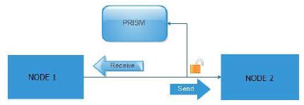
    Security agencies have access in data of any internet and smartphone subscribers in the world. This practice on several countries is legal and is based on several privacy protection laws This laws gives the freedom to governmental agencies to store and to process huge amounts of data without exception if these people are criminalized or not. The PRISM project became quite popular and reinforced worries of the world on the violation of privacy and data. The Project PRISM named after the word outlet means mirror – reflection and this is because the data pass through an internet node continue their route but the items are copied (reflection) from the PRISM project without harming their quality neither have been some form of alteration to worry the subscriber that something is wrong. Below in Figure 2 is a simple example of Project PRISM.

    FIG 2

    Figure 2: Example of Project PRISM

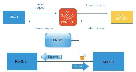
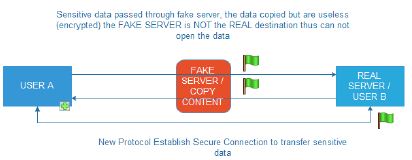
    As shown in the figure above, to make a data breach between nodes the data must be copied without the subscriber knowledge. Usually this makes it coherent with telecoms operators and other services that there are active subscribers. All of the above would be useless if there was not the necessary data mining tool and statistical tests, called XKEYSCORE by pressing a few keys are able to know everything related to a human, such as telephones, e-mails, habits, searches has done in search engines, behaviors, internet of Things activities (IOT) and of course building electronic profile “e-profile”. The security services have advanced already on potential networking devices controls and firewalls of known companies manufacturing such devices. The XKEYSCORE, used in conjunction with another program called TURBULENCE, the TURBULENCE are two subsystems the TURMOIL and TURBINE. Briefly mention that TURMOIL is an information collection system of satellite and cable communications, while TURBINE unleashes attacks on serial systems (Greenwald, 2015). From the above could not be missing collection of information from social media, Cookies, Internet services, Internet of Things, etc. Once the target is locked: the next step is the QUANTUMTHEORY attacks and QUANTUMNATION which will give full control of the remote device, even is a smart phone, Internet of Things, computer, or anything else. In Figure 3 shows how is working the intermediary fake Server.

    FIG 3

    Figure 3: Working of intermediary fake Server

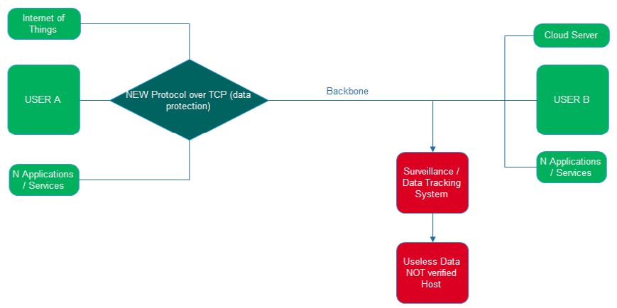
    The data collection and processing is performed by each device connected to the Internet, the mobile network providers collect personal data transferred to Servers of their infrastructures, telecommunications providers know all about digital life and human conversations, applications for smart phones collect personal data and data about subscribers behavior. Imagine a Smart phone devices which will send personal data to the manufacturer or other organizations and then those organizations to have remote access to Smartphone and opened the camera, or perform real time data analysis. Many automated bot trying to break different kinds of access codes to gain access. interception of data is shown in Figure 4 and 5 below. A smartphone that work exclusively on 4G/5G and not in 2G/3G networks would be the best option. No security method can prevent the monitoring of a smartphone, as long as the subscriber tries to protect his/her smartphone, as the methods presented in this chapter are practically impossible, even if the subscriber uses an old mobile phone device no that is, a smartphone. No cell phones and no security methods should be considered secure, subscribers should consider their smartphones to be insecure and that their activities are not confidential, even if point-to-point encryption communication applications are used [1-5].

    FIG 4

    Figure 4: Interception of data

    FIG 5

    Figure 5: Interception of data

    References

    1. Glenn Greenwald (2015) No Place to Hide: Edward Snowden, the NSA, and the U.S. Surveillance State.
    2. Christos Beretas (2018) Security and Privacy in Data Networks. Research in Medical & Engineering Sciences.
    3. Christos Beretas (2018) Internet of Things and Privacy. Journal of Industrial Engineering and Safety.
    4. Christos Beretas (2020) How Really Secure is TOR and the Privacy it Offers. Nanotechnology and Advanced Material Science.
    5. Christos Beretas (2020) Cyber Hybrid Warfare: Asymmetric threat. Journal of Nanotechnology and Advanced Material Science.

Advances, Prospects and Concerns in Nanomaterials for Food Processing

DOI: 10.31038/NAMS.2022532


The advent of nanotechnology has boosted various sectors of human endeavors including food processing and packaging, the medical sector, and environment amongst others. It has improved the food industry significantly through enhanced food packaging, nutrient bioavailability and food preservation [1,2]. Nanomaterials generally are considered as those particles having particle dimensions lower than 100 nanometers at which their novel physicochemical properties make them significantly different from their bulk counterparts making them useful for various applications. Various investigations have been carried out and many more are in progress focusing on the potential usage of nanotechnology in the area of food packaging. Nanofood is a concept that is applied to food materials generated through the use of nanotechnology in production, processing, securing and packaging. Nanotechnology has outstanding potential in post-harvest processing of food [3,4]. The application of nanomaterials ensures food consistencies, which is attained through particle size modification, formation of desired clusters as well as the surface charges of the food nanoparticles. Furthermore, the emergence of nano-mediated delivery systems for nutraceuticals, and the synergistic actions of nanoparticles in food security as further aided the advancement of this area. More recently, there are numerous consumable nano-coatings that are used in fruits, cheese, meat, bakery foods and fast food. Nanofilters have been adopted for the removal of undesired colours from beetroot as well as lactose from milk to make nano-modified milk available for patients that are lactose intolerant [5]. The introduction of bio-active nanomaterials into polymeric matrices further enhances the efficiency of the packaging materials and makes provision for enhanced functional components and attributes such as scavenging, antimicrobial and antioxidant potentials. The formulation of various nanocrystalline particles has aided in the reduction of carbon dioxide inflow in beers. Oxygen carrier in alcohol production is enhanced through the use of clay nanomaterials. There foregoing shows that nanotechnology has remarkable superiority over the conventional approaches to food processing with an enhanced shelf life of the products, production of high quality food and prevention of contamination [6].

Presently, millions of dollars are spent at a global scale for food security and safety. The utilization of nanomaterials has the unique potential of improving food processing and packaging and also enhancing their taste and nutritional values. Nanotechnology can be utilized for the enhancement of food texture and flavor, reduction of the contents of fat, encapsulation of various nutrients, and prevent the degradation of vital nutrients and bioactive components during storage. The introduction of intelligent food packaging through the integration of nanosensors can further provide consumers with reliable details on the food present. The food packages are enclosed with nanomaterials thereby giving an immediate alert to the consumers on the safety levels of the food [7].

Despite of the recent advances in this direction, most of such applications are at present in their elementary stage, with most aimed at products of high value. Also, the evaluation of nanofood safety has not been fully established. It is paramount for various regulatory agencies to set pragmatic standards to be followed in the evaluation of food safety, packaging as well as the supplementary usage of nanomaterials [8]. There are limited studies on the exploration of naturally occurring nanostructures and their potential benefits in food processing. It is therefore not easy to conclude in the overall merits and demerits of nanotechnology in this regard. Currently, nano-modified foods are not usually labeled making it difficult for potential consumers to identify which is modified or not and make the decision to consume such food products. It is vital for standard testing of such nano-modified food prior to their discharge into the market. There is therefore pressing need for novel and reliable approaches to studying the effects of nanomaterials on human cells with the intent of assessing their inherent hazardous impacts. It is envisaged that nano-modified food products will be increasingly available to consumers at a global scale [9].

More recently, there are numerous emerging concerns in the application of nanomaterials in the food industry.  One of the major areas of interest to researchers is their toxicological and environmental impacts. Although the nanomaterials present in the surfaces of food do not pose harm directly from some available studies, however their transportation and integration in the food can affect human health adversely. The toxicological concerns associated with these nanomaterials are primarily due to their non-dissolvable nature, non-biodegradability and persistency in living cells and the environment [10]. Their poor consumer awareness, government policies, and methods of detection for risk assessments of nanomaterials deter detailed understanding of toxicity inherent with the utilization of nanotechnology. The toxicity associated with a nanomaterial has been reported to increase with a decrease in the sizes of the nanoparticle. Due to the high reactivity of nanoparticles they readily passed through the membrane bringing about various toxicodynamic and toxicokinetics effects. There is a need for extensive studies in this regard. Although there are wide applications of nanomaterials only a highly limited study exists on the in vivo toxicological effects using nanoparticles in mammalian models [11,12].

Nanomaterials are currently being applied in various parts of the world, although there are few countries in the world with reliable regulatory policies and standards for the use of nanotechnology in the food sector. There is a dearth in the scientific exploration of the various nanosystems thereby creating challenges in reaching a good conclusion about their efficacy. There is a paramount need for efficient labeling of packaged nanofood so that consumers can make their choice. The utilization of nanotechnologies if effectively managed and regulated would play a remarkable role in the improvement of food processing technology for the well-being of humans.

References

  1. Inobeme A, Adetunji CO, Ajai AI (2022) Future perspectives in nanomaterials for environmental and medical applications. Int J Nanomater Nanotechnol Nanomed 8: 005-006.
  2. Assey G, Malasi W (2021) Advances in Nanomaterials Sciences and Nanotechnology for Sustainable Development: A Review Tanzania Journal of Science 47: 1450-1463
  3. Noah N (2020) Design and Synthesis of Nanostructured Materials for Sensor Applications, Journal of Nanomaterials.
  4. Adetunji CO, Ogundolie FA, Olaniyan OT, Mathew JT (2022) Nanobiomaterials for Food Packaging Sensor Applications.
  5. Sharma C, Dhiman Rokana R, Panwar H (2017) Nanotechnology: An Untapped Resource for Food Packaging. Front. Microbiol Sec. Food Microbiology.
  6. Peidaei Ahari H. Anvar S, Maryam Ataee (2021) Nanotechnology in Food Packaging and Storage: A Review 2: 123-153.
  7. Sharma P, Pandey V, Sharma M, Patra A, Husen A (2021) A Review on Biosensors and Nanosensors Application in Agroecosystems. Nanoscale Res Lett. 16: 136. [crossref]
  8. Peidaei M, Panigrahi C, Vishwakarma S, Kumar J (2022) A Review on Nanotechnology: Applications in Food Industry, Future Opportunities, Challenges and Potential Risks. J Nanotechnol Nanomaterials. 3: 28-33.
  9. Tirado-Kulieva V, Sánchez-Chero M, Jimenez D. Sánchez-Chero J. Garcia L.(2022) A Critical Review on the Integration of Metal Nanoparticles in Biopolymers: An Alternative for Active and Sustainable Food Packaging.
  10. Inobeme A, Mathew JT, Adetunji CO (2023) Recent advances in nanotechnology for remediation of heavy metals. Environ Monit Assess 195: 111.
  11. Subhan M, Choudhury K, Neogi N (2021) Advances with Molecular Nanomaterials in Industrial Manufacturing Applications. Nanomanufacturing 1: 75-97.
  12. Adetunji CO, Ogundolie FA, Olaniyan OT, Mathew (2022) Bio-and Nano-sensing Technologies for Food, Royal Society of Chemistry. Nanobiomaterials for Food Packaging Sensor Applications. 167-180.

Development of Rapid Pre and Post Mortem On-farm Diagnostic Test Kit for Porcine Cysticercosis (Pork Tapeworm)

DOI: 10.31038/MIP.2022321

Executive Summary

The goal of this project is to enhance sustainable productivity, value added and competitiveness of the pig industry in Uganda through easier, user-friendly and more accurate diagnosis, control and prevention of Taenia solium cysticercosis. The enhanced control and prevention of the infection is also expected to increase pork trade and food safety, prevent human infections and eliminate a health risk that has both social and economic implications.

Background

A zoonotic tapeworm Taenia solium transmitted among humans and between humans and pigs causes cysticercosis. Humans acquire taeniosis (tapeworm infection) when they eat raw or undercooked pork meat contaminated with cysticerci, the larval from of T. solium. When ingested, the cysticerci establish in the intestine of humans, become adult tapeworms and shed eggs in human feces that can infect in turn other humans and pigs by direct contact or by indirect contamination of water or food.

Epidemiological studies of porcine cysticercosis (pork tapeworm) require identification of pigs harbouring viable Taenia solium cysticerci and estimates of the degree of exposure to the parasite in the pig population destined for human consumption. Stool microscopy for diagnosis of taeniasis is inefficient and thus it is not recommended unless there is a specific indication and no suitable alternative. Even with multiple samples and concentration of large volumes of stool sample, sensitivity of stool microscopy does not exceed 60 to 70% (Allan et al. 1993). Diagnosis of teania eggs and proglottids in definitive hosts doesn’t distinguish between T. solium and T. saginata of pigs and cattle respectively. However, given that the prevalence of infection with either species is usually low, the role of parasitologic diagnosis in control programmes is relatively minor. For diagnosis of cysticercosis, histological confirmation of excised cysts is rarely required, nor easily undertaken except in a small proportion of patients with subcutaneous nodules where biopsy can provide diagnostic support. Currently, few copro-PCR techniques and non-commercial copro-Ag-ELISA assays are available. In contrast to the PCR assay, most copro-Ag-ELISA assays are genus, not species specific and thus cross-react with T. saginata (beef tapeworm). Antibody detection tests require parasitic cysts or tapeworm excretory/secretory material as a source of antigen. Assays using recombinant or synthetic antigens if available would be more suitable. Intermediate hosts diagnosis in pigs (porcine cysticercosis) can be made by tongue inspection, antibody or antigen detection, or by postmortem inspection at slaughterhouses. Rapid lingual examination for cysts is an inexpensive but insensitive test (Willingham 2006). Likewise, diagnosis by detection of cysts at slaughter of pigs is also insensitive. Uganda is ranked the biggest consumer of pork in the world. Roast pork with beer is a booming business in Uganda. This half cooked pork is a high risk for the transmission of porcine cysticercosis yet routine deworming is not commonly practiced by Ugandans. Diagnosis of porcine cysticercosis in humans and pigs ante-mortem is not developed in Uganda hence many cases go untreated. We intend to develop and evaluate a recombinant antigen for rapid diagnosis of porcine cysticercosis, lateral flow assay and LAMP assay. This will contribute to the control of this zoonotic disease in Uganda for instance neurocysticercosis; epilepsy [1-4].

Porcine cysticercosis is a zoonotic disease, it is highly prevalent in humans, livestock (12.2-25.7%) and wild suidae. Uganda is the topmost consumer of pork in the world. Mostly in form of roast pork with beer. This is half cooked pork with the risk of transmission of neuro-cysticercosis in humans. This increases the incidence of epilepsy in Uganda. Hence controlling porcine cysticercosis improves not only livestock health & productivity but socio-economics and public health.

Given the magnitude of the problem of porcine cysticercosis in Uganda, The physical tests applied for its diagnosis are not sensitive, hence the need for more sensitive point of care and field tests. Physical tests are done at the point of slaughter or postmortem. There is however need for field based diagnostics to direct treatment and control, thus improving livestock health and production as well as human health. Research has been done mostly on the prevalence of porcine cysticercosis in Uganda. However, no effort has been done to improve the disease diagnosis. The overall apparent sero-prevalence (12.2%) reported by Kungu 2015; while previous reports (25.7%) by Nsadha and others (2014) in Lake Kyoga basin. Effective disease control depends on accurate diagnosis.

Keywords

Animal health, Animal production, Pigs, Zoonotic diseases, Biotechnology, Uganda

References

  1. Allan JC, Mencos F, Garcia-Noval J, Sarti E, Flisser A, et al. (1993) Dipstick dot ELISA for the detection of Taenia coproantigens in humans. Parasitology 107: 79-85. [crossref]
  2. Arve Lee Willingham III, Dirk Engels (2006) Control of Taenia solium Cysticercosis/Taeniosis. Advances in Parasitology 61: 509-566.
  3. Kungu JM, Dione MM, Ejobi F, Ocaido M (2015) Status of Taenia solium cysticercosis and predisposing factors in developing countries involved in pig farming. Int J One Health 1: 6: 13.
  4. Nsadha Z, Thomas FL, Fèvre ME, Nasinyama G, Ojok L, Waiswa C (2014) Prevalence of porcine cysticercosis in the Lake Kyoga Basin, Uganda. BMC Veterinary Research 10: 239. [crossref]
FIG 1

How to Nail Down Trace Proteins in Any Sample

DOI: 10.31038/MGJ.2022514

Abstract

Proteins present in most biological mixtures are expressed over a vast concentration range (up to 10-12 orders of magnitude in human sera), the most abundant ones complicating the detection of low-abundance species or trace components, Classical approaches, such as pre-fractionation and immuno-depletion methodologies are frequently used to remove the most abundant species. Unfortunately, these methods not only are unsuccessful in concentrating trace components, which could remain below the detection limits of analytical approaches, but also may cause a non-specifically depletion of other components (including low abundance ones). In case of immuno-depletion, the situation is less than brilliant: untargeted proteomic analyses using current LC-MS/MS platforms with immuno-depletion cannot be expected to efficiently discover low-abundance, disease-specific biomarkers in plasma, since the increment in detection of these trace components after such a treatment results in a meagre 25% increase, accounting for only 5-6%of total protein identifications in depleted plasma. The characterization of minor components in complex protein systems, has been revolutionized by the introduction of the combinatorial peptide ligand libraries technology. This new methodology is based on the use of hexa-peptide baits to capture and normalize the relative concentrations of the components of any proteome under investigation. The major advantage of this technique, in comparison with other pre-fractionation methods, is that it not only diminishes the concentration of the more abundant proteins, but also concentrates low-abundance and even trace components, thus providing access to the “invisible” proteome. In addition, the loss of low-abundance species that may be accidentally eliminated by co-depletion using immuno-subtraction methods, is avoided.

The Problems with Current Depletion Methods

The elimination of high-abundance proteins is operated by immunodepletion using specific solid-phase antibodies against the proteins to be suppressed. The method is quite effective; however, it suffers from a vicious circle which starts from the use of small volumes of expensive immunosorbents which accept only small samples to deplete. In small samples the amount of targeted markers is very low and they are additionally diluted during the process, thus rendering their detection even more challenging without any post concentration. Naturally concentration is possible but it contributes to protein losses. Immunosorbents are also limited for sample treatment due to their species specificity; immunosorbents available for proteomics are limited for the treatment of only human blood plasma. Conversely the use of enrichment methods based on solid-phase adsorption of targeted species or groups (e.g. glycoproteins, phosphoproteins and other classes) or the use of solid-phase combinatorial affinity ligands are by far more effective since they allow much larger initial biological samples and hence larger quantities of targeted low-abundance proteins. The Combinatorial Peptide Ligand Library (CPLL) is a technology for sample treatment that repeatedly demonstrated its capability to allow detecting proteins that are most of the time ignored because well below the level of sensitivity of proteomics equipments and methods. It is additionally of general use for various biological material and various species. This original procedure, that takes its origin from affinity chromatography mechanisms, when used under overloading conditions, contributes not only to improve the knowledge in proteomics but, more importantly, to detect dilute proteins that are expressed at the early-stage of metabolic diseases. It is after years of applications in various conditions and various sample situations that low-abundance protein detection by CPLL in early-stages of diseases is gaining momentum as a potential discovery allowing the design of diagnostic tools. Explanation of the mechanism of action is given in the following sections as well as examples of detection of panels of exclusive low abundance proteins present in various diseases [1,2].

Biomarker Discovery, a Major Target in Proteomics Investigations

This original procedure, that takes its origin from affinity chromatography mechanisms, when used under overloading conditions, contributes not only to improve the knowledge in proteomics but, more importantly, to detect dilute protein that are expressed at the early-stage of metabolic diseases. It is after years of applications in various conditions and various sample situations that low-abundance protein detection by CPLL in early-stages of diseases is gaining momentum as a potential discovery allowing the design of diagnostic tools. Explanation of the mechanism of action is given in the following sections as well as examples of detection of panels of exclusive low abundance proteins present in various diseases. Proteins and their variants are produced in a very large number and their individual concentration is extremely large, ranging throughout at least a dozen of orders of magnitude if not more. This situation renders the detectability of low- and very low-abundance species very challenging or clearly impossible in practice. Without any kind of sample treatment the large majority of proteins cannot be detected because their concentration is either below the detectability levels or because their signal is suppressed by the presence of most abundant proteins. the use of enrichment methods based on solid-phase adsorption of targeted species or groups (e.g. glycoproteins, phosphoproteins and other classes) or the use of solid-phase combinatorial affinity ligands are by far more effective since they allow much larger initial biological samples and hence larger quantities of targeted low-abundance proteins. The Combinatorial Peptide Ligand Library (CPLL) is a technology for sample treatment that repeatedly demonstrated its capability to allow detecting proteins that are most of the time ignored because well below the level of sensitivity of proteomics equipment and methods. This original procedure, that takes its origin from affinity chromatography mechanisms, when used under overloading conditions, contributes not only to improve the knowledge in proteomics but, more importantly, to detect dilute protein that are expressed at the early-stage of metabolic diseases. It is after years of applications that low-abundance protein detection by CPLL in early-stages of diseases is gaining momentum as a potential discovery allowing the design of diagnostic tools. Explanations of the mechanism of action is given in the following sections as well as examples of detection of panels of exclusive low abundance proteins present in various diseases.

The CPLL Capabilities to Detect Low-abundance Proteins from Early Stage Gene Expression

A combinatorial peptide ligand library (CPLL) is a quite recent technology now extensively described for successful applications in animal and plant proteomics investigations. Many applications have been reported with a major interest in the discovery of low- and very low-abundance proteins that are undetectable even after the use of immuno-depletion of major species. In practice the CPLL procedure allows compressing the dynamic concentration range of protein components by simultaneously decreasing the concentration of high-abundance species and enriching for low- and very low-abundance ones (for reviews see references This concept has been coined several years ago and since then had not reduced its interest for many applications including the discovery of markers of diagnostic and prognostic interest. The library is composed of millions of spherical gel porous beads each of them covalently carrying many copies of a single hexapeptide structure. The library is made via a combinatorial synthesis process that uses natural amino acids grafted the one after the other (split-and-pool procedure). Each bead can be considered as an affinity chromatography sorbent addressing a single or a group of proteins from the crude biological sample with a common affinity for the same peptide structure. Considering that the mixture of beads carries millions of different affinity beads, most, if not all, proteins are adsorbed. Under large overloading sample conditions, concentrated proteins (high abundance species) saturate rapidly the corresponding affinity beads while the excess remains free in solution. Conversely very dilute proteins (very low-abundance species) converge towards their specific beads and are thus concentrated. Upon completion of the binding process, dominated by not only adsorption, but also by quite intensive displacement effects, the beads are washed and all proteins in solution, mainly the excess of large abundance proteins, are eliminated. The adsorbed proteins are then desorbed using dissociation compounds such as those adopted in affinity chromatography; the collected sample thus comprises all captured proteins where their respective dynamic concentration range is much reduced. In this sample low-abundance proteins are detectable because first they are concentrated by the affinity process and also because their signal is not any longer obscured by the high-abundance species that are now largely diluted. The intense competition effect among proteins during the adsorption phase is the result of numerous molecular interactions singularly or collectively present generated by the mixed-mode affinity ligand library (the peptide). Among them are hydrophobic associations, electrostatic interactions and hydrogen bonding. The interaction forces are governed by the mass action law for systems that associate together by molecular affinity; the association and dissociation of partners depend on environmental conditions such as the pH, the ionic strength of the buffer, the temperature, the presence of competitors, their concentration and the extent of overloading. All these physicochemical parameters need to be considered with care in order to get the maximum reproducibility between samples. The two major success factors are (i) the enrichment of low abundance species, which is dependent on the availability of biological sample (the larger the sample, the higher level of enrichment) and (ii) the ability to desorb all proteins captured by the beads. In comparison to the so-called “depletion” or “immune-depletion” technologies, CPLLs show large distinctive characteristics. While depletion does not concentrate the low-abundance proteins, the CPLL main property is to concentrate most of very dilute species to bring them to the level of detectability by current analytical methods. High abundance proteins are not eliminated as this is the case with depletion methods, but rather maintained at a certain level of concentration conserving thus the property to carry other interacting polypeptides that are not The risk of protein losses due to non-specific binding on solid supports is prevented with CPLLs; adsorbed proteins necessitate a complete elution using various appropriate dissociation methods. Alternatively, after extensive washings the beads loaded with proteins can be directly trypsinized in order to produce peptides that are collected and streamlined within a LC-MS/MS equipment for protein identification.

Expected Upcoming Developments with CPLL

Recent technological developments about identifying early-stage modifications of protein expression for various critical pathologies are a promise for a great future. Statistical observations of low-abundance proteins expressed during the development of some cancers will improve the reliability of selection of marker candidates.

Post-translational modifications such as truncations, mis-glycosylations, mistaken phosphorylations and others, that are also tracked as potential biomarkers, could eventually be circumvented if the enzymes that are at the origin of such modifications are identified as very low-abundance proteins that are dependent on a bad or modified regulation of the expression system. Although the performance of CPLLs has largely contributed to the progress of novel discoveries, other complementary approaches associated to modern and more sensitive equipment, will increase the probability of novel reliable and affordable findings.

On CPLL technology itself possible developments are envisioned that could be advantageously associated to specific enrichment technologies. Thus the general enrichment governed by this multi-affinity principle could be enhanced by adding to the libraries various accurately selected adsorbents in order to either increase the low-abundance species or to further enrich a special group of proteins. Main principles for this approach were already suggested in 2015. The mode of use of CPLLs could also be progressively normalized as a function of the type of biological samples. For the discovery phase of novel biomarkers, two main general routes are used today: (i) the direct comparative expression difference of previously fluorescently labelled samples by 2DDIGE separation analysis and (ii) the indirect comparative tryptic digests of enriched samples, classified as bottom up approach.

Both are more rapid than other intricate protein capture methods and multiple sequential elutions from beads followed by technological clean-up and/or fractionation methods with the additional risk of protein losses. Although to date the return from massive efforts in proteomics is quite scarce in terms of diagnostic tests, the search for early-stage protein expression modifications continues. The acceleration of exploitable results in view of bringing findings to clinical practice is contingent upon deep collaborations between laboratories having complementary skills and also complementary interests including industrial organizations as well as bio-banks and clinicians. In this endeavour it is believed that CPLLs as are described or enriched by additional features may contribute to novel discoveries for early-stage potential protein biomarkers allowing differentiation of patients subgroups to fit with the current trends in personalized medicine.

Conclusions

In the early years of CPLL applications, we felt the growth of the methodology was in a stage of “Andante moderato”, like in the second theme of the third movement of the famous Symphony No. 9 by Ludwig van Beethoven (LvB). Yet, as the years went by, and as witnessed by the graph in Figure 1, it would appear that now CPLLs have reached the stage of “Andante maestoso”, as in the fourth movement of the Symphony, which ends with the Hymn to Joy from Friedrich Schiller (FS). It is hoped that more and more scientists will pick up the technique, given its high performance (Figure 2).

FIG 1

Figure 1: Progression of the number of publications over years mentioning the use of CPLL. A: representation of the number of published papers from 2005 to 2016. The last year represents an incomplete count. B: progression of published reports on CPLL evidencing or mentioning their use within the domain of biomarker discovery. Each bar is expressed in % of the total number of published papers shown above in panel A.

FIG 2

Figure 2: Two dimensional polyacrylamide gel electrophoresis of serum from healthy women (left panel) and from epithelial ovarian cancer (right panel). The first dimension separation was performed by using a relatively narrow pH gradient from 4 to 7. Both serum samples were treated by CPLL. Three spots of significantly different density between the groups were found (see arrowed indications) and then identified by MALDI mass spectrometry.

References

  1. Righetti PG, Boschetti E (2013) Combinatorial peptide libraries to overcome the classical affinity-enrichment methods in proteomics. Amino Acids 45: 219-229. [crossref]
  2. Boschetti E, Righetti PG (2013) Low-abundance Protein Discovery: State of the Art and Protocols, Elsevier. 978-0-12-401734-4, 2013.
fig 9

Excogitation of AgNPs Employing Pulicaria jaubertii Leaf Extract and Evaluation of Its Antibacterial and Cytotoxic Activity

DOI: 10.31038/NAMS.2022531

Abstract

Plant-mediated fabrication of nanomaterials has gained remarkable recognition due to its environmental friendliness and economic efficiency. In the present study, a green synthesis protocol, based on Pulicaria jaubertii leaves decoction, was employed to manufacture silver nanoparticles (AgNPs). The constituents of the leaf extract served a two-fold role, first as an agent to reduce ionic silver to silver nanoparticles (AgNPs); secondly by stabilizing the as-synthesized AgNPs by capping their surface. The as-fabricated silver nanoparticles (AgNPs) were scrutinized for biophysical characterization using UV-VIS spectroscopy, dynamic light scattering (DLS), Fourier transform infrared (FT-IR) spectroscopy, transmission electron microscopy (TEM), scanning electron microscopy (SEM), and energy-dispersive X-ray (EDX) spectroscopy. Next, we explored the anti-bacterial activity of AgNPs against two micro-organisms namely Escherichia coli (E. coli) and Staphylococcus aureus (S. aureus). We have also deciphered the antibiofilm potential of the as-fabricated AgNPs against E. coli and S. aureus. Additionally, the as-fabricated AgNPs manifested the least toxicity to peripheral blood mononuclear cells (PBMC) and human red blood cells (RBCs), signifying that as-synthesized nanoparticles ‎are innocuous to be brought into use as future medicinal agents.

Keywords

Silver nanoparticles, Pulicaria jaubertii, Green synthesis, Cytotoxic, Microbes, Antimicrobial

Introduction

Nanomaterials, such as silver nanoparticles, have been researched intensely across the world. The average size of the metal-based nanoparticles including AgNPs has been reported to be less than 100 nm. In general, nanoparticles, compared with their large bulk precursor material, exhibit reformed or enhanced properties, in terms of their characteristic features including morphology, dimensions, size distribution, etc [1]. ‎Nanomaterials have entertained considerable attention for their special biological, chemical, physical, and optical properties. They have been widely exploited in multiple fields including biomedicine, drug delivery, agriculture, water treatment, topical ointments, cosmetics, electronics, textile industry, etc [2,3]. Nanoparticles can be synthesized through biological, physical, and chemical approaches. Nanoparticle synthesis using the chemical method is advantageous because it requires less time for the generation of large numbers of nanoparticles, however, this method is full of hazardous issues associated with the involved chemicals. Moreover, it fails to provide capping factors that can help in the stabilization of as-synthesized particles [4]. Further, the nanoparticles which are synthesized by the chemical method are incongruous for biological activities due to their toxicity [5,6]. The biological synthesis of nanoparticles using eco-friendly methods has invited significant attention to protect the environment from hazardous by-products of the nanoparticle fabrication process [7]. The biological materials such as the extract of fungi, bacteria, and algae on one hand and various types of plants, on the other, have been extensively employed in the green synthesis of a variety of nanoparticle manufacturing processes [8,9]‎.‎ The ‘filtered plant extract’, upon its incubation with the solution of a metal-salt (cf. AgNO3) at a temperature of 25°C can mediate the nanoparticle synthesis. The progress of the synthesis can be followed on the basis of the changes in the physicochemical properties such as the colour change of the reaction mixture within a few minutes to several hours. The green synthesis method has remained stupendous in the synthesis of silver, gold, and other metallic nanoparticles [10]. The parameters such as character and potency of the plant extract as well as the concentration of the metal salt, pH, and temperature have a great deal of impact on the kinetics of nanoparticle synthesis. These factors also affect other physicochemical properties of the as-fabricated nanoparticles [11].

The plant Pulicaria genus belongs to Asteraceae family of plants and includes more than 100 species widespread ‎around the world. Locally known as (Anssif), the plant is indigenous to Yemen and has been widely used in traditional ‎medicine as‎an antipyretic, and diuretic. The leaves of this plant ‎are used as a flavoring agent. The plant is also found in various other parts of Asia, Europe, and North Africa and is known for its odoriferous nature. Several properties, including anti-leukemic, anti-inflammatory, and cytotoxic activity, have been associated with Pulicaria species [12]. Some researchers have reported that the plant has antioxidant, antimicrobial, ‎antimalarial, and insecticidal properties as well [13].‎

The primary goal of the present study was to generate AgNPs using Pulicaria jaubertii leaf extract. The method avoided the involvement of high heat input in the synthesis or generation of toxic by-products formed during the chemical synthesis approach. The aqueous extract of Pulicaria jaubertii\dried leaf powder was used to synthesize AgNPs. Finally, the as-fabricated AgNPs were tested for their antimicrobial efficacy against bacterial microbes namely E. coli and S. aureus. In addition, toxicity studies of as-fabricated AgNPs were carried out on human erythrocytes (RBCs) and peripheral blood mononuclear cells (PBMCs).

Materials and Methods

Materials

Chemicals such as silver nitrate salt (AgNO3) and MTT [3-(4,5-dimethylthiazol-2-yl)-2, 5 diphenyltetrazolium bromide] were procured from Merck, India. Agar powder, Luria Broth (LB), Brain Heart Infusion (BHI), and Muller-Hinton agar (MHA) were provided by Hi-media-India. FITC dye was procured from Sigma-Aldrich. The bacterial strains were a kind from Prof. Mohammad Shahid, provided by JNMC, Aligarh Muslim University (AMU), Aligarh, India.

Plant Collection and Preparation of the Leaf Extract

The Pulicaria jaubertii leaves were collected on 3rd September 2018 at Shameer district (one of the villages surrounding Taiz city, ‎the Republic of Yemen). In the extraction process, the green leaves of Pulicaria jaubertii were washed 2-3 times with plain water and dried in ‎a shadowed ‎area. The dried leaves were pulverized to get a fine powder. Subsequently, about 10 grams of leaf powder was poured into ‎an ‎Erlenmeyer flask (500 ml) with 300 ml of distilled water and boiled till the volume gets halved. Whatman filter paper (pore size 0.45 microns) was used to filter the extract that was kept at 4°C for subsequent use.‎ The aqueous leaf extract was used for the green synthesis of AgNPs [14].

AgNPs Green Synthesis

AgNPs were manufactured using a previously published procedure that was further standardized in our lab [15]. To execute the formation of metallic nanoparticles, Pulicaria jaubertii leaf extract was used as a reducing agent to convert the ionic state of Ag+ to Ag0. The leaf extract simultaneously functioned as a stabilizing and capping agent for the as-formed nanoparticles. Briefly, the aqueous solution of AgNO3 was prepared as a stock and used to synthesize AgNPs at a concentration of (10 mM). A fixed volume (500 μl) of Pulicaria jaubertii leaf extract was mixed with 2.5 ml silver nitrate solution (10 mM), and the volume was made to 5 ml by using distilled water. The solution was incubated for an extended time period in an incubator shaker at room temperature (25 ℃). A color change was witnessed wherein the light yellow color of reactant AgNO3 gets intensified to form a dark brown shade which implied that Ag+ of AgNO3 was being reduced to metallic silver (Ag0) [16].

Characterization Studies of As-fabricated AgNPs

UV-VIS Absorption Spectroscopy‎

UV-VIS double beam spectrophotometer was used for the identification, characterization, and analysis ‎of ‎metallic ‎nanoparticles. The colored product was scanned in a range of wavelengths (300-700 nm) to follow the synthesis of as-fabricated AgNPs by ‎using ‎a UV-VIS spectrophotometer‎‎ (Perkin-Elmer spectrophotometer) [17].

FTIR Spectroscopy Studies

Shimadzu IR Affinity-1 Fourier transforms infra-red spectrometer (IRAffinity-1, Japan), was used to analyze the functional groups of organic and inorganic compounds, in Pulicaria jaubertii leaf extract, the factors responsible for the reduction and stability of the as-formed AgNPs. The spectra were obtained by taking readings in the range of (4000 cm-1 to 400 cm-1) wavelength. As-synthesized AgNPs were utilized in the co-preparation of KBr crystals, which served as a beam splitter.

DLS and Zeta-potential Determination

DLS was used to assess the size distribution and average size of the as-fabricated AgNPs using the Malvern Zetasizer Nano-ZS90 (ZEN3590, UK). After passing the solution through a 0.22 m filter (Millipore), the nanoparticle suspension was lyophilized to get a powder. The powder form of the NPs was suspended in PBS and characterized to investigate the size and dispersal pattern of bio-fabricated AgNPs. The zeta potential of as-fabricated AgNPs was deciphered by the same instrument (ZEN3590, UK). The electrophoretic mobility (EPM) of nanoparticles was determined by employing (gel electrophoresis) after the samples were reconstituted in a buffer solution (5 mM PBS, pH 7.3). The AgNPs samples were analyzed in triplicate.

Electron Microscopic Studies

The surface properties of the as-fabricated AgNPs were characterized in terms of size, dimension, and surface morphology by TEM and SEM analysis. The TEM grid was made by adding one drop of the bio-reduced diluted solution to a copper grid that had been covered with carbon and dried under light (Dynopro-Tc-04 instrument protein solution). The surface morphology studies of as-synthesized AgNPs were carried out using TEM (JEOL model JSM67500F). We have also used Energy Dispersive X-Ray Analyzer for examining the elemental composition of the as-fabricated AgNPs.

Antibacterial Potential of As-fabricated AgNPs

Bacterial Strains

Antibacterial efficiency of as-fabricated AgNPs fabricated by using Pulicaria jaubertii leaf extract was tested on two common microbes, gram-positive, S. aureus and gram-negative, E. coli.

MIC of As-fabricated AgNPs against Bacterial Pathogens

MIC is the minimum concentration of a drug (antimicrobial agent) needed to inhibit obvious microbial growth. The MIC value of the as-manufactured AgNPs was evaluated using the broth microdilution procedure. Briefly, 100 μl of fresh media was added into the wells of a round-bottomed 96-well microtiter plate. Subsequently, 100 μl as-fabricated AgNPs (from a stock of 1 mg/ml) was added to the 1st well and serially diluted up to the last well. Subsequently, 100 μl of stock of each bacterial culture i.e., E. coli and S. aureus having106 CFU/ml was added into each well of 96 well micro-dilution culture plates. The wells that contained both medium and bacterial culture functioned as positive controls, whereas the wells that contained only media served as negative controls. Next, the microtiter plate was incubated for 24 hrs at 37°C. The bacterial growth was evaluated based on the turbidity of the broth, while the potential antimicrobial susceptibility was indicated by the decrease in the magnitude of turbidity of the broth. The MIC of AgNPs that can effectively inhibit bacterial growth was indicated by the lowest concentration where the broth was clear.

Assessing the Antibacterial Potential of AgNPs Using Agar Well Diffusion

The well diffusion assay was employed for the assessment of the antibacterial effect of as-fabricated AgNPs [18]. The bacterial test organisms were cultured overnight in fresh sterile LB broth (E. coli) and BHI broth (S. aureus) to achieve a colony-forming unit (CFU)‎‎of 106 per ml. A fixed volume (100 μl) of each bacterial culture was uniformly spread on the agar plate with the help of a plastic spreader. Wells were punched on agar plates. Varying concentration of AgNPs and gentamycin (as a standard antibiotic) using 1 mg/ml of stock solution were dispensed into different wells. The antibacterial potential of a combination of AgNPs and antibiotics was evaluated against standard microbes. The agar plates were incubated for 18 hrs at 37°C. The zone of inhibition was deciphered by determining the amount of clear zone that surrounded each well.

Time-kill Kinetic Studies

The as-fabricated AgNPs showing antibacterial activity were also assessed for their antimicrobial potential by employing time-kill kinetic studies. Both the bacterial strains namely, Escherichia coli and Staphylococcus aureus were cultured overnight at a temperature of 37°C in LB and BHI media, respectively. The selected isolated colonies were suspended in sodium chloride (NaCl 0.9%) and the turbidity of a growing bacterial suspension was estimated with the McFarland standard, which was equal to a value of 0.5. In the culture tubes containing medium (growth control), AgNPs were dispensed at 1X or 2X of the MIC, and tubes without AgNPs served as a control. Culture tubes inoculated with the bacterial suspension were incubated until a cell count of ∼106 CFU/ml was attained. In order to achieve this, the cultures were kept at 37°C for 24 h [19]. Aliquots were removed for serial dilutions at the indicated time and the number of CFUs/ml was determined on LB/BHI agar. The absorbance at 600 nm wavelength (O.D600) was determined after serial dilution with PBS [20]. ‎

Biofilm Inhibition Assay

To determine the antibiofilm potential of as-fabricated AgNPs, overnight cultures of E.coli and S. aureus were grown in fresh sterile LB and BHI broth. Sterile broth (500 μl) was dispensed on a glass coverslip positioned inside wells of the 6-well microtitre plate and inoculated with log-phase bacterial culture aliquots (106 CFU/ml). The biofilm was allowed to grow for 24 hours at 37°C. To eliminate non-adherent cells from a mature biofilm, sterile PBS was employed to wash the film. The AgNPs suspension (corresponding to MIC value) was spread onto the biofilm and incubated for 3 h at 37°C without agitation. The same concentration of AgNO3 solution was used as a control. For staining the bacterial biofilm grown on the coverslip, it was first treated with 1% paraformaldehyde (PFA) followed by exposure to the fluorescein isothiocyanate dye (FITC). The biofilm was washed gently using sterile phosphate-buffered saline (PBS). The anti-biofilm activity of as-synthesized AgNPs was examined based on the determination of residual fluorescence acquired by the bacterial biofilm using a Zeiss Axiocam fluorescence microscope (40x magnification) [21].

AgNPs Cytotoxicity: Haemolytic Effect on Erythrocytes (RBCs) for Assessing Intrinsic Cytotoxicity of As-formed AgNPs

The hemolytic activity of as-fabricated AgNPs against RBCs was determined to assess their cytotoxic effects. We followed a standard protocol, described elsewhere, to determine the hemolytic potential of as-formed AgNPs [22,23]. A known volume of blood (3 ml) was taken from a healthy person and collected in ethylene-diamine tetra-acetic acid (EDTA) containing PBS. The sample was centrifuged for 10 minutes at 1500×g. The supernatant, along with the buffy coat, was removed and the pellet (containing the RBCs) was washed with PBS by centrifugation at 1500×g for 5 minutes. A 50% hematocrit was prepared by diluting the ‎RBCs pellet (packed cells) with an equal volume of PBS. The incurred RBC lysis was measured by incubating the RBC suspension for 1 hr at 37°C with the concentrations of AgNPs varying from 0.5 to 250 µg/ml. The incubated samples were then subjected to centrifugation at 1500g, and the obtained supernatant was assessed and analyzed for the released hemoglobin using a UV spectrophotometer (λmax = 576 nm).

The following equation was used to calculate the percent hemolysis:‎

% Haemolysis={(Abs (t) – Abs(c))/ (Abs (100%)-Abs(c))} ×100

Where Abs (t) is the absorbance of supernatants obtained from samples mixed with AgNPs, Abs(c) is the absorbance of supernatant from control (PBS), and Abs (100%) is the absorbance of supernatant of the control mixed with (1% Triton X-100), which causes ‎the total lysis of ‎RBCs [24].‎

Cell Viability (MTT) Assay for Assessing Cytotoxicity of AgNPs

The MTT test is employed to determine the cell viability, proliferation, and cytotoxicity of the living cells. The MTT assay estimates the mitochondrial functioning of the healthy live cells, where the enzyme present in the mitochondrion helps in the conversion of MTT (3-(4, 5-dimethylthiazol-2-yl) – 2, 5-diphenyl tetrazolium bromide) into the formazan crystals. Following a standard protocol, PBMCs were isolated and seeded in a 96-well plate for an overnight period. An increasing amount (concentration) of AgNPs were dispensed into PBMCs harbouring wells. The treated PBMCs were incubated in a CO2 incubator at a temperature of 37°C with 5% of CO2 and 90% relative humidity for 24 hours. After 24 h, 10 μl of MTT solution (5 mg/ml) was put in each well and further incubated for another 4 h. The formazan crystals formed were solubilized in 200 µl DMSO. A Genetix580 microplate reader was used to measure absorbance at 570 nm after 10 minutes (USA). The untreated sets of PBMCs served as a control. To validate the data, the MTT experiment was repeated at least three times. The following formula was used to convert OD values of culture into percentage viability.

Cell viability (%)=[ODSample /ODControl] ×100

Results

Characterization of As-fabricated AgNPs

UV-VIS Spectroscopic Study

The reduction of silver ions (Ag+) into silver particles (Ag0) was observed after the addition of AgNO3 to freshly prepared Pulicaria jaubertii leaf extract at room temperature. The green synthesis of the AgNPs can be followed by determining the colour change (from the original yellow to ‎light brownish to dark brown)as a function of the surface plasmon resonance (SPR) phenomenon. The AgNPs were scanned in the range of ‎‎300–700 nm using UV-VIS spectroscopy. Due to the concordant oscillation of the core metal electrons (associated with nanoparticles) in resonance with the light wave; the associated free electrons participate in the SPR with the incident electromagnetic radiation. The λmax  of as-fabricated AgNPs was observed to be approximately 465 nm (Figure 1).

fig 1

Figure 1: UV-VIS spectrum showing the characteristic features of the as-synthesized AgNPs fabricated by Pulicaria jaubertii leaf extract (λmax ~465 nm).

FTIR Spectroscopic Characterization of As-synthesized AgNPs

The FTIR study was executed to identify metabolites that might be participating in the reduction of Ag+ ions. FTIR spectroscopic study specified the presence of various functional groups in the extract that worked as reducing and capping/stabilizing agents during nanoparticle production (Ag+ to Ag0). The extract exhibited bands at 3200, 2855, 1602, 1514, 1438 and 597 cm−1, whereas the AgNPs exhibited bands at 3399, 2367, 1596, 1244, 1120, and 597 cm−1 (Figure 2). Slight differences were observed between the relative intensities of the neat extract and the AgNPs, where the spectrum shoulder was found to be slightly shifted. The peak at 3399 cm−1 in the FTIR spectrum of AgNPs was ascribed to the stretching vibration of the O-H stretch, a functional group in alcohols/phenols. The absorption band at 2367 cm−1 was ascribed to a thiol as a medium single-bonded S-H stretching. The appearance of an absorption peak at 1596 cm−1, which might be an N-H bend attached to primary amines. The spectral peak at 1244 cm−1 was assigned to a single-bonded C-H wag. The absorption peak at 1120 cm−1 was specific for the vibration of the C-N stretch attributed to aliphatic amines. The absorption band at 597 cm−1 was assigned to a medium single-bonded C-Br stretch attributed to alkyl halides.
fig 2

Figure 2: FTIR spectrum of as-fabricated AgNPs showing the characteristic features of associated functional groups accountable for the reduction and capping/stabilization of AgNPs. A) FTIR spectrum of Pulicaria jaubertii leaf extract. B) FTIR spectrum of as-fabricated AgNPs synthesized by incubation of plant leaves extract with precursor AgNO3 solution.

DLS and Surface Charge Analysis of As-synthesized AgNPs

The size of AgNPs as determined by DLS was found to be in the range of 40 to 90 nm (Figure 3A). The zeta potential (AgNPs surface charge) value of as-fabricated AgNPs was around -21 mV as shown in Figure 3B. The stability of the as-synthesized AgNPs has a great deal of correlation with the zeta potential value lying between +30 mV and -30 mV. The nanoparticles with a specific surface charge experience a repulsive force, which in turn prevents agglomeration of the nanoparticles.
fig 3

Figure 3: DLS and zeta potential of as-fabricated AgNPs. A) The particle size distribution of as-fabricated AgNPs was assessed by DLS analysis depicting particle size of the as-synthesized AgNPs ranging from 40 to 90 nm. B) The zeta potential of as-fabricated AgNPs was found to be around -21 mV which is representative of the surface charge of AgNPs.

TEM and SEM Electron Microscopic Studies

The size and shape of as-fabricated AgNPs were analyzed through TEM and SEM (Figure 4A and 4B). SEM analysis of as-fabricated ‎AgNPs revealed a spherical shape, while some populations had ovoid or elliptical shapes (Figure 4A). The TEM micrograph showed that the as-fabricated AgNPs had size dimensions between 20 and 50 nm (Figure 4B). The particles were arranged in a clustered pattern. Based on EDX analysis, the elemental composition and relative abundance of the synthesized AgNPs were determined (Figure 4C and 4D). The total chemical composition and purity of as-synthesized AgNPs were assessed by the EDX spectrum. The percentage of the silver metal as compared to other chemical elements was found to be significant. The reduced AgNPs were analysed using EDX, which showed an optical absorption-specific peak at 20 keV.

The EDX analysis displayed the relative percentage composition of many components, including silver (Ag) 50.73%, oxygen (O) 39.58%, gold (Au) 7.75%, and silicon (Si) 1.94%. The residual elements act as capping agents on the surface of AgNPs (Figure 4 and Table 1).

fig 4

Figure 4: TEM and SEM analysis of as-synthesized AgNPs. A) Representative TEM image of as-fabricated AgNPs fabricated by employing Pulicaria jaubertii leaf extract. TEM image showed that the as-fabricated AgNPs had predominantly spherical, ovoid, or elliptical shapes with particle sizes of 20-50 nm. B) SEM analysis of the as-fabricated AgNPs. C) Determination of elemental compositions of as-fabricated AgNPs. The percentages of Ag, O, Au, and Si in the as-fabricated AgNPs were found to be 50.73%, 39.58%, 7.75%, and 1.94% respectively by employing EDX (equipped with SEM-EDX). D) The elemental composition of the as-fabricated AgNP, is depicted in the form of a bar graph.

Table 1: Characterization of as-fabricated AgNPs fabricated by employing Pulicaria jaubertii leaf extract

Sample

UV-VIS Spectroscopy

λmax

DLS (Size range)

Zeta-potential (mV)

PDI

TEM (Size-range)

AgNPs

465 nm

40-90 nm

-21

0.4

20-50 nm

Antibacterial Activity of As-fabricated AgNPs

MIC value of As-fabricated AgNPs

In order to assess their antimicrobial efficacy, we estimated the MIC of the as-fabricated AgNPs against gram-negative and gram-positive bacterial pathogens. The MIC value was found to be 12.5 μg/ml for E. coli and 25 μg/ml for S. aureus. The as-synthesized AgNPs showed significant antibacterial potential at par with positive drug control (Gentamycin). Table 2 shows the MIC of as-fabricated AgNPs against the two test bacterial microbes.

Table 2: MIC value of AgNPs against bacterial strains

Bacteria

MIC

E. coli

12.5 ‎μg‎/ml‎

S. aureus

25 μg‎/ml‎

Antibacterial Potential of As-synthesized AgNPs as Determined by Agar Well Diffusion Method

The antibacterial efficacy of as-synthesized AgNPs against E. coli and S. aureus was also assessed by ‎the agar well diffusion method. We determined the antibacterial activity of AgNPs at increasing concentrations (12.5, 25, 50, and 75 μg/ml). The drug gentamycin (at a concentration of 10 μg/ml) was used as a standard control antibiotic (Figure 5A and B). The antibacterial activity of as-fabricated AgNPs was deciphered from the zone of inhibition (mm) (Table 3). It was found that the antibacterial activity of as-fabricated AgNPs increased significantly in a dose-dependent manner. The relatively large zone of inhibition in E. coli in comparison to S. aureus suggests that AgNPs are more effective against the former. The co-exposure of the bacteria, with a combination of as-synthesized SNPs and standard antibiotic (gentamicin), was executed to establish the synergistic effect of the combination against both E. coli and S. aureus. The AgNPs combination with gentamicin showed a significant increase in the inhibition zone in agar plates.

fig 5

Figure 5: Antibacterial activity of increasing concentration of as-synthesized AgNPs at various concentrations, such as (A) 12.5 µg/ml, (B) 25 µg/ml, (C) 50 µg/ml, (D) 75µg/ml, (E) antibiotic (10µg/ml), and (F) combination of AgNPs (75 µg/ml) + gentamicin (5 µg/ml), against both bacteria i.e., E.coli and S. aureus was checked by using the agar well diffusion method.(A) Zone of inhibition (in mm) was measured for increasing concentrations of as-fabricated AgNPs against the bacterial strain E. coli (i) and E. Aureus (ii). (B) Representative bar graph showing zone of inhibition (mm) corresponding to increasing concentrations of AgNPs against E. coli and S. aureus. The combination therapy (AgNPs along with antibiotics) led to synergistic effects of the mixture by several folds as compared to the precursor formulation.

Table 3: The antibacterial effect of as-fabricated AgNPs was deciphered from the zone of inhibition (in mm) by employing the agar well diffusion method

AgNPs Concentration (μg/ml)

Zone of inhibition (mm)

E. coli

S. aureus

12.5 ‎

17

12

25 ‎

19

15

50

23

16

75

25

19

10 (Gentamicin)

27

23

AgNPs (75) + Gentamicin (5)

30

27

Analysis of Time-kill Kinetics of As-synthesized AgNPs

The rate of bacterial killing by the as-fabricated AgNPs was assessed by the time-kill curves. The AgNP formulations were used at 1×MIC and 2×MIC in both the bacteria i.e., E. coli and S. aureus. After 12 hours of incubation, the AgNPs induced a 99% loss of bacterial viability ‎(Figure 6A and 6B). In other words, the ‎AgNP treatment showed a decrease in the number of cells. This was assessed by measuring the ‎absorbance (O.D.) at 600 nm compared to the untreated control.

fig 6

Figure 6: Time-kill curve plot of as-fabricated AgNPs. A) Time-kill curve plot against E. coli in presence of AgNPs 1× or 2× MIC. B) Time-kill curve plot against S. aureus in presence of AgNPs 1× or 2× MIC. The wells without AgNPs served as a control.

Biofilm Inhibition Potential of As-fabricated AgNPs

To evaluate the antibiofilm potential of as-fabricated AgNPs, a biofilm inhibition assay was performed. The fluorescence microscopic images revealed a significant reduction in the formation or maturation of biofilm when bacterial cells were treated with AgNPs (at MIC value).  In contrast, there was profuse biofilm formation in the case of control (without treatment of AgNPs), suggesting unhindered proliferation and maturation of the bacterial biofilm (Figure 7).

fig 7

Figure 7: Antibiofilm activity of as-fabricated AgNPs (at MIC value). (A) E.coli biofilm inhibition by AgNPs. Panels [A], [B], and [C] are representative (phase-contrast, fluorescence, and merged) images of untreated(control) biofilm of E. coli, while the images corresponding to panels [D], [E], and [F] show AgNPs treated biofilm of E. coli. (B) The anti-biofilm potential of AgNPs against S.aureus. Panels [A], [B], and [C] are the representative images of untreated (control) biofilm of S. aureus, [D], [E], and [F] panels represent the cells upon their treatment with AgNPs.

As-synthesized AgNPs Manifested Minimal Toxicity on Normal Human RBCs

The RBC lysis (hemolysis) studies ‎showed‎ that as-synthesized AgNPs are safe to be utilized as a therapeutic agent. The detrimental effect of as-fabricated Pulicaria jaubertii leaves decoction fabricated AgNPs on human RBCs was tested by performing a hemolysis assay employing the concentration of AgNPs close to MIC. The AgNPs mediated hemolysis was less than 12%. The toxicity appears to be quite low when compared to the dose equivalent to the MIC value in the instance of E. coli (12.5 µg /ml). However, the hemolysis was ‎‎~39% at a maximum concentration (256 µg/ml) of the as-synthesized AgNPs. These findings unequivocally demonstrate that as-synthesized nanoparticles are safe for direct use at low doses (Figure 8).

fig 8

Figure 8: Hemolytic activity of as-fabricated AgNPs. The RBCs were incubated with an increasing concentration of as-synthesized AgNPs. PBS only and Triton X-100 (1%) treated RBCs were referred to as negative and positive control, respectively. The level of damage induced by the AgNPs formulation to human RBCs was evaluated based on the percentage of total erythrocytes that were lysed in each sample. The results are described as mean ± SD; ***P-value ≤ 0.001 and *P-value ≤ 0.05; experiments were performed in triplicate.

Cytotoxicity Assays on PBMC

The as-fabricated AgNPs exhibited minimum cytotoxicity to human peripheral blood mononuclear cells (PBMCs). The results of the MTT assay affirmed that ‎AgNPs did not impose cytotoxic effects on human PBMCs. The AgNPs exhibited a non-toxic nature towards mammalian cells exhibiting a cell survival rate of more than 90% at 32 μg/ml concentration of as-synthesized AgNPs (Figure 9). Even at the maximum concentration of 512 μg/ml, the viability of mammalian cells was maintained at ~45%. However, this amount of AgNPs is not likely to be achieved at a therapeutic level, yet it has little effect on cell viability. The toxicity data establishes the safety aspects of the as-produced AgNPs.

fig 9

Figure 9: Cytotoxicity assay of as-fabricated AgNPs. Thiazolyl blue tetrazolium bromide (MTT) assay was used to measure the dose-dependent effect of AgNPs on PBMCs.The data are shown as mean ± SD; ***p-value ≤ 0.001; **p-value ≤ 0.01; and; *p-values ≤ 0.05 where, the experiments were performed in triplicate.

Discussion

In the present study, we have presented an environmentally green synthesis method to fabricate silver nanoparticles by making use of Pulicaria jaubertii leaf extract. Pulicaria jaubertii is a medicinally active plant. The data of the present study establish the potential of Pulicaria jaubertii leaf extract to mediate the green synthesis of AgNPs by reduction of core Ag+ salt.  Metal salts are prone to undergo reduction by the components of plant extract and eventually get transformed into nanosized particles of varying sizes and shapes. Pulicaria jaubertii leaf extract was found to transform silver salt into nanosized AgNPs. The leaf extract also helped AgNPs formation and its subsequent capping (to avoid aggregation). The nanocrystal nuclei collide with each other to form relatively large-sized nanoparticles [25]. The synthesis of the nanoparticles was found to be accompanied by a change in the color of the suspension [26]. The UV-VIS absorption spectrophotometric study of as-prepared AgNPs revealed a λmax at 465 nm.

FTIR analysis revealed the existence of many functional groups in both Pulicaria jaubertii leaf extract and their subsequent adsorption onto the surface of as-fabricated AgNPs, including carboxylates, hydroxyl, amines, and phenolic functional groups. The occurrence of the functional groups on the surface of AgNPs indicates that they were derived from leaf extract. This also in turn suggests that the above-specified chemicals are not only playing a potential role in particle fabrication, however, also acted as a capping agent to stabilize as-synthesized particles.

TEM analysis revealed that the nanoparticle’s dimension ranges from 20 to 50 nm. Additionally, DLS measurements showed that the nanoparticle size ranges from 40 to 90 nm. In the DLS method, the particle’s average hydrodynamic diameter is determined by using the Stokes-Einstein equation. This is mostly considered a possible explanation for the inconsistency between DLS and TEM-based size determinations. In the former, the size corresponds to solvent-free form, whereas DLS measurements involve hydrodynamic and electro-kinetic parameters [27].

Next, as-fabricated AgNPs were assessed for antimicrobial potency towards gram-positive and gram-negative bacterial pathogens. After 12 hours of incubation with AgNPs, the AgNP formulations induced a 99% reduction of bacterial viability when tested at 1× MIC or 2× MIC. The inhibition of microbes was further confirmed by an agar disc diffusion assay, which revealed a distinct inhibition zone in the agar plate. Gentamicin was used as a standard antibiotic control. The efficacy of as-fabricated AgNPs alone and in combination with gentamycin was also evaluated. When standard antibiotic was supplemented with AgNPs, the antimicrobial potential of antibiotic against microbes increased significantly. The zones of inhibition increased significantly with increasing concentrations of AgNPs.

The biofilm inhibition assay showed enhanced antibiofilm activity of as-fabricated AgNPs against both bacterial strains. The AgNPs demonstrated higher antibacterial efficacy against pathogenic microorganisms E coli and S. aureus. The mechanism behind the antibacterial activity is most likely because of the binding of AgNPs with the bacterial cell wall and causing the generation of free ‎radicals. The nanoparticles disrupt cells by reacting with phosphorus- and sulfur-containing compounds like DNA and proteins [28]. The release of silver ions from the nanoparticles is another potential explanation for the bactericidal effects of silver; that is how the AgNPs degrade the bacterial cell.

Next, a toxicity assay of AgNPs on RBCs and PBMCs was also performed. Despite the very high dose of as-fabricated AgNPs (256 μg/ml), there was less than ‎‎~39% lysis‎of RBCs (Figure 8). Similarly, after being exposed to 32 μg/ml, PBMCs demonstrated cell viability of about 90%. The toxicity study supports the safety aspects of as-synthesized AgNPs.

Conclusion

The proposed bio-mediated synthesis of AgNPs has the adeptness to serve as a potential method for the effective, inexpensive, and non-toxic large-scale manufacturing of AgNPs. Because of their benign and stable nature, and also their antibacterial properties, the as-fabricated AgNPs may be successfully employed in industrial and therapeutic use and could pave the way as an alternative medicine for several disease therapies. However, more studies are needed to investigate its mechanism of action and also to assess its toxicity in in-vivo animal models as well.

Acknowledgement

We are thankful to the co-ordinator, IBU, AMU, Aligarh, for allowing us to avail the department instrument facilities. We are also thankful to Prof. M. Shahis for his help and supply of the microbial strains.

References

  1. Lee, Sang Hun, Bong-Hyun Jun (2019) Silver nanoparticles: synthesis and application for nanomedicine. International journal of molecular sciences 20: 865. [crossref].
  2. Mittal, Amit Kumar, Yusuf Chisti, Uttam Chand Banerjee (2013) Synthesis of metallic nanoparticles using plant extracts. Biotechnology advances 31: 346-356.
  3. Tran, QuangHuy, Anh-Tuan Le (2013) Silver nanoparticles: synthesis, properties, toxicology, applications and perspectives. Advances in Natural Sciences: Nanoscience and Nanotechnology 3: 033001.
  4. Ojo, OluwafemiAdeleke, et al. (2021) Nanoparticles and their biomedical applications. Biointerface Res Appl Chem 11: 8431-8445.
  5. Bhattacharya, Tanima, et al. (2022) Novel Green Approaches for the Preparation of Gold Nanoparticles and Their Promising Potential in Oncology. Processes 10: 426.
  6. Prabhu, Sukumaran, Eldho K. Poulose (2012) Silver nanoparticles: mechanism of antimicrobial action, synthesis, medical applications, toxicity effects. International nano letters 2: 32.
  7. Moustafa, Hesham, et al. (2019) Eco-friendly polymer composites for green packaging: Future vision and challenges. Composites Part B: Engineering 172: 16-25.
  8. Gahlawat, Geeta, Anirban Roy Choudhury (2019) A review on the biosynthesis of metal and metal salt nanoparticles by microbes. RSC advances 9: 12944-12967.
  9. Albukhari, Soha M, et al. (2019) Catalytic reduction of nitrophenols and dyes using silver nanoparticles@ cellulose polymer paper for the resolution of waste water treatment challenges. Colloids and Surfaces A: Physicochemical and Engineering Aspects 577: 548-561.
  10. Ragab, Menna M, Ahmed G. Hassabo (2021) Various uses of natural plants extracts for functionalization textile based materials. Journal of Textiles, Coloration and Polymer Science.
  11. Dwivedi, AmarendraDhar, Krishna Gopal (2010) Biosynthesis of silver and gold nanoparticles using Chenopodium album leaf extract. Colloids and Surfaces A: Physicochemical and Engineering Aspects 369: 27-33.
  12. Dubaie, A. S, A. A. El-Khulaidi (2005) Medicinal and aromatic plants in Yemen, deployment-components of effective-uses. EbadiCenter for studies and Publishing Sana’a-Yemen 127.
  13. Dubaie, A. S, A. A. Al‐Khulaidi (1993) Studies on the flora of Yemen on the flora of Tihama plain with one figure. FeddesRepertorium 104: 259-265.
  14. Zhang, Yu, et al. (2021) Green synthesis of NiO nanoparticles using calendula officinalis extract: chemical charactrization, antioxidant, cytotoxicity, anti-esophageal carcinoma properties. Arabian Journal of Chemistry 14: 103105.
  15. Ashraf, Jalaluddin M, et al. (2016) Green synthesis of silver nanoparticles and characterization of their inhibitory effects on AGEs formation using biophysical techniques. Scientific reports 6: 1-10. [crossref]
  16. Kelly, Fern M, James H. Johnston (2011) Colored and functional silver nanoparticle− wool fiber composites. ACS applied materials & interfaces 3: 1083-1092. [crossref]
  17. Roy, Swarup, Tapan Kumar Das (2015) Plant mediated green synthesis of silver nanoparticles-A. A Review Int J Plant Biol Res 3: 1044-1055.
  18. Saravanan, M, Anil Kumar Vemu, Sisir Kumar Barik (2011) Rapid biosynthesis of silver nanoparticles from Bacillus megaterium (NCIM 2326) and their antibacterial activity on multi drug resistant clinical pathogens. Colloids and Surfaces B: Biointerfaces 88: 325-331. [crossref]
  19. Zhao, Tong, Michael P. Doyle, Ping Zhao (2004) Control of Listeria monocytogenes in a biofilm by competitive-exclusion microorganisms. Applied and Environmental Microbiology 70: 3996-4003. [crossref]
  20. Kumar, TKM Prashantha, et al. (2015) Highly efficient performance of activated carbon impregnated with Ag, ZnO and Ag/ZnO nanoparticles as antimicrobial materials. RSC Advances 5: 108034-108043.
  21. She, Pengfei, et al. (2015) The effects of d-Tyrosine combined with amikacin on the biofilms of Pseudomonas aeruginosa. Microbial pathogenesis 86: 38-44. [crossref]
  22. Oves, Mohammad, et al. (2013) Antibacterial and cytotoxic efficacy of extracellular silver nanoparticles biofabricated from chromium reducing novel OS4 strain of Stenotrophomonasmaltophilia. PloS one 3: e59140.
  23. Rauf, MohdAhmar, et al. (2017) Biomimetically synthesized ZnO nanoparticles attain potent antibacterial activity against less susceptible S. aureus skin infection in experimental animals. RSC Advances 58: 36361-36373.
  24. Vijayan, Smitha, et al. (2016) Antibacterial and Cytotoxicity Studies of Silver Nanoparticles Synthesized by EndophyticFusariumsolani Isolated from Withaniasomnifera (L. ) Journal of Water and Environmental Nanotechnology 1: 91-103.
  25. [25] Arya, Geeta, et al. (2018) Green synthesis of silver nanoparticles using Prosopisjuliflora bark extract: reaction optimization, antimicrobial and catalytic activities. Artificial cells, nanomedicine, and biotechnology 46: 985-993.
  26. Kumar, R, et al. (2017) Rapid green synthesis of silver nanoparticles (AgNPs) using (Prunuspersica) plants extract: exploring its antimicrobial and catalytic activities. J NanomedNanotechnol 8: 2.
  27. Premasudha, Paramasivam, et al. (2015) Biological synthesis and characterization of silver nanoparticles using Ecliptaalba leaf extract and evaluation of its cytotoxic and antimicrobial potential. Bulletin of Materials Science 38: 965-973.
  28. Hamouda, Ragaa A, et al. (2019) Synthesis and biological characterization of silver nanoparticles derived from the cyanobacterium Oscillatorialimnetica. Scientific Reports 9: 1-17. [crossref]
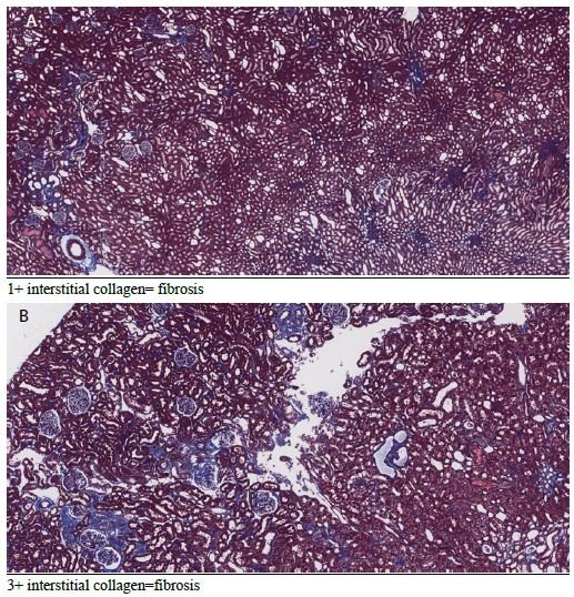
fig 1

Weight Change in Tertiary Students: Implications for Academic Performance

DOI: 10.31038/NRFSJ.2022524

Abstract

Background: Although adequate nutrition and good health are known to promote academic success, the tuition and non-tuition expenses often force most students change their eating patterns after starting tertiary education. The unresolved dilemma is that there are high rates of obesity alongside high rates of hunger on campuses in Jamaica. This study among students in three tertiary institutions in Jamaica examined the risk factors for weight gain and weight loss. More importantly, the aim was to determine whether these weight changes among students affected their academic performance.

Results: While overall weight gain and weight loss were similar (34-37%), older students experienced more weight gain (39.7%) and males had more weight loss (41.7%). Significantly less fully employed students (24.6%) lost weight than those partially employed (43.9%) or those with no job (43.2%). Disordered eating was high (39.2%) and was associated mainly with weight loss. Lower GPA scores were correlated with weight loss. Key independent factors related to weight change were age, gender, disordered eating, the amount, and type of food consumed, depression and anxiety.

Conclusion: The large proportion of students with weight change cannot be ignored by campus administrators. On-going programs are clearly not sufficient to halt this behaviour. For tertiary institutions to meet their education mandate, authorities must provide an enabling environment for students at risk of major weight changes. Policies and programs such as regular screening of students, and education to impart relevant nutritional knowledge and improve practices, are vital to promote student health and ultimately their academic success.

Keywords

Weight change, Eating disorders, Risk factors, Policies, Higher education

Introduction

University life is a critical period for cementing healthy and sustainable eating habits [1]. Not only do adequate nutrition and good health promote academic success, but in the long run they also reduce government expenditure on management and treatment of chronic diseases. In Jamaica, chronic disease prevalence has increased steadily among the adult population and for the last 3 decades they have been responsible for most of the illness and deaths. In addition, the total economic burden on individuals has been estimated at over US$600 million [2].

While recent research has focused on “the Freshman 15” – a term used to describe the rapid and dramatic increase in weight of college students [3], there is also evidence that food insecurity significantly affects students as well [4,5]. This study therefore examined the apparent paradox between weight gain and weight loss among tertiary students in Jamaica.

National surveys in Jamaica show overweight/obesity trends for adults as 34% in 2000; 49% in 2008 and 54% in 2016 [6]. This trend is astounding – showing a 59% increase in 16 years and clearly calls for bold and sustained corrective action. Further, other surveys have documented an increase in the major behavioural risk factors and NCDs such as diabetes, hypertension, and obesity among adults [7]. In 2017, PAHO estimated that 78% of all deaths are caused by NCDs. Of all NCD deaths 30% occurred prematurely between 30 and 70 years of age [8]. Coupled with such high rates of chronic disease is the fact that almost half (46.0%) of the adult population are classified as having low physical activity and approximately 99.0% of adults consume well below the daily recommended portions of fruits and vegetables [6].

Graduates from tertiary institutions are expected to be the main drivers of sustainable development. These institutions should therefore obtain a better understanding of food insecurity and eating behaviours among students as they not only have the potential to influence academic performance, student retention and graduation rates but also allow such institutions to provide key evidence needed to advocate for and develop policies at the national and regional levels [5].

Methods

A quantitative survey was used to capture the dynamics that can affect student eating behavior and also gain insights into the mechanism through which weight change can harm student academic performance. Three tertiary institutions participated in this self-reporting study: University of Technology, Jamaica, the University of the Commonwealth Caribbean and Shortwood Teachers’ college. About 300 students from each of these institutions were randomly selected to participate. Efforts were made to stratify by faculty in each institution. A pilot test of the questionnaire was done on 20 students at different levels to assess clarity and understanding. The test results were used to modify the structure and content of questions where necessary. To solicit maximum honesty and confidentiality the students were not required giving their names, identification numbers, or any information that can be traced to them individually. After ethical clearances and permissions from the university authorities, coordinators from each institution were assigned to administer the questionnaire. No payment was given to the students for completing the questionnaire. Student responses were scrutinized for completeness and quality. Analysis was planned to reveal several descriptors of weight gain and weight loss. Independent factors were identified using weight gain and weight loss as dependent variables.

Results

Analysis of data from the three institutions combined showed that the proportion of students who reportedly gained weight (34.0%) was close to those who lost weight (36.9%). Stark differences were however found in weight change with various variables.

Figure 1 shows that among those less than 22 years old, 44.4% lost weight and 31.1% gained weight. In the 22-28-year-old category, more students also reported that they had lost weight (37.9%) compared to those who had gained (33.3%). In the over 28 years old category, however, more reported that they had gained weight (39.7%) and 22.6% lost weight.

fig 1

Figure 1: Weight change by age of tertiary students

Among males, the greatest percentage (41.7%) reported that they had lost weight while 24.6% had gained weight. Among females, weight gain was found in 36.6% of them and 35.4% lost weight.

Some students with full-time employment reported that they had gained weight (35.5%) while 24.6% reported they had lost weight. Among students who were employed part-time, the greatest percentage (43.9%) had lost weight, while 35.1% had gained weight. A larger proportion of unemployed students (43.2%) reported that they had lost weight while 32.5% had gained weight (Table 1).

Table 1: Relationship between Employment Status and Weight Change

Weight Change

Employment

Full Time

Part Time

No Job

Gained (%)

35.5

35.1

32.5

Lost (%)

24.6

43.9

43.2

No Change (%)

39.9

21.1

24.3

Total

338

171

465

P<.001

As expected, more students (40.8%) who ate 3 times per day gained weight whereas 18.9% lost weight. In contrast, most students who reported that they ate once per day had lost weight (54.0%) while 27.7% had gained weight (Table 2).

Table 2: Relationship between Meal Frequency and Weight Change


Weight Change

Meals per day

3+

2

1

Gained (%)

40.8

33.9

27.7

Lost (%)

18.9

37.0

54.0

No Change (%)

40.3

29.1

18.3

Total

201

570

202

P<.001

Among all students 39.2% indicated that their eating changes were due to a disorder. Of those who reported a disorder 41.6% had lost weight, 37.4% gained and 21.1% had no change in weight. Of those whose consumption changes were not due to a disorder the difference was not as large; 34.6% reported no weight changes, 33.7% reportedly lost weight and 31.7% indicated they had gained weight (Table 3).

Table 3: Changes in eating habits due to a disorder and its relation to weight change

Weight Change

Eating changed due to disorder

Yes

No

Gained (%)

37.4

31.7

Lost (%)

41.6

33.7

No Change (%)

21.1

34.6

Total

380

590

P<.05

Figure 2 shows that of those who were undernourished or normal, 50.3% said they lost weight. Of those overweight, 44% said they gained weight. Of those who were obese 49.7% said they gained weight.

fig 2

Figure 2: Weight change according to weight status

Figure 3 shows that when the type of food changed there was an overall loss in weight. Students were also asked whether anxiety and stress caused changes in their consumption habits. Most students indicated this was the case (59.9%). Of these, 44.3% reported that they had lost weight, 32.3% had gained weight. Of those whose eating had not changed because of stress and anxiety, 36.7% gained weight and 25.6% reportedly had lost weight.

fig 3

Figure 3: Other Causes of weight change

A minority of students (39.3%) indicated that they were involved in planned physical activity. Among these students, 42.1% reported that they had lost weight, 30.6% had gained weight (Figure 3). The duration of physical activity was categorized as less than 1 hour and 1-2 hours per week. There was no difference in weight change according to the duration of physical activity. Most students reported that they were not involved in planned physical activity (60.7%). Of these, 35.9% reportedly gained weight, 33.6% lost weight and 30.5% experienced no change in weight. The grade point averages (GPAs) for students were used to denote academic performance. Figure 4 shows that students with lower GPAs (<3.2) experienced more weight loss than the high achievers (GPA >3.2).

fig 4

Figure 4: Weight change in relation to academic performance (GPA)

The likelihood ratio test was used to identify the independent factors related to weight gain and weight loss. Table 4 shows that the full-time employed student was the main factor related to weight gain, whereas eating one meal per day was the main factor related to weight loss.

Table 4: Independent factors related to weight change

Factors and weight change

Weight gain vs. no change

Weight loss vs. no change

Full-time employed student (p> 001)

Older student (p<.05)

Female student (p<.05)

Eating disorder (p<.05)

Type of food consumed p<.05


1 vs. 3 Meals per day (p<.001)

Depression & anxiety (p<.05)

Type of food consumed (p<.05)

Discussion

Previous studies in Jamaica revealed high levels of food insecurity among university students [9]. The natural hypothesis from this observation is that it will express itself as weight loss. However, with obesity on the rise in Jamaica, that hypothesis needed to be tested. Weight gain among students who are food insecure may be the result of poor food choices, lifestyle habits (harmful use of alcohol or lack of engagement in physical activity) or the body’s response to stress including finances, academic pressures and work life. While evidence from the Caribbean is sparse and mostly anecdotal, a review of the literature shows that in several countries a double-burden of sorts exists – both food insecurity and overweight/obesity, particularly among women and youth [10].

Evidence from other parts of the world suggests that university students consume more unhealthy foods (processed foods with high total and saturated fats) and have lower intakes of fresh fruit and vegetables [10-12]. Such behaviours may carry on to adulthood thereby contributing to the burden of disease seen among middle- and older-aged adults at the population level.

The self-reported weight changes in this study indicate significantly increasing weight gain with age. This is consistent with the observation that the 18-29 year-old age group is often viewed as being at high risk for weight-related behaviour change as the transition from adolescence to adulthood is made and there is more freedom regarding the type and amount of food consumed [11]. Even when students are aware of the health consequences of overconsumption of unhealthy foods, food choices have been shown to be heavily influenced by convenience and taste [12].

Most published studies have focused specifically on weight gain, but this study shows that weight loss is relatively high, particularly among males. Globally, studies examining sex-related changes in weight have shown that both males and females experience weight changes when they enter university with the greatest changes in weight taking place in the first semester [13].

This study found higher weight gain among students fully employed. Working full time has been thought to be associated with unhealthy eating and consequently weight gain [14].

Research shows that stress can result in changes in food intake patterns and among university students has been linked to maladaptive eating behaviours such as consumption of unhealthy foods and overeating [15]. In fact, there is an apparent link between the types of foods that are viewed as ‘comfort foods’ which are used to cope with stressors and age – with younger persons seeming to prefer snack-related foods such as ice cream, candy, and sweet breads [16]. As university enrolment often results in major life changes for example new living arrangements, new social situations and increased academic and time demands, stress and its consequent effect on dietary habits may be commonplace [16]. As expected in this study, students who ate more meals achieved more weight gain. But this relationship is complex. The effect of meal frequency plays an important role not just in academic achievement but also long-term health consequences. The omission of one or more meals from the diet has been linked to poorer diet quality, increased risk of abdominal adiposity and increased BMI [17-21]. Reasons for meal skipping among university students include a lack of hunger, depression, lack of time, lack of money, and lack of cooking skills [22,23].

The cost of food has been shown to be a key factor that influences what students purchase [24]. Foods that are relatively cheaper also tend to be high in salt, sugar, fat and flavour additives which have been identified as contributory factors to the obesity epidemic. Disordered eating is associated with weight gain, overweight and obesity among adolescents/young adults [25,26]. The high percentage of disordered eating among Jamaican students is worrisome. And it resulted mainly in weight loss. Studies have shown that disordered eating is more prevalent among those who may be experiencing feelings of anxiety, loneliness and stress, all of which are common among university students [27].

Among university students, research shows that physical activity levels continue to decline while engagement in sedentary activities continue to increase [28]. This study shows a minority of students involved in planned physical activity. Several factors have been proposed regarding this decline among university students e.g. university students have greater control of their daily lives and are not mandated to participate in physical activity, the impact of residence (on- or off-campus), time demands (including time spent on social media) and access to facilities where physical activities are offered or where physical activity can be engaged in safely [29]. Data from the United Kingdom suggest that up to 60% of university students are not meeting physical activity recommendations [30].

Campus administration has an obligation to equip students not only with knowledge for the world of work but also for a healthy lifestyle which is integrally linked to work performance. Efforts can include: (1) Screening students at the start of each school year for food insecurity to gauge the type and quantity of support that is required. (2) Collaborating with food manufacturers and supermarkets for food donations to university/college campuses. (3) Conduct of an interactive “Cooking on a Budget” program during the semester which teaches students how to cook quick, cheap, and healthy meals on a budget. (4) Meal Program – Provision of a student space which can be accessed by students and provides coffee/tea, affordable and healthy snacks, sandwiches etc. (5) Pantry Program – Installation of a student-run pantry that students can access which includes toiletries and grocery vouchers for students in need.

Acknowledgment

We thank the University of Technology, Jamaica, for providing funding through the Research Development Fund, managed by the University’s Research Management Office, the School of Graduate Studies, Research & Entrepreneurship. Gratitude is expressed to Mr. Kevin Powell (University of the Commonwealth Caribbean) and Ms. Ava-Marie Reid (Shortwood Teachers’ College who coordinated the data collection at their respective institutions.

References

  1. Kim HS, Ahn J, No JK (2012) Applying the Health Belief Model to college students’ health behavior. Nutrition Research and Practice 6: 551-558. [crossref]
  2. Ministry of Health and Wellness Jamaica (2013). National strategic and action plan for the prevention and control of NCDs in Jamaica 2013-2018. Kingston, Jamaica Retrieved from http://moh.gov.jm/wp-content/uploads/2015/05/National-Strategic-and-Action-Plan-for-the-Prevention-and-Control-Non-Communicable-Diseases-NCDS-in-Jamaica-2013-2018.pdf
  3. Sharif MR, Sayyah M (2018) Assessing physical and demographic conditions of freshman” 15″ male medical students. International Journal of Sport Studies for Health 1: e67421.
  4. Freudenberg N, Goldrick-Rab S, Poppendieck J (2019) College students and SNAP: The new face of food insecurity in the United States. American Journal of Public Health 109: 1652-1658. [crossref]
  5. Payne-Sturges DC, Tjaden A, Caldeira KM, Vincent KB, Arria AM (2018) Student hunger on campus: Food insecurity among college students and implications for academic institutions. American Journal of Health Promotion 32: 349-354. [crossref]
  6. JHLS (2000). Jamaica Health and Lifestyle Survey. Ministry of Health 2000.
    ______ (2008). Jamaica Health and Lifestyle Survey. Ministry of Health 2008
    ______ (2018). Jamaica Health and Lifestyle Survey. Ministry of Health 2018
  7. Ministry of Health (2013) National Strategic and Action Plan for the Prevention And Control Non-Communicable Diseases (NCDS) in JAMAICA 2013-2018. Retrieved from: https://www.moh.gov.jm/wp-content/uploads/2015/05/National-Strategic-and-Action-Plan-for-the-Prevention-and-Control-Non-Communicable-Diseases-NCDS-in-Jamaica-2013-2018.pdf
  8. PAHO (2017) Regional mortality estimates 2000-2015. PAHO 2017.
  9. Henry FJ, Nelson M, Aarons R (2020) Learning on empty. In University Student Life and learning: Challenges for Change. Ed: Fitzroy J. Henry, University of Technology, Jamaica Press 102-112.
  10. Au L, Zhu S, Ritchie L, Nhan L, Laraia B, et al. (2019) Household Food Insecurity Is Associated with Higher Adiposity Among US Schoolchildren Ages 10–15 Years (OR02-05-19). Current Developments in Nutrition 149: 1642-1650. [crossref]
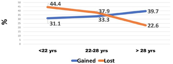
  11. Nelson MC, Story M, Larson NI, Neumark-Sztainer D, Lytle LA (2008) Emerging adulthood and college-aged youth: an overlooked age for weight-related behavior change. Obesity 16: 2205-2211. [crossref]
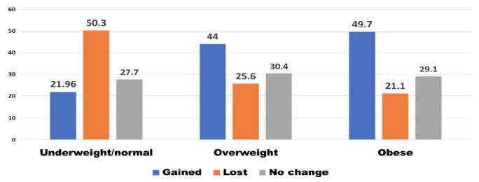
  12. Abraham S, Noriega B, Shin J (2018) College students eating habits and knowledge of nutritional requirements. Journal of Nutrition and Human Health 106: 46-53. [crossref]
  13. Lloyd-Richardson EE, Bailey S, Fava JL, Wing R, Network TER (2009) A prospective study of weight gain during the college freshman and sophomore years. Preventive Medicine 48: 256-261. [crossref]
  14. Escoto KH, Laska, MN, Larson N, Neumark-Sztainer D, Hannan PJ (2012) Work hours and perceived time barriers to healthful eating among young adults. American Journal of Health Behavior 36: 786-796. [crossref]
  15. Lyzwinski LN, Caffery L, Bambling M, Edirippulige S (2019) The mindfulness app trial for weight, weight-related behaviors, and stress in university students: randomized controlled trial. JMIR mHealth and uHealth 7: e12210. [crossref]
  16. Kandiah J, Yake M, Jones J, Meyer M (2006) Stress influences appetite and comfort food preferences in college women. Nutrition Research 26: 118-123.
  17. Chung H-Y, Song M-K, Park M-H (2003) A study of the anthropometric indices and eating habits of female college students. Journal of Community Nutrition 5: 21-28.
  18. Kerver JM, Yang EJ, Obayashi S, Bianchi L, Song WO (2006) Meal and snack patterns are associated with dietary intake of energy and nutrients in US adults. Journal of the American Dietetic Association 106: 46-53. [crossref]
  19. Ma Y, Bertone ER, Stanek III EJ, Reed GW, Hebert JR, et al. (2003) Association between eating patterns and obesity in a free-living US adult population. American Journal of Epidemiology 158: 85-92. [crossref]
  20. Musaiger A, Radwan H (1995) Social and dietary factors associated with obesity in university female students in United Arab Emirates. Journal of the Royal Society of Health 115: 96-99. [crossref]
  21. Timlin MT, Pereira MA (2007) Breakfast frequency and quality in the etiology of adult obesity and chronic diseases. Nutrition Reviews 65: 268-281. [crossref]
  22. Afolabi W, Towobola S, Oguntona C, Olayiwola I (2013) Pattern of fast-food consumption and contribution to nutrient intakes of Nigerian University students. Int J Educ Res 1: 1-10.
  23. Lee J-E, Yoon W-Y (2014) A study of dietary habits and eating-out behavior of college students in Cheongju area. Technology and Health Care 22: 435-442. [crossref]
  24. Tam R, Yassa B, Parker H, O’Connor H, Allman-Farinelli M (2017) University students’ on-campus food purchasing behaviors, preferences, and opinions on food availability. Nutrition 37: 7-13. [crossref]
  25. Barrack MT, West J, Christopher M, Pham-Vera A-M (2019) Disordered eating among a diverse sample of first-year college students. Journal of the American College of Nutrition 38: 141-148. [crossref]
  26. Goldschmidt AB, Aspen VP, Sinton MM, Tanofsky-Kraff M, Wilfley DE (2008) Disordered eating attitudes and behaviors in overweight youth. Obesity 16: 257-264. [crossref]
  27. Costarelli V, Patsai A (2012) Academic examination stress increases disordered eating symptomatology in female university students. Eating and Weight Disorders-Studies on Anorexia, Bulimia and Obesity 17: e164-e169. [crossref]
  28. Lauderdale ME, Yli-Piipari S, Irwin CC, Layne TE (2015) Gender differences regarding motivation for physical activity among college students: A self-determination approach. The Physical Educator 72.
  29. Calestine J, Bopp M, Bopp CM, Papalia Z (2017) College student work habits are related to physical activity and fitness. International Journal of Exercise Science 10: 1009. [crossref]
  30. Aceijas C, Bello-Corassa R, WaldhäuslN S, Lambert N, Cassar S (2016) Barriers and determinants of physical activity among UK university students Carmen Aceijas. European Journal of Public Health 26.