Archives

Featured Image2

A Short Note on Climate Change: An Alternative View

DOI: 10.31038/AFS.2021331


It is the general view of the Media that the cause of climate change is directly related to air pollution from coal burning and vehicle exhaust. Undoubtedly this cause of air pollution is a worrying factor but whether it is the cause of climate change can’t go unchallenged.

In the geological past evidence of glacial and interglacial periods span a minimum of 400 million year and evidence of glaciation in the Lower Paleozoic, 4-500 million years ago, is seen in the Garevellach Islands and Islay, South West Highlands of Scotland. This event is well documented substantiates the concept of worldwide glaciations throughout the past. This geological evidence appears not to concern the media, apparently due to the lack of understanding of geological science.

In geologically recent times, late 7th to early 19th century, the River Thames periodically froze over to allow the so-called Frost Fairs to take place. No automobiles or electricity generators existed therefore these events could not have been caused by air pollution from these sources.

The controlling factor is more likely to be changes in solar radiation due to alterations in the Earth’s orbital around the Sun with time, as this would affect the solar radiation falling on the Earth. In which case there is nothing man can do to alter the situation.

Twins with Juvenile Hyaline Fibromatosis

DOI: 10.31038/PSC.2021111

Abstract

Background: Juvenile hyaline fibromatosis (JAF) is a rare autosomal-recessive disease in which patients progressively develop cutaneous tumoral fibroblastic proliferations, and joint contractures with bone involvement. JAF is caused by aberrant synthesis of glycosaminoglycans by fibroblasts due to a mutation of the capillary morphogenesis factor-2 gene (CMG2). Limited treatment options are available.

Method: We report monozygotic twins who presented with multiple, recurrent, painless cutaneous nodules.

Result: The presence of twins with JAF is extremely rare. A lesion on the head of one boy had ruptured, and pathological analysis indicated benign spindle cells in a periodic acid-Schiff (PAS)-positive hyaline background. One of twins had much more severe clinical presentation than the other, including more frequent diarrhea, larger nodules, more severe joint involvement, and more easily ruptured masses.

Conclusion: Monozygotic twins who present with JAF may have different severity of symptoms despite the presence of identical mutations in CMG2.

Keywords

juvenile hyaline fibromatosis, twins, subcutaneous mass

Introduction

Juvenile hyaline fibromatosis (JHF) is a rare autosomal-recessive disease caused by mutations in capillary morphogenesis gene-2 (CMG2). There have been fewer than 70 cases reported so far in the literature [1–2], and there has only been one report of twins with JHF [3].The clinical onset usually occurs before 5 years of age, and this disorder is slightly more common in boys [4]. JHF is characterized by cutaneous tumoral fibroblastic proliferation, joint contractures, and bone involvement [2].

Materials and Methods

Clinical Features

Two one-and-a-half year-old male monozygotic twins presented with multiple, progressive, painless, variable-sized nodules all over their bodies. The parents had the same family name and lived in the same remote village in Henan province of China, but were not first-degree relatives. In the year prior to presentation, both infants had recurrent diarrhea of unknown etiology, and in the 6 months prior to presentation, both had multiple, painless, variable-sized cutaneous nodules all over their bodies. These nodules were first noticed around the joints, then on the trunk and head, and eventually all over their bodies. The size of nodules increased gradually over time. One year ago, a nodule on one child’s finger was given an incisional biopsy at a local hospital, and the pathological result was “benign”, but there was no definite diagnosis or further investigation. When they were hospitalized, one of masses on the head had decayed spontaneously and the joint contractures had deteriorated gradually, which crippled both of them. They both had very poor appetites. One of them (patient A) had more severe signs and symptoms than the other (patient B) [Table 1]. They both achieved normal mental development milestones.

Table 1. Clinical feature difference between the twins


patien A

patient B

clinical onset

earlier

later

maximium diameter of nodules

25

15

mass rupture

yes

no

gingival proliferation

svere

moderate

joint constracture

svere

moderate

perianal tissue proliferation

yes

no

nostril narrow

yes

no

diarrhea

conatantly

intermittent

On physical examination, there were multiple, painless, variable-sized nodules all over the bodies, which were soft upon palpation [Figure 1]. The largest nodule was on the head of patient A, whose diameter was 25 cm [Figure 2]. One mass on the head had ruptured and had a terrible odor [Figure 3]. Cutaneous tumoral fibroblastic proliferation around the lips and gingival hyperplasia [Figure 4] were also present. Patient A had a narrow nostril caused by subcutaneous proliferation, but no shortness of breath [Figure 5]. There were many nodules on their hands and fingers [Figure 6], elbows [Figure 7], and abdomens [Figure 8]. There were also flexion contracture deformities of the limbs [Figure 9], scales on the feet [Figure 10], and proliferation in the perianal area [Figure 11], but no significant lymphadenopathy or hepatosplenomegaly.

PSC 2019-101 - Deng Gaoyan China_f1

Figure 1. Appearance of the twins.

PSC 2019-101 - Deng Gaoyan China_f2

Figure 2. Mass on the head.

PSC 2019-101 - Deng Gaoyan China_f3

Figure 3. Decayed mass on the head.

PSC 2019-101 - Deng Gaoyan China_f4

Figure 4. Cutaneous proliferation around the lips and gingival hyperplasia.

PSC 2019-101 - Deng Gaoyan China_f5

Figure 5. Patient A had a narrow nostril.

PSC 2019-101 - Deng Gaoyan China_f6

Figure 6. Nodules on hands and fingers

PSC 2019-101 - Deng Gaoyan China_f7

Figure 7. Nodules on elbow.

PSC 2019-101 - Deng Gaoyan China_f8

Figure 8. Nodules on abdomen.

PSC 2019-101 - Deng Gaoyan China_f9

Figure 9. Flexion contracture deformities of the knee.

PSC 2019-101 - Deng Gaoyan China_f10

Figure 10. Scales on the foot.

PSC 2019-101 - Deng Gaoyan China_f11

Figure 11. Proliferation in the perianal area.

The complete blood counts and electrolyte levels were within normal limits. An X-ray indicated osteoporosis in vertebrae of patient A [Figure 12]. Computed tomography (CT) results showed no masses in the abdominal cavities, except for cutaneous nodules in the abdominal wall [Figure 13]. A head CT of patient A showed  a large subcutaneous mass but no intracranial abnormalities [Figure 14].

PSC 2019-101 - Deng Gaoyan China_f12

Figure 12. Osteoporosis in vertebrae.

PSC 2019-101 - Deng Gaoyan China_f13

Figure 13. Cutaneous nodules in the abdominal wall.

PSC 2019-101 - Deng Gaoyan China_f14

Figure 14. Subcutaneous mass without intracranial abnormalities.

After consent from the parents, a biopsy of the large mass on patient A’s head was performed. Hyaline subcutaneous tissue was present beneath the skin [Figure 15], which was sent for pathological investigation. The ruptured head mass was covered with dressing and required regular changes.

PSC 2019-101 - Deng Gaoyan China_f15

Figure 15. Hyaline subcutaneous tissue beneath the skin.

Pathological Findings

Gross examination indicated that the nodules had a gelatinous white and hyaline appearance [Figure 16]. Microscopically, there were poorly circumscribed lesions composed of many uniform spindle cells embedded in an abundant homogenous fibrous matrix. This matrix was eosinophilic and tested positive on periodic acid-Schiff (PAS) staining, findings characteristic of JHF [Figure 17]. The parents did not consent to surgical procedures on the other child.

PSC 2019-101 - Deng Gaoyan China_f16

Figure 16. Gross pathological examination.

PSC 2019-101 - Deng Gaoyan China_f17

Figure 17. Microscopical pathological examination.

RNA isolation and cDNA amplification of samples from each patients’ blood were performed. Cloning and sequencing of the CMG2 gene showed that both patients had the same 4 mutations: c.294C→A, c.1069G→C, 1074delT, and c.1153G→C.

Follow up

The twins were followed up for 4 years after the initial presentation. At the last follow-up, the decayed head lesion of patient A was still unhealed, there were more nodules all over their bodies, and the masses on the head of patient A were larger. The parents refused further therapy.

Discussion

JHF is a rare autosomal-recessive disease that was first described by McMurray [1] as molluscum fibrosum, but renamed by Kitano [2]. Patients with JHF present with multiple cutaneous nodules or masses, gingival hypertrophy, joint contractures, and osteolytic lesions [4]. Skin lesions are the most prominent symptoms [8]. Normal motor development is impaired if joint contractures occur during infancy. Eating and delayed dentition can be caused by severe gingival hyperplasia [1].

Histological examination can confirm a diagnosis of JHF [10]. In particular, lesions from the dermis, subcutis, gingivae, bone, and joints contain abundant homogeneous eosinophilic matrix, which is embedded with cords of spindle-shaped cells. The matrix stains positively with PAS and alcian blue, but not with toluideine blue or Congo red [10]. Nodules are calcified occasionally, but have no elastic tissue. The etiology is unknown. The differential diagnosis includes neurofibromatosis, fibromatosis, amyloidosis, infantile systemic hyalinosis, lipoid proteinosis, and Winchester syndrome [2].

The JHF gene is located on gene 4q21 [5], and previous studies have described the effects of mutations in CMG2 [6]. Previous researchers have described JHF as a connective tissue disease that is characterized by aberrant synthesis of glycosaminoglycans. Dermatan sulfate is the predominant glycosaminoglycan in the skin of patients with JHF, which also has chondroitin sulfate and hyaluronan, but hyaluronan is the most abundant glycosaminoglycan in normal skin [7].

There are no specific treatments for JHF, but cosmetic surgery and those that limit orthopedic disability may be employed. This disease has a progressive course, and most patients only survive up to the 4th decade [1–2]. Relapses are common after tumor removal. A previous report indicated good cosmetic results after removal of more than 100 tumors over a period of 19 years [9]. Physiotherapy may be performed to prevent flexion contractures.

The occurrence of twins with JHF is extremely rare, and there has only been one previous case report [3], and the researchers did not report differences between these twins. It is generally accepted that twins will have the same presentations, because JHF is a genetic disease. However, our twins had very different presentations. Patient A had more severe symptoms [Table 1], and a head mass that ruptured and bled, and was refractory to treatment. Patient A also had more frequent diarrhea, larger nodules, and more severe joint involvements.

We do not know the reasons for the different presentations of these twins. They had identical mutations in CMG2, and might be expected to eventually develop the same signs and symptoms. It may be hypothesized that the differences between the normal and genetically mutated somatic cells led to the differences in these twins. It is possible that patient B will eventually develop the same severe symptoms as patient A. A close follow-up of these children is warranted.

Statement

There are no prior publications or submissions with any overlapping information, including studies and patients. The manuscript has not been and will not be submitted to any other journal. There have no financial support or relationships that may pose conflict of interest. Gaoyan, Deng wrote the first draft of the manuscript. No honorarium, grant, or other form of payment was given to anyone to produce the manuscript. Each author listed on the manuscript has seen and approved the submission of this version of the manuscript and takes full responsibility for the manuscript.

References

  1. Nischal KC, Sachdev D, Kharkar V, Mahajan S. (2004) Juvenile hyaline fibromatosis. J Postgrad Med 50: 125–6.
  2. Haleem A, Al-Hindi HN, Juboury MA, Husseini HA, Ajlan AA. (2002) Juvenile hyaline fibromatosis: Morphologic, immuno histochemical and ultrastructural study of three siblings. Am J Dermatopathol 24: 218–24.
  3. Habibeddine S, Khadir K, Azzouzi S, Skalli S, Lakhdar H. (2003) Juvenile hyaline fibromatosis: 2 twin brothers affected. Ann Dermatol Venereol 130: 43–6.
  4. Weiss SW, Goldblum JR. (2001) Fibrous tumors of infancy and childhood. In: Strauss M, editor. Enzinger and Weiss’s Soft Tissue Tumors. 4th ed. St. Louis: Missouri Mosby; 363–7.
  5. Nazneen Rahman, Melanie Dunstan M. Dawn Teare. (2002) The gene for juvenile hyaline fibromatosis maps to chromosome 4q21. Am J Hum Genet 71; 975–980.
  6. Sandra Hanks, Sarah Adams, Jenny Douglas. (2003) Mutations in the gene encoding capillary morphogenesis protein 2 cause juvenile hyaline fibromatosis and infantile systemic hyalinosis. Am J Hum Genet 73; 791–800.
  7. Katagiri K, Takasaki S, Fujiwara S, Kayashima K, Ono T, et al. (1996) Purification and structural analysis of extracellular matrix of a skin tumor from a patient with juvenile hyaline fibromatosis. J Dermatol Sci 13: 37–48.
  8. Sanzalin H, Kiyozuka Y, Uemura Y, Shikata N, Ueda S, et al. (1998) Juvenile hyaline fibromatosis: A report of two unrelated adult sibling cases and a literature review. Pathol Int 48: 230–6.
  9. Woyke S, Domagala W, Markiewicz D. (1984) A 19-year follow-up of multiple juvenile fibromatosis. J Pediatr Surg 19: 302.
  10. Arnold H, Odom RB, James WD. (1990) Andrews’ diseases of the skin, Clinical dermatology. 8th ed. Philadelphia: Saunders, 712.
Featured Image2

Physical Activity in Our Modern World: An Evolutionary Necessity

DOI: 10.31038/CHBT.2021111


In our modern world obesity is a causal factor in numerous metabolic and endocrine disorders including heart disease, non-alcoholic fatty liver disease (NAFLD), type 2 diabetes mellitus (T2DM), bone and joint disorders and some types of cancer [1]. According to the World Health Organization in 2016, globally more than 1.9 billion adults were overweight and of these over 650 million were obese [2]. Childhood obesity is a more disturbing trend; in 2019 thirty eight million children under the age of 5 were overweight or obese and in 2016 over 340 million children and adolescents aged 5-19 were overweight or obese [3]. Obesity is not a cosmetic issue but rather an affliction that places a tremendous burden to quality of life on individuals and health care systems. It is not without exaggeration to say that obesity due to physical inactivity is the biggest public health problem of the 21stcentury [4]. However, obesity is preventable and reversible and for interpreting the obesity pandemic afflicting modern societies we need to understand our evolutionary history and examine the environmental, physical, and physiological conditions that impelled our ancestral gorilla-chimpanzee lineageto shift from a sedentary, plant and fruit diet existence to a foraging, meat eating, hunter-gatherer existence some 2.5 million years ago [5].

The transition of our ancestral lineage from the gorilla-chimpanzee line to hunter gatherer necessitated changes in diet, metabolism and physiology that are conducive for high levels of physical activity for survival. Although the environmental, physical, and physiological conditions have changed, our physiology remains genetically adapted for a nomadic forager, hunter gatherer life style [6,7]. Our physiology adapted to storing lipids and carbohydrates as fuels to cope with the uncertainties of food availability in our past. This may have been a survival advantage 2.5 million years ago, but in our modern world it is a liability that leads to increased morbidity and mortality due to increased circulating triglycerides, cholesterol, blood pressure, T2D, NAFLD, Alzheimer’s disease, cardiovascular disease (CVD), and several types of cancer [8,9]. For most of our evolutionary history, as conditions changed so did our physiology. Our lungs, brain, muscles, blood vessels and all our organs and physiological organ systems have evolved to support a high activity level demanded for survival and our bodies have retained this adaptation to the present day. However, we no longer have to forage 15-20 kilometers per day, nor do we have to fend off predators or perform exhaustive farming practices to necessitate high energy diets. In evolutionary terms, the modern norms of an inactive lifestyle and a diet based on an excess of energy-dense, nutrient-deficient, foods such animal products, processed foods, alcohol, sugar and sugar derivatives are not conducive for the purpose for which we evolved.

To prevent or reverse the obesity epidemic and minimize our proclivity to obesity and the subsequent metabolic disorders we need to modify our diet and increase our levels of physical activity. We need to exercise. Our physiology is a dynamic product of evolution that adjusts to changes in living conditions. Regular exercise reduces chronic inflammation which constitutes risk factor for CVD, it reduces insulin resistance which is a harbinger for T2D and helps to shuttle glucose into muscle glycogen stores instead of fat, thus helping reduce triglycerides from the circulation [10]. Additionally, it lowers resting levels of testosterone, estrogen and progesterone, which may explain the reduced rate of reproductive cancers among adults who exercise regularly, improves the effectiveness of immune function, which helps stave off infection especially as we age, and may blunt the morning rise of the stress hormone cortisol [11]. Other than maintaining health, physical activity contributes to healthy aging by reducing the accumulation of senescent cells and the of secretion of a complex mixture of different inflammatory cytokines, chemokines, growth factors and proteases such as IL1a, IL1b, IL6, IL8 and metalloproteinases (MP-1, MP-3) that contribute to the aging process [12]. Also, evidence supports an association between exposure to regular exercise and reduced risk for the development of several cancers that might improve clinical outcomes following a diagnosis of primary disease [7,13].

In order to prevent or reverse the obesity pandemic afflicting modern societies the WHO recommends adults undertake at least 150 min moderate or 75 min vigorous intensity physical activity per week, conduct muscle strengthening activities twice a week, and minimize time spent being sedentary [14]. In an effort to counteract the risks of a sedentary lifestyle the “Exercise is Medicine initiative” in collaboration with the American College of Sports Medicine and American Medical Association was introduced in 2007 to advance the implementation of evidence-based strategies and incorporate exercise as part of a standard treatment for conditions related to sedentary lifestyle such as T2D, CVD, and obesity. It calls for the assessment of the every patient’s physical activity program and prescribing a regimen tailored to each individual’s condition and needs. The implementation of this initiative may change patient behaviour, lighten the burden placed on the quality of life and healthcare systems. The benefits of a structured, regular physical activity regimen for health and disease prevention are unequivocal. Natural selection has tailored our bodies for movement and our physiology has retained the metabolic hunter-gatherer characteristics that do not conform to the sedentary way of life of modern societies. Our contemporary lifestyle and dietary habits conflict with our evolutionary outcome. A quote attributed to Hippocrates (460-370 BC) states “If we could give every individual the right amount of nourishment and exercise, neither too much nor too little, we would have found the safest way to health”. A more recent comment by Manini TM (2015) in the JAMA-Internal Medicine testifies that “there is no single medication treatment that can influence as many organ systems in a positive manner as can physical activity” [15].

Author Contributions

Peter Lembessis: Conceptualization of topic, literature search, writing of original draft.

Anastassios Philippou: Review & editing, literature search.

Stamatis Mourtakos: Literature search on nutrition and dietetics, review and editing of draft.

References

  1. Mantovani A, Allavena P, Sica A, Balkwill F (2008) Cancer-related inflammation. Nature 454: 436-444.
  2. WHO (2020) Obesity and overweight.
  3. Saklayen MG (2018) The Global Epidemic of the Metabolic Syndrome. Curr Hypertens Rep 20: 12. [crossref]
  4. Cowan RE (2016) Exercise Is Medicine Initiative: Physical Activity as a Vital Sign and Prescription in Adult Rehabilitation Practice. Arch Phys Med Rehabil 97: S232-237. [crossref]
  5. Sarich VM, Wilson A C (1967) Immunological time scale for hominid evolution. Science 158: 1200-1203.
  6. Hambrecht R, Gielen S (2005) Essay: Hunter-gatherer to sedentary lifestyle. Lancet 366: S60-61. [crossref]
  7. Koelwyn GJ, Quail DF, Zhang X, White RM, Jones LW (2017) Exercise-dependent regulation of the tumour microenvironment. Nat Rev Cancer 17: 620-632. [crossref]
  8. Freese J, Klement RJ, Ruiz-Núñez B, Schwarz S, Lötzerich H (2017) The sedentary (r)evolution: Have we lost our metabolic flexibility? F1000Res 6: 1787. [crossref]
  9. Rezende LFM, Sá TH, Markozannes G, Rey-López JP, Lee IM, et al. (2018) Physical activity and cancer: an umbrella review of the literature including 22 major anatomical sites and 770 000 cancer cases. Br J Sports Med 52: 826-833. [crossref]
  10. Handschin C, Spiegelman BM (2008) The role of exercise and PGC1α in inflammation and chronic disease. Nature 454: 463-469. [crossref]
  11. Kelly RS, Kelly MP, Kelly P (2020) Metabolomics, physical activity, exercise and health: A review of the current evidence. Biochim Biophys Acta Mol Basis Dis 1866: 165936. [crossref]
  12. Schafer MJ, White TA, Evans G, Tonne JM, Verzosa GC, et al. (2016) Exercise Prevents Diet-Induced Cellular Senescence in Adipose Tissue. Diabetes 65: 1606-15. [crossref].
  13. Ruiz-Casado A, Martín-Ruiz A, Pérez LM, Provencio M, Fiuza-Luces C, et al. (2017) Exercise and the Hallmarks of Cancer. Trends Cancer 3: 423-441. [crossref]
  14. Sallis JF, Bull F, Guthold R, Heath GW, Inoue S, et al. (2016) Progress in physical activity over the Olympic quadrennium. Lancet 388: 1325-1336. [crossref]
  15. Manini TM (2015) Using Physical Activity to Gain the Most Public Health Bang for the Buck. JAMA Intern Med 176: 968-9. [crossref]
fig 1

Viscum album Therapy for Treating Lymphoma in an FIV and FeLV Positive Cat (Felis catus) – Case Report

DOI: 10.31038/IJVB.2021524

Abstract

Lymphoma is the hematopoietic tumor of higher incidence in domestic cats. Myelosuppression is the most remarkable side effect of the conventional treatments for this disease, especially for patients with debilitating infections such as FIV and FeLV. In this context, alternative therapies could be recommended for treating lymphoma as a sole therapy or even in association with conventional therapies to provide a better quality of life to the patients and fewer side effects.

Material and Methods: A 4-year-old neutered male mixed breed cat was seen at NaturalPet Clinic in Brazil. The cat was positive for FIV and FeLV, diagnosed with lymphoma, and had relative difficulty breathing and swallowing. On the physical examination, a nodule was observed in the right ventral part of the tongue. An incisional biopsy was performed for histopathological analysis, and the lymphoma diagnosis was confirmed. The disease was treated with homeopathic medicines, such as daily applications of Viscum album in the D3, D6, D9, D12, and D30 dilutions. Complete remission of clinical signs was observed after completing the treatment period with the reestablishment of the patient’s health.

Conclusion: The homeopathic treatment used in this case allowed the disease resolution, had no side effects on the patient and increased his life expectancy.

Keywords

Cancer, FeLV, FIV, Viscum album

Introduction

Lymphoma can be defined as neoplasia of lymphoid tissues or lymphosarcoma. It primarily attacks lymph nodes and/or other solid organs, such as the spleen, liver, or thymus. It can also affect no lymphoid tissues [1]. Neoplasms of hematopoietic origin can be considered the most frequent among domestic cats [2,3], and the most frequently diagnosed neoplasms representing about 90% of tumors in this species [4,5].

The possible accepted etiologies for the occurrence of feline lymphoma are viral immunosuppression, and the feline leukemia virus (FeLV) stands out as one of the most significant inducers of this disease [6,7]. This virus was cited in approximately 70% of cats with lymphoma. However, this reality is modifying since only 25% of cats with lymphoma are FeLV-positive [2,8]. Another virus possibly involved with the emergence of lymphomas is the feline immunodeficiency virus (FIV). It is known that approximately 20% of animals with lymphoma in the United States are seropositive for antibodies against FIV [2].

The average age of patients diagnosed with lymphoma is 8 to 10 years [3,9]. However, it varies with the form of the disease, and its etiology can strongly influence the onset of clinical signs [9]. According to [5], there is no evidence for racial or sex predisposition. These authors also claim that neutered males are slightly more affected by lymphoma, especially the alimentary type.

The lymphoma diagnosis is performed by clinical history, physical examination, laboratory and imaging tests, aspiration cytology, biopsy, and immunohistochemistry [8]. The treatment consists of chemotherapy protocols, usually with combined medicines, with no evidence that the associated surgical procedure is more effective than chemotherapy alone [9].

Nevertheless, antineoplastic medicines cause different toxic effects, especially regarding liver parenchyma [10] and animals with viral diseases, such as FIV and FeLV. The hepatotoxicity of chemotherapeutic medicines generally coincides with the increase in serum enzymes, such as alanine aminotransferase (ALT) and alkaline phosphatase (AP) [11]. Therefore, the elevation of the ALT and AP enzymes in the first chemotherapy sessions may indicate the toxic action of these medicines on the liver [11,12]. Myelotoxicity is another frequent and severe limiting factor of chemotherapy, impairing treatment efficacy and increasing the chances of metastases [13]. Additionally, it can lead the animal to death due to septic neutropenia [14]. Neutropenia is dose-limiting and can provoke septicemia with a risk of death for the patient [15].

Within this context, complementary therapies gain ground both as a primary or complementary treatment for cancer patients since they do not present side effects and they stimulate the immune system, improving life quality and, as a consequence, increasing the patient’s survival [16]. Viscum album is the plant most used in the world for the complementary treatment of cancer, with promising results recorded both as a curative or palliative strategy [17-19]. It is used in diverse injectable commercial forms, presenting a bidirectional effect to the treated patient. This medicine presents selective cytotoxicity, being aggressive only against the tumoral cells. It also presents immunomodulatory and anti-inflammatory activities [16].

[20] associated Viscum album with the traditional chemotherapy in dogs and observed a decrease in the total treatment time, reducing the chemotherapy side effects, such as leukopenia.

The objective of this study was to report the case of a domestic feline positive for FIV and FeLV, diagnosed with lymphoma, and successfully treated by injectable homeopathy.

Case Report

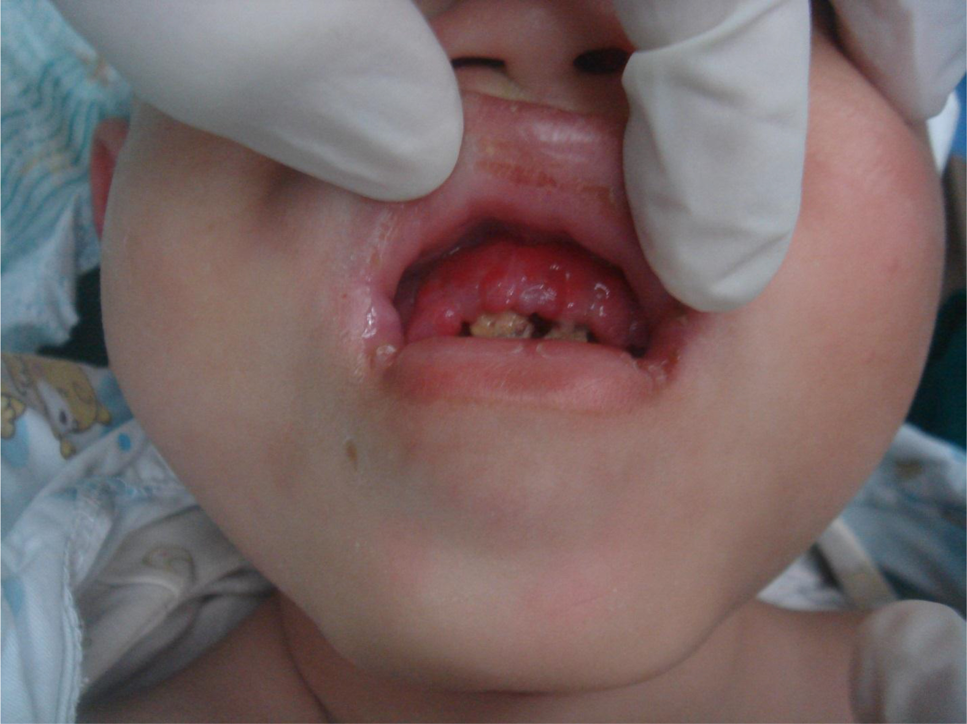
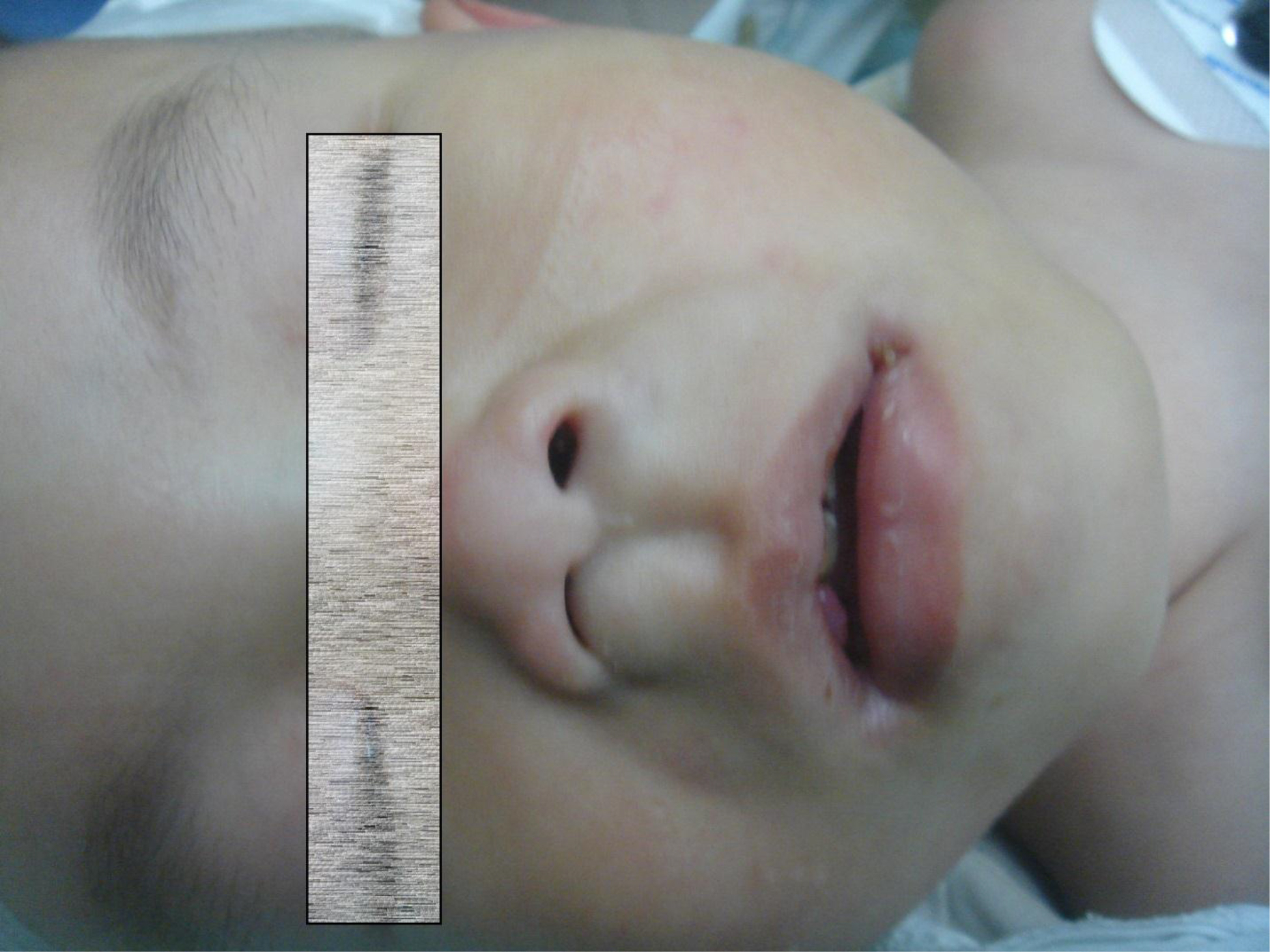
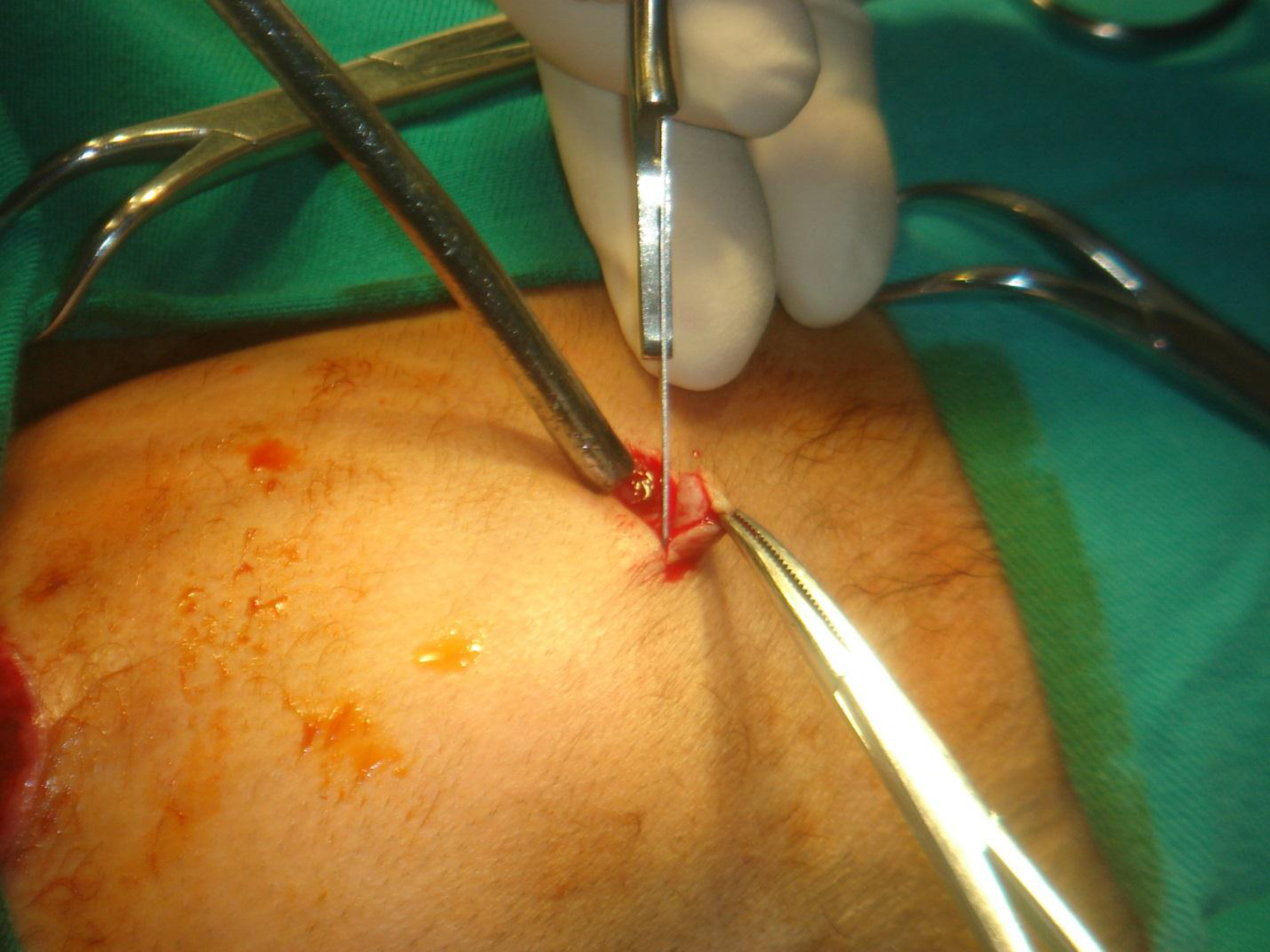
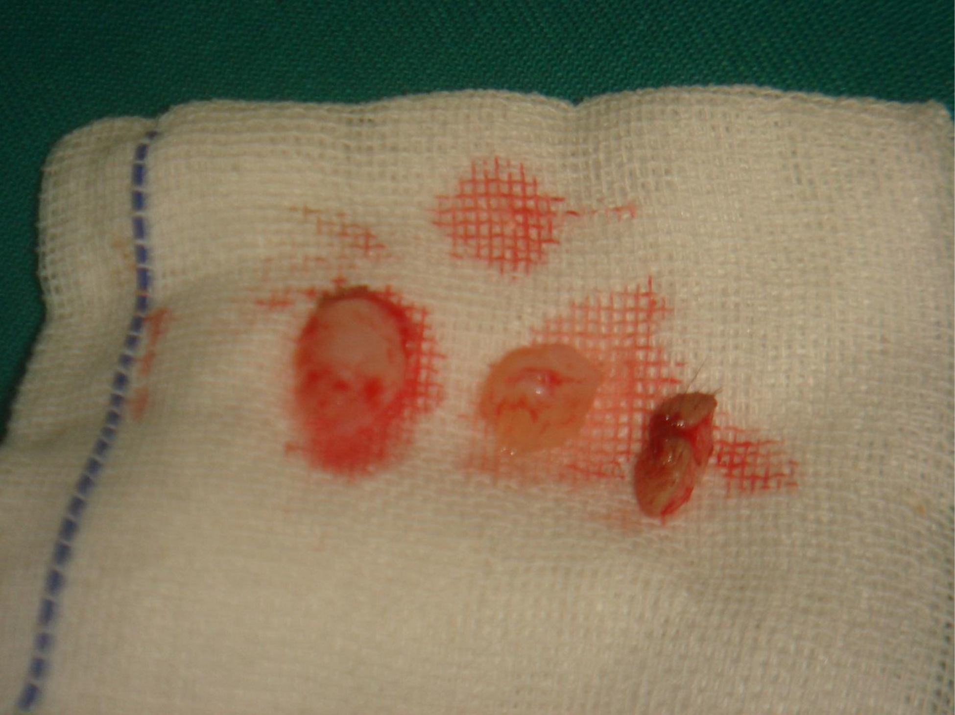
A 4-year-old male feline weighing 4.6 Kg, and positive for FIV and FeLV was seen at NaturalPet Clinic in Brasilia, Brazil. The animal has a history of difficulty swallowing concomitant with inappetence and respiratory difficulty. On physical examination, the animal was tachypneic with abdominal breathing, presented pale mucosa, CRT 3″, cardiac auscultation within the normal range considering age and species, mild dehydration, infarcted bilateral submandibular lymph nodes, and absence of pain on abdominal palpation. The examination of the oral cavity showed increased volume on the lower right side of the soft palate, small ulcerated portion, and hyperemic mucosa with apparent edema (Figure 1). Blood was collected for laboratory tests of complete blood count and biochemical measurements of alanine aminotransferase (ALT), alkaline phosphatase (AP), urea, and creatinine. The patient was also referred for a contrast-enhanced computed tomography. After the test results, the animal was referred for punch incisional biopsy (4 mm) and dental treatment, and the material was directed to histopathology. Subsequently, the treatment protocol was established through veterinary homeopathy. The following medicines were also prescribed, subcutaneously: Viscum album D3 (1×10⁻³), D6 (1×10⁻⁶), D9 (1×10⁻⁹), D12 (1×12-12), and D30 (1×10-30) (Injectcenterâ), a combination of two ampoules, once a day, for 60 days; Arnica montana D4 (1×10⁻⁴) (Injectcenterâ), one ampoule, once a day, for seven days; Apis melifera D4 (1×10⁻⁴) (Injectcenterâ), one ampoule, once a day, for seven days; Mercurius solubilis D18 (1×10-18) (Injectcenterâ), one ampoule, once a day, for 20 days. In addition, Stomorgylâ 10, ½ tablet every 24 hours, for seven dias; Omega 3 1000 mg, one capsule, once a day, for 60 days; and Vitamin C 500 mg, one tablet, once a day, for 30 days, were orally prescribed. After five months, the animal returned to the clinic complaining of discomfort in chewing. On the physical examination, the animal presented an ulcerated lesion on the contralateral side of the initial lesion (Figure 2). The same initial treatment was initiated for additional 12 weeks. The day after the evaluation, the animal was submitted to dental treatment to extract molar and premolar teeth and cauterize the injured region.

fig 1

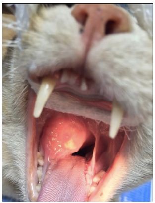
Figure 1: Presence of slightly ulcerated and contaminated nodule in the oral cavity of the cat.

fig 2

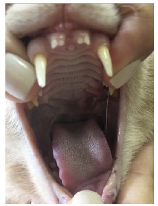
Figure 2: Presence of ulcerative lesions, represented by the white color arrow, in the initial portion of the soft palate and final portion of the hard palate (left side). On the right side of the image, represented by the green arrow, healed tissue previously affected by lymphoma.

Results

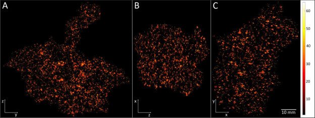
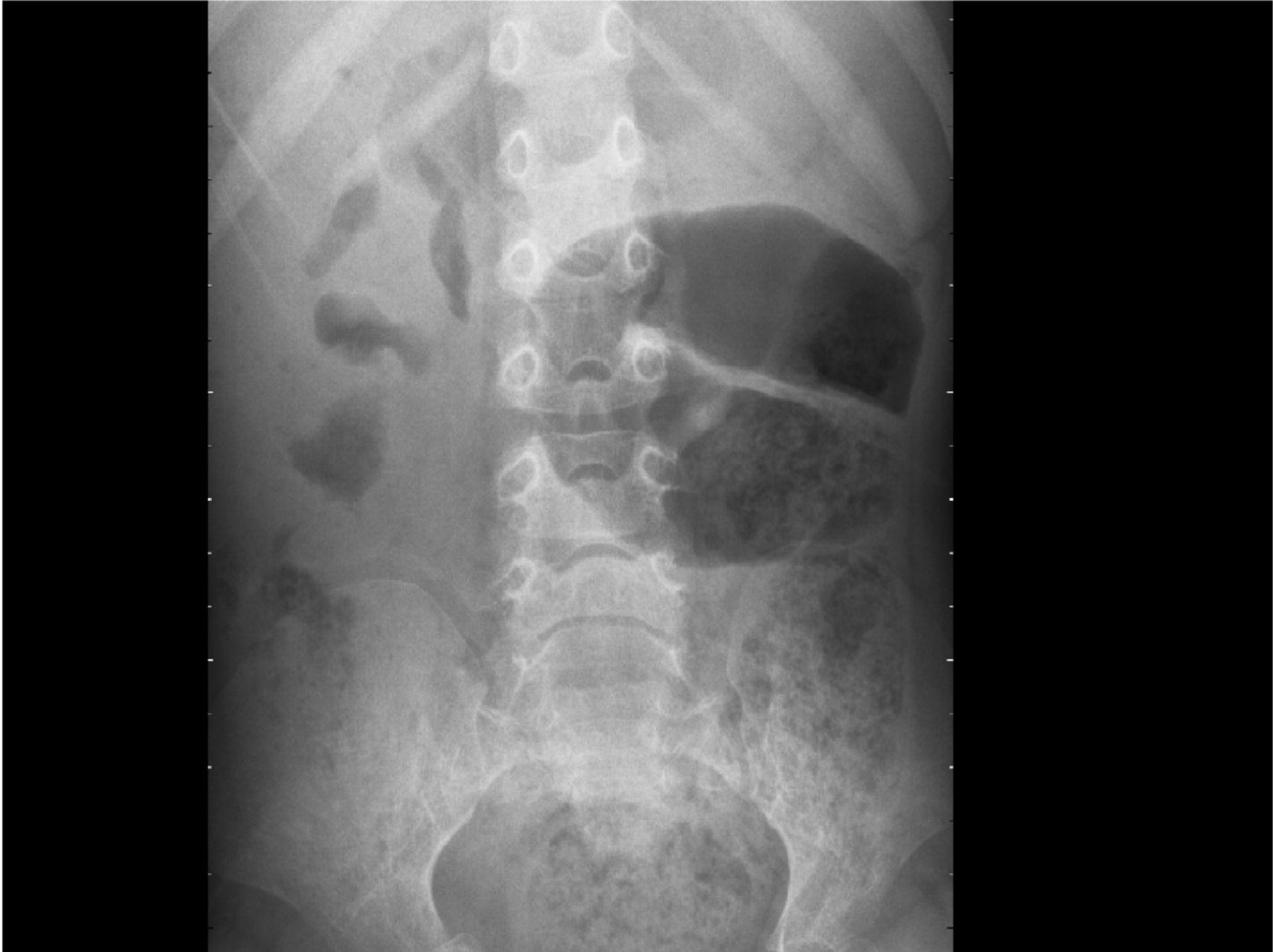
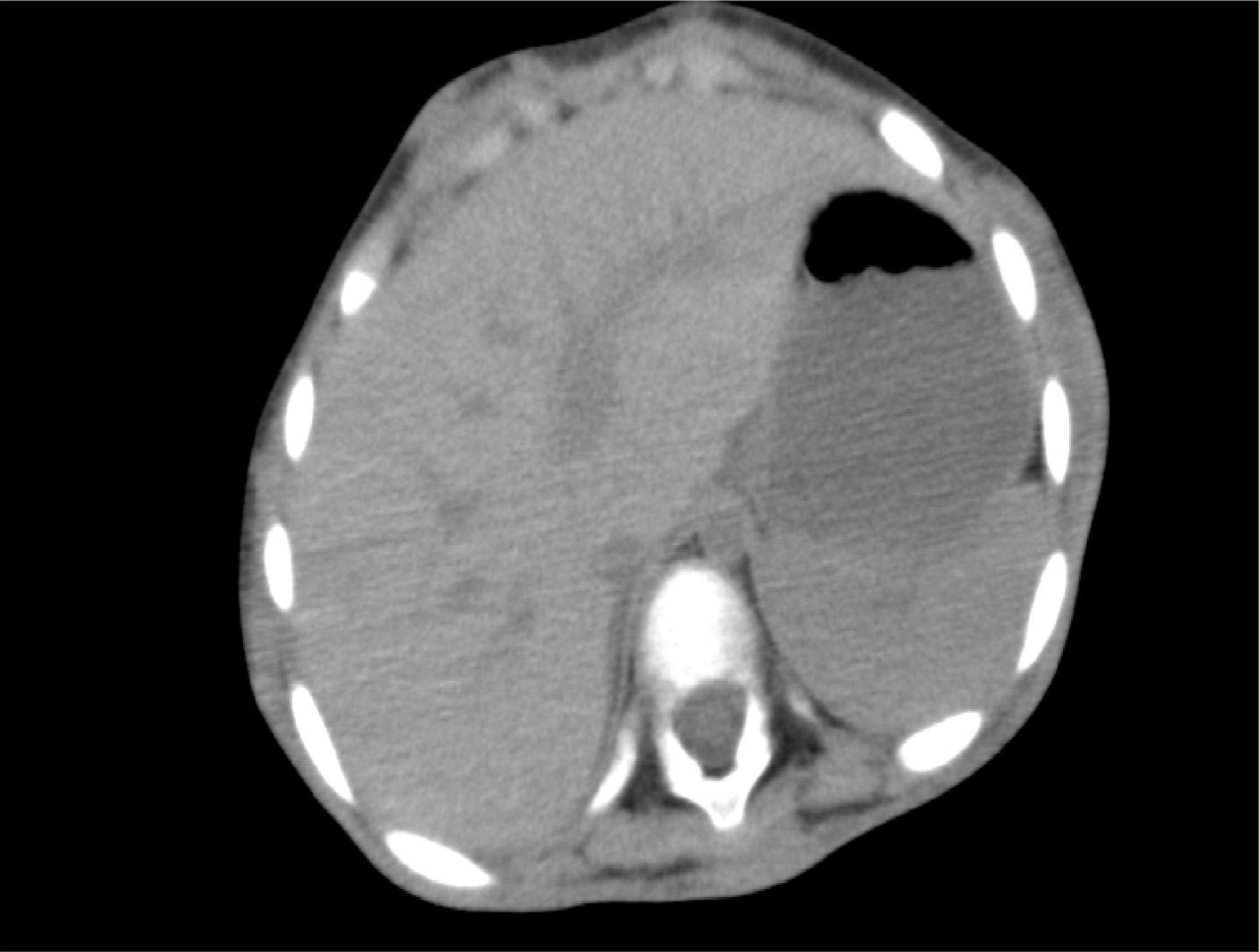
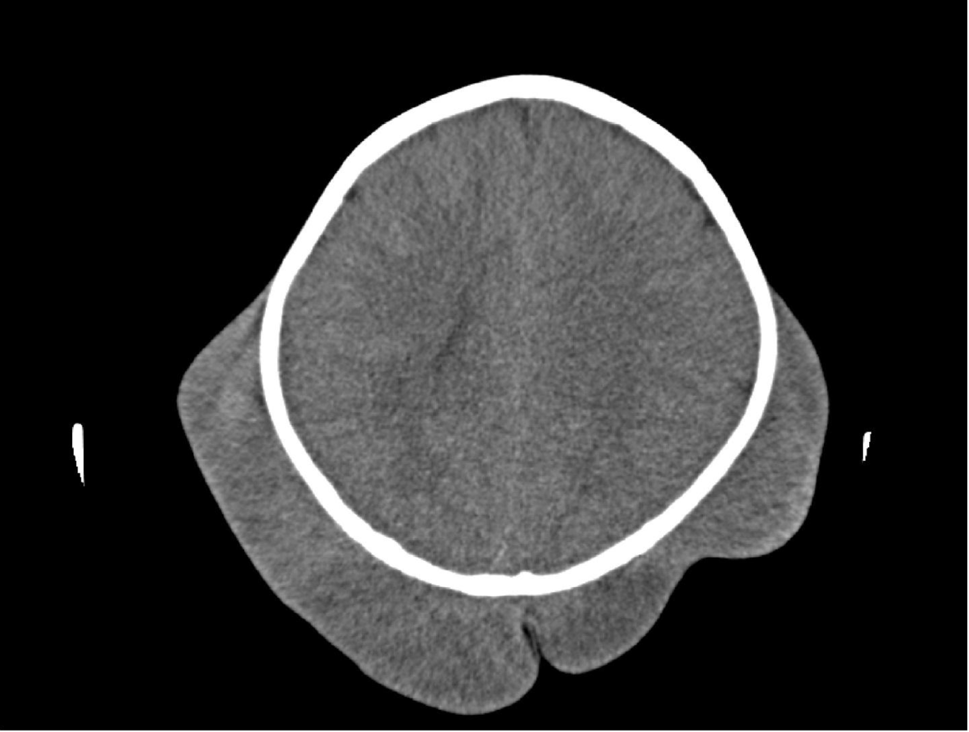
Contrast-enhanced computed tomography images (Figure 3) showed neoformation with an irregular shape, with venous contrast hyper-uptake presenting hypodensity areas in its interior, measuring approximately 3.0 cm in height x 3.5 cm wide x 4.8 cm in length, and located in the right submandibular lymph nodes topography. Signs of invasion of the neoformation mentioned above into the oropharyngeal region were observed, presenting signs of partial obstruction of the nasal choanae. The conclusion of the exam was suggestive of neoformation in the right submandibular lymph nodes topography, followed by the obstruction of the nasal choanae by a mass, as previously described. The results of the blood tests are shown in Table 1.

fig 3

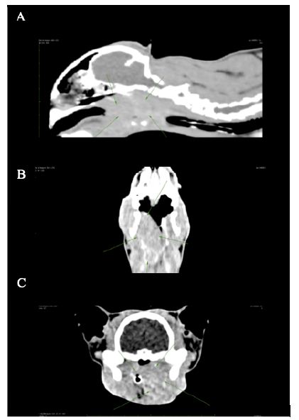
Figure 3: (A) Image in right longitudinal section (head and neck) – presence of a mass. (B) Ventral cross-sectional image of the animal’s head – presence of a mass on the left side. (C) Cross-sectional image of the sinus of the face – presence of a mass in the left sinus.

Table 1: Blood test data of a 4-year-old male feline, positive for FIV and FeLV, seen at NaturalPet Clinic in Brasilia, Brazil.


May/2016

July/2016 September/2016 October/2016 January/2017

April/2017

Red blood cells (mL)

7,690,000

8,460,000 8,020,000 6,050,000 7,200,000

6,450,000

Hemoglobin (g/dL)

11

11.5 11.3 9.2 10.3

9.3

Hematocrit (%)

31.3

36.6 35 25.8 31.1

26.2

MCV (g/dL)

40.75

42.25 43.64 42.64 43.19

40.62

MCHC (g/dL)

35.14

31.51 32.29 35.66 33.12

35.5

Leukocytes (mL)

4,500

6,700 9,500 4,600 6,000

7,000

Eosinophils (mL)

585

402 0 506 720

630

Lymphocytes (mL)

1,800

2,278 855 1,794 1,440

1,750

Platelets (mL)

16,000

301,000 230,000 128,000 264,000

368,000

TPP (dL)

11.2

7.0 9.8 8.0 6.4

8.8

ALT (U/L)

55

** ** ** 33

53

Creatinine (mg/dL)

1.5

** ** ** 1.34

1.76

AP (U/L)

13

** ** ** 17

24

Urea (mg/dL)

51

** ** ** 53

31

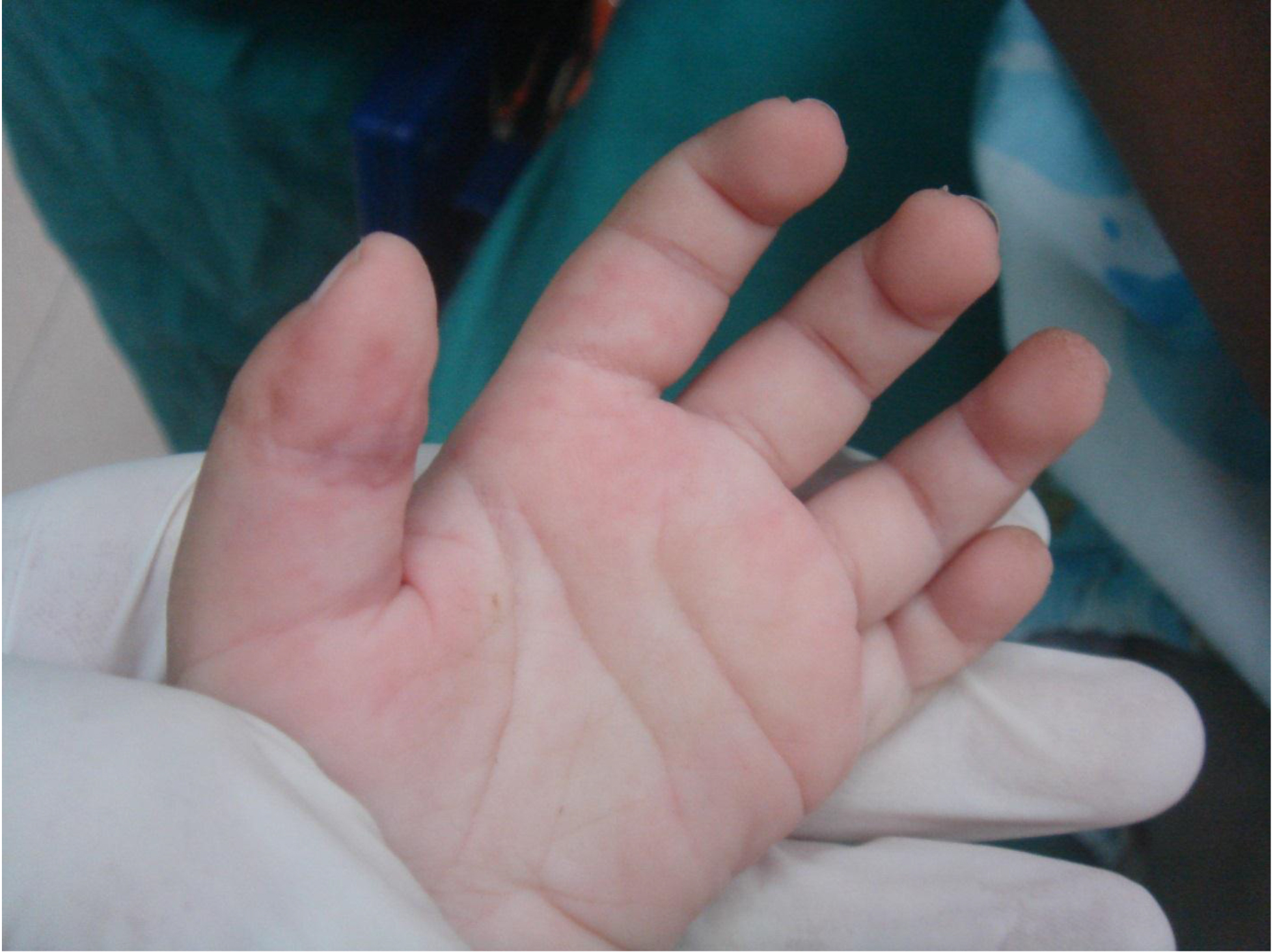
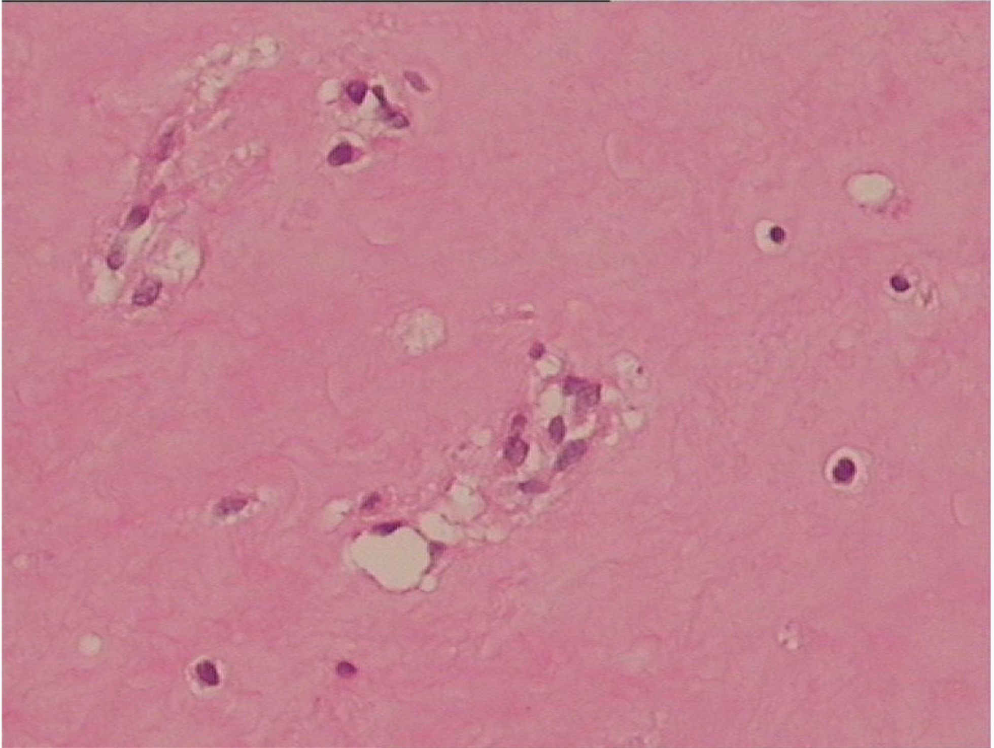
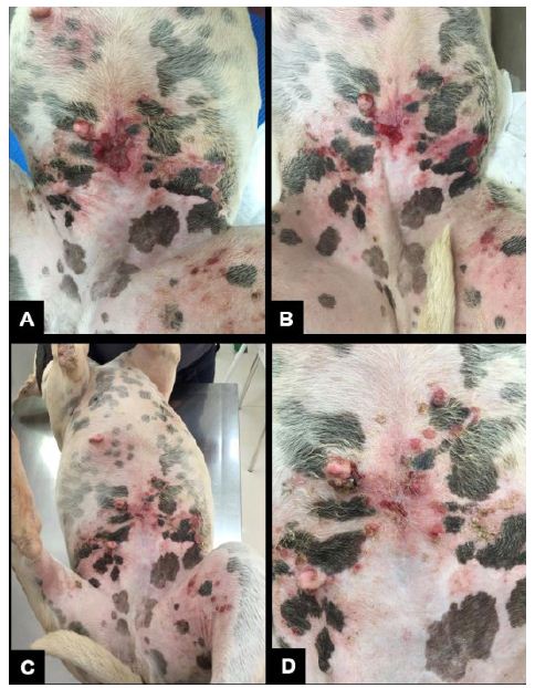
One week after the biopsy procedure and treatment initiation, the animal returned to the clinic, and the lesion was much smaller (Figure 4A). The clinical improvement can be seen in Figure 4, from the first week after the Viscum album therapy initiation until the 28th day of treatment, when the initial lesion was healed. The biopsy result was conclusive for lymphoma. The patient was followed up with complete blood count tests. The Viscum album applications should have happened daily, as per the veterinarian’s recommendation. However, the tutor of the animal discontinued the treatment, on her own, from the moment the lesion healed.

fig 4

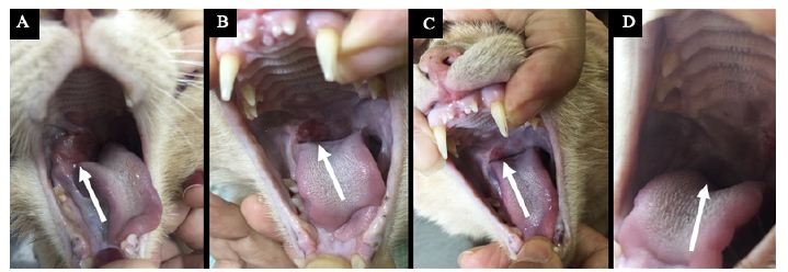
Figure 4: Lesion 1 (right side) – Evolution of the treatment and its response. (A) Lesion appearance seven days after treatment initiation. (B) Lesion appearance 14 days after.

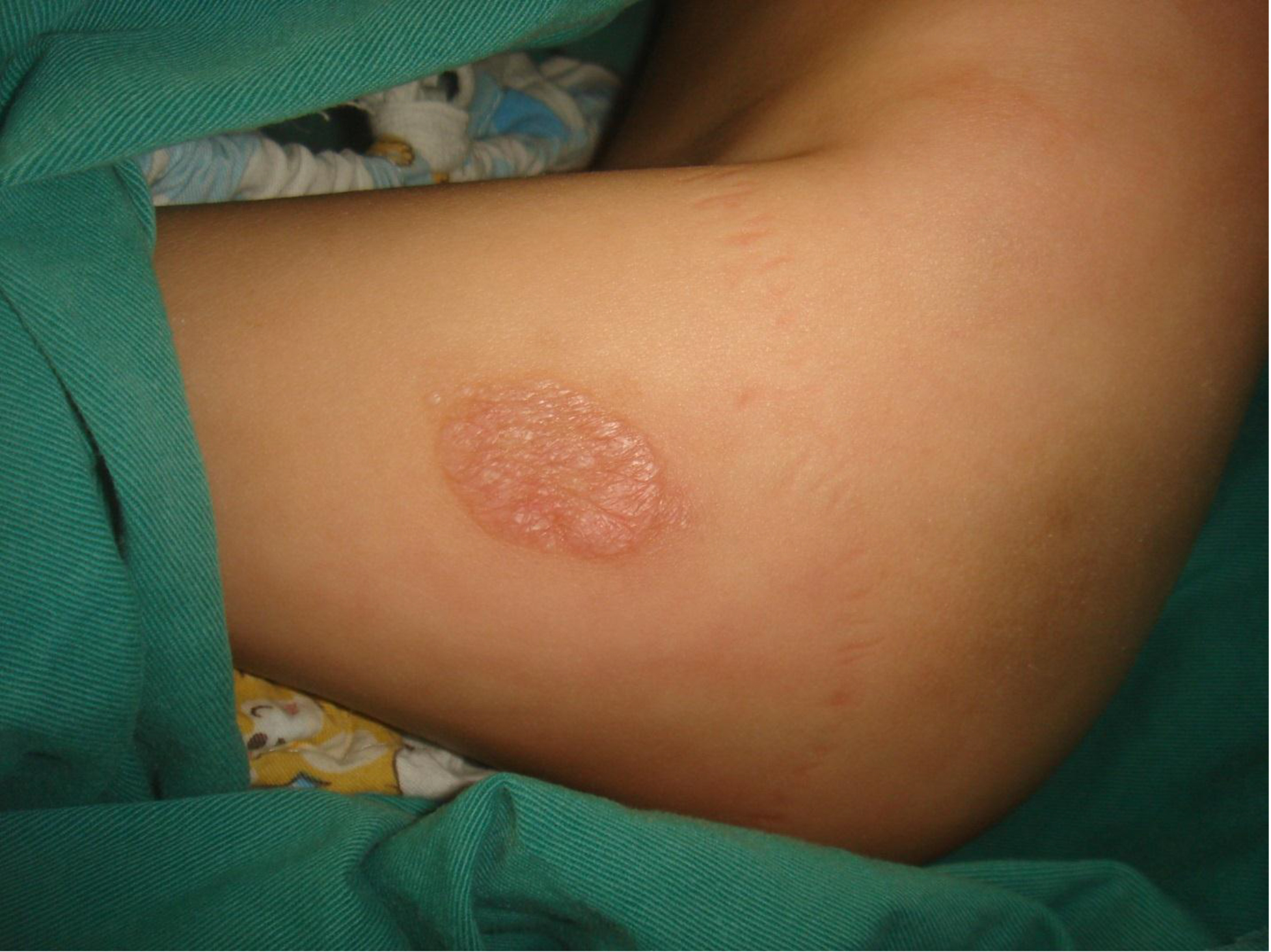
After restarting the treatment protocol and extracting the teeth, treatment evolution occurred in three weeks (Figure 5). The tutor continued the treatment for 60 days, and complete lesion healing was observed. Also, the patient remained well for another 36 months and presented normal appetite, water intake, urination, and defecation until he died with severe anemia associated with stage 3 kidney disease.

fig 5

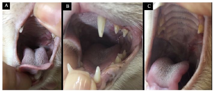
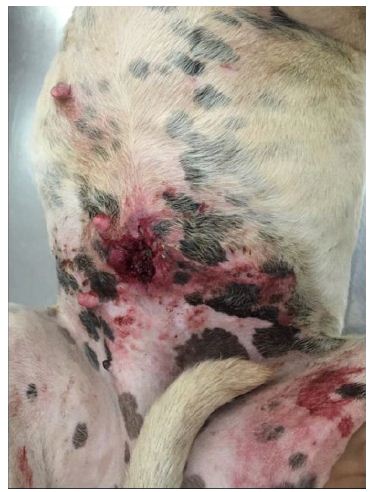
Figure 5: Lesion 1 (left side) – Evolution of the treatment and its response. (A) Lesion appearance seven days after treatment initiation. (B) Lesion appearance 15 days after treatment initiation. (C) Lesion appearance 28 days after treatment initiation.

Discussion

Lymphoma is the hematopoietic neoplasm most commonly diagnosed in domestic cats, comprising approximately one-third of all tumors in this species. FeLV-positive animals continue to be potential candidates for lymphoma and feline leukemia involvement [21]. Lymphoma is a systemic neoplasm, and the basis of treatment is chemotherapy. Its proper use promotes increased longevity and quality of life of cancer patients, and in some cases, even achieves the complete cure of cancer [22]. The chemotherapeutic treatment aims to induce complete and lasting remission of the neoplasm, and in cases of recurrence, reinduce its remission [23].

Numerous treatment protocols have been reported for feline lymphoma with varied results. The reported literature includes a wide variety of chemotherapy protocols associated or not with corticosteroids that are typically extended for a year or more [21]. Many of these protocols have different side effects, which are limiting factors for FIV and FeLV patients due mainly to the immunosuppression caused by chemotherapy and corticosteroids. In contrast to these authors, the present report describes a case of lymphoma in an FIV and FeLV positive cat treated by the Viscum album therapy with satisfactory results. Furthermore, it is important to emphasize that the therapy used did not have side effects for the patient, nor does it have contraindications.

Also, according to [24-26], overall responses reported for feline lymphoma treated with various chemotherapy protocols vary widely, with median survival times ranging from 50 to 388 days. On the other hand, the present study reports that besides being FIV and FeLV positive, the animal was treated by complementary medicine, basically by the Viscum album therapy. Under this protocol, the patient presented a survival three times greater than the maximum survival recorded for patients treated exclusively by the conventional treatment in FIV and/or FeLV positive or negative animals.

Additionally, our findings are corroborated by [18], who also recorded disease resolution, increased life expectancy, and no side effects when using injectable Viscum album and Magnesia phosphorica for treating feline alimentary lymphoma. Therefore, these are promising data that point to the importance of ongoing studies on the effects of the Viscum album therapy on cancer patients to allow for meaningful decision-making and efficient disease treatment.

Conclusion

This study raises another possibility for treating patients to whom conventional therapies do not contemplate the quality of life associated with survival. Homeopathy is an excellent therapeutic tool for patients with pre-existing diseases, debilitated, or even as a complementary therapy for conventional treatments. Therefore, we propose further studies in the area so that the results obtained in this work are better validated.

References

  1. Vail DM, Ogilvie GK (1998) Lymphoid neoplasias. In: Manual saunders: small animal clinic, Eds., Bichard, S. J. and Sherding, R.G. Roca: São Paulo, Brazil. 218-225.
  2. Court EA, Watson ADJ, Peaston AE (1997) Retrospective study of 60 cases of feline lymphosarcoma. Aust Vet J 75: 424-427. [crossref]
  3. Gabor LJ, Malik R, Canfield PJ (1998) Clinical and anatomical features of lymphossarcoma in 118 cats. Aust Vet J 76: 725-732. [crossref]
  4. Vonderhaar MA, Morrison WB, Lymphosarcoma (2002) In Cancer in dogs and cats: medical and surgical management, Ed., Morrison, W.B. Jackson Hole, WY: Teton New Media. 641-670.
  5. Norsworthy GD, Grace SF, Crystal MA, Tilley LP (2011) The feline patient. ed. Wiley – Blackwell: Iowa.
  6. Lutz H, Addie D, Belák S, Boucraut-Baralon C, Egberink H, et al. (2009) Feline leukaemia. ABCD guidelines on prevention and management. J Feline Med Surg. 11: 565-574. [crossref]
  7. Hartmann K (2012) Feline leukemie virus infection. In Infections diseases of the dog and cat, Ed., Greene, C.E. Missouri, USA: Elsevier.
  8. Bado AS (2011) Alimentary lymphoma in cats. Undergraduate thesis (Undergraduation in Veterinary Medicine) – Universidade Federal do Rio Grande do Sul, Porto Alegre, Brazil. 38p.
  9. Wilson HM (2008) Feline Alimentary Lymphoma: Demystifying the Enigma. Top Companion Anim Med. 23: 177-184. [crossref]
  10. Barger AM, Grindem CB (2000) Hematologic abnormalities associated with cancer therapy. In Schalm’s Veterinary Hematology, Eds., Feldman, B.F., Zinkl, J.G., and Jain, N.C. 5 ed. Canada: Lippincott Williams.
  11. Rodaski S and Werner J (2008) Skin neoplasia. In Oncology in dogs and cats, Eds., Daleck, C.R., De Nardi, A.B. and Rodaski, S. Sao Paulo, Brazil: Roca. 253-279.
  12. Araujo GG (2009) Feline lymphoma. Undergraduate thesis (Undergraduation in Veterinary Medicine) – Universidade Federal do Rio Grande do Sul, Porto Alegre, Brazil. 45 p.
  13. Hernandez JDT (1994) Uso clínico de los factores de crescimento hematopoyético. Iatreia 7: 173-180.
  14. Lanore D, Delprat C (2004) Anticancer chemotherapy. Roca: São Paulo, Brazil.
  15. Nelson RW, Couto CG (2003) Chemotherapy complications of cancer. Small animal internal medicine. 3 ed. Mosby: St. Louis.
  16. Valle ACV, Andrade RV, Lopes D, Sibata M, Carvalho AC (2018) Ultradiluted Viscum album in the treatment of myeloma in a dog – case report. In the Proceedings of the Supportive care makes excellent cancer care possible – MASCC. Viena, Austria.
  17. Valle ACV, Lima L, Bonamin L, Brunel H, Barros A, et al. 2020. Use of Viscum album in the Integrative Treatment of Cholangiocarcinoma in a Dog (Canis familiaris) – Case Report. Adv Complement Alt Med 5: 476-481.
  18. Carvalho A, Valle ACV (2021) Treatment of Alimentary Lymphoma in Cat (Felis catus) by Injectable Homeopathy – Case Report. Integr J Vet Biosc 5: 1-6.
  19. Valle ACV, Carvalho AC (2021) Ultra-diluted Viscum album in the treatment of cutaneous melanoma in a dog (Canis familiaris) – Case report. Paripex Indian J Res 10: 1-4.
  20. Lefebvre GNF, Bonamin LB, Oliveira CM (2007) Treatment of transmissible venereal tumor (TVT) using Viscum album in combination with chemotherapy. Rev Clin Vet 70: 78-86.
  21. Collette SA, Allstadt SD, Chon EM, Vernau W, Smith AN, et al. (2016) Treatment of feline intermediate‐ to high‐grade lymphoma with a modified University of Wisconsin–Madison protocol: 119 cases (2004–2012). Vet Comp Oncol 14: 136-146.
  22. Couto CG (2000) Advances in the treatment of the cat with lymphoma in practice. J Feline Med Sur 2: 95-100. [crossref]
  23. Withrow SJ, Vail DM (2007) Withrow & MacEwen’s Small Animal Clinical Oncology. 4. ed. St. Louis, MO: Saunders Elsevier.
  24. Simon D, Eberle N, Laacke-Singer L, Nolte I (2008) Combination chemotherapy in feline lymphoma: treatment outcome, tolerability, and duration in 23 cats. J V Intern Med 22: 394-400. [crossref]
  25. Krick EL, Moore RH, Cohen RB, Sorenmo KU (2011) Prognostic significance of weight changes during treatment of feline lymphoma. J Feline Med Surg 13: 976-983. [crossref]
  26. Krick EL, Cohen RB, Gregor TP, Salah PC, Sorenmo KU (2013) Prospective Clinical Trial to Compare Vincristine and Vinblastine in a COP‐Based Protocol for Lymphoma in Cats. J Vet Intern Med 27: 134-140. [crossref]

Infant Oral Health: Focus on Caries Prevention during Pregnancy

DOI: 10.31038/AWHC.2021434

Abstract

Tooth decay or dental caries is the most common chronic disease of childhood. However, caries is also highly preventable through a combination of oral hygiene, the use of fluoride and dietary measures. Prevention of caries should start in the prenatal period because the maternal caries status is strongly associated with the caries status of their children. This short review will address the importance of early risk assessment for identification of parent-infant groups who are at high risk for Early Childhood Caries (ECC) and would benefit significantly from early preventive intervention.

Keywords

Dental caries, Early childhood caries, Pregnancy, Infant oral health

Introduction

Tooth decay or dental caries continues to be a major public health problem worldwide. Caries affects nearly 23% of U.S. children aged 2-5 years and 56% of those aged 6-8 years [1]. Early childhood caries (ECC) can have life-long consequences on the overall health of children due to severe pain, abscesses, chewing difficulty, malnutrition, poor speech articulation, low self-esteem, and increased risk for future caries [2,3]. Importantly, pregnant women are at higher caries risk due to reasons such as increased acidity in the oral cavity, sugary dietary cravings, and limited attention to oral health [4]. The lack of maternal dental care and the transmission of caries pathogens from caries-active mothers to infants lead to increased risk for childhood caries. Appropriate dental care and caries prevention during pregnancy may decrease infant and childhood caries. Therefore, early risk assessment is critical- for identification of parent-infant groups who are at high risk for ECC and would benefit significantly from early preventive intervention.

The Caries Process

The term caries lesions are the scientific term used to describe the signs and symptoms of the disease of dental caries (or tooth decay). A caries lesion is the result of a localized destruction (or demineralization) of the tooth surface caused by acids produced from the fermentation of dietary carbohydrates (mainly free sugars) by dental plaque bacteria. The destruction can affect tooth enamel, dentin and cementum. The first sign of a caries lesion on enamel that can be detected with the naked eye is called a white spot lesion. The caries process can be stopped or reversed at this point because enamel can repair itself by using minerals from saliva, and fluoride from toothpaste or other sources. But if the caries process continues, more minerals will be lost. Over time, the enamel is weakened and destroyed, forming a cavity. A cavity is a permanent damage on the tooth that a dentist has to repair with a filling. Unquestionably, a combination of oral hygiene, the use of fluoride and dietary measures must take place to avoid the onset of white spot lesions and their consequent progression into cavities.

Caries Prevention

Preventing caries and promoting oral health begins with establishing healthy habits in infancy. Dental caries is preventable through a combination of three major behaviors that are proven effective: 1) oral hygiene methods, 2) frequent exposure to fluoride, and 3) a diet that limits the intake of sugary components [5]. Oral Hygiene (OH) is key to remove the acid-producing dental plaque that can demineralize the teeth. The best OH recommendation includes tooth brushing twice a day (especially at bedtime) followed by flossing. In addition, fluoride is a mineral that can prevent, stop the progression, and even reverse early white spot lesions. Fluoride prevents mineral loss in tooth enamel and replaces lost minerals. Sources of fluoride include: drinking fluoridated water from a community water supply (about 74% of Americans served by a community water supply system receive fluoridated water, brushing teeth with a fluoride toothpaste, rinsing your mouth daily with a fluoride mouth rinse, and application of a fluoride gel or varnish to tooth surfaces at dental clinics by dental professionals. Also extremely important in caries prevention is a diet low in foods and drinks containing sugar and starches, which can be metabolized into acids by caries pathogens.

Infant Oral Health

The American Academy of Pediatric Dentistry (AAPD) recognizes infant oral health care as a foundation for offering preventive education and dental care that can enhance the opportunity for a lifetime free from preventable oral diseases [3]. The AAPD recommends that every infant receive an oral health risk assessment from his/her primary health care provider or qualified health care professional by 6 months of age [3], and that parents establish a dental home for infants by 12 months of age [6]. Caries risk assessment for infants allows for the implementation of appropriate prevention and management strategies as the primary dentition begins to erupt [3].

This initial oral health risk assessment should include: 1) evaluation of medical (infant) and dental (parent and infant) histories; 2) a thorough oral examination; 3) an assessment of the patient’s risk of developing oral diseases of soft and hard tissues; 4) education on infant oral health, oral hygiene measures (such as age-appropriate tooth brushing demonstration), nonnutritive sucking habits, teething, injury prevention, and the dietary effects on the dentition; 5) anticipatory guidance regarding dental and oral development; 6) evaluation and optimization of fluoride exposure; 7) development of a preventive treatment plan that includes prophylaxis, fluoride varnish and periodic re-assessment appointments; and 8) referral to the appropriate health professional if specialized intervention is necessary.

The ultimate goal of early assessment is the timely delivery of educational information to populations at high risk for developing caries and prevention of later surgical intervention. Despite the importance of early risk assessment, there is an unfortunate lack of access to dental services among young children in the United States, coupled with a limited workforce to address their oral health needs. In fact, the implementation of Infant Oral Health Programs (IOHP) in dental schools is not only an important community resource for preventive dental care for young children at high risk for caries but should increase the workforce providing care to this population [7].

References

  1. Dye BA, Thornton-Evans G, Li X, Iafolla TJ (2015) Dental caries and sealant prevalence in children and adolescents in the United States, 2011-2012. NCHS Data Brief 1-8. [crossref]
  2. Ramos-Gomez FJ (2005) Clinical considerations for an infant oral health care program. Compend Contin Educ Dent 26: 17-23. [crossref]
  3. Dentistry AAoP (2014) Guideline on Infant Oral Health Care. Pediatr Dent 36: 14-15. [crossref]
  4. Silk H, Douglass AB, Douglass JM, Silk L (2008) Oral health during pregnancy. Am Fam Physician 77: 1139-1144.
  5. Kidd E, Fejerskov O (2013) Changing concepts in cariology: forty years on. Dent Update 40: 277-278, 80-82, 85-86. [crossref]
  6. Dentistry AAoP (2011) Policy on the dental home. Pediatr Dent 33: 24-25. [crossref]
  7. Nascimento MM, Mugayar L, Tomar SL, Garvan CW, Catalanotto FA, et al. (2016) The Impact of an Infant Oral Health Program on Dental Students’ Knowledge and Attitudes. J Dent Educ 80: 1328-1336. [crossref]

Cometary Origin of COVID-19

DOI: 10.31038/IDT.2021212

Abstract

The evidence for the cometary origin then rapid global spread of COVID-19 through 2020 is critically reviewed. We outline why it is an alternative plausible scientific explanation to the current bat/pangolin animal jump theories. In our view this explanation is consistent with all the available temporal unfolding scientific data (genomic, immunologic, epidemiologic, geophysical, astrophysical and astrobiological). Thus COVID-19 arrived as infective cryopreserved virions in cometary meteoritic dust clouds from space in a bolide strike in the stratosphere over China on October 11 2019. Prevailing high-level and low-level wind systems then globally distributed the infective viral dust clouds, striking different regions at different times. Given this possibility, a new space challenge for mankind is to develop near-Earth early warning biological surveillance (and mitigation) systems for incoming cosmic in-falls of micro-organisms and viruses from the cometary dust and meteorite streams that our planet routinely encounters as it orbits the Sun.

Since it first emerged in Wuhan, China in late November into December 2019 the coronavirus pandemic due to COVID-19 (SARS-CoV-2) has been engulfing the world with considerable economic and health impact targeting mainly our elderly co-morbid citizens with clear deficits mainly in type I and type III interferon inducible anti-viral immunity [1-5].

How credible then is our conclusion, embodied in our title, that the COVID-19 pandemic could have arrived from space? The journal has invited us to outline this evidence for an extra-terrestrial origin, which we began publishing from early 2020 [6-10] and then in a mid-year review which appeared in November 2020 [11]. Our initial focus has been to explain the key events of the first months of the pandemic so as to understand its origin and rapid global spread. We marshalled not only the geophysical and temporal global epidemiological evidence [6-8,12], the prior knowledge from astrophysical and astrobiological evidence [13-16] but also gained insight into the genetic adaptation strategy of the virus, based on APOBEC and ADAR deaminase driven responses in infected subjects. These host innate immune responses designed to mutate and thus cripple the viral RNA genome actually helps steer viral haplotype diversification for optimal replicative efficacy. We view this as a ribo-switching host-parasite selection process for the fittest transmissible RNA haplotypic genome in a subject host [12,17].

Our short narrative summary of one possible scenario not considered as mainstream thinking goes like this: A life-bearing loosely held carbonaceous cometary bolide arrived in the stratosphere over Jilin in North East China on the night of Oct 11 2019. This well-documented and widely observed event is recorded at the Space.com website. The viral-laden cometary dust particles and clumps (typically micron size) were released prior to the fireball in a fragmentation process, and they began their expected slow descent from the stratosphere in the 40° N Latitude band (30-50°). Over the next month some of this cometary-meteorite dust cloud was brought down to ground by local weather precipitation (rain) targeting the central Chinese city of Wuhan in Hubei province. This event, within another month, ignited by mass simultaneous infective exposure caused the explosive rise of COVID-19 cases through January in Wuhan and its wider contaminated regions. About 30% of all such infections were demonstrably not connected to any food or wet market [18,19]. However much of the upper troposphere/stratosphere viral laden dust remained there in the East to West (E-W) jet streams and was distributed around the globe at great speed (jet-streams circle the globe in ~ 3 days at speeds 150-200 km/hr). We speculate that explosive outbreaks on the ground of human person-to-person (P-to-P) passaged COVID-19 viruses could have created a secondary rising plume of viral-laden pollution and dust [20,21] over Wuhan and Hubei province containing trillions of dust-associated COVID-19 virions. We think that this plume was then carried by the lower level West to East (W-E) prevailing wind systems across the Pacific (Max Wallis, pers comm) engaging cruise ships through February in the South China Sea and Sea of Japan (Diamond Princess, Westerdam). This mode of transfer, including early first wave in-falls in South Korea and Japan and, by mid-February, to the US West Coast [7], could explain the virus outbreak on the Grand Princess cruise ship sailing out of San Francisco. This scenario for the Grand Princess outbreak has supportive genetic evidence as the main COVID-19 haplotype in infected passengers was identical to the unmutated (and lightly mutated) L haplotype that dominated the Wuhan outbreak [12,22]. The deposits in the higher E-W jet streams could have been brought down by capricious local weather conditions during early-mid March 2020 in Tehran/Qom, Lombardy/Italy and Spain, and then on the 40° N Latitude band to engage New York City as our earlier analysis had predicted [7,8].

The explosive outbreaks at widely dispersed global sites are consistent with this scenario. Further, they happened at great speed, with exponential growth rates of case numbers per day, that defied initial expectations of P-to-P spreading with the expected 1-2 week infection incubation periods. Aerial infective in-fall by viral laden dust over large population centres seems the most logical explanation for the simultaneous infections with little evidence of delay due to incubation time. The same explosive apparent simultaneous large-scale outbreaks occurred in clearly documented cases of deck crew members on ships at sea [17,23] and in the remote Chilean Bernardo O’Higgins Army Station in Antarctica in late December 2020 [24]. In a similar vein, the island of Sri Lanka flat lined in case numbers for many months with hardly any cases, and became suddenly engaged in a mass outbreak of over a 1000 COVID-19 cases in the period Oct 4-6 [10] and see also Sri Lanka at the Google URL search link below [25].

By March into April part of the E-W 40° N jet stream transporting the dust clouds was diverted into South America, particularly Brazil by prevailing Atlantic ocean wind systems [9]. From there on the prevailing wind systems of the Southern Hemisphere became engaged as the principal carriers of the infalling viral laden dust clouds. The high profile infective outbreaks were those in June through September occurring predominantly in South Africa and Victoria Australia, which coincidently are on the same prevailing W-E 40° S Latitude band (the French Polynesian Islands became engaged several months later from September). The latter interpretation for Victoria, Australia, is at odds with the prevailing local belief that the 2nd Wave outbreak was caused by hotel quarantine “escapees into the community”. This claim infers that infected travellers to Melbourne in March-April from Northern Hemisphere zones inadvertently spread the virus into the community where after several months these “escapee” variants then ignited the 2nd Wave in late June 2020. However, our evaluation of the publicly available Victorian case incidence data and the publicly available COVID-19 genomic sequence data leads to a qualified and quite different explanation which is also consistent with the viral laden dust cloud in-fall interpretation.

Interested readers can perform their own survey of the global COVID-19 cases per day patterns since early 2020 to the present at the Google URL site listed below to verify our claims [25]. It is evident that the disease has now spread all over the globe to all major continents and regions. At the time of writing (April-May 2021) a major in-fall has occurred over India and regions which we speculate is a down draft off the 40° N latitude band on the southern side of the Himalayan Mountains. But it is an exceedingly patchy pattern worldwide as discussed extensively in Hoyle and Wickramasinghe [13] again recently in Wickramasinghe et al [10] and as is evident in the Figures and Supplementary data to be found in Steele and Lindley [12] and Steele et al. [11,17]. Thus epidemics begin and end at different times in different regions and reach different intensities. In our view this behaviour reflects globally dispersed fragmented viral laden dust clouds brought haphazardly and capriciously to ground by local meteorological conditions. Certain regions may not experience a real in-fall event, and this is borne out in islands like Taiwan despite its closeness to China. The vagaries of local weather, prevailing winds, and chance in-fall of viral-laden dust clouds combine to present a capricious pattern of attack.

So in the ongoing pandemic, outbreaks around the globe have their own sudden beginnings and endings, yet major regions on the N 40° Latitude band (North America, Europe Asia Minor and the Indian subcontinent) have experienced several “Waves” already, or by the scenario considered here, separate “wash down” events from the troposphere. An interesting feature across all regions where a clear single mode of in-fall can be discerned is an unmistakable symmetrical bell-shaped curve, such as happened for the 2nd Wave in Victoria Australia, 1st Wave in South Africa, and the 1st Wave in Pakistan. There are many instances of this type if one cares to Google survey the case incident patterns per day across the globe [25]. Despite the height of the peak or intensity of the epidemic in individual instances, it is remarkable that the base of the symmetrical curve stretches typically over 2-3 months. The simplest interpretation is that this reflects the decay time of the virions in the physical environment, which remains remarkably the same across the globe.

With respect to the symmetrical nature of the bell-shaped curves describing the distributions of cases per day seen in such well documented epidemics such as the Victorian 2nd Wave an important deduction can be drawn about the impact of extreme ‘lockdown’ social distancing measures aimed at reducing viral reproduction rate Ro to less than 1. We have statistically analysed the Gaussian features of the Victorian 2nd Wave (which peaked on August 1-2, 2020). The best Gaussian fit with R2 gives 0.8999 which implies an almost perfect statistical fit to a symmetrical bell-shaped curve. Such a result would be consistent with the epidemic curve being overwhelmingly dominated by the growth and decay of a localised atmospheric in-fall event. The hard Stage 4 lockdown in Victoria came into effect on August 2, 2020. Given this perfect symmetry we conclude that the hard lock down measures had little impact, if any, on the course of the 2nd Wave COVID-19 epidemic in Victoria, Australia. This conclusion is consistent with the independent analyses of the impact of extreme lockdown measures on the course of the COVID-19 lockdowns introduced in a number of States in the USA during 2020 [26].

The patterns we have discussed apply generally to the manner in which suddenly emergent pandemics run their course throughout history [13]. It seems plausible to us that many might be a combination of wash down from the troposphere, leading to population wide exposures, eventually inducing herd immunity and a natural decay of the virions in the environment. However, we admit that there are many unknowns and that there are alternative explanations for these effects. Yet, on some specific details presented to us, the scenario presented here is a possible new reality that will need much future research.

Thus, from our viewpoint, the bulk of the key global evidence is consistent with an extra-terrestrial origin of COVID-19. Are there alternative explanations for the sudden origin and rapid global spread of the pandemic? We have dealt with this issue at some length [17], and there is one possible yet highly unlikely scientific explanation, and a bio weapon conspiracy theory explanation. Both are discussed in detail in Steele et al [17], but it is the zoonotic theory that can be dealt with rationally and scientifically, namely that COVID-19 arose in a jump from an animal reservoir, either in one or two steps or in combination, with SARS CoV related variants growing in bats and/or pangolins. In reviewing all the known related bat and pangolin SARS-CoV-like sequences the closest possible precursors are 96.2 % similar to the COVID-19 Hu-1 reference sequence (29903 nt). To get an exact match, in the normal haplotype range for globally dispersed COVID-19 of 99.98% sequence similarity [12,17] this involves>1100 specific nucleotide changes in the precursor to get such an exact COVID-19 match. These numbers imply super astronomical odds against a successful jump. Indeed, even if we are generous and assume only a 1% difference (which has not been seen in the wild) this gives odds of one successful mutational jump in 10180 trials, which also is a super astronomical number, far exceeding the molecular, and statistical, resources of the known universe. In our view a zoonotic explanation for the origin of COVID-19, although a valid scientific concept, is implausible on the current evidence [17].

This leaves us then with an alternative plausible scientific explanation that is consistent with the available data: the arrival of COVID-19 as infective cryopreserved virions in cometary meteoritic dust clouds from space. Given this possibility, a new space challenge for mankind is to develop near-Earth early warning biological surveillance (and mitigation) systems for incoming cosmic in-falls of micro-organisms and viruses from the cometary dust and meteorite streams that our planet routinely encounters as it orbits the Sun.

Acknowledgement

We acknowledge for discussion and contributions in development of ideas in this and earlier articles, Stephen G Coulson, Max K Wallis, Brig Klyce, Predrag Slijepcevic, Alexander Kondakov, Dayal T Wickramasinghe, George Howard, Herbert Rebhan, Pat Carnegie, Ananda Nimalasuriya and Milton Wainwright

References

  1. Acharya D, Liu G-Q, Gack MU (2020) Dysregulation of type I interferon responses in COVID-19. Nat Rev Immunol 20: 397- [crossref]
  2. Blanco-Melo D, Nilsson-Payant BE, Liu WC, Uhl S, Hoagland D, et al. (2020) Imbalanced Host Response to SARS-CoV-2 Drives Development of COVID-19. Cell 181: 1036-1045. [crossref]
  3. Hadjadj J, Yatim N, Barnabei L, Corneau A, Boussier J, et al. (2020) Impaired type I interferon activity and exacerbated inflammatory responses in severe Covid-19 patients. Science 369: 718-724. [crossref]
  4. Netea MG, Giamarellos-Bourboulis EJ, Domı ́nguez-Andre ́s J, Curtis N, van Crevel R, et al. (2020) Trained Immunity: a tool for reducing susceptibility to and the severity of SARS-CoV-2 infection Cell 181: 969- 977. [crossref]
  5. Lucas C, Wong P, Klein J, Castro TBR, Silva J, et al. (2020) Longitudinal analyses reveal immunological misfiring in severe COVID-19. Nature. 584: 463-469.
  6. Wickramasinghe NC, Steele EJ, Gorczynski RM, Temple R, Tokoro G, et al. (2020) Comments on the Origin and Spread of the 2019 Coronavirus. Virology: Current Research 4:1.
  7. Wickramasinghe NC, Steele EJ, Gorczynski RM, Temple R, Tokoro G, et al. (2020) Growing Evidence against Global Infection-Driven by Person-to-Person Transfer of COVID-19. Virology: Current Research 4:1.
  8. Wickramasinghe NC, Steele EJ, Gorczynski RM, Temple R, Tokoro G, et al. (2020) Predicting the Future Trajectory of COVID-19. Virology: Current Research 4:1.
  9. Wickramasinghe NC, Wallis MK, Coulson SG, Kondakov A, Steele EJ, et al. Intercontinental Spread of COVID-19 on Global Wind Systems. Virology: Current Research 4:1.
  10. Wickramasinghe NC, Steele EJ, Nimalasuriya A, Gorczynki RM, Tokoro G, et al. (2020) Seasonality of Respiratory Viruses Including SARS-CoV-2. Virology: Current Research 4:2.
  11. Steele EJ, Gorczynski RM, Lindley RA, Tokoro G, Temple R, et al. (2020) Origin of new emergent Coronavirus and Candida fungal diseases- Terrestrial or Cosmic? Advances in Genetics 106: 75-100. [crossref]
  12. Steele EJ, Lindley RA (2020) Analysis of APOBEC and ADAR deaminase-driven Riboswitch Haplotypes in COVID-19 RNA strain variants and the implications for vaccine design. Research Reports Vol 4.
  13. Hoyle F, Wickramasinghe NC (1979) Diseases from Space. JM Dent Ltd, London.
  14. Hoyle F, Wickramasinghe NC (2000) Astronomical Origins of Life: Steps Towards Panspermia. Klower Academic Publishers, Dordrechl, Netherlands.
  15. Steele EJ, Al-Mufti S, Augustyn KA, Chandrajith R, Coghlan JP, et al. (2018) Cause of Cambrian Explosion: Terrestrial or Cosmic? Prog Biophys Mol Biol 136: 3-23. [crossref]
  16. Steele EJ, Gorczynski RM, Lindley RA, Liu Y, Temple R, (2019) et al. Lamarck and Panspermia – On the Efficient Spread of Living Systems Throughout the Cosmos. Prog Biophys Mol Biol 149: 10 -32.
  17. Steele EJ, Gorczynski RM, Rebhan H, Carnegie P, Temple R, et al. (2020) Implications of haplotype switching for the origin and global spread of COVID-19. Virology: Current Research 4: 2. Supplementary data at: https://www.hilarispublisher.com/open-access/implications-of-haplotype-switching-for-the-origin-and-global-spread-of-covid19.pdf
  18. Huang C, Wang Y, Li X, Zhao J, Ren L, et al. (2020) Clinical Features of Patients Infected with 2019 Novel Coronavirus in Wuhan. Lancet 395: 497-506.
  19. Cohen J (2020) Wuhan seafood market may not be source of novel virus spreading globally. Science Vol 367.
  20. Coccia M (2020) Factors determining the diffusion of COVID-19 and suggested strategy to prevent future accelerated viral infectivity similar to COVID. Science of the Total Environment 729: 138474. [crossref]
  21. Martelletti L, Martelletti P (2020) Air Pollution and the Novel Covid-19 Disease: a Putative Disease Risk Factor. SN Compr Clin Med 2: 383-387.
  22. Andersen K (2020) Clock and TMRCA based on 27 genomes. Novel 2019 coronavirus. http://virological.org/t/clock-and- tmrca-based-on-27-genomes/347
  23. Howard GA, Wickramasinghe NC, Rebhan H, Steele EJ, Reginald M, et al. (2020) Mid-Ocean Outbreaks of COVID-19 with Tell-Tale Signs of Aerial Incidence Virology: Current Research 4: 2.
  24. Antartica Base: The remote Chilean Army Base in Antartica suddenly became engaged in late December 2020 by multiple simultaneous COVID-19 cases https://www.bbc.com/news/world-latin-america-55410065; https://www.abc.net.au/news/2020-12-23/more-covid-cases-linked-to-chilean-antarctic-base/13009706
  25. Google: “Coronavirus disease statistics” URL is https://bit.ly/3vxbD5i This gives you the “Australia” dashboard (from there you can choose your country in the menu bar scroll)
  26. Luskin DL (2020) The failed experiment of COVID-19 lockdowns. The Wall Street Journal.
FIG 1

Accumulation Rates Using the 210Pb Dating Method in a Sediment Core of the Cispatá Bay, a Marine Protected Area in the Southwestern Colombian Caribbean

DOI: 10.31038/GEMS.2021322

Abstract

Sedimentation rates in coastal environments are controlled by different natural processes and could be affected by anthropic activities. To evaluate how was the evolution of the sedimentation along recent time, one sedimentary core (P01-BDC) from the Cispata bay was collected and analyzed to determine the sediment accumulation rates by 210Pb dating. Using the constant flux (CF) model and validating by the activity of 137Cs, the mass accumulation rates varied from 0.02 g cm-2 year-1 during 1888 until 0.29 g cm-2 year-1 in 2019. Increasing low sedimentation rates in a transitional estuarine -marine environment with variable fluvial sediment supply was explained by changes in the salinity due to the relocation of the main tributary of the bay.

Keywords

Sediment accumulation rates, Pb-210, Marine protected area, Colombian Caribbean

Introduction

Coastal zones are essential and integral components of the land since they constitute critical areas for environmental, economic, and social well-being. These environments have unique characteristics because of the exchange of matter and energy between the atmosphere, the land, and the sea, those which promote the development of coastal ecosystems and habitats such as estuaries, deltas, beaches, among others [1,2]. Estuaries are inlets open to coastal oceans that receive freshwater inputs and increasingly face the effects of climate change, including sea-level rise, habitat loss, hurricanes, and anthropic effects such as changes in land use, pollution, among others [1-3]. These stressors affect the distribution and behavior of animal and plant species, chemical components, and sedimentation processes [2] Sedimentation in estuaries is driven by runoff from hydrographic basins and tidal currents that vary with different time scales, it is one of the most relevant management challenges since it can negatively affect the environment by modifying flood regimes, and circulation and water quality [3]. On the Colombian Caribbean coast, in the southwest of the Morrosquillo Gulf is the Cispatá Bay, an estuary made up of fine sediments and some coral fossil deposits [4]. The Cispatá bay was formed from the evolution of the deltaic lagoon system of the Sinú River, whose flow is thrown into the Caribbean Sea through three mouths, Corea, Tinajones, and Los Llanos [5]. This river ended up in the Cispatá Bay until 1938, beginning its avulsion in the Tinajones area, whose mouth opened completely around 1945. Since then, different disturbances to the flow regime of the Sinú River have caused changes in the hydrodynamics and the contribution of sediments to the Cispatá Bay [6]. Using 210Pb technique, we dated a sediment core collected in Cispatá bay in 2019 to evaluate the temporal trends of sediment accumulation rates (SAR) in the last century, under the hypothesis that changes in the input of sediments to the bay are related to the changes in the channel of the Sinú River. The results achieved will be valuable to understand the role of changes in the Sinú River delta, erosive processes, textural features, and pollution trends.

Methodology

Area of Study

Cispatá Bay is located in the Colombian Caribbean Coast, enclosed in The Mestizos peninsula on the western side of the Morrosquillo Gulf (Figure 1), Cordoba department, between 09°25′12″–09°20′8″N and 75°47′37″–75°55′30″W [7,8]. The Bay is a Holocene depositional landform formed by Rio Sinú before its diversion occurred between 1937 and 1945, when the Tinajones delta started to grow [6]. Rio Sinú is one of the most important fluvial systems of the Colombian Caribbean, draining the Andes with a total length of about 415 km and is a very intervened catchment area of 17,000 km2. Its mean discharge is about 398.09 m3/s (max. 858.2 m3/s, min. 29.1 m3/s) [9] and its sediment load is estimated to be in the order of 4.2 million t/y [10]. The bay has a tropical climate affected by Intertropical Convergence (ITC) annual displacements between the latitudes 5°S and 15°N, which produces an arid xerophytic savanna climate, with annual mean temperatures of 28.3°C (max. 28.8°C in January and April; min. 27.9°C in October and November). Mean annual rainfall is about 1230 mm, with 3.3 mm in January and 177.6 mm in September (data for Monteria from IDEAM [11]. During the dry season (from December to April), north-east and north Trade winds prevail with speeds between 4 and 23 kt, whereas the wet season (from August to November) is characterized by calms and slow winds mostly from the west and south-west. Cispatá Bay is surrounded by a dense mangrove forest where some urban settlements live on fishing and wood production [12]. The surrounded areas have shown in the last years an increment in anthropic activities like agriculture, large variety of commercial fishing and tourism, as well as, some industrial activities such as an oil port which is located approximately 12 km from the Cispatá Bay [13]. This mangrove ecosystem also has the potential of saving high amounts of blue carbon [14], being in this way an important area for climate change politics.

FIG 1

Figure 1: Dated core in the study area, Cispatá Bay Colombian Caribbean.

Sample Collection

Sediment cores were collected in three different points of the Bahia de Cispatá in September of 2019, using a gravity corer UWITECTM with a transparent liner (1,2 m long, 8.5 cm inner diameter). The depth of the water was around 1,8 m. The cores were immediately transported to the laboratory, where they were extruded and subsampled every 1 cm. The mass of each section was recorded before and after drying at 40°C. Sediments were ground to powder by using agate mortar and pestle. The samples were stored in polyethylene bags and the analysis were carried out at the Marine Environmental Quality – LABCAM of INVEMAR. The core collected at 9°24’54,2” (N) – 75°48’35,2” (W) (point P01-BDC, Figure 1) had a length of 72 cm and was dated with 210Pb. The activities are expressed on a dry weight basis.

Laboratory Analyses

Total 210Pb activities (210PbTot) in the core were estimated by measuring the activity of its daughter product 210Po assuming secular equilibrium between the two isotopes by alpha spectrometry [15,16]. Accuracy was evaluated by measuring a certified reference material DL1-A (Uranium -Thorium Ore DL1-A, Canada Centre for Mineral and Energy Technology) for 210Pb. Polonium isotopes were measured using a silicon surface barrier (EG&G Ortec Mod. ENS-U450) α-spectrometer. This detector is characterized by high energy resolution, low background, and stability. Discs were measured until achieving less than 5% of uncertainty in the 210Po counting rate, according to IAEA [20]. Standard gamma spectrometry was used to measure 137Cs via its emission at 662 keV and 226Ra (supported 210Pb) by the activity of 214Pb at 295 keV and 351 keV [17]. Samples were placed and measured in a coaxial type (8 cm diameter) high-purity germanium detector (HPGe) from CANBERRA and counted for one week. Energy and efficiency calibrations were made using a certified reference material DL1-A (Uranium -Thorium Ore DL1-A, Canada Centre for Mineral and Energy Technology) for 210Pb and 226Ra, IAEA-375 (Radionuclides and trace elements in soil) for 137Cs, and 40K in a high purity (≥99.5%) salt KCl salt (manufactured for Merck)

Data Processing

Constant flux (CF) of 210Pb model was used. In the CF model non-linearities of the 210Pb profile are interpreted assuming a constant net rate of supply of unsupported 210Pb (210Pbuns) from sea-water to the sediment, irrespective of changes which may have occurred in the net dry mass sedimentation rate [18]. Supported value (210Pbsup) is determined by two methods: first one by averaging the 210Pb activities in the base of the core where they become constant [19] and second one by the activity of 226Ra measured by gamma spectrometry. Then, unsupported value (210Pbuns) is obtained by subtracting 210Pbsup from 210PbTot. This model allows to estimate the age of the sediment as well as the mass accumulation rates (MAR) and sediment accumulation rates (SAR). More information about age models and calculations are explained in detail by Sanchez [20].

Results

The core shows an exponential decay of the total 210Pb (210PbTot) activity with mass depth (Figure 2a), with values ranging between 13.0 ± 1.1 Bq kg-1 and 43.7 ± 4.5 Bq kg-1. and an average 210PbTot activity of 24.78 ± 7.3Bq kg-1 However, there are clear differences mainly after 15 g cm-2 where the profile shows a peak with the lowest activity of 13.0 Bq kg-1, increasing to 25.5 Bq kg-1 at 17.7 g cm-2. The activities behavior until the end of the core is erratic. Values for 210Pbsup are very similar by the two methods of calculation, i.e. via 214Pb (226Ra) emission peaks (Table 1) and by averaging the activities of 210PbTot (Figure 2a) at the bottom of the core (20.9 ± 3.1 Bq kg-1). Due to the uncertainties associated to both methods, the average activity determined by alpha spectrometry was used for calculations of 210Pbex. The 210Pbexs activity (Figure 2b) shows values between 22.7 and 4.3 Bq kg-1 with an average value of 13.8 Bq kg-1 fitted to an exponential decay profile (r2=0.75). The 210Pbexs activities in the first 15 cm of the sediment section were used to calculate the age sediments applying the CF model (Figure 2c), allowing to date the period since 1888 until 2019. The model was validated by gamma detection of 137Cs in the core at 7.7 g cm-2 which was dated at 1983 ± 4, with this uncertainty the result could be associated with the Chernobyl accident in 1986. MAR and SAR profiles (Figure 3) present the same trends with time, a low and constant rate in the bottom increasing little by little until the top of the core. MAR increase from 0.02 g cm-2 year-1 in 1888 to 0.30 g cm-2 year-1 in 2019. By the other hand, SAR increase from 0.02 cm year-1 in 1888 to 0.45 cm year-1 in 2018 and decreasing to 0.38 cm year-1 in 2019. Due to the low sedimentation rates each analyzed section enclose several years having decadal or five-years period resolution.

Table 1: Gamma emission for 226Ra (210Pbsup) for the bottom sediment section for the core P01-BDC, Cispatá Bay Colombian Caribbean.

Core section

214Pb295kev (Bq kg-1)

214Pb351kev (Bq kg-1)

67-68

17.99

17.25

70-71

18.54 18.38
71-72 17.85

20.30

Average

18.39

Std. Dev

1.04

fig 2

Figure 2: Dated core P01-BDC in Cispatá Bay Colombian Caribbean. a) total activity profile of 210Pb, b) excess activity profile of 210Pb, c) Age model.

fig 3

Figure 3: Sediment rates for core P01-BDC: a) Mass accumulation rates, b) Sediment accumulation rates.

Discussion

Activity of 210Pbtot is comparable with the reported values of 12.8 ± 0.4 to 46.6 ± 1.1 Bq kg-1 in Soledad Lagoon [21] which is part of the Cispatá swampy system. This low activity values are expected in some marine areas previously explained as the result of low atmospheric 210Pb fluxes and low production of 210Pb [22]. Sedimentation rates (MAR ad SAR) are higher in Cispatá bay compare with those calculated for Soledad Lagoon MAR 0.08 ± 0.01 g cm-2 year-1 and SAR 0.154 ± 0.018 cm year-1 [21], due to the differences in the type of system, being Cispatá bay a more dynamic system influenced directly by the Caribbean sea and the fluctuations of the Sinú river mouth relocation, that control the sediment supply.

The Sediment rates in Cispatá bay showed changes associated with the geomorphological variations during the last 100 year. The relocation of the principal mouth after 1938 is evidenced in the increase of MAR and SAR. Before this time, the system was an estuarine system with high input of fresh water [6] and with variable hydrodynamics because of the changes in the Sinú river mouth which have been occurring since 1762 according to the available registers [6,11]. The transitional events occurring between an estuarie and a marine system in which salinity changes could form a salt wedge or significant differences in density, causing the fine sediment to remain more time in the water column. Thus, the sedimentation rates are low even with high sediment supply. The gradually increase trend of the sedimentation rates are in agreement with [6] who showed bathymetrically that during the period 1762-1849 the area where the core was recollected, presented erosive processes which implies a minor sediment input, meanwhile for the period 1849-1938 occurred a siltation process. According with the MAR and SAR calculated between 1938 and 2019, the sedimentation process has increased slowly evidencing that sedimentary behavior of Cispatá Bay is driven by: the morphodynamical characteristics of the surrounding environment, the input of water and sediment from the Sinú River remaining channels, the possible income of sediments from the erosion process occurring in the previous river mouth in Punta Terraplen, and by changes in land use.

Conclusion

The age model, based on 210Pb activities, give valuable information about the sedimentary rates (MAR and SAR) in Cispatá Bay during the last ~100 year, evidencing changes in the sedimentary regime associated with geomorphological events which are important for the management of this marine protected area with high ecological potential, especially in blue Carbon sequestration. These preliminary results should be part of complementary research about flux of organic matter, pollutants as heavy metal, hydrocarbons, and other variables of environmental interest.

Acknowledgment

This research was financed by the National Hydrocarbons Agency (ANH) of Colombia, Ministry of Environment and Sustainable Development and the Institute for Marine and Coastal Research (INVEMAR). The authors thank to the Ministry of Science and Technology of Colombia, who support the post doctorate of Dr. Pedro Vallejo-Toro. The authors acknowledge the fishermen Domingo Rodríguez and Nairo Mendoza for their support to collect the sample cores, the staff of INVEMAR Marine Environmental Quality Laboratories Unit, for collect the samples and carry out the laboratory analysis, and Andrea Beltran of the INVEMAR Laboratory for Information Services, for preparing the map. Finally, the authors thank the anonymous reviewers whose suggestions improved the manuscript. Scientific Contribution of INVEMAR No. 1303.

Reference

  1. Morzaria Luna H, Turk Boyer P, Polanco Mizquez EI, Downton Hoffmann C, Cruz Piñón G, et al. (2020) Coastal and Marine Spatial Planning in the Northern Gulf of California, Mexico: Consolidating stewardship, property rights, and enforcement for ecosystem-based fisheries management. Ocean & Coastal Management
  2. Rippel TM, Tomasula J, Murphy SM, Wimp G J (2021) Global change in marine coastal habitats impacts insect populations and communities. Curr Opin Insect Sci 47: 1-6. [crossref]
  3. Brand C, Götschi T, Dons E, Gerike R, Anaya Boig E, et al. (2021) The climate change mitigation impacts of active travel: Evidence from a longitudinal panel study in seven European cities. Global Environmental Change
  4. López Sánchez, Clara Marcela, Mancera Pineda, José Ernesto (2019) Structural Parameters of Two Populations of Crassostrea rhizophorae (Ostreidae) in Bahía Cispatá, Colombian Caribbean. Acta Biológica Colombiana 24: 361-371.
  5. Molina A, Molina C, Giraldo L, Parra C, Cevillot P (1994) Dinámica marina y sus efectos sobre la geomorfología del golfo de Morrosquillo. Boletín Científico CIOH 15: 93-113.
  6. Serrano Suarez B E (2004) The Sinú river delta on the northwestern Caribbean coast of Colombia: Bay infilling associated with delta development. Journal of South American Earth Sciences 16: 623-631.
  7. Piccardi M, Correa ID, Pranzini EJ (2020) Cispatá Bay and Mestizos Evolution as Reconstructed from Old Documents and Maps (16th–20th Century). J Mar Sci Eng
  8. Sánchez Páez H, Ulloa Delgado G, Tavera Escobar H, Gil Torres, W JB, et al. (2005) Ministerio de Ambiente, Vivienda y Desarrollo Territorial, , Plan de manejo integral de los manglares de la zona de uso sostenible del sector estuarino de la Bahía de Cispatá departamento de Córdoba-Colombia.
  9. Restrepo, JC, Ortíz, JC, Pierini J, Schrottke K, Maza M, et al. (2014) Freshwater discharge into the Caribbean Sea from the rivers of Northwestern South America (Colombia): Magnitude, variability and recent changes. Journal of Hydrology 509: 266-281.
  10. Restrepo JD, López SA, Restrepo JC (2009) The effects of geomorphic controls on sediment yield in the Andean rivers of Colombia. Latin American Journal of Sedimentology and Basin Analysis 16: 79-92.
  11. IDEAM (1998) Morfodinámica, Población y Amenazas Naturales en el Litoral Caribe Colombiano: Valle del Sinú-Morrosquillo-canal del Dique, in IDEAM. ed.: Santa Fe de Bogotá.
  12. Sánchez Páez H, Alvarez León R, Guevara Mancera O, Zamora Guzmán A, Rodríguez Cruz H, et al. (1997) Diagnóstico y zonificación preliminar de los manglares del Pacífico de Colombia.
  13. Burgos-Núñez S, Navarro-Frómeta A, Marrugo-Negrete J, Enamorado-Montes G, Urango Cárdenas (2017) Polycyclic aromatic hydrocarbons and heavy metals in the Cispata Bay, Colombia: A marine tropical ecosystem. Mar Pollut Bull 120: 379-386.
  14. Yepes A, Zapata M, Bolivar J, Monsalve A, Espinosa SM, et al. (2016) Ecuaciones alométricas de biomasa aérea para la estimación de los contenidos de carbono en manglares del Caribe Colombiano. A J R d B T 64: 913-926.
  15. Flynn W (1968) The determination of low levels of polonium-210 in environmental materials. Analytica chimica acta, 43: 221-227. [crossref]
  16. Hamilton TF. Smith JD (1986) Improved alpha energy resolution for the determination of polonium isotopes by alpha-spectrometry: International Journal of Radiation Applications and Instrumentation. Part A. Applied Radiation and Isotopes 37: 628-630.
  17. Appleby P (2001) Chronostratigraphic techniques in recent sediments, Tracking environmental change using lake sediments. Springer 171-203.
  18. Appleby P, Oldfield F (1992) Applications of lead-210 to sedimentation studies. Uranium-series disequilibrium: applications to earth, marine, and environmental sciences 2. ed.
  19. Michael WB (1990) Calculation and uncertainty analysis of 210 Pb dates for PIRLA project lake sediment cores. Journal of Paleolimnology 3: 253-267.
  20. Sanchez-Cabeza JA, Díaz-Asencio M, Ruiz-Fernández AC (2012) Radiocronología de sedimentos costeros utilizando 210Pb: modelos, validación y aplicaciones. Organismo Internacional de Energía Atómica, Centro Internacional de Viena.
  21. Ruiz-Fernández AC, Marrugo Negrete JL, Paternina Uribe R, Pérez-Bernal LH (2011) 210 Pb-derived Sedimentation Rates and C org Fluxes in Soledad Lagoon (Cispatá Lagoon System, NW Caribbean Coast of Colombia): Estuaries and Coasts. Estuaries and Coasts 34: 1117-1128.
  22. Ruiz Fernández A, Hillaire Marcel C (2009) 210 Pb-derived ages for the reconstruction of terrestrial contaminant history into the Mexican Pacific coast: potential and limitations. Marine Pollution Bulletin 59: 134-145.
fig 1

Seroprevalence of Cytomegalovirus Antibodies in a Group of Bangladeshi Women in Child-Bearing Age: A Pilot Study

DOI: 10.31038/AWHC.2021433

Abstract

Background: Human Cytomegalovirus (HCMV) is the most common cause of congenital infections and can be life-threatening in immune compromised individuals. We aimed to shed light on the Seroprevalence of HCMV antibodies in women of child-bearing age in a tertiary care hospital and in a health science university of Bangladesh.

Methods: A total of 84 apparently healthy 20-40 years old women (42 pregnant and 42 non-pregnant) were screened for anti-CMV IgG and IgM antibodies using Enzyme Linked Immunosorbent Assay (ELISA). Serum levels of bilirubin, and liver enzymes (alanine aminotransferase, aspartate aminotransferase) were evaluated by biochemistry auto analyzers.

Results: All the participants were found positive for anti-CMV IgG (100%) while 1 pregnant woman revealed positivity for both IgM and IgG justifying recent infection. CMV IgG antibody was found positive in 48 (68.6%) and 14 (100%) relatively younger volunteers in married (n=70) and unmarried (n=14) group respectively, and in 22 (31.4%) elderly volunteers of married group. Significantly higher mean value of Sample Optical Density (SOD) for CMV IgM was observed in pregnant than that of non-pregnant counterpart (p=0.023; 95% CI=0.032-0.002). Again, the mean SOD of CMV IgG was significantly lower in non-pregnant women compared to that of the pregnant women in the employed group [p=0.029; 95% CI=1.176-(-0.006)].

Conclusion: The present study demonstrates that the women of child bearing age are very much exposed to CMV infection. Pregnancy and working outside (employment) are two important risk factors for repeated exposure to infection as indicated by higher measured Optical Density (OD) for CMV IgG. A comprehensive study with a long-term follow-up of offspring born to HCMV IgM-positive mothers would provide estimates of an accurate percentage of symptomatic congenital HCMV infection in Bangladesh.

Keywords

CMV IgM, CMV IgG antibody, ELISA, Child-bearing age

Introduction

Human Cytomegalovirus (CMV), a double-stranded DNA virus belonging to the Herpesviridae family, is widespread throughout the world. The seroprevalence rates of CMV vary in countries with an estimated seroprevalence of 45% to 100% in the general population [1-3]. Globally, the disease burden is highest because of congenital CMV infection. Non-primary maternal infection among seropositive women is the cause of most congenital infections in populations with high CMV seroprevalence [4] though there are potential risk factors also for pregnant seronegative women in these settings. Intrauterine transmission of this virus has been associated with both primary and non-primary maternal CMV infection resulting in birth defects and long-term developmental disabilities [4]. Permanent sequelae include Sensorineural Hearing Loss (SNHL), microcephaly, seizures, neurologic deficits, and retinitis. The virus becomes latent after primary infection and sporadic recurrence with intermittent viral shedding may last throughout the life an important source of infection [5]. Studies in high-income and middle-income countries revealed that sensorineural hearing loss and neurological damage due to CMV infection is driven by maternal infection that occurs before 14 weeks of pregnancy [6,7]. Majority of the patients are asymptomatic but can cause life-threatening complications in immunocompromised individuals like patients with AIDS and other immune disorders, transplant recipients, individuals admitted to intensive-care units, and to some extent in elderly people [8]. In these patients, high viral loads in the urine are associated with viraemia, dissemination to multiple organs, and end‐organ diseases such as pneumonitis, retinitis, hepatitis, or gastroenteritis [9]. Transmission of CMV via blood transfusion and blood component is a matter for concern among blood bank professionals and blood transfusion recipient, particularly in cases of transfusion to neonates and immunocompromised patients.

Few women are aware of this public health burden [10-12] which can be alleviated by widespread education relating CMV transmission and preventive hygiene behavior and thereby can reduce congenital CMV infections [13-17]. Active and passive immunization strategies would be necessary to prevent in utero infection. The tendency for infection with multiple different virus strains and high virus diversity pose a vital biological barrier to the progress of effective vaccines [18-22]. Though there is no licensed vaccine available so far that protects against CMV, still several vaccine candidates are being tested now in clinical trials [23-25]. To address public health issue and for primary prevention through immunization, epidemiological data on CMV susceptibility of the population are crucial so that undesirable consequences in infants could be circumvented. Since there is insufficient population-based CMV-specific IgG seroprevalence data available for adults of Bangladesh, the aims of this study were to estimate CMV seroprevalence in the women of child bearing age in Bangladesh and to identify socio-demographic factors that are potentially associated with CMV seropositivity.

Methods and Materials

Study Design, Setting, and Type of Participants/Materials Involved

This cross-sectional study spanned between July 2019 to June 2020 which included total 84 (42 pregnant, 42 non-pregnant) women. Pregnant women of child-bearing age (20-40 years) attending at the Obstetrics and Gynaecology out-patient department of Bangladesh Institute of Health Sciences General Hospital, Mirpur, Dhaka for routine antenatal checkup and non-pregnant women (faculty members, staffs and senior students) of equal age group from the Bangladesh University of Health Sciences (BUHS) were enrolled as study subjects. Pregnancy status and duration of pregnancy was confirmed by Obstetricians diagnosis and ultrasonography report (USG of lower abdomen) respectively.

Specimen Collection and Serum Preparation

Under aseptic precaution, 4 ml of blood sample was collected from each volunteer by venipuncture and transferred to a vacutainer (plain red–top tube). Prior to sample collection, a written informed consent was obtained from each participant. Sample was allowed to stand for one hour to clot and then centrifuged at 3000 rpm for 15 minutes. Separated serum was aliquoted into three microcentrifuge tubes (0.5 ml in each). One aliquot was used for biochemical liver function test, another for determination of IgM and IgG antibodies for CMV. The third aliquot was preserved at -20º C for future laboratory analyses (if any).

Liver Function Test, Anti-CMV IgM and Anti-CMV IgG Detection

Liver function tests; serum bilirubin, ALT (Alanine Transaminase) and AST (Aspartate Transaminase) levels were estimated by biochemistry auto analyzer (Dimension, Siemens, Germany). Anti-CMV IgM and anti-CMV IgG antibodies were determined by Enzyme Linked Immunosorbent Assay (ELISA) method using commercially available qualitative ELISA kits (DRG, Germany).

Statistical Analysis

Data were expressed as mean ± SD, number (percent) as appropriate. Statistical Package for Social Sciences (SPSS Inc., Chicago, IL, USA) version 20.0 was used to carry out statistical analysis. Unpaired Student’s ‘t’-test and Chi-squared test with 95% Confidence Interval (CI) were applied to calculate statistical difference and association for continuous and qualitative data respectively. Means and standard deviations were calculated for continuous variables while frequencies and percentages were calculated for categorical variables. Data were presented by tables and figures. A p value of less than 0.05 was considered as level of significance.

Ethical Aspects

The study was carried out following the international codes for ethical use of human subjects. The study was approved by the Ethical Review Committee (ERC) of Bangladesh University of Health Sciences and proper ethical guidelines were followed during sample collection.

To meet the ethical consideration an information sheet explaining the purpose of the study was provided to the subjects and a written informed consent from each of the participants was sought to recruit them in this study.

Results

Socio economic characteristics of the volunteers were shown in Table 1. The study included adult women (pregnant 42 and non-pregnant 42) of child bearing age in Dhaka city of age range from 20 to 40 years. Mean ( ± SD) age (years) of the pregnant cases was 27.62 ( ± 4.57) and non-pregnant 28.21 ( ± 5.41). Most of the participants (67%) of the pregnant women group were homemakers and rests involved in job of different type (33%). Of the non-pregnant counterpart majority (67%) were working outside which demonstrated statistically significant association (p=0.002).

Table 1: Socio-economic characteristics in respect to the pregnancy status (N=84).

Variables

Pregnant (n=42) N (%) Non-pregnant (n=42) N (%) χ2

p value

Educational Level Upto class 12

12 (38)

20 (62)

3.23

0.072

Graduate and Higher

30 (58)

22 (42)

Occupational Status Employed

14 (33)

28 (67) 9.33

0.002

Unemployed

28 (67)

14 (33)

Income group Below cut off

30 (52)

28 (48) 0.223

0.637

Above cut off

12 (46)

14 (54)

Results were expressed as number and percentage. Chi square test was performed to calculate significant statistical association. p< 0.05 was considered as level of statistical significance.

Income group taking per-capita income USD 2064/- to date; Unemployed include homemakers (26 pregnant & 5 non pregnant) and students (2 pregnant & 9 non pregnant).

All the participants were found to be seropositive for CMV IgG antibody irrespective of pregnancy status, whereas only one pregnant woman of 28 years was found to be seropositive for CMV IgM antibody at her third trimester of pregnancy.

CMV IgG antibody was found positive in 48 (68.6%) and 14 (100%) relatively younger volunteers in married (n=70) and unmarried (n=14) group respectively, and in 22 (31.4%) elderly volunteers of married group (Figure 1).

fig 1

Figure 1: Percentage of positive CMV IgG antibody among different age group of married and unmarried women.

Mean ± SD of Age, Bilirubin, ALT and AST levels in pregnant and non-pregnant group did not show any statistically significant difference (Table 2). It was observed that majority of the subjects had no history of blood transfusion and jaundice (pregnant 83.3% and 95.2% respectively and non-pregnant women 90.5% and 90.5% respectively). It was also found that majority of the pregnant women (83.3%) had no history of major surgery previously. Although 69% of non-pregnant women had no history of major surgery in their previous days, almost one-third (31%) of this group had positive history of major surgery previously (Data not shown).

Table 2: Age and biochemical variables of the subjects in respect to Pregnancy status (N=84).

Variables

Pregnant (n=42) Non-pregnant (n=42) p value 95% CI
Upper

Lower

Age (yrs)

27.62 ± 4.57

28.21 ± 5.41 0.59 1.58

-2.78

S bilirubin (mg/dl)

0.31 ± 0.13

0.37 ± 0.17 0.07 -0.13

0.01

S ALT (U/l)

22.63 ± 8.51

22.41 ± 6.82 0.89 3.56

-3.13

S AST (U/l)

23.78 ± 12.19

20.67 ± 5.43 0.14 7.2

-0.99

Results were expressed as Mean ± SD. Unpaired Students’ t-test was performed to calculate statistical difference between the two groups. p<0.05 was considered significant.

When we measured Optical Density (OD) for CMV IgM, it was observed significantly higher in pregnant than that in the non-pregnant women (p=0.023). No such significant difference was seen in case of CMV IgG (Table 3).

Table 3: Mean values of optical density (OD) of CMV IgG and CMV IgM with Pregnancy status (N=84).

Optical density (OD)

Pregnant (n=42) Non pregnant (n=42) P value 95% CI
Upper

Lower

CMV IgG

2.78 ± 1.08

2.70 ± 0.73 0.711 0.477

0.327

CMV IgM

0.040 ± 0.044

0.023 ± 0.17 0.023 0.032

0.002

Results were expressed as Mean ± SD. Unpaired Students’ t-test was performed to calculate statistical difference between the two groups. p<0.05 was considered significant.

Upon comparison between employed and unemployed status, the OD for measurement of CMV IgG was significantly higher in pregnant women than that of the non-pregnant women in the employed group (p=0.029). Among the unemployed group, it is quite similar (2.57 ± 1.22 in pregnant and 2.49 ± 0.75 in non-pregnant) (p=0.184). No statistically significant difference was observed in OD for measurement of CMV IgM (Table 4).

Table 4: Optical density values of CMV IgG and IgM assay among those employed and unemployed of pregnant and non-pregnant subjects (N=84).

Variable

Pregnant (n=42) Non pregnant (n=42) P value 95% CI
Upper

Lower

CMV IgG assay Employed (14/28)

3.19 ± 0.55

2.81 ± 0.71 0.029 1.176

-0.066

Unemployed (28/14)

2.57 ± 1.22

2.49 ± 0.75 0.184 0.799

-0.158

CMV IgM assay Employed (14/28)

0.043 ± 0.07

0.020 ± 0.01 0.817 0.033

-0.026

Unemployed (28/14)

0.039 ± 0.03

0.029 ± 0.02 0.098 0.001

0.02

Results were expressed as Mean ± SD. Unpaired Students’ t-test was performed to calculate statistical difference between the two groups. p<0.05 was considered significant.

Discussion

All the subjects of childbearing age (20-40 years), pregnant and non-pregnant, recruited in the study demonstrated 100% seropositivity for anti-CMV IgG antibody. It suggests that these women had been exposed with cytomegalovirus previously at some points of their life. They were of different occupations ranging from being unemployed housewives to pursuing high careers and also students. The level of education also ranged widely up to post-graduation. This vast diversity of participants being positive for CMV IgG represents that the virus is well spread through the population of Bangladesh. The present study is supported by report carried out in a tertiary care hospital of Dhaka which also demonstrated 100% pregnant women were seropositive for anti CMV IgG antibody in their first antenatal visit, reflecting a high seroprevalence of CMV IgG in Bangladesh [26]. Another recent study in Bangladesh revealed 91% seroprevalence of CMV IgG among the blood donors [27]. Finding of this study is also consistent with a report from India where seropositivity of anti-CMV IgG was 87% in women of child bearing age [28]. Higher seroprevalence of CMV IgG in pregnant women was also found in other countries like 98.5% in Turkey [29], 89.6% in Mexico [30] and 98% in Brazil [31]. A systematic review and meta‐analysis estimated global mean seroprevalence for the general population was 83% and the maximum mean seroprevalence was found 90% in the Eastern Mediterranean region, while the lowest was 66% in the European countries [1].

Majority of the participants from both pregnant and non-pregnant group in this study had no previous history of blood transfusion and never underwent a major surgery although it is proven that CMV infection is not directly related with blood transfusion which can be justified by a study where CMV DNA was rarely identified in healthy blood donors validated by Polymerase Chain Reaction (PCR) assay [32]. Most of the subjects in our study never had jaundice.

One pregnant woman in her third trimester was found to be positive for both anti-CMV IgM and IgG antibodies. Being positive for both types of antibodies makes it impossible to determine whether it was a primary infection, a re-infection, or a super-added infection during the pregnancy. Although the low Sample Optical Density for IgM (SOD: 0.262) in comparison to that of IgG (SOD: 3.14) was indicative for convalescence stage of a primary infection earlier in her pregnancy or there may be a possibility of low level of re-infection. Our study does not correlate with findings of a hospital study in Dhaka where the researchers found 60% of the pregnant women were positive for anti-CMV IgM antibody, whereas 1.3% newborns of CMV IgM positive mothers were also found positive for CMV IgM antibody [26]. We did not perform such screening in the newborns in our study. However, the CMV IgM positive mother in our study neither showed any symptom of viral infection nor had a suggestive history during blood collection. Again, her USG of pregnancy profile revealed a normal pregnancy going on. Since this pregnant woman was moderately anemic (Hb level: 9 gm/dl, data not shown), it probably exposes her vulnerability to viral infections due to poor immune status though single finding is not enough to prove. Her serum Bilirubin, ALT and AST levels were all within normal range possibly for the mild infection. Since, anemia significantly correlated with pregnancy status, our findings did not support its relationship with CMV infection. Especially iron deficiency anemia affects humoral immunity adversely which subsequently renders increased chance of viral infection. Although high incidence of anemia in pregnancy is probably the result of inadequate intake of nutritious food and increasing demand for iron and folic acid making them more vulnerable to different viral infections [33]. CMV has an uncommon link with anemia since it is one of the features of Chronic Kidney Disease (CKD) and a complication of renal transplantation because of impaired production of erythropoietin [34].

Both married and unmarried participants who were relatively younger (20-30 years) showed increased seropositivity for CMV IgG than the older (above 30 years) age group. A longitudinal study in Germany also exhibited the maximum seroprevalence in the 16-20 years age group [35].

Significantly, higher mean SOD value of CMV IgM in pregnant women compared to non-pregnant women reflects probable reactivation of the virus since CMV IgM is highly sensitive but has poor specificity for identifying primary CMV infection. One drawback of this study was that we could not measure Avidity Index (AI) which is a suitable diagnostic tool for the detection of primary infection or reactivation/reinfection. Low CMV IgG avidity indicates primary infection whereas high avidity exhibits past infection or reactivation excluding primary infection [36-38].

Another limitation was the small sample size which was confined to only one selected hospital and health Science University at same premise. Newborns of infected mothers could not be screened for the presence of CMV antibodies due to time constraints.

In this study, we observed that the pregnant women who were employed, more likely to demonstrate significantly higher mean SOD value for CMV IgG than the employed but non-pregnant women. This higher measurement for CMV IgG assay perhaps may be caused by repeated exposure (for working outside) to the CMV infection during pregnancy or by development of cross-reactive antibodies in pregnant women. Higher prevalence of CMV IgG among working women was observed than homemakers though the difference was not significant demonstrated by Aljumaili et al., [39]. Moreover, immunocompromised state during pregnancy makes the women much more vulnerable to any kind of infection like infection by CMV.

A major strength of this study was the use of a representative population-based sample to determine CMV seroprevalence in women of reproductive age. But longitudinal analysis with large sample size and factors associated with it would be essential to figure out the true picture of CMV seroprevalence in women.

Conclusion

Data suggest that high endemicity of CMV infection is present in women of child bearing age in Bangladesh. To reduce the risk of CMV infection, measures like CMV screening during pregnancy and educating seronegative women could prevent congenital CMV infections with its serious consequences. Advanced laboratory techniques like PCR may be applied for strain detection and following up treatment outcome. Moreover, due to mutation, a strain of CMV is possible to emerge with devastating effects such as seen in case of COVID-19.

Acknowledgement

We are thankful as well as grateful to the BUHS authority for financial support to perform the study and to all the subjects who participated voluntarily in this study.

References

  1. Zuhair M, Smit GSA, Wallis G, Jabbar F, Smith C, et al. (2019) Estimation of the worldwide seroprevalence of cytomegalovirus: A systematic review and meta‐analysis. Rev Med Virol [crossref]
  2. Lachmann R, Loenenbach A, Waterboer T, Brenner N, Pawlita M, et al. (2018) Cytomegalovirus (CMV) seroprevalence in the adult population of Germany. PLoS ONE 13: e0200267. [crossref]
  3. Cannon MJ, Schmid DS, Hyde TB (2010) Review of cytomegalovirus seroprevalence and demographic characteristics associated with infection. Rev Med Virol 20: 202-213. [crossref]
  4. Manicklal S, Emery VC, Lazzarotto T, Boppana SB, Gupta RK (2013) The “silent” global burden of congenital cytomegalovirus. Clin Microbiol Rev 26: 86-102.
  5. Cannon MJ, Stowell JD, Clark R, Dollard PR, Johnson D, et al. (2014) Repeated measures study of weekly and daily cytomegalovirus shedding patterns in saliva and urine of healthy cytomegalovirus-seropositive children. BMC Infect Dis 14: 569. [crossref]
  6. Chatzakis C, Ville Y, Makrydimas G, Dinas K, Zavlanos A, et al. (2020) Timing of primary maternal cytomegalovirus infection and rates of vertical transmission and fetal consequences. Am J Obstet Gynecol 223: 870-883. [crossref]
  7. Faure-Bardon V, Magny J-F, Parodi M, Couderc S, Garcia P, et al. (2019) Sequelae of congenital cytomegalovirus following maternal primary infections is limited to those acquired in the first trimester of pregnancy. Clin Infect Dis 69: 1526-1532. [crossref]
  8. Griffiths PD (2012) Burden of disease associated with human cytomegalovirus and prospects for elimination by universal immunisation. Lancet Infect Dis 12: 790-798. [crossref]
  9. Emery VC, Sabin CA, Cope AV, Gor D, Hassan‐Walker AF, et al. (2000) Application of viral‐load kinetics to identify patients who develop cytomegalovirus disease after transplantation. Lancet 355: 2032‐2036. [crossref]
  10. Willame A, Blanchard-Rohner G, Combescure C, Irion O, Posfay-Barbe K, et al. (2015) Awareness of Cytomegalovirus Infection among Pregnant Women in Geneva, Switzerland: A Cross sectional Study. Int J Environ Res Public Health 12: 15285-15297. [crossref]
  11. Thackeray R, Magnusson BM (2016) Women’s attitudes toward practicing cytomegalovirus prevention behaviors. Prev Med Rep 4: 517-524. [crossref]
  12. Binda S, Pellegrinelli L, Terraneo M, Caserini A, Primache V, et al. (2016) What people know about congenital CMV: an analysis of a large heterogeneous population through a web-based survey? BMC Infect Dis 16: 513.
  13. Picone O, Vauloup-Fellous C, Cordier AG, Parent Du Chatelet I, Senat MV, et al. (2009) A 2-year study on cytomegalovirus infection during pregnancy in a French hospital. BJOG 116: 818-823. [crossref]
  14. Reichman O, Miskin I, Sharoni L, Eldar-Geva T, Goldberg D, et al. (2014) Preconception screening for cytomegalovirus: an effective preventive approach. Biomed Res Int 2014: 135416.
  15. Adler SP, Finney JW, Manganello AM, Best AM (2004) Prevention of child-to-mother transmission of cytomegalovirus among pregnant women. J Pediatr 145: 485-491. [crossref]
  16. Vauloup-Fellous C, Picone O, Cordier AG, Parent-du-Chatelet I, Senat MV, et al. (2009) Does hygiene counseling have an impact on the rate of CMV primary infection during pregnancy? Results of a 3-year prospective study in a French hospital. J Clin Virol 46: S49-53. [crossref]
  17. Revello MG, Tibaldi C, Masuelli G, Frisina V, Sacchi A, et al. (2015) Prevention of Primary Cytomegalovirus Infection in Pregnancy. EBioMedicine 2: 1205-1210. [crossref]
  18. Boppana SB, Rivera LB, Fowler KB, Mach M, Britt WJ (2001) Intrauterine transmission of cytomegalovirus to infants of women with preconceptional immunity. N Engl J Med 344: 1366-1371. [crossref]
  19. Hansen SG, Powers CJ, Richards R, Ventura AB, Ford JC, et al. (2010) Evasion of CD8+T cells is critical for superinfection by cytomegalovirus. Science 328: 102-106. [crossref]
  20. Pignatelli S, Dal Monte P, Rossini G, Landini MP (2004) Genetic polymorphisms among Human Cytomegalovirus (HCMV) wild-type strains. Rev Med Virol 14: 383-410. [crossref]
  21. Ross SA, Arora N, Novak Z, Fowler KB, Britt WJ, Boppana SB (2010) Cytomegalovirus reinfections in healthy seroimmune women. J Infect Dis 201: 386-389. [crossref]
  22. Yamamoto AY, Mussi-Pinhata MM, Boppana SB, Novak Z, Wagatsuma VM, et al. (2010) Human cytomegalovirus reinfection is associated with intrauterine transmission in a highly cytomegalovirus-immune maternal population. Am J Obstet Gynecol 202: 297.e291-298. [crossref]
  23. Smith LR, Wloch MK, Chaplin JA, Gerber M, Rolland AP (2013) Clinical Development of a Cytomegalovirus DNA Vaccine: From Product Concept to Pivotal Phase 3 Trial. Vaccines (Basel) 1: 398-414. [crossref]
  24. Bernstein DI, Munoz FM, Callahan ST, Rupp R, Wootton SH, et al. (2016) Safety and efficacy of a cytomegalovirus glycoprotein B (gB) vaccine in adolescent girls: A randomized clinical trial. Vaccine 34: 313-319. [crossref]
  25. Pass RF, Zhang C, Evans A, Simpson T, Andrews W, Huang ML, et al. (2009) Vaccine prevention of maternal cytomegalovirus infection. N Engl J Med 360: 1191-1199. [crossref]
  26. Jahan M, Sultana N, Asma R, Tabassum S, Islam MN (2017) Birth Prevalence of Congenital Cytomegalovirus (CMV) infection in a cohort of pregnant women in Bangladesh. Bangladesh Med Res Counc Bull 43: 77-81.
  27. Shaheen SSI, Hoque MA, Ferdous J (2020) Seroprevalence of Cytomegalovirus among Blood Donor in Transfusion Medicine: Study from Bangladesh. International Journal of Innovative Research in Medical Science
  28. Sheevani, Jindal N, Aggarwal A (2005) A pilot seroepidemiological study of cytomegalovirus infection in women of child bearing age. Ind J Med Microbiol 23: 34-36. [crossref]
  29. Satilmiş A, Güra A, Ongun H, Mendilcioğlu I, Colak D, et al. (2007) CMV seroconversion in pregnants and the incidence of congenital CMV infection. Turk J Pediatr 49: 30-36. [crossref]
  30. Esquivel CA, Terrones-Saldivar MC, Hernandez-Tinoco J, Munoz-Terrones MDE, Gallegos-Gonzalez RO, et al. (2018) Seroepidemiology of Cytomegalovirus Infection in Pregnant Women in the Central Mexican City of Aguascalientes. J Clin Med Res 10: 337-344. [crossref]
  31. Mussi-Pinhata MM, Yamamoto AY, Aragon DC, Duarte G, Fowler KB, et al. (2018) Seroconversion for Cytomegalovirus Infection During Pregnancy and Fetal Infection in a Highly Seropositive Population: “The BraCHS Study”. The Journal of Infectious Diseases 218: 1200-1204. [crossref]
  32. Roback JD, Drew WL, Laycock ME, Todd D, Hillyer CD, et al. (2003) CMV DNA is rarely detected in healthy blood donors using validated PCR assays. Transfusion 43: 314-321. [crossref]
  33. Hassan TH, Badr MA, Karam NA, Zkaria M, El Saadany HF, et al. (2016) Impact of iron deficiency anemia on the function of the immune system in children. Medicine [crossref]
  34. Butler LM, Dzabic M, Bakker F, Davoudi B, Jeffery H, et al. (2014) Human cytomegalovirus inhibits erythropoietin production. J Am Soc Nephrol 25: 1669-1678. [crossref]
  35. Hoehl S, Berger A, Ciesek S and Rabenau HF (2020) Thirty years of CMV seroprevalence-a longitudinal analysis in a German university hospital. European Journal of Clinical Microbiology & Infectious Diseases 39: 1095-1102.
  36. Grangeot-Keros L, Mayaux MJ, Lebon P, Freymuth F, Eugene G, et al. (1997) Value of Cytomegalovirus (CMV) IgG avidity index for the diagnosis of primary CMV infection in pregnant women. J Infect Dis 175: 944-996. [crossref]
  37. Bodeus M, Feyder S, Goubau P (1998) Avidity of IgG antibodies distinguishes primary from non-primary cytomegalovirus infection in pregnant women. Clin Diagn Virol 9: 9-16. [crossref]
  38. Lazzarotto T, Spezzacatena P, Pradelli P, Abate DA, Varani S, et al. (1997) Avidity of immunoglobulin G directed against human cytomegalovirus during primary and secondary infections in immunocompetent and immunocompromised subjects. Clin Diagn Lab Immunol 4: 469-473. [crossref]
  39. Aljumaili ZKM, Alsamarai AM, Najem WS (2014) Cytomegalovirus seroprevalence in womenwith bad obstetric history in Kirkuk, Iraq. Journal of Infection and Public Health 7: 277-288.


fig 2

Homeopathic Viscum Album on the Treatment of Scamous Cell Carcinoma Lesion in a Dog (Canis familiaris) – Case Report

DOI: 10.31038/IJVB.2021523

Abstract

Squamous Cell Carcinoma (SCC) is characterized by the uncontrolled growth of abnormal skin cells. The disease affects dogs and cats generally with advanced age and has a high incidence in white and/or depigmented animals. In some situations, the conventional treatments do not contemplate the cure nor the improvement of life quality of the patients affected by this disease. For this reason, the Viscum album therapy has been indicated for treating cancer patients when conventional medicine is not an option to be considered anymore, as well in Palliative Care aiming to maintain the patient’s quality of life. This study aimed to report the healing process of a difficult resolution lesion, due to a squamous cell carcinoma, in a 9-year-old dog (Canis familiaris), PitBull breed. The disease was treated by the intravenous Viscum album therapy associated with Hamamellis virginiana, which was orally administered. The patient showed excellent response to the prescribed treatment with complete healing of a lesion derived from a SCC in only four weeks. Improvement in appetite and overall disposition were also recorded.

Keywords

Squamous cell carcinoma, Hamamellis virginiana, Viscum album, Therapy

Introduction

Squamous cell carcinoma (SCC) is a malignant neoplasia relatively common in dogs and cats [1]. The disease development is directly related to skin exposition to UV rays. However, it can also be related to burnings, previous non-malignant lesions, and chronic inflammatory diseases [2]. SCC has a higher incidence in dogs with advanced age among 6-10 years. There is no sex predisposition. The breeds Collie, Basset Hound, Schnauzer, Dalmatian, Pitbull, and Beagle seem to have a higher risk for developing this disease. Dogs of white and short fur with white or spotted ventral body parts also seem to be predisposed [2]. Diagnosis occurs by histopathological analysis of the injured tissue. The treatment of choice is by surgical resection, when possible, intralesional chemotherapy, phototherapy, cryosurgery, and electrotherapy according to the localization, evolution time, and progression of the disease [2]. Nevertheless, all treatments considered by conventional medicine cause moderate to severe side effects to the patients. Also, many times, they are not efficient. Complementary therapies have gained notoriety in cancer treatment either as a single or complementary therapy. Within this context, the Viscum album therapy is the complementary treatment most indicated by the medical doctors in Germany and Switzerland for treating cancer patients. One of the main benefits of this medicine is its selective toxicity activity on tissues affected by cancer. Viscum album presents a bidirectional activity for the patients since it can also immunomodulate the host organism [3].

Therefore, this study aimed to report the healing process of a lesion of difficult resolution due to a squamous cell carcinoma in a 9-year-old dog, PitBull breed.

Material and Methods

A 9-year-old female PitBull with white fur, 29 Kg, fed with commercial dog food, was seen at NaturalPet Clinic in Brasilia, DF, Brazil. The patient was referred from another colleague with the main complaint of a difficult to heal lesion in the abdomen and the impossibility of its surgical removal (Figure 1). The previous diagnosis was squamous cell carcinoma. The lesion had already been treated by conventional chemotherapy but with no success. On physical examination, the animal was alert to stimuli, had normal mucous membranes, and CRT 2”. The cardiac auscultation and cardiac and respiratory frequencies were within the normal range considering age and species. The patient was overweight, in normal hydration conditions, and had opaque and dry fur. Blood was collected for complete blood count and biochemical measurements. The treatment was initiated on the same day using the Viscum album therapy. One ampoule of Viscum album D3 (1×10⁻³) (1.1 mL) (Injectcenterâ) was intravenously administered. Additionally, Hamamellis virginiana 30CH (1×10-60), three drops, SID, for 30 days; and Omega 3 2000 mg, SID, for 60 days, were orally prescribed. It was recommended for the animal to begin a low-carb diet containing 10% carbohydrate. The tutor was also advised that the animal should come back at a 7-day interval for Viscum album D3 intravenously applications for 60 days.

fig 1

Figure 1: Overall abdomen appearance of the animal with an ulcerated and difficult to heal abdominal lesion.

Results

The result of the blood tests showed: Red blood cells – 7,750,000/uL; Hemoglobin – 17.7 g/dL; Hematcrit – 53.5%; Leukocytes – 7,600/mL; Eosinophils – 152/mL; Lymphocytes – 1,444/mL; Platelets – 324,000/mL; Albumin – 3.26 g/dL; Alanine aminotransferase – 57 U/L; Aspartate aminotransferase – 87 U/L; Creatinine – 1.23 mg/dL; Alkaline phosphatase – 29 U/L; Total proteins – 7.14 g/dL; and Urea – 29 mg/dL.

Seven days after treatment initiation, the patient returned for the endovenous Viscum album administration, and the lesion already had another appearance. The edema was smaller than the observed in the first evaluation, and so was the lesion size (Figure 2A). The progressive improvement was observed every seven days throughout four weeks (Figures 2B-D).

fig 2

Figure 2: Lesion appearance seven (A), 14 (B), and 21 (C) and (D) days after treatment initiation.

The tutor reported that the animal was more cheerful than before previously, and its appetite had improved. It also restarted playing with the other dogs of the house, which has not happened for months. This patient was followed up by the total healing period of the lesion. After the lesion has healed, the owner did not want to continue the treatment. Six months after the lesion has healed, another colleague took care of the animal, within the principles of conventional medicine. The complaint was claudication in the left posterior limb. A new investigation was initiated, and osteosarcoma was diagnosed. The patient died in 30 days by euthanasia.

Discussion

Squamous Cell Carcinoma is characterized by the uncontrolled growth of abnormal cells that appear in the squamous cell layer of the epidermis [1]. The conventional treatment protocols include surgical intervention, chemotherapy, and radiation, which possess various adverse effects [4].

Many homeopathic and phytotherapeutic medicines (Complementary Medicine) with anticancer properties, similar to the conventional medicines for treating cancer, are currently available [4], such as Viscum album. The use of complementary therapies is common among patients with advanced cancer. The role of the therapies used in Palliative Care is to guarantee the best quality of life for patients to whom healing is no longer an option. On the other hand, comfort and welfare must be reached as much as possible until the moment of death.

Within this context, Kienle and Kiene [5] had already recorded in a systematic review the best quality of life of patients using the Viscum album therapy, especially in cases of advanced cancer. Similarly, Valle and Carvalho [6] reported enhanced life quality, including improved overall condition, appetite, and activity in a patient with cutaneous melanoma under Palliative Care. Our findings are corroborated by these authors and described a clear improvement in the patient’s quality of life, which was verified not only for the lesion clinical improvement but also for enhancing the animal’s disposition and appetite. The animal started to do activities that she did not use to do for a long time, such as playing and inviting other dogs to play. She also became more active, less sleepy, and less tired.

In this study, the animal was systematically treated for approximately one month until lesion healing and had another six months of survival until the osteosarcoma diagnosis in the posterior limb. Its tutor opted for euthanasia. Among the various roles of the Viscum album therapy for the cure and treatment of cancer patients, it plays a critical action within Palliative Care. Under this perspective, this therapy aims to maintain the quality of life of patients in cases when the cure of the disease is no longer possible, but only the disease control, providing welfare until the moment of the death.

Conclusion

The Viscum album therapy associated with Hamamellis virginiana proved to be efficient in its purpose of healing an ulcerated lesion derived from the SCC development. This therapy also improved the patient’s life quality and clearly showed that Integrative therapies could be intrinsic to Palliative Care. Additional studies must be developed for the confirmation of such effects.

References

  1. Ciani F, Tafuri S, Troiano A, Cimmino A, Fioretto BS, et al. (2018) Anti- proliferative and pro-apoptotic effects of Uncaria tomentosa aqueous extract in squamous carcinoma cells. J Ethnopharmacol 211: 285-294. [crossref]
  2. Daleck CR, De Nardi AB (2017) Oncology in dogs and cats. 2nd ed, Rocca, Rio de Janeiro. 343p.
  3. Valle ACV (2020) In vitro and in vivo evaluation of the ultra-diluted Viscum album efficacy and safety. Doctorate dissertation. Catholic University of Brasilia – UCB, Brasilia-DF, Brazil. 78p.
  4. Magadi VP, Ravi V, Arpitha A, Litha, Kumaraswamy K, Manjunath K (2015) Evaluation of cytotoxicity of aqueous extract of Graviola leaves on squamous cell carcinoma cell-25 cell lines by 3-(4,5-dimethylthiazol-2-Yl) -2,5-diphenyltetrazolium bromide assay and determination of percentage of cell inhibition at G2M phase of cell cycle by flow cytometry: An in vitro Contemp Clin Dent 6: 529-533. [crossref]
  5. Kienle GS, Kiene H (2010) Review article: Influence of Viscum album L (European mistletoe) extracts on quality of life in cancer patients: a systematic review of controlled clinical studies. Integr Cancer Ther 9: 142-157. [crossref]
  6. Valle ACV, Carvalho AC (2021) Ultra-diluted Viscum album in the treatment of cutaneous melanoma in a dog (Canis familiaris) – Case report. Paripex Indian J Res 10: 1-4.

Awareness of Ultrasonography, Preconception during Pregnancy and Use of Sonography by Tribal Women- Rural Community Based Study

DOI: 10.31038/IGOJ.2021415

Abstract

Background: Ultrasonography (USG) has become part of everyday care of pregnant women in most of the countries of the globe. However like any other technology, it has potential to raise social, ethical, economic dilemmas about benefits, challenges for health providers, beneficiaries of the services. Awareness, utilization of USG by rural tribal women who live in extreme poverty with access problems is not well known.

Objective: Community based study was carried out to know awareness of USG amongst rural, tribal, preconception, pregnant women and use of USG during pregnancy.

Material Methods: Study was conducted in tribal communities of 100 villages where community based mother child care services were initiated after having developed a health facility in one of 100 villages. Total 2400 preconception, 1040 pregnant women of 15-45 years, were interviewed in villages for knowing their awareness about USG, whether pregnant women had USG during pregnancy.

Results: Of 2400 preconception women, 626 (26.08%) were not aware of Sonography. Of those who knew, 694 (39.1%) said Sonography helped in confirmation of pregnancy, 1080 (60.88%) said it helped in knowing fetal age and position. Of 1040 pregnant women also 271 (26.1%) were not aware of USG. Those who knew, sources of information, were Accredited Social Health Activists (ASHAs) in 208 (27%), nurse midwives in 170 (22.1%), family members in 311 (40.4%), doctors in 80 (10.4%). Only 258 (33.5%) of 769 women who knew about USG had got USG done. Of them 82 (31.8%) were told that something was wrong without any details.

Conclusion: Study revealed that many rural tribal women did not even know about USG. Community health workers, ASHAs did create awareness of USG in some. Only 25% pregnant women had USG done but without knowing any details of findings.

Keywords

Preconception, Pregnancy, Awareness, Ultrasonography Finding

Background

Ultrasonography is now a integrated part of pregnancy care in most of the countries around the world. Diagnostic ultrasound during pregnancy may be employed for variety of reasons to see image of the baby, placenta and amonite fluid even for the woman and her family to see in addition to sonologist. Actually some clinicians are replacing clinical examination of pregnant women by USG, may be for confirmation of pregnancy, duration of pregnancy, number of foetuses, fetal growth and development, abnormalities of fetus, placenta and liquor by direct visualization, amniocentesis and/or cordocentesis. It can be used for foetal therapy too and even foetal foeticide and also for prediction of maternal disorders which affect mother as well as the baby [1]. However if abnormalities are detected during pregnancy it might lead to stress for the woman and the family, sometimes problems may be detected in women who do not have any risk factors, creating a lot of stress which has sequlae. Unfortunately there is likelihood of false alarm too specially when USG is performed by a person who lacks desired skill and knowledge or lack of time or desired attitude too. Assumptions are made that routine USG will prove beneficial by enabling earlier detection and improved management of pregnancy complications [2]. Routine screening may be done in early or late pregnancy, or both. Use of USG early in pregnancy is increasing, but there is limited information about linkage decision-making and impact on expectant women/couples. It is essential to know because globally there has been increasing medicalization of pregnancy [3]. However the awareness and utilization of USG by rural tribal women especially those with extreme poverty are not well known.

Material and Methods

After approval of ethics committee, which works on the principle of Helsinki Declaration, the study was conducted in tribal communities of 100 villages of rural, hilly and forestry, Melghat of Amravati, Maharashtra, India. In these villages community based mother and child care services were initiated after having created a health facility in one of the villages. Information was collected visiting every 5th house randomly, minimum 20 preconception women from each village total 2400 and 1040 pregnant women of 15-45 years. Interviews were conducted taking consent using a pretested tool in the language understood by women. Some questions needed yes or no answers and others short open answers.

Results

Of total 2400 preconception study subjects, 27% did not know anything and 1774 (73.9%) women were aware about sonography. Overall 694 (39.1%) of those who were aware said sonography helped in knowing about pregnancy, 1080 (60.88%) of those who knew about USG said it helped to know the fetal age and position. Overall 336 (14%) of 2400 women were of 15-19 years age, 271 (80.65%) of them were aware of sonography during pregnancy and 245 (90.41%) of them said USG helped in knowing fetal age with position and only 26 (9.59%) said it can confirm pregnancy. Of 74 women of 40-45 years age, 59 (79.3) were aware of USG similar to young women and 50 (84.75%) of them said it helped in knowing fetal age and position and only 9 (15.25%) said for confirmation of pregnancy. Of 953 (39.70%) of 2400 women were illiterate, 726 (76.18%) of them were aware of sonography and 539 (74.24%) said it helped to know fetal age and position and 187 (25.76%) said it confirmed pregnancy. Of 60 (65.93%) of 91 women with higher secondary education, 54 (90%) said USG helped in knowing fetal age and position and only 6 (10%) said confirmation pregnancy. Of 275 housewives 211 (76.72%) were aware of USG, 182 (86.26%) women said sonography helped in estimation of fetal age and position and 29 (13.74%) said for confirmation of pregnancy. Of 958 labourer 681 (71.86%), knew about sonography and 444 (65.2%) of those who knew about USG, said it helped to know the fetal age and position and 237 (34.8%) for confirmation pregnancy. Of 2400 preconception women, 662 (27.58%) belonged to upper lower economic class, [economic status was divided in five], 553 (83.53%) of them were aware of sonography, 340 (61.48%) said it helped in knowing fetal age and position and 213 (38.52%) confirmation of pregnancy. Seventy-four (50.34%) of 147 women who belonged to upper economic class, knew about sonography, significantly less (P Value 0.0127) and 39 (52.7%) said it helped in confirmation of pregnancy and 35 (47.3%) said to know about fetal age and position. Overall 85 (81%) of 105 who had no child were aware of sonography, 75 (88.24%) said it helped in estimation of fetal age, position and only 10 (11.8%) said confirmation pregnancy. Overall 421 (82.7%) of 509 who had five or more births were aware of sonography, similar to those with no child, 250 (59.4%) said it helped in confirmation of pregnancy and 171 (40.62%) estimation of fetal age and position. Total 626 (26.08%) 2400 preconception women did not know that there was something like sonography (Tables 1-3).

Table 1: Awareness of Ultrasonography in Preconception Women.

Variables

Total

Awareness

Age

No % Yes

%

15 to 19

336

65 19.35 271

80.65

20 to 24

828

181 21.86 647

78.14

25 to 29

736

243 33.02 493

66.98

30 to 34

333

75 22.52 258

77.48

35 to 39

93

47 50.54 46

49.46

TOTAL

74

15 20.27 59

79.73

Education

2400

626 26.08 1774

73.92

Illiterate
Primary

953

227 23.8 726

76.18

Secondary

850

282 33.2 568

66.82

Higher secondary

506

86 17.0 420

83

Graduate

91

31 34.1 60

65.93

Post graduate

0

0 0.0 0

0

Total

2400

626 26.08 1774

73.92

Economic status
Upper

275

64 23.27 211

76.73

Upper middle

958

277 28.91 681

71.09

Upper lower

468

121 25.85 347

74.15

Lower middle

699

154 22.03 545

77.97

Lower

2400

626 26.08 1774

73.92

Total
Profession

147

73 49.66 74

50.34

Housewife

183

59 32.24 124

67.76

Own farm labour

544

170 31.25 374

68.75

Labourer

662

109 16.47 553

83.53

Other work

864

215 24.88 649

75.12

Total

2400

626 26.08 1774

73.92

Parity
P. 0

105

20 19.05 85

81

P. 1

411

131 31.87 280

68.1

P. 2

672

218 32.44 454

67.6

P. 3

453

123 27.15 330

72.8

P. 4

250

46 18.4 204

81.6

P. 5 Above

509

88 17.29 421

82.7

Total

2400

626 26.08 1774

73.9

Table 2: Awareness of Ultrasonography Pregnant Women and Source of Information.

Variables

Total Awareness

Source of information

Age

NO

% YES % ASHA % ANM % Doctor % Family Member

%

15 to 19

323

107 33.1 216 66.9 129 59.7 49 22.7 21 9.7 17

7.9

20 to 24

536

130 24.3 406 75.7 166 40.9 152 37.4 66 16.3 22

5.4

25 to 29

109

22 20.2 87 79.8 19 21.8 41 47.1 3 3.4 24

27.6

30 to 34

68

12 17.6 56 82.4 21 37.5 19 33.9 4 7.1 12

21.4

35 to 39

4

0 0.0 4 100.0 1 25.0 0 0.0 0 0.0 3 75.0

TOTAL

1040 271 26.1 769 73.9 336 43.7 261 33.9 94 12.2 78

10.1

Education
Illiterate

56

19 33.9 37 66.1 21 56.8 13 35.1 3 8.1 0 0.0

Primary

321 42 13.1 279 86.9 134 48.0 97 34.8 33 11.8 15

5.4

Secondary

358

58 16.2 300 83.8 102 34.0 186 62.0 6 2.0 6 2.0

Higher secondary

196 58 29.6 138 70.4 41 29.7 29 21.0 13 9.4 55

39.9

Graduate

66

54 81.8 12 18.2 2 16.7 3 25.0 2 16.7 5 41.7
Post graduate

43

40 93.0 3 6.97 1 33.3 0 0.0 0 0.0 2 66.7

Total

1040 271 26.1 769 73.9 301 39.1 328 42.7 57 7.4 83

10.8

Economic status
Upper

43

42 97.7 1 2.3 1 100.0 0 0.0 0 0.0 0 0.0

Upper middle

51 49 96.1 2 3.9 2 100.0 0 0.0 0 0.0 0

0.0

Upper lower

142

42 29.6 100 70.4 56 56.0 20 20.0 11 11.0 13 13.0

Lower middle

186 61 32.8 125 67.2 67 53.6 30 24.0 12 9.6 16

12.8

Lower

618

77 12.5 541 87.5 294 54.3 166 30.7 52 9.6 29 5.4

Total

1040 271 26.1 769 73.9 420 54.6 216 28.1 75 9.8 58

7.5

Profession
Housewife

943

227 24.1 716 75.9 322 45.0 109 15.2 103 14.4 182 25.4

Own farm labour

53 24 45.3 29 54.7 19 65.5 6 20.7 1 3.4 3

10.3

Labourer

40

19 47.5 21 52.5 17 81.0 2 9.5 0 0.0 2 9.5

Other work

4 1 25.0 3 75.0 2 66.7 1 33.3 0 0.0 0

0.0

Total

1040

271 26.1 769 73.9 360 46.8 118 15.3 104 13.5 187

24.3

Parity
P.1

117

6 5.1 111 94.9 11 9.9 52 46.8 21 18.9 27

24.3

P.2

103

4 3.9 99 96.1 66 66.7 21 21.2 8 8.1 4

4.0

P.3

155

6 3.9 149 96.1 41 27.5 64 43.0 11 7.4 33 22.1

P.4

204 15 7.4 189 92.6 49 25.9 21 11.1 29 15.3 90

47.6

P.5 Above

461

240 52.1 221 47.9 41 18.6 12 5.4 11 5.0 157 71.0
Total

1040

271 26.1 769 73.9 208 27.0 170 22.1 80 10.4 311

40.4

ASHA: Accredited Social Health Activist.
ANM: Auxiliary nurse midwife.

Table 3: Ultrasonography during Pregnancy by Rural Tribal Women.

Variables

Total Ultrasound done

If YES Abnormality Informed

Age

NO

% YES % Yes % No

%

15 to 19

323

220 68.1 103 31.9 25 24.3 78 75.7

20 to 24

536 413 77.1 123 22.9 42 34.1 81

65.9

25 to 29

109

91 83.5 18 16.5 7 38.9 11 61.1

30 to 34

68 57 83.8 11 16.2 6 54.5 5

45.5

35 to 39

4

1 25.0 3 75.0 2 66.7 1 33.3

Total

1040 782 75.2 258 24.8 82 31.8 176

68.2

Education
Illiterate

56

40 71.4 16 28.6 4 25.0 12 75.0

Primary

321 282 87.9 39 12.1 19 48.7 20

51.3

Secondary

358

307 85.8 51 14.2 15 29.4 36 70.6

Higher secondary

196 136 69.4 60 30.6 16 26.7 44

73.3

Graduate

66

11 16.7 55 83.3 14 25.5 41 74.5

Post graduate

43 6 14.0 37 86.0 14 37.8 23

62.2

Total

1040

782 75.2 258 24.8 82 31.8 176

68.2

Economic status
Upper

43

1 2.3 42 97.7 11 26.2 31 73.8

Upper middle

51 10 19.6 41 80.4 6 14.6 35

85.4

Upper lower

142

82 57.7 60 42.3 12 20.0 48 80.0

Lower middle

186 130 69.9 56 30.1 27 48.2 29

51.8

Lower

618

559 90.5 59 9.5 26 44.1 33 55.9

Total

1040 782 75.2 258 24.8 82 31.8 176

68.2

Profession
Housewife

943

718 76.1 225 23.9 66 29.3 159 70.7

Own farm labour

53 34 64.2 19 35.8 16 84.2 3

15.8

Labourer

40

29 72.5 11 27.5 0 0.0 11 100

Other work

4 1 25.0 3 75.0 0 0.0 3

100

Total

1040

782 75.2 258 24.8 82 31.8 176

68.2

Parity
P.1

117

88 75.2 29 24.8 7 24.1 22 75.9

P.2

103 62 60.2 41 39.8 11 26.8 30

73.2

P.3

155

87 56.1 68 43.9 24 35.3 44 64.7

P.4

204 135 66.2 69 33.8 22 31.9 47

68.1

P.5 Above

461

410 88.9 51 11.1 18 35.3 33 64.7

Total

1040 782 75.2 258 24.8 82 31.8 176

68.2

Total 769 (73.9%) of 1040 rural tribal pregnant women, knew about USG in pregnant women but 271 (26.1%) did not know. The sources of information were Accredited Social Health activists (ASHA) 208 (27%), nurse midwives 170 (22.1%), family members 311 (40.4%) and in 80 (10.4%) doctors. Of 1040 study subjects, 406 (75.7%) of 536 of 20-24 years were aware of USG, Sources of information were ASHAs in 166 (40.9%), Nurse Midwives in 152 (34.4%), 66 (16.3%) Doctors and 22 (5.4%) family members. As age increased more women were found to be knowing about USG, 216 (66.9%) of 323 of 15 to 19 year, 60 (83.33%) of 72 of 30-39 years old. (P Value 0.3776) It seemed to be related to increased parity too. Out of 1040 pregnant women, 43 (4.13%) were postgraduate studied still only 3 (6.97%) were aware, one (33.3%) was toldby ASHA and 2 (66.7%) by family members. Overall 56 (5.38%) illiterate women, 34 (66.1%) were aware of USG, by ASHAs 21 (56.8%), Nurse midwives 13 (35.1%) and Doctors 3 (8.1%). Only 2 (3.9%) of 51who belonged to middle economic class were aware of USG, ASHAs, being the source of information in both. Of 1040 pregnant women, 618 belonged to lower economic class and 541 (87.5%) of them were aware of USG, ASHAs were the source in 294 (54.3%), nurse midwives in 166 (30.7%), doctors in 52 (9.6%) and family members in 29 (5.4%). Among 1040 pregnant women, 943 were housewives and of them 716 (75.9%) were aware of USG. ASHAs were the source in 322 (45.0%) and family members in 182 (25.4%). Overall 21 (52.5%) of 40 labourers were aware of USG and 17 (81%) were told by ASHAs.

As the parity increased number of women with awareness increased, 27 (24.3%) of 111 primigravida and 157 (71%) of 221 fifth gravida said they were told by family members. Only 258 (24.8%) of 1040 pregnant women themselves had USG and 782 (75.2%) did not. 258 (33.5%) of 769 women who knew about USG had USG done. Of them 82 (31.8%) were told of possibilities of some abnormalities but they did not know any details. There seemed to be no communication in most of the cases in whom USGs was done, probably because USG were done in camps at Primary Health Centers or Sub District Hospital with crowds around. Of 1040 study subjects, 536 (51.53%) were of 20-24 year, 123 (22.9%) got USG done, 42 (34.1%) said some abnormalities were told but did not know any details. 14 (19.44%) of 72 of 30-39 year had USG, of which 8 (57.14%) were told of abnormalities without details. Of 66 graduates, 55 (83.3%) had USG and 29 (52.7%) were told of some abnormalities. Only 16 (28.6%) of 56 illiterate had USG and 4 (25%) were told of some abnormalities. Of 43 (4.13%) who belonged to middle economic class, 41 (80.4%) had USG and 9 (22%) of them were told of some abnormalities. Only 59 (9.5%) of 618 women who belonged to lower economic class had USG. Twenty (33.9%) said some abnormalities were told without any details. Of 943 (90.67%) of 1040 pregnant housewives, 225 (23.9%) had USG and 66 (29.3%) were told about abnormalities, but they did not know any details. Only 11 (27.5%) of 40 labourers had USG and no one said they were told of any abnormalities. Total 117 (11.25%) were primipara, only 29 (24.8%) of them had USG and 7 (24.1%) said some abnormalities were told without any details. Overall 188 (29.74%) of 632 women who had 3 or more births in 64 (34.04%) were told of some abnormalities with no details.

Discussion

In the present day clinical practice the discussion is on evidence-based guidelines disseminated to physicians, obstetrician, nurses and sonologists for antenatal ultrasound scans with advocacy of guidance about the appropriate use of ultrasound scans to be shared with women in order to discourage unreasonable expectations, demands and apprehensions. On one side USG is done many times during pregnancy by urban women for various reasons, many rural women do not even know about USG, do not even think of diagnostic antenatal checkup. Bashour et al. reported that private doctors, who looked after 80% of pregnant women, offered ultrasound primarily to attract women to their clinics and increase their income [4]. Kozuki et al. reported that the utilization of obstetric USG in rural women of Nepal was very limited. Researchers reported that more research was necessary to assess the potential of health impact of obstetric USG in low-resource settings, while addressing limitations such as cost and misuse [5]. Cherniak et al. reported that women could be motivated to attend antenatal clinics when offered the incentive of seeing their baby through USG [6]. Huang et al. reported high use of antenatal ultrasound in rural Eastern China, influenced by socio-demographic and clinical factors [7]. Torloni et al. reported that USG in pregnancy was not associated with adverse maternal effects, impaired physical or neurological development or increased risk to children [8]. However with over diagnosis some stress is always likely. With under diagnosis there are many problems and it is essential that there is awareness and understanding of use and misuse.

Whitworth et al. also reported that early USG helped in the detection of multiple pregnancies with improved gestational dating which resulted in fewer inductions for post maturity [2]. This can only happen if women know and can use the technology. It does not seem to be happening for rural women. Abramowicz et al. reported that USG carried some risks of misdiagnosis on the one hand and possible undesired effects on the other [9]. The general belief existed that diagnostic USG did not pose any risk, neither to the pregnant women nor to the fetus. But risk-benefit analysis may also be important, as well as education of the end users to assure safety. Fact remains false diagnosis might give mental stress even dilemmas when not knowing anything as happened in the present study. USG were done during camps at PHCs and Sub-District Hospital (SDH) and women did not know details of abnormalities. Phutke et al. reported it is essential to re-examine and update the use of diagnostic, USG widely available even the most peripheral health facilities [10]. Studies showed that pregnant women generally value routine ultrasounds in the first two trimesters because they get reassurance and chances to see their unborn baby.

Although growing, evidence on the impact, access, utility, effectiveness, and cost-benefit of obstetric ultrasound in resource-constrained settings is still somewhat limited, questions around the purpose and the intended benefits as well as potential challenges across various domains must be carefully reviewed prior to implementation and scale-up of obstetric USG in Low-and Middle-Income Countries (LMICs). Whitworth et al. reported that some (but not all) benefits described in the literature have been validated by evidence-based analysis [2]. Unlike other modes for prenatal screening and diagnosis, USG offers parents direct access to images of the fetus. This makes obstetric ultrasound popular and attractive among expectant mothers so they want to use it often [11,12]. Women see prenatal USG as means for reassurance about the health and well-being of their fetuses. However, sometimes USG may yield unexpected findings which may have adverse effects on the mental health of mother and may provoke emotional crisis [13,14]. Significant psychological harm from antenatal ultrasound as well as positive psychological effects have been reported [15]. Counselling is needed to further enhance the USG experience and to reduce anxiety and dispel any misconceptions and irrational expectations regarding the antenatal USG. In the present study quite a few preconception as well as pregnant women did not know anything about USG. Those pregnant women who knew also did not get USG done during pregnancy due to various reasons. Those who had USG did not know details of abnormalities as any discussion or communication took place.

Of 2400 preconception study subjects, 1774 (73.9%) women were aware about sonography, 694 (39.1%) said sonography helped knowing about pregnancy, 1080 (60.88%) of those who knew said it helped in knowing fetal age and position. Overall 1774 of 2400 preconception women, 626 (26.08%) were not aware of USG, of 953 (39.70%) illiterate, 726 (76.18%) were aware of sonography, 539 (74.24%) said sonography helped to know fetal age and position and 187 (25.76%) said for confirmation pregnancy. So it was word of mouth which more often made women aware.

Of 1040 pregnant women, 271 (26.1%) were not even aware about USG during pregnancy, women with more than one birth too did not know. Most women got information from ASHAs 208 (27.0%), 170 (22.1%) NM of Sub center. Of 1040 study subjects, 258 of 769 (33.5%) of those who knew had USG, 82 (10.7%) of them were told about some abnormalities, but without any details. Rest did not know anything about what was found. Communication and counseling are essential.

Halle et al. reported that USG in the first half of pregnancy were in high use in Iceland and apparently became part of a broader pregnancy culture, encompassing both high- and low-risk pregnancies [16]. Whether this is a favourable development or to some extent represents unwarranted medicalization needs further discussion. More balanced information might be provided prior to early screening for foetal anomalies. In rural community women start care by mid pregnancy.

Yadav et al. reported that in their study 72.41% pregnant women felt USG was done for knowing fetal anomalies and 27.93% for sex detection, majority (93.1%) had USG more (43.45 %) in second trimester mainly on advice of doctors (91.03%) [17]. Nearly half of them (50.69%) considered it as expensive procedure and 50.69%% of them opined it should be done twice in pregnancy. Almost 94.83% considered USG as safe and beneficial. Awareness regarding the uses of USG during pregnancy and attitude towards USG was neither negative possible. Westerneng et al. reported that pregnant women seemed to appreciate a third trimester routine ultrasound, but it did not seem to reduce anxiety or improve bonding with their baby [18]. Women’s appreciation of a third trimester routine ultrasound might arise from getting used to routine ultrasounds throughout pregnancy. Results of such findings should be taken into consideration when balancing the gains, which are as yet not clear, of introducing a third trimester routine ultrasound against unwanted side effects and costs.

Ikeako et al. did a study and reported that the number of respondents who had USG in their previous pregnancies was 58.7% [19]. Although many reasons were given for personal USG requests, 19.7% women who had obstetric scan in their previous pregnancies thought it was a normal booking test done for every pregnant woman. When compared with other booking investigations, 60.1%, mainly civil servants said that USG in pregnancy was costly, 24.4% felt it was cheap, 9.1% said it was very costly and remaining 2.4% thought it was not affordable. Apart from visualizing the images of their babies, 17.8% of the cases wanted to know the gender and 15.4% said it was for knowing of fetal position.

Total 52.9% were of the opinion that women could decide when to request for sonography. Majority of Nigerian women requested ultrasound for looking at fetus and gender determination. Gururaj et al. reported that care providers and government officials perceived ultrasound diagnosis as critical to deciding whether to refer women who might need high-risk support from higher-level centres that are often geographically remote [20]. Findings suggested a strong need to re-evaluate the evidence base for routine obstetric ultrasound in rural LMIC settings and include more stakeholders in participatory, co-design approaches to innovation. Firtha et al. opined that ultrasound would increase Antenatal Care (ANC) attendance [21]. Kim et al. opined that as cost of obstetric ultrasound became more affordable in LMICs, it is essential to assess the benefits, trade-offs and potential drawbacks of large-scale implementation [22]. Additionally, there was a need to more clearly identify the capabilities and the limitations of ultrasound, particularly in the context of limited training of providers, to ensure that the purpose, for which an ultrasound was intended, was actually feasible. Researchers also reported that there was evidence that ultrasound was not associated with reducing maternal, perinatal or neonatal mortality, also reported various studies revealed both positive and negative perceptions and experiences related to ultrasound and lastly, illegal use of ultrasound for determining fetal sex raised a concern. Saleh et al. reported that most of the participants were aware of ultrasound scan and also believed that the procedure was safe, and the main purpose was for fetal wellbeing and viability [23].

Ugwu et al. did a study and reported that 73% women got their information from antenatal centres. Over 20% were interested in the lies and presentation of their foetus [24].

Conclusion

The role of prenatal sonography in obstetric care should be real with preconception awareness in antenatal centres, and initiating mother/sonographers interaction is necessary.

References

  1. Ville YG, Bault JP (2016) Prenatal Diagnoses of Fetal Malformations by Ultrasound. Genetic Disorders and the Fetus 5: 121-126.
  2. Whitworth M, Bricker L, Mullan C (2015) Ultrasound for fetal assessment in early pregnancy. Cochrane database of systematic reviews. [crossref]
  3. Miller S, Abalos E, Chamillard M, Ciapponi A, Colaci D, et al. (2016) Beyond too little, too late and too much, too soon: a pathway towards evidence-based, respectful maternity care worldwide. The Lancet 388: 2176-2192.
  4. Bashour H, Hafez R, Abdulsalam A (2005) Syrian women’s perceptions and experiences of ultrasound screening in pregnancy: implications for antenatal policy. Reproductive Health Matters 13: 147-154. [crossref]
  5. Kozuki N, Katz J, Khatry SK, Tielsch JM, LeClerq SC, et al. (2016) Community survey on awareness and use of obstetric ultrasonography in rural Sarlahi District, Nepal. International Journal of Gynecology & Obstetrics 134: 126-130. [crossref]
  6. Cherniak W, Anguyo G, Meaney C, Kong LY, Malhame I, et al. (2017) Effectiveness of advertising availability of prenatal ultrasound on uptake of antenatal care in rural Uganda: A cluster randomized trial. Plos One 12. [crossref]
  7. Huang K, Tao F, Raven J, Liu L, Wu X, et al. (2012) Utilization of antenatal ultrasound scan and implications for caesarean section: a cross-sectional study in rural Eastern China. BMC Health Services Research 12: 93.
  8. Torloni MR, Vedmedovska N, Merialdi M, Betràn AP, Allen T, et al. (2008) OC196: Safety of ultrasonography in pregnancy: WHO systematic review of the literature and meta‐analysis. Ultrasound in Obstetrics and Gynecology 32: 307-.
  9. Abramowicz S, Susarla HK, Kim S, Kaban LB (2013) Physical findings associated with active temporomandibular joint inflammation in children with juvenile idiopathic arthritis. Journal of Oral and Maxillofacial Surgery 71: 1683-1687. [crossref]
  10. Phutke GA, Laux T, Jain P, Jain YO (2018) Ultrasound in rural India: A failure of the best intentions. Indian J Med Ethics 18: 1-7. [crossref]
  11. Bricker L, Gacia J, Henderson J, Mugford M, Neilson J, et al. (2000) Ultrasound screening in pregnancy: a systematic review of the clinical effectiveness, costeffectiveness and women’s views. Health Technol Assess 4: 1-193. [crossref]
  12. Georgsson Ohman S, Waldenstrom U (2008) Second-trimester routine ultrasound screening: expectations and experiences in a nationwide Swedish sample. Ultrasound Obstet Gyneco 32: 15-22.
  13. Gammeltoft T, Thi H, Nguyen T (2007) The Commodification of Obstetric Ultrasound Scanning in Hanoi, Viet Nam. Reprod Health Matters 15: 163-171. [crossref]
  14. Sommerseth E, Sundby J (2010) Women’s experiences when ultrasound examinations give unexpected findings in the second trimester. Women and Birth 23: 111-116. [crossref]
  15. Harris G, Connor L, Bisits A, Higginbotham N (2008) “Seeing the Baby”: Pleasures and Dilemmas of Ultrasound Technologies for Primiparous Australian Women. Med Anthropol Q 18: 23-47. [crossref]
  16. Halle KF, Fjose M, Kristjansdottir H, Bjornsdottir A, Getz L, et al. (2018) Use of pregnancy ultrasound before the 19th week scan: an analytical study based on the Icelandic Childbirth and Health Cohort. BMC Pregnancy and Childbirth 1; 18: 512.
  17. Yadav JU, Yadav DJ (2017) Ultrasonography awareness among pregnant women attending medical college hospital in Kolhapur District of Maharashtra, India. International Journal of Research in Medical Sciences 5: 2612.
  18. Westerneng M, Diepeveen M, Witteveen AB, Westerman MJ, Van Der Horst HE, et al. (2016) Experiences of pregnant women with a third trimester routine ultrasound-a qualitative study. BMC Pregnancy and Childbirth 19: 1-0.
  19. Ikeako LC, Ezegwui HU, Onwudiwe E, Enwereji JO (2014) Attitude of expectant mothers on the use of ultrasound in pregnancy in a tertiary institution in South East of Nigeria. Annals of Medical and Health Sciences Research 4: 949-953. [crossref]
  20. Gururaj A (2017) Exploring the role of a semi-automated ultrasound technology in rural Indian antenatal care (Doctoral dissertation, University of Oxford).
  21. Firtha ER, Mlay P, Walker R, Sill PR (2011) Pregnant women’s beliefs, expectations and experiences of antenatal ultrasound in Northern Tanzania. African Journal of Reproductive Health 15: 91-107. [crossref]
  22. Kim ET, Singh K, Moran A, Armbruster D, Kozuki N (2018) Obstetric ultrasound use in low and middle income countries: a narrative review. Reproductive Health 15: 129.
  23. Saleh AA, Idris G, Dare A, Yahuza MA, Suwaid MA, et al. (2017) Awareness and perception of pregnant women about obstetrics ultrasound at Aminu Kano Teaching Hospital. Sahel Medical Journal 20: 38.
  24. Ugwu AC, Udoh BE, Eze JC, Eze PC (2011) Awareness of information, expectations and experiences among women for obstetric sonography in a south east Nigeria population. East African Journal of Public Health 8: 142-144. [crossref]