Archives

Telemedicine: Enabling Patients with Arrhythmias in Self-Care Behaviors

DOI: 10.31038/JCRM.2020326

Abstract

The study, Telemedicine: Enabling Patients with Arrhythmias in Self-Care Behaviors study is designed for early recognition and treatment of an arrhythmia and optimizing patients’ medication, activity, and arrhythmia self-efficacy. Telemedicine is a method which allows health care professionals to evaluate, diagnose, and treat patients within their homes and remote locations [1]. Connecting with patients via video and telephone visits allows the caregiver access and assists the patient in improving self-care behaviors and self-efficacy in managing arrhythmias [1,2]. This pilot telemedicine study provides earlier diagnosis of abnormal arrhythmias and increased patient involvement and self-efficacy of one’s health care solutions [2]. The Telemedicine: Enabling patients in Self-Care Behaviors study started in February 2020, prior to the onset of the Covid 19 pandemic. The study has been placed on hold since March 17, 2020. In a response to the Covid-19 pandemic (separate from this study) multiple medical and nursing practices have adopted telemedicine to maintain ongoing care appointments [3]. The study displays the complementing use of three survey tools (Medication Understanding and Self-Efficacy Tool, Functioning Self Efficacy Scale, and Arrhythmia Specific questionnaire in Tachycardia an Arrhythmia) with monitoring devices (loop recorders, Kardia-TM, pacemakers and cardioverter defibrillators-ICDs) coupled with telephone and video visits to pinpoint arrhythmia changes and exact patient reactions and discussion to reinforce self-efficacy behaviors.

Keywords

Telemedicine, arrhythmia, self-efficacy, behavior.

Introduction

The purpose of the study, Telemedicine: Enabling Patients with Arrhythmias in Self-Care Behaviors (T:EPASB) is to provide an alternative to in person visits, decreased the time of diagnosis and treatment of an arrhythmia via the internet, and enable patients to improve self-efficacy of arrhythmia care behaviors. Self-efficacy can be defined as the individual’s belief in oneself to handle a set of circumstances or changes in physical or mental well-being [2].

The first outcome of the study is to determine if subjects in a telemedicine program for the care of cardiac arrhythmias have  any difference in [1] time of arrhythmia recognition [2], time of arrhythmia diagnosis by a healthcare provider, and [3] time of treatment initiation compared with patients enrolled in standard care for cardiac arrhythmias. The second outcome of the study examines subjects’ self-efficacy of medication use, functional self-efficacy, and arrhythmia self-efficacy. A data collection tool was utilized with a simplistic check off system used to mark when one recognized changes in symptoms such as increased palpitations, fatigue, activity intolerance, shortness of breath, and any other change in symptoms associated with an arrhythmia. The tool allowed for quick responses to these symptoms with self-initiated blood pressure check, heart rate check, increased fluids, or taking an additional beta blocker, sitting down and resting, and calling the electrophysiology (EP) office for advice (Appendix A).

Background Information

University based tertiary care clinics, which  treat  irregular heart rhythms, are known as arrhythmia clinics and formally called electrophysiology departments [4]. These departments have been in existence prior to the early 1960’s and their technology has continued to evolve over time. The need to meet with patients and discuss  their abnormal and irregular heart rhythms has entailed prescribing medications to slow the heart rate, prescribing medications to eliminate abnormal heart rhythms, and implanting devices to further control the heart rhythms, known as pacemakers (PPM) and implantable cardioverter-defibrillators (ICDs) [4]. The continued improvement in technology and expansion of such departments has led to a need for increased numbers of patient appointments, dual appointments for arrhythmia management and pacemaker or ICD management, and coordinated appointments with other cardiology sub-specialties [5]. This increased frequency and duration of appointments places stress upon patients with longer drives, wait times, financial stressors of parking, food, and gas costs in reaching such appointments [6]. With such stressors, a need for computer assisted video visits has evolved [6]. The monitoring  of  arrhythmias  involves  home  monitoring via external disposable monitors which are affixed on  the chest wall, small implanted monitors (loop recorders), and utilizing the monitoring features of permanent pacemakers (PPMs) or implantable cardioverter defibrillators (ICDs).

Review of literature

The T:EPASB is based upon studies showing improved clinical outcomes with the use of telemedicine. The TRUST trial compares the use of a telephone video conference to conventional in person visits with individuals with ICDs. The TRUST trial determined the efficacy and safety for monitoring ICDs and the reduction of in person visits [7,8]. This study displayed an adverse event rates of 10.4 for each group [7,10] and no difference in the telemedicine versus the in person visit group.

The Poniente trial determined there was no difference in arrhythmia detection and functional capacity in monitoring elderly patients with pacemakers via home monitoring compared with in person monitoring [9]. The CHOICE AF was a pilot study to test the feasibility of brief telephone-based program to target improving cardiovascular risk factors and health related quality of life in patients with atrial fibrillation [11], showing great potential for a telephone- based program.

A study by Ryan et. al. (2018) [13] verified the efficacy of theory based Integrated Theory of Health Behavior Change (ITHBC) intervention utilizing a cellular phone application to increase women’s initiation and long-term maintenance of osteoporosis self-management behaviors. This study takes a chronic disease state, osteoporosis, and combines ITHBC prompted behaviors with a cellular phone application to assist women in behavior change. Suter et. al. (2011) [14] used self-efficacy as a key component in managing one’s health noting patient empowerment in the management of chronic disease conditions such as diabetes mellitus and heart failure. The study identified the essence of telemedicine in its ability to empower patients with skills in managing one’s chronic health condition.

Theoretical Framework

Integrated Theory of Health Behavior Change (ITHBC) was used in guiding this study as it notes the importance in assisting individuals in becoming increasingly involved in their own health care [2]. This theory links a relationship between the way one views one’s own health care and an overall sense of wellness. Dr. Ryan’s study of those with chronic health care diagnosis’ and improving specific health care behaviors highlighted the need to 1) have a change in how one reacts followed by 2) one’s resultant behavior with an improved sense of wellness (when assisted with behavior changes). Essential components for behavior change include a desire to change, self- reflection, positive social-influence and support required in creating the change [2].

Methods and Materials

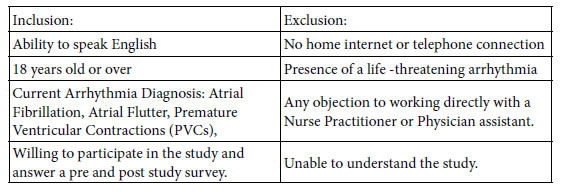
The study is a prospective randomized controlled study, in which informed consent was obtained. Randomization included subjects picking from sealed envelopes which were numbered, labelled with a folded card within each envelop stating either standard versus telemedicine visits. The University of Michigan Hospital IRB number: IRB00001995.

Inclusion/Exclusion Criteria:

JCRM-3-2-307-g001

Methods

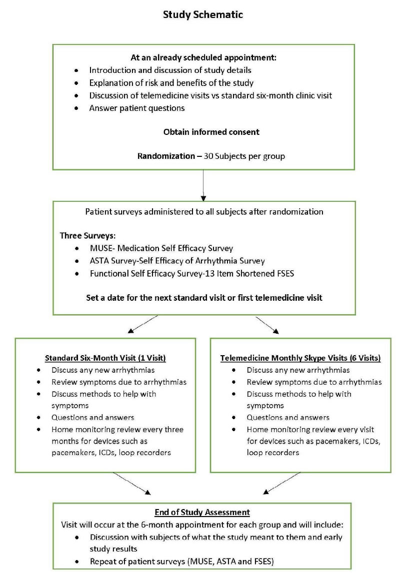
The study was introduced to the subjects during an initial meeting with an explanation of the study and an explanation of the consent. After the informed consent was obtained, the subjects were randomized into telemedicine or standard in person six- month visits.

With the initial visit, surveys were completed with telemedicine and standard visit groups. Telemedicine subjects received monthly visits for three consecutive months and standard received a six month return visits (Appendix B-study schematic). Interventions provided to the telemedicine group included discussion and reinforcement of medication, functional activity and arrhythmia self-efficacy, guided discussion, and social support.

The surveys utilized were the Medication Understanding and Self-Efficacy Tool, Functioning Self Efficacy Scale, and Arrhythmia Specific questionnaire in Tachycardia an Arrhythmia (MUSE, FSES, ASTA) surveys. All three surveys were provided on the first day of the study to each study group subject and on the last day of the study for each study group subject. Key questions were compared with a calculation of the mean for these questions, comparing the standard group with the telemedicine group. (Appendix C, D, E– MUSE, FSES, and ASTA surveys).

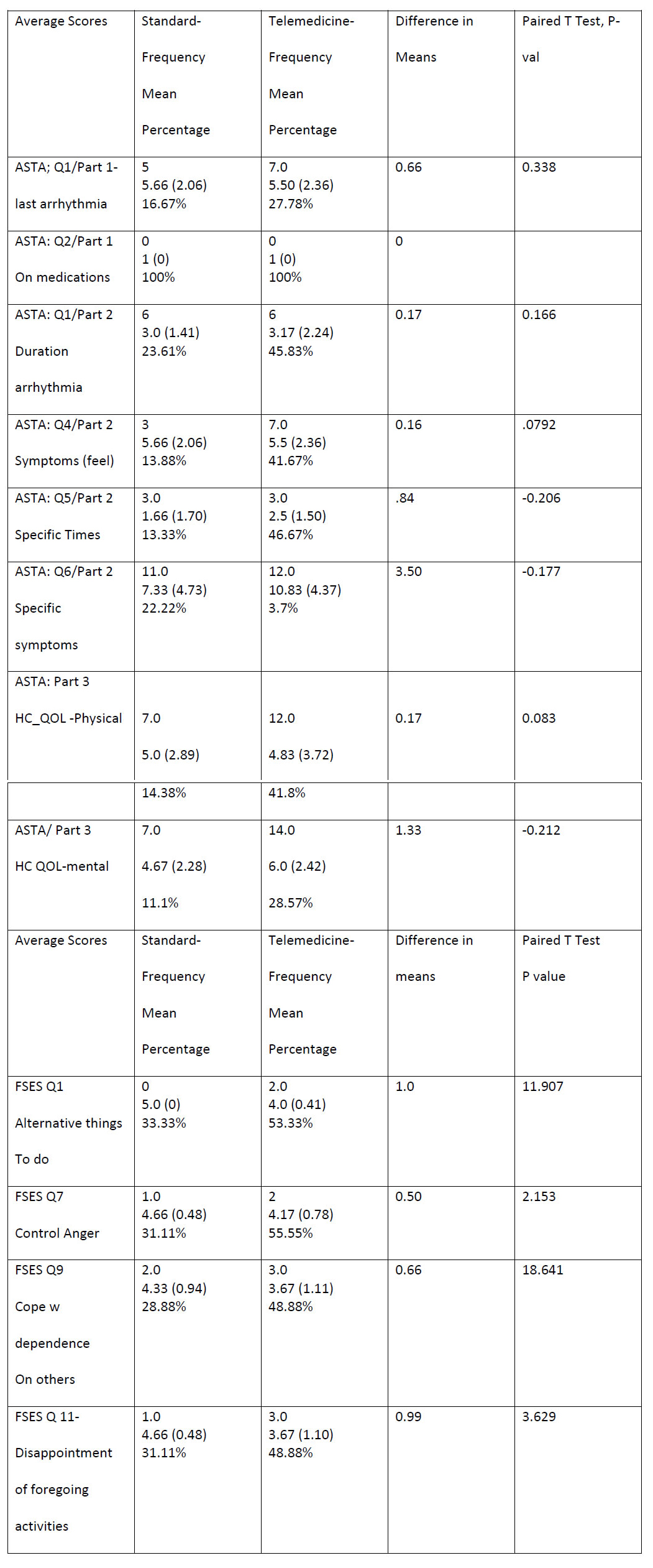
There were chart reviews and analysis of monitored data from devices  revealing  onset  of  arrhythmias,  times  of  diagnosis’  and treatments in the telemedicine group compared with the standard group. The T:EPASB utilizes the null hypothesis to demonstrate no difference in time of recognition of an arrhythmia, time to diagnosis and treatment of the arrhythmia, between the telemedicine group as compared with the standard group. The null hypothesis is be used in the Medication Understanding and Use Self- Efficacy (MUSE), ASTA (Arrhythmia Specific questionnaire in Tachycardia and Arrhythmia) and FSES (Shortened Functional Self Efficacy Scale) surveys. A paired T test with the difference in the means of answers to survey questions was utilized in calculating a P value for select survey questions (Appendix C).

Measures

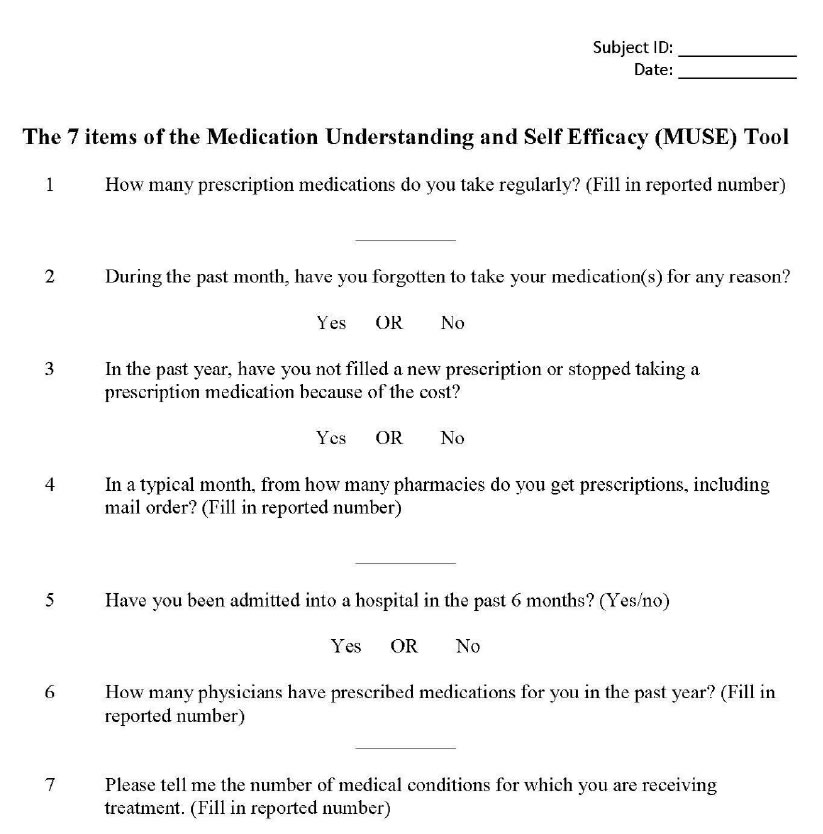
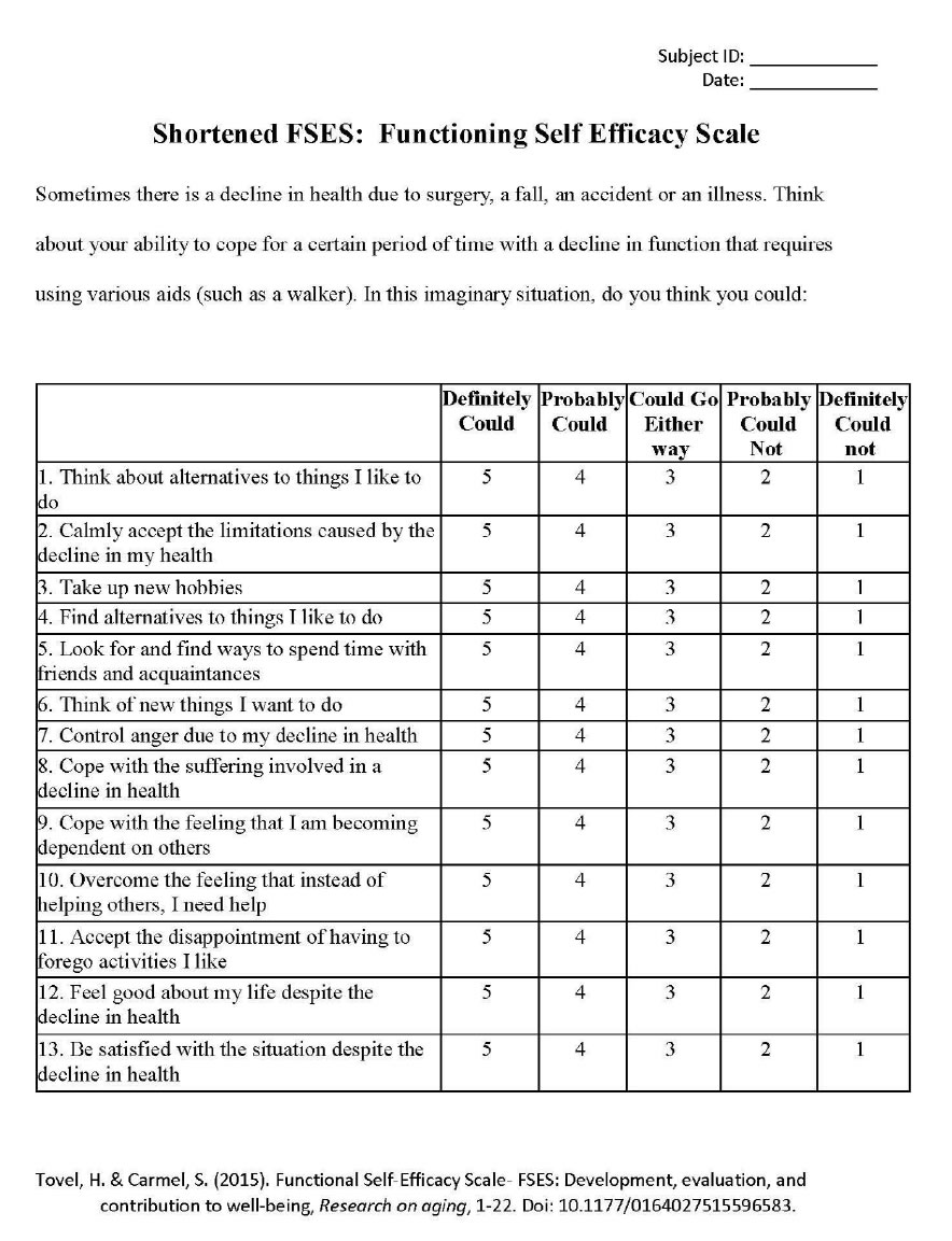
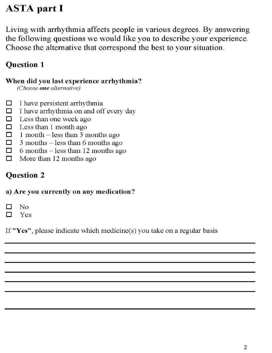
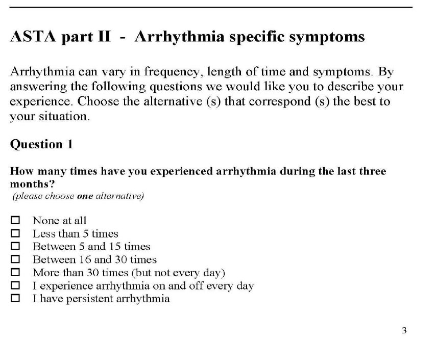
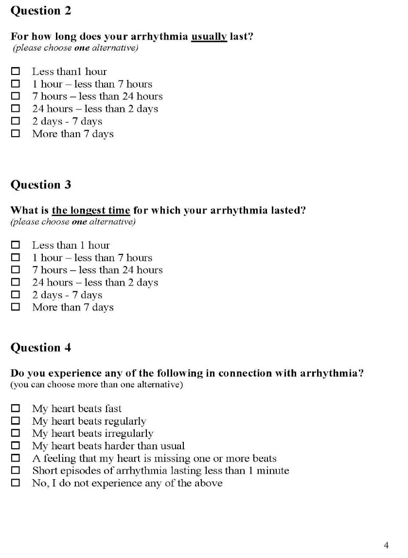
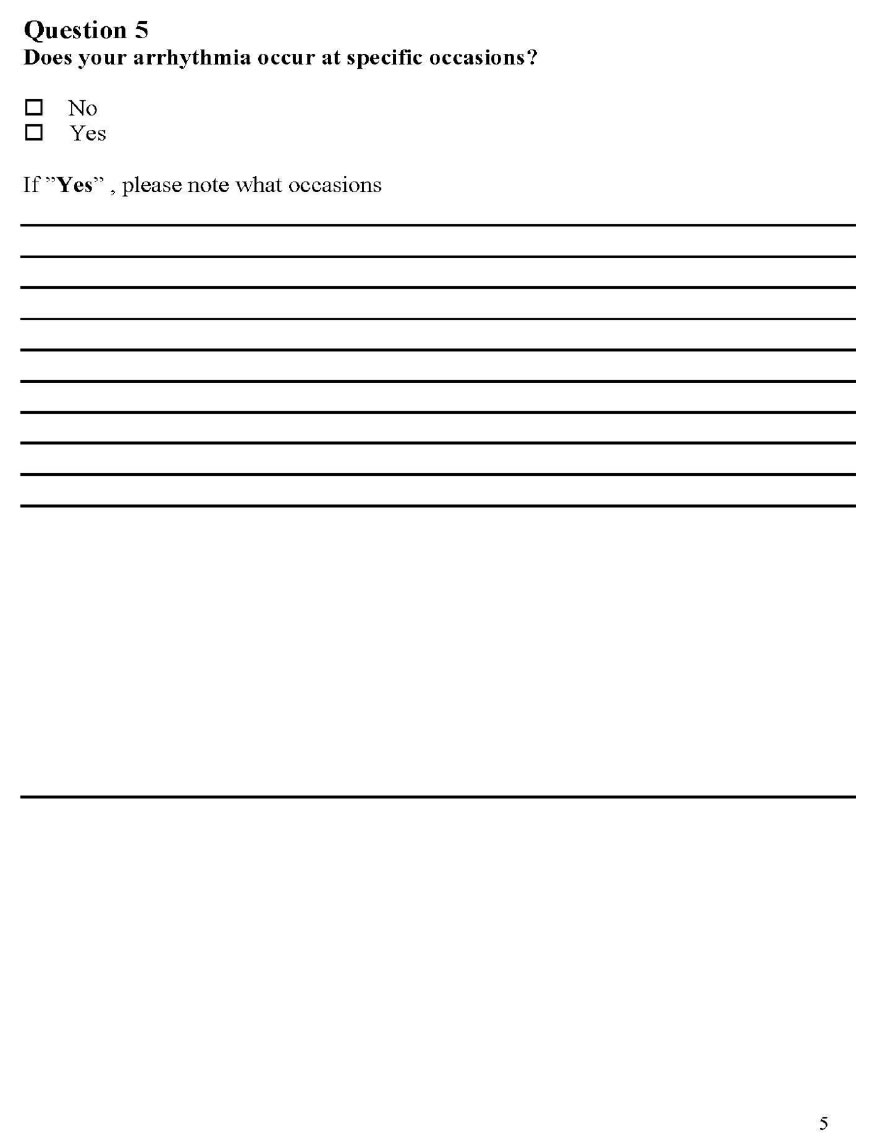
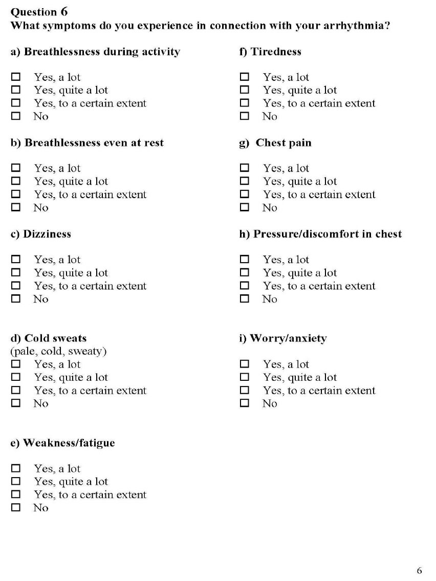
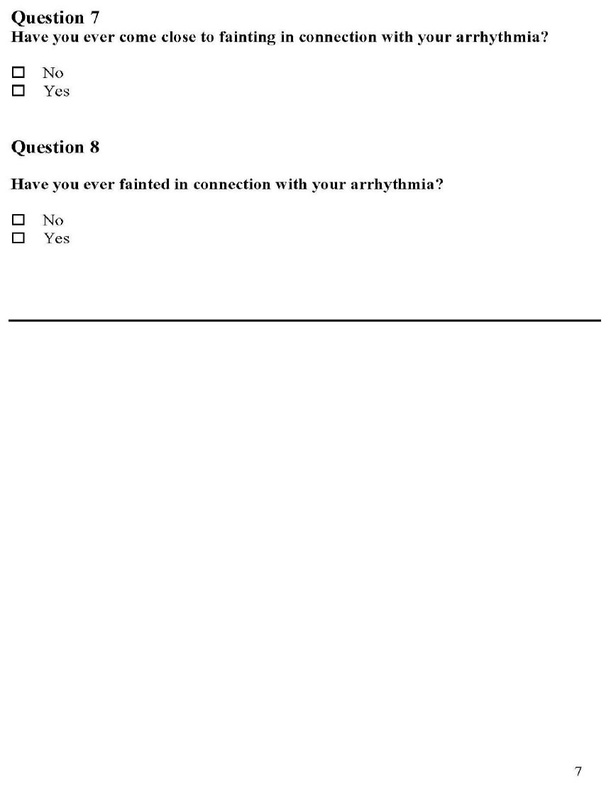
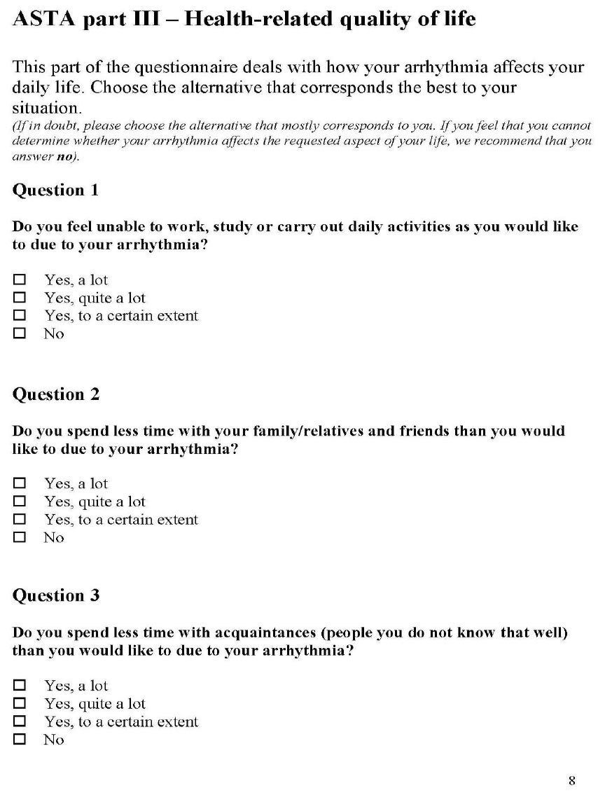
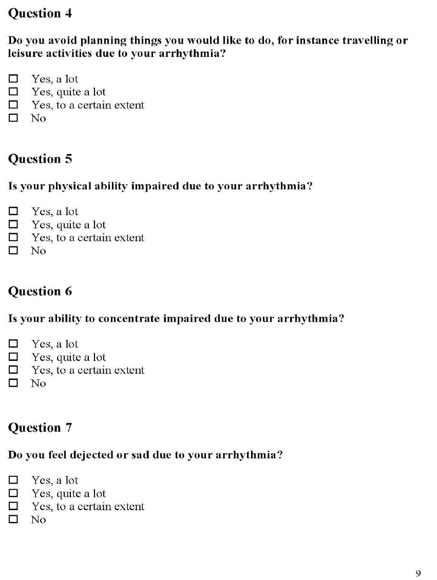
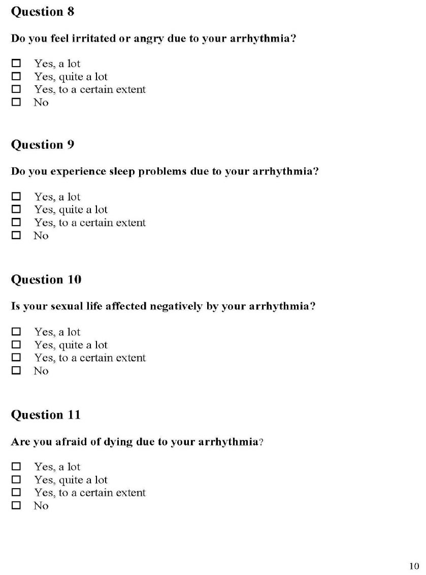
Arrhythmias can be multifactorial and can cause no perceived symptoms versus serious symptoms such as palpitations, fast and pounding heart beats, sweating, chest pain and or pressure, anxiety, fear, and depression [15]. One single survey may not capture the data experienced by the subject and not every survey relates to the self- efficacy of these perceived events. The MUSE survey gives information on medication compliance, cost barriers, number of medications, physicians and pharmacies and hospitalizations. The FSES gives a scale of the subject’s self- efficacy to cope with the arrhythmia and day to day functioning. The ASTA survey is the most specific survey to arrhythmias and the symptoms associated with arrhythmias; but does not reflect the medications or functional capabilities.

The MUSE survey was tested for validity and reliability in measuring   patients’ self-   efficacy   in   understanding   and   using prescription medications [12]. FSES displayed good internal consistency and satisfactory criterion and convergent validity in assessing the degree of confidence self-functioning while facing decline in health and function [16]. ASTA, displayed content validity for all items, and internal consistency [17].

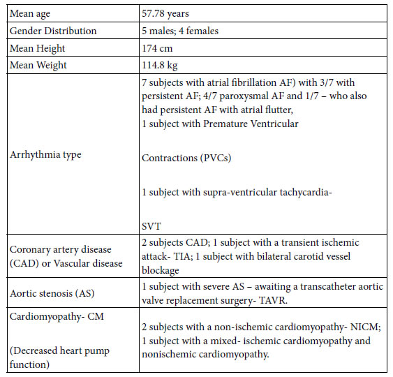
Data Collection Sheet and Demographics:

JCRM-3-2-307-g002

Pilot Study Results

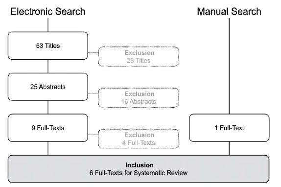
From late February 2020 to March 2020, 9 patients were enrolled in the Telemedicine study and randomized to either standard visits or telemedicine visits. Three patients declined the study, one patient noted he would join the study, but only if he received a Kardia monitoring device (he was not enrolled in the study as this could not be guaranteed and he noted his intention of simply gaining the Kardia device) and was not enrolled due to ethical concerns.

All subjects signed the informed consent and received copies of the protocol and consent, including the clause that they may drop out of the study. Each subject was given instruction on filling out surveys and were given the opportunity to answer questions by the nurse practitioner (NP) in clinic and the research assistant. The surveys were reviewed and scored by the research assistant and double scored with the author of the study. The surveys pinpointed the overall arrhythmia burden, degree to which subjects felt the arrhythmia, and physical and mental coping levels in relation to the arrhythmia. The surveys gave the caregiver (Nurse Practitioners and Physician Assistants within the EP clinic) specific areas to discuss, reinforce, and empower the subject in arrhythmia behavior change. The time of recognition, diagnosis and treatment of arrhythmias was deferred due to the Covid 19 pandemic and this monitoring data is attained only for daily arrhythmia management.

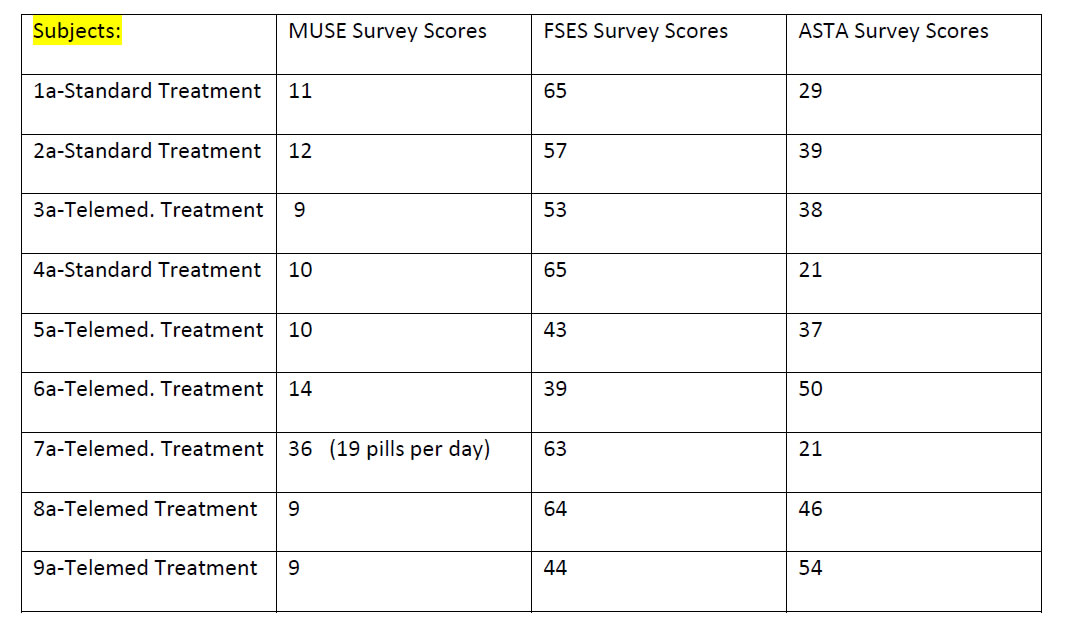
Appendix D gives the overview of all survey results for T:EPASB. The overview gives the researcher a quick glimpse of any problem areas such with decreased self-efficacy of medication use, or functional capacity or arrhythmia knowledge and understanding.

Results of the MUSE survey show near complete compliance in medication use with only one missed dose of medications from one subject. MUSE tallied results also show no financial constraints to medication obtainment in all nine subjects. There was a 0.8679:1 ratio with number of number of physicians treating to number of medical diagnosis; with a mean of 2.89 physicians prescribing medications to a mean of 3.33 medical diagnosis for the 9 subjects evaluated (Appendix B). When combining the surveys, key data becomes clear. The subjects scoring the lowest functional status self-efficacy, subject 5a and subject 9a with scores of 43 and 44 respectively, scored 37 and 54 respectively on the ASTA arrhythmia burden survey. One can note poor functional self-efficacy, but not necessarily related to arrhythmia burden in subject 5a, while poor functional self-efficacy may be related to a higher arrhythmia burden in subject 9a. Another complementing data point will be specific arrhythmia logs within monitoring- devices; once this deferred data is allowed within the study. Another interesting finding is the subject 8a who has a high arrhythmia burden noted with ASTA of 46, but a very good FSES score of 64.

Discussion

The T:EPASB pilot study has shown the importance of offering an alternative to conventional in person visits, offering counseling in  managing  one’s  self-efficacy  for  arrhythmia  care,  and  providing reinforcement and social support in managing one’s arrhythmia care. The study illustrates the importance of gathering complementing data on arrhythmia management including medication use and understanding, functional self-efficacy, and arrhythmia self-efficacy. The triad of these surveys provides an excellent overview of one’s arrhythmia self-efficacy. With such data, the medical and nursing provider may offer patient specific counseling. There is an advantage of having a specifically timed event, match a corresponding subject’s complaint. The use of implanted and portable monitoring data gives an excellent overview of the subject’s associated heart rhythm abnormality. The MUSE survey used alone gives a false sense that there may not be any need for any reinforcement of self-efficacy of medication or arrhythmia understanding. When this survey is coupled with the FSES and ASTA, trends begin to develop and specific areas of intervention, such as improved daily activity levels, decreased arrhythmia burdens via medications or activity, utilization of beta or calcium channel blockers, increased fluids, and or activity training to improve arrhythmias may be discussed. The surveys when used  together  display  specific  areas  to  improve  subject’s  knowledge, one’s confidence and self-efficacy in arrhythmia management. Those with higher arrhythmia burdens in which the subject feels palpitations, fatigue, and side effects have a greater need for intervention which strengthen self-efficacy and social support for medication use, functional activities and arrhythmia management [16,17]. This pilot T:EPASB study continues to show great potential and will likely mimic the TRUST, CHOICE-AF and Poinete trials in identifying, diagnosing, and treating arrhythmias with no difference in timing of these events with telemedicine compared with in person follow up visits with prompt device monitoring. The study has already helped to pinpoint areas of difficulties with arrhythmias, medications, and functional capacity. Via interventions such as affirming knowledge, counselling medication usage, and validating activity and exercise efforts and knowledge, the caregiver  may  help  improve  the  subjects’  overall  functional  capacity in coping with one’s arrhythmia. The study’s evaluated questions have not reached significant p values, as the study has been Anecdotally, the overall response to telemedicine has been very positive with comments like, “this is so much better”, “I can concentrate on what you are teaching me, without the long drive” and “this information seems to stick much better, when I learn it at home” and “can we make more telemedicine appointments”. The T:EPASB study can in no way be fully assessed at this early point, but its potential to assist in improving patients’ self-efficacy via increased patient interaction, reinforcement of arrhythmia details, and social support will surely lead to further studies using telemedicine and a triad of survey tools.

Authorship:

Kathleen Fasing, DNP-c, MS,

ACNP Madonna University,

Livonia MI

University of Michigan, Staff ACNP, Ann Arbor, Michigan

kfasing@med.umich.edu

DNP Project Chair: Patricia Clark, DNP, RN, ACNP-BC,

ACNS- BC, CCRN, Madonna University,

pclark@madonna.edu

DNP Project Member: Rachel Mahas, PhD, MS, MPH,

Madonna University, rmahas@madonna.edu

DNP Project Member: Vicki Ashker, DNP, MSA, RN,

CCRN, Madonna University, vashker@madonna.edu

Milwaukee- Self Management Science for your great research on self- efficacy and your ITHBC theory and allowing its use.

Sara Carmel- Ben Gurion University of Negev- Public Health Faculty Member- for your behavior research and functional self- efficacy tool and allowing its use.

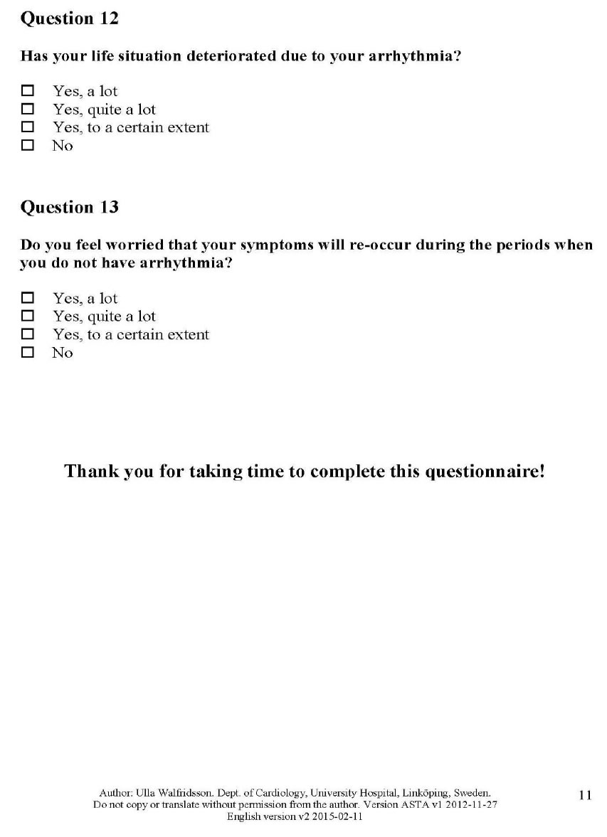
Ulla Walfridsson RN, PhD-Division of Nursing Science; Dept of Medicine & Health Sciences, Linkping, Sweeden- for sharing your incredible arrhythmia assessment tool and allowing its use.

Sangeeta Lathkar-Pradhan- Research Assistant for ongoing support and patience.

Rachel Wessel- Research Assistant for exacting perseverance.

Hakan Oral, MD- University of Michigan EP Director- thanks for believing in me.

Dr. Patricia Clark- DNP committee lead and advisor and patience extraordinaire.

My husband- Gregory Fasing BSN, RCIS- for his forever support.

Acknowledgements

Thank you so much for all who assisted in this project including and in equal acknowledgement.

Polly Ryan PhD, RN, CNS-BC – University of Wisconsin

References

  1. Kay, Misha, Santos, Takane (2010) Telemedicine opportunities and development in member states, Global observatory for eHealth series, virtualhospital.org.uk. (1,2). [crossref]
  2. Ryan, P. (2009) Integrated theory of health behavior change: Background and intervention development, Clinical Nurse Specialist, 23 (3): 161-172. (3, 5,18,19). [crossref]
  3. Lovett-Rockwell, K. & Gilroy, A. (2020) Incorporating telemedicine as part of COVID-19 Outbreak response systems, The American Journal of Managed Care, 26 (4): 147-148. Doi.org/10.37765/ajmc.,(4). [crossref]
  4. Fozzard (2011) History of basic science in cardiac electrophysiology, Cardiac electrophysiologycinics, 3, 1, 1-10. Doi:10.1016/j.ccep.2010.10.010,(6,7). [crossref]
  5. Phend, C. (2020) Telehealth shaping up for Covid-19- Cardiology illustrates what specialties can do to be ready, Medpage today.,(8).
  6. Maffei, R., Hudson, Y. & Skim Dunn, M. (2008) Telemedicine for urban uninsured: A pilot Framework for specialty care planning sustainability, J E health, 14(9), 925- 931 [crossref]
  7. Dalouk,  K.,  Gandhi,  N.,  Jessel,  P.,  MacMurdy,  K.  et.  al.  (2017)  Outcomes   of telemedicine  videoconferencing  clinic  versus   in-person   clinic   follow-up for implantable cardioverterdefibrillator recipients, Circulation arrhythmia electrophysiology, 10, (11,13).
  8. Varma, N., Epstein, A., Irimpen, A., Schweikert, R., & Love, C. (2010) Efficacy and safety of automatic remote monitoring for implantable cardioverter-defibrillator follow-up: The Lumos-T safely reduces routine office device follow-up, TRUST trial, Circulation, 122: 325332.,(12). [crossref]
  9. Lopez-Villegas, A., Catalan-Matamoros, D., Robles-Musso, E., & Peiro, S. (2015) Effectiveness of pacemaker tele-monitoring on quality of life, functional capacity, event detection and workload: The PONIENTE trial, Geriatrics and gerontology international, 16 (11).,(14). [crossref]
  10. Varma, N. & Ricci, R. (2013) Telemedicine and cardiac implants: what is the benefit? European heart journal, 34 (25), 1885-1895.(15).
  11. Lowres, N., Redfern, J., & Freedman, S. (2014) Choice of health options in prevention of cardiovascular events for people with  atrial  fibrillation  (CHOICE  AF):  A  pilot study, European journal of cardiovascular nursing, doiorg.proxy.lib.umich. edu/10.1177/14745114549687. (16). [crossref]
  12. Cameron, K., Ross, E., Clayman, M., Bergeron, A., et. al. (2010) Measuring patients’ self-efficacy in understanding and using prescription medication, PatientEducation Couns, 80 (3); 372376. Doi: 10.1016/j.pec.2010.06.029.,(17,21). [crossref]
  13. Ryan, P., Papanek, P., Csuka, M., Brown, M., et. al. (2018) Background and method of the striving to be strong study, a RCT test the efficacy of a mhealth self-management intervention, Contemporary Clinical Trials, 71, 80-87.,(16). [crossref]
  14. Suter, B., Suter, W. N., & Johnston, D. (2011) Theory-based telehealth and patient empowerment, Population Health Management, 14 (2).,(19). [crossref]
  15. Withers, K., Wood, K., Carolan-Rees, G, Patrick, H., et.al. (2015) Living on a knife edge- the daily struggle of coping with symptomatic cardiac arrhythmias, Health quality life outcomes, 13:86.,(20). [crossref]
  16. Tovel, H. & Carmel, S. (2015) Functional Self-Efficacy Scale- FSES: Development, evaluation, and contribution to well-being, Research on aging, 1-22. Doi: 10.1177/0164027515596583. (16,22,24). [crossref]
  17. Walfridsson, U., Arestedt, K., & Stromberg, A. (2012) Development and validation of a new arrhythmia-specific questionnaire in tachycardia and arrhythmia (ASTA) with focus on symptom burden, Health quality life outcomes, 10-44, doi: 10.1186/1477- 7525-10-44.,(23,25). [crossref]

Appendix A. Data Collection Tool- Aid for patient at home.

JCRM-3-2-307-g003

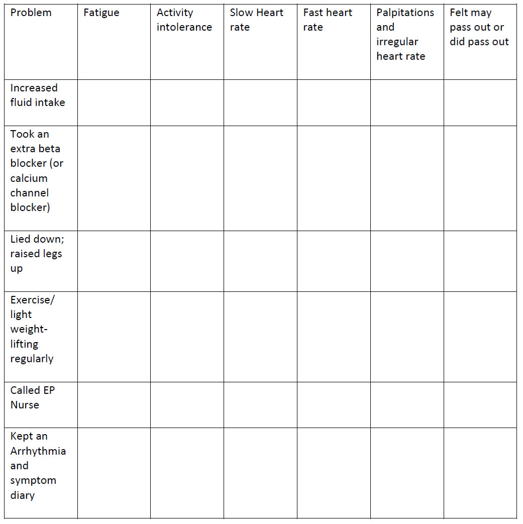
2 points each for every activity noted along the vertical axis; showing an activity taken due to the symptom noted on the horizontal axis. Subject to keep weekly log of number of symptoms and number of points for response activities.

Appendix B: Study Schematic

JCRM-3-2-307-g004

Appendix C: Three Surveys

JCRM-3-2-307-g005

JCRM-3-2-307-g006

Walfridsson, U., Arestedt, K., & Stromberg, A. (2012) Development and validation of a new arrhythmia-specific questionnaire in tachycardia and arrhythmia (ASTA) with focus on symptom burden, Health quality life outcomes, 10-44, doi: 10.1186/1477- 7525-10-44.,(23,25).

JCRM-3-2-307-g007

JCRM-3-2-307-g008

JCRM-3-2-307-g009

JCRM-3-2-307-g010

JCRM-3-2-307-g011

JCRM-3-2-307-g012

JCRM-3-2-307-g013

JCRM-3-2-307-g014

JCRM-3-2-307-g015

JCRM-3-2-307-g016

JCRM-3-2-307-g017

JCRM-3-2-307-g018

Appendix D: Scores of completed surveys:

Survey Scores: Key- Muse– Shows any difficulty in taking; or understanding medications. FSES

highest the better functional status; highest possible =65. ASTA– the highest the worse arrhythmia burden, symptoms, and mental and physical QOL.

JCRM-3-2-307-g019

Appendix D. – Answers to the above scores

Muse Survey Answers

Question 1– How many prescriptions medications do you take regularly?

Mean= 5.33 medications; Median 4 Medications; Mode 5 medications with a low answer of 2 and high answer of 19 medications.

Question 2– During the past have you forgotten to take any medication? “Only 1 yes.

Question 3– In the past did you not fill or stop taking the prescription due to cost? All answered no.

Question 4– In a typical month how many pharmacies do you use; including mail order? Six subjects answered 1/ 3 subjects answered 2.

Question 5– Have you been admitted to the hospital in the past six months? –Three subjects -yes.

Question 6– How many physicians prescribed medications for you in the past year? – mean answer 2.89.

Question 7– How many medical conditions which you are receiving treatment? – mean answer 3.33

FSES Shortened Survey * Higher scores showing better functional status

This survey gained very differing results for patient. Two subjects scored the total possible of 65 points indicating the best functional status and answered 5 (maximal score) for all 13 questions. One subject answered 3 for each of the 13 questions with a score of 39 and indicating day to day function was exactly in the middle of the survey. Other subjects gave a variable scoring with specific areas and gave a scattered response, depending upon the question. Two of the subjects gave responses in the 2 range, or lower level of functional capabilities. Scores listed in 1a-9a order: 65/ 57/ 53/ 65/ 43/ 39/ 63/ 64/ 44.

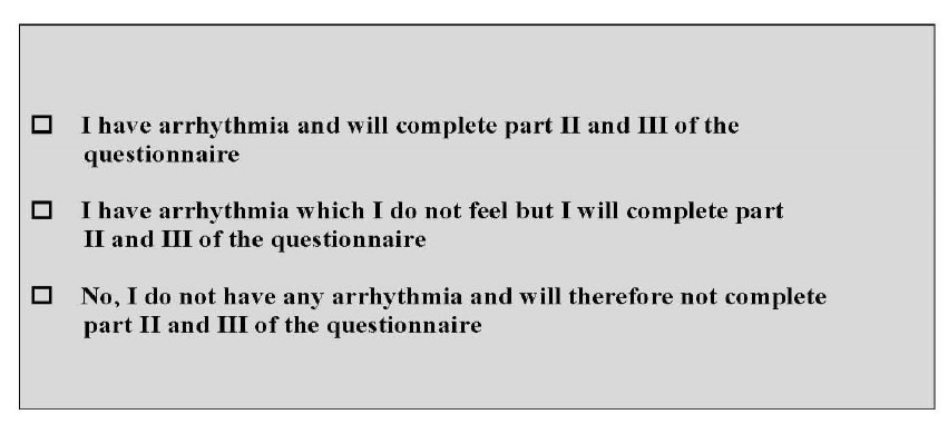
ASTA Survey The survey is scored into three categories- presence of arrhythmia, symptoms associated with the arrhythmia and Health related Quality of life (QOL)- both mental and physical. Higher the score- the higher the arrhythmia burden, more symptoms and more impact on the health related QOL. Scores listed in 1a- 9a order: 39/39/38/21/37/50/21/46/54.

Appendix E.

Average Scores Pre and Post (n=9)

JCRM-3-2-307-g020

Evidence-Based Guideline for COVID-19 Infection Control in Dental Medicine: A Systematic Review

DOI: 10.31038/JCRM.2020325

Abstract

Objective: COVID-19 became a pandemic and has caused a global emergency in the healthcare sector. Dental professionals pose high risk of viral spreading. This systematic review focused on COVID-19 infection control protocols in oral and dental medicine to derive an evidenced-based guideline.

Method: An electronic and manual search was performed for articles related to the outbreak of COVID-19 published between 2019/11/01 and 2020/03/31.

Results: Of the 53 titles retrieved by the systematic search, a total of 6 full-texts were included for data extraction.

Conclusions: Dental treatment must be limited to emergencies during the pandemic. Strict adherence to the hygiene chain is a prerequisite for the control of further transmission and protection of dental professionals. The use of aerosol-producing therapies must be minimized. Establishing regional dental emergency centers can help maintain the necessary capacities for society while maintaining appropriate hygiene standards. Personal protective equipment must be used in a resource-saving manner for dental emergencies of infectious patients.

Keywords

Corona virus, SARS-CoV-2, Pandemic, Dentistry, Public health

Introduction

The previously unknown coronavirus disease 2019 (COVID-19) and the pathogen SARS-CoV-2 (severe acute respiratory syndrome) with its specific virulence and pathogenesis quickly became  a  global pandemic [1]. The rapid spread from human-to-human through saliva, blood, and other body fluids have made it difficult to precisely determine its transmission pattern, extent of spread, and dangerousness. These factors, along with the fact that symptoms of COVID-19 infection are similar to types of viral influenza, led to an initial underestimation of the scale of the problem [2,3].

COVID-19 emerged in Wuhan City – capital of Hubei Province in China – and rapidly spread through South Korea, Japan, and Australia, then to Europe and the Middle-East, and recently North and South America [4]. The outbreak has spiraled into a global emergency for the healthcare sector, with wider economic damage and social restrictions, and unpredictable consequences in the future [5,6]. The most important characteristics of a pandemic are the spreading dynamics in time and space, the transmission rate and pathways, the incubation period, and the proportion of severe cases and mortality rate. The apparently high virulence of SARS-CoV-2, including the asymptomatic incubation period (up to 14 days), is a particular problem in this age of globalization [7,8].

Individuals with jobs that put them in close physical contact with many other people are at the greatest risk of becoming infected by

SARS-CoV-2 and for transmitting the infection onwards. The New York Times calculated the risk to be infected by SARS-CoV-2 for various occupations based on the risk of “exposure to disease” and “proximity to other humans” [9]. Healthcare workers were found to be at the greatest risk of encountering diseases and infections and typically work in close relationship to one another and their patients. In particular, dental care providers had the highest overall risk for SARS-CoV-2 infection, based on both exposure and physical proximity to others [9]. This clearly reflects the facts that dental care providers work directly with one of the known transmission routes  of SARS-CoV-2, the oral cavity, and this is exacerbated by the use of rotating instruments or ultrasonic tips with aerosol production.

The key questions currently facing dental care workers are therefore: how should we act upon the COVID-19 crisis? And what are the adequate standards required for the well-being of patients and dental staff, considering different job groups, and also for the wider protection of society?

This review aimed to systematically screen the current literature on COVID-19 infection control protocols in oral and dental medicine to derive an evidenced-based guideline for stakeholders in the dental healthcare sector.

Methods and Meterials

The systematic review was conducted in accordance with the guidelines of Preferred Reporting Items of Systematic Reviews and

Meta-Analyses (PRISMA) [10]. An electronic search strategy of PubMed was performed for all types of publications reporting on COVID-19 in the oral medicine setting. The search was limited to articles published from around the time of the start of the outbreak to the present day (2019/11/01 to 2020/03/31). All types of publications were included. Search syntax comprised a combination of Medical Subject Headings [MeSH-Terms] and free-text words in simple or multiple conjunctions: ((“COVID-19” [All Fields] OR “2019-nCoV” [All Fields] OR “SARS-CoV-2” [All Fields] OR “coronavirus” [All Fields] OR “corona” [All Fields]) AND  (“dentistry”  [All Fields] OR “dental medicine” [All Fields] OR (“dental” [All Fields] AND “medicine” [All Fields]) OR “oral medicine” [All Fields] OR (“oral” [All Fields] AND “medicine” [All Fields]) OR “oral healthcare” [All Fields] OR (“oral” [All Fields] AND “healthcare” [All Fields]))) AND (“2019/11/01” [PDAT] : “2020/03/31” [PDAT]). An additional manual search of the bibliographies of all retrieved full-text articles and related reviews, selected from the electronic search, was conducted.

Two reviewers (T.J. and N.U.Z.) independently reviewed the titles of articles retrieved by the systematic literature search. Following this, the abstracts of all agreed titles were screened to identify articles that reported COVID-19 infection control protocols in oral medicine. The full texts of selected articles were obtained and data were extracted independently by the two reviewers using a data extraction form. Disagreements at each stage of the screening process were resolved by discussion. The following information was collected from the full text articles: a) authors including year of publication; b) country; c) publication type; and c) details of the COVID-19 infection control protocols reported in the articles.

Results

Of the 53 titles retrieved by the systematic PubMed search, 25 abstracts were further screened, and the full text of nine potentially relevant articles were obtained and reviewed. A total of six full-texts were included for data extraction, comprising four full-texts obtained from the electronic search plus one additional full-text obtained from the manual search (Figure 1) [11-16].

JCRM-3-2-312-g001

Figure 1. Systematic search strategy.

Included publications were categorized as four Narrative Reviews (Li & Meng 2020; Peng et al. 2020; Tang et al. 2020; Yang et al. 2020), one Discovery Report (Meng et al. 2020), and one Letter to the Editor (Sabino-Silva et al. 2020). Key recommendations for the management of COVID-19 infection control from each publication were summarized and tabulated along with the first author name, year of publication, and country of publication (Table 1).

Table 1: Data extraction for included studies (n=6).

Authors (year)

Country

Study design

COVID-19 infection control protocol

Li and Meng (2020) [11]

China

Narrative Review

• Use of effective and strict disinfection measures in both clinical settings and public area.
• Adequate management of contaminated consumables.

Meng et al. (2020) [12]

China

Discovery Report

• Dental care provider should measure the temperature of every staff and patient as a routine procedure.
• Dental staff should strictly use personal protective equipment (PPE).
• Preoperative antimicrobial mouth rinse reduces the number of microbes in the oral cavity.
• Aerosol-generating procedures should be minimized as much as possible.
• The use of rubber dam and saliva ejectors with high volume can reduce the production of aerosols.
• Extra-oral dental radiography is an appropriate alternative during the outbreak of COVID-19.

Peng et al. (2020) [13]

China

Narrative Review

• Dental professional should be able to identify patients with suspected COVID-19 infections.
• Hand hygiene has been considered the most critical measure for reducing the risk of transmitting COVID-19.
• Procedures that are likely to induce coughing should be avoided or performed cautiously.
• The use of rubber dam can significantly minimize the production of saliva- and blood-contaminated aerosol.
• Droplet and aerosol transmission of COVID-19 are the most important concerns in dental treatments; and therefore, should be reduced whenever possible.

Sabino-Silva et al. (2020) [14]

Canada/ Brazil

Letter to the Editor

• It is crucial for dentists to refine preventive strategies to avoid the COVID-19 infection by focusing on patient placement, hand hygiene, all personal protective equipment (PPE), and caution in performing aerosol-generating procedures.

Tang et al. (2020) [15]

China

Narrative Review

• Dental professionals play great role in preventing the transmission of COVID-19 by following infection control measures during dental practice to block the person-to-person transmission routes.

Yang et al. (2020) [16]

China

Narrative Review

• Use of and strict personal disinfection of maxillofacial professionals in a huge hospital.

• Responsible handling of infectious consumables and sterilization of contaminated instruments.

Discussion

COVID-19 is a newly identified respiratory disease. The first case can be tracked back to 17 November 2019, according to Chinese government data [17]. Therefore, this systematic PubMed search was limited to publications reporting on infections caused by SARS-CoV-2 that were published after 1 November 2019. Publications focused on other SARS-related diseases were excluded.

Considering the recent and rapid development of the COVID-19 outbreak, it was not surprising that the identified publications from the systematic search were not clinical studies, but rather reports on self-learned experiences and trial and error management, mainly from research groups in China (five publications out of six) [11-16]. As COVID-19 continues to spread globally, it is assumed that confirmatory and/or new recommendations, and retrospective analyses of the effectiveness of different infection control protocols, will continue to be published from Europe and then from the USA [1,6,13].

The oral healthcare sector in particular has been hit hard by COVID-19 [12]. Confusion prevails due to the flood of information from policy stakeholders, statements from national dental associations, and press releases, accompanied by various (fake) posts on social media [17-21]. What infection control protocols should dental care provider follow during the COVID-19 crisis? And what lessons can the dental society learn from the COVID-19 pandemic to guide the precautions and preventative measures that may be required for future infectious diseases?

Based on the findings of this systematic review [11-16] and involving the expertise and experience of colleagues at the University Center for Dental Medicine in Basel (UZB), Switzerland, an evidence- based guideline was developed (Figure 2). This guideline was implemented on 2020/03/31 and will be reviewed and updated as new information on the COVID-19 outbreak comes to light. This guideline considers both the external and the internal dental ecosystems. On the one hand, the institutional infrastructures of dental care providers must be differentiated from an external point of view: i) Single Offices;Clinical Centers; and iii) Dental Hospitals with/without University setting including under- and postgraduate education, and clinical trials with voluntary participants and patients under dental treatment. On the other hand, possible routes of microbial transmission within the dental environment must be analyzed internally: i) dental and oral diagnostics including maxillofacial radiology and in particular aerosol-producing treatments; ii) patient flows plus accompanying relatives in waiting areas, restrooms and toilet facilities; as well as iii) employees’ facility rooms of daily use.

JCRM-3-2-312-g002

Figure 2. Patient flow in a centralized setting of a Dental Hospital.

In common with the rest of society, “physical distancing” is the top priority. In this context, the catchment radius of potential patients and dental staff must be taken into account for the decision who should maintain the dental care mandate during a crisis. Reducing the number of employees needed to deliver emergency dental treatments is the ultimate goal to reduce contact between people and diminish the risk of viral spreading. This will also help to preserve the supply of personal protective equipment for the protection of the high-risk group of dental care providers. Moreover, the range of dental treatments that will be delivered during the pandemic should be transparently communicated to both the dental society and to patients.

Centralization of dental emergency treatments in regional Dental Hospital settings is the key for maintain dental healthcare standards during pandemic crisis. In contrast to primary healthcare, dental procedures are usually elective. All non-urgent dental treatments must be postponed. Supplies of personal protective equipment needed by clinical staff who are treating critically ill patients in General Hospitals are critically low. If all dental care providers store and use these items for non-urgent treatments, frontline healthcare workers will be left unprotected. In case of COVID-19, it is not possible to reliably identify patients who are asymptomatically infected and most of the dental treatments produce aerosols that is known to increase exposure potential if patients are infected.

We recommend that dental care providers should focus on the management of conditions that require immediate attention to relieve severe pain and/or risk of infection and to alleviate the burden on hospital emergency departments. These comprise:

• Severe dental pain from pulpal inflammation;

• Acute exacerbation of chronic apical periodontitis;

• Acute necrotizing ulcerative gingivitis or periodontitis;

• Painful mucosal changes;

• Pericoronitis of third molars;

• Post-operative osteitis and dry-socket phenomenon;

• Infections with intra-/extra-oral swelling or abscess, particularly those potentially compromising the patient’s airway;

• Facial and/or dental trauma with avulsion, dislocation and/ or fracture;

• Uncontrolled bleeding;

• Dental treatment required prior to critical medical procedures;

• Fixation or removal of broken prosthodontic or orthodontic appliances to avoid injuries or foreign body aspiration.

If dental diagnostics and/or treatments cannot be delayed and have to be carried out related to the above list of indications, strict adherence to the hygiene chain is mandatory. In addition to the routine patient history, contactless temperature measurement using infrared thermometers should now be employed as an initial step to identify patients who are potentially infected with SARS-CoV-2.

For essential emergency treatment of patients with confirmed COVID-19, or patients with a strong suspicion of an acute infection, the following protocol should be applied by the dental care providers:

• Patients should wear masks in public and in the hospital setting;

• Patients with suspected or confirmed infection should wait in separate rooms and use indicated rest-rooms;

• Dental treatment should be conducted in an isolated and well- ventilated room (100% fresh air supply of the ventilation or air condition system instead of recirculating air is pursued);

• Use of personal protective equipment is mandatory, comprising respirators for filtering inspired air, face masks and goggles, double-layered gloves, and protective clothing, including safe disposal of used consumables;

• Patients should use pre-operative antimicrobial mouth rinse with 1% hydrogen peroxide for 30 seconds (peroxide dissolves the virus protein);

• Extra-oral dental radiographs can be used as alternatives to intra-oral imaging during the outbreak of COVID-19 to avoid any coughing of the patients;

• Emergency treatments should be as minimally invasive as possible and reduced in length and in spread of oral fluids (rubber dam and high-volume saliva ejectors to minimize aerosol);

• Treatment rooms and other areas, and used instruments, should be cleaned with appropriate virostatic agents;

• The number of dental care providers in the facility should be limited; and staff should work in weekly shifts and should be separated from household family members if they are at risk;

• Tele-healthcare should be used (whenever possible).

In addition, human resource management should consider different scenarios for work deployment plans in the Dental Hospital settings during the crisis. Tandems consisting of one dentist and one dental assistant should work together in blocks of weekly shifts to reduce interactions with other colleagues and the risk of infection. If a member of a team becomes infected, or an infection is suspected, the team should be replaced by a new tandem. Those dental personnel who have previously been infected with SARS-CoV-2, and have evidence of immunity in the form of antibodies to the virus, could take over the emergency treatments to protect the susceptible colleagues. Moreover, it must be considered whether routine testing of the dental staff should or could be carried out to clarify the infectious status of the staff in order to help prevent further transmission of the virus.

Conclusion

What lessons can dental stakeholders learn from the current COVID- 19 pandemic?

The dental profession and its special environment pose high risk of viral spreading and cross infections between patients and dental care providers. Strict adherence to the hygiene chain is a prerequisite for the control of further transmission. Dental treatment must be limited to emergencies during the pandemic. Positively tested patients or symptomatic patients with a strong suspicion of an acute infection must be treated under personal protective conditions of the dental professionals. Any aerosol-producing therapy must be reduced to a minimum.

After the successful fight against the pandemic, international standards need to be defined by a task force with experts from the public health sector, health economists, and politicians, in order to develop standard operating procedures for quickly coordinating and coping with future public health emergencies. Dental treatments are usually elective procedures. During a pandemic, it is important to establish regional dental emergency centers as quickly as possible to provide the necessary capacities for society and to ensure appropriate hygiene standards. Personal protective equipment must be used for dental emergencies of infectious patients and in a resource-saving manner.

Author Contributions

Conceptualization, Methodology, Writing-Original Draft, Writing-Review and Editing, Supervision and Project Administration:

T.J. and N.U.Z.; both authors have read and agreed to the published version of the manuscript.

Declaration of Interests

The authors declare no conflict of interest.

Acknowledgements

The authors express their gratitude to Mr. James Ashman for proofreading the final manuscript. This research received no external funding.

References

  1. Tuite AR, FismanDN(2020) Reporting, Epidemic Growth, and Reproduction Numbers for the 2019 Novel Coronavirus (2019-nCoV) Epidemic. Ann Intern Med,[Crossref]
  2. Cheng, ZJ,ShanJ (2020) 2019 Novel coronavirus: where we are and what we know. Infection, [Crossref]
  3. Habibzadeh P,StonemanEK (2020)The Novel Coronavirus: A Bird’s Eye View. Int J Occup Environ Med, 11(2): 65-71. [Crossref]
  4. Hui DS(2020)The continuing 2019-nCoV epidemic threat of novel coronaviruses to global health – The latest 2019 novel coronavirus outbreak in Wuhan, China. Int J Infect Dis, 91: 264-266. [Crossref]
  5. Dey SK(2020) Analyzing the epidemiological outbreak of COVID-19: A visual exploratory data analysis approach. J Med Virol, [Crossref]
  6. Wu, JT, LeungK, LeungGM(2020) Nowcasting and forecasting the potential domestic and international spread of the 2019-nCoV outbreak originating in Wuhan, China: a modelling study. Lancet, 395(10225): 689-697. [Crossref]
  7. Yang W(2020) Clinical characteristics and imaging manifestations of the 2019 novel coronavirus disease (COVID-19):A multi-center study in Wenzhou city, Zhejiang, China. J Infect, 80(4): 388-393. [Crossref]
  8. Zhou F(2020) Clinical course and risk factors for mortality of adult inpatients with COVID-19 in Wuhan, China: a retrospective cohort study. Lancet, [Crossref]
  9. Gamio L (2020)The workers who face the greatest coronavirus risk. The New York Times,
  10. Moher D(2009) Preferred reporting items for systematic reviews and meta-analyses: the PRISMA statement. PLoS Med, 6(7):e1000097. [Crossref]
  11. Li ZY,MengLY (2020) [The prevention and control of a new coronavirus infection in department of stomatology]. Zhonghua Kou Qiang Yi XueZaZhi, 55(0): E001. [Crossref]
  12. Meng L,Hua F,BianZ (2020) Coronavirus Disease 2019 (COVID-19): Emerging and Future Challenges for Dental and Oral Medicine. J Dent Res. [Crossref]
  13. Peng X(2020) Transmission routes of 2019-nCoV and controls in dental practice. Int J Oral Sci, 12(1): 9. [Crossref]
  14. Sabino-Silva R,JardimACG, SiqueiraWL (2020) Coronavirus COVID-19 impacts to dentistry and potential salivary diagnosis. Clin Oral Investig,
  15. Tang, HS, YaoZQ,WangWM (2020) [Emergency management of prevention and control of novel coronavirus pneumonia in departments of stomatology]. Zhonghua Kou Qiang Yi XueZaZhi, 55(0): E002. [Crossref]
  16. Yang Y(2020) Experience of Diagnosing and Managing Patients in Oral Maxillofacial Surgery during the Prevention and Control Period of the New Coronavirus Pneumonia. Chin J Dent Res, 23(1):57-62. [Crossref]
  17. Davidson H (2020) First Covid-19 case happened in November, China government records show. Guardian Weekly,
  18. COVID-19 Infection Control Protocols and Procedures. 2020; Available from: https://www.ada.org/en/member-center/coronavirus-resource-toolkit-for-ada-members/covid-19-infection-control-protocols-and-procedures-webinar?utm_source=adaorg&utm_medium=vanityurl&utm_content=safety&utm_campaign=covid-19.
  19. Zhejiang University School of Medicine C, Handbook of COVID-19 Prevention and Treatment. 2020.
  20. Coronavirus disease (COVID-19) Pandemic. 2020; Available from: https://www.who.int/emergencies/diseases/novel-coronavirus-2019.
  21. Dossier: AktuelleInformationenzum Coronavirus. 2020; Available from: https://www.sso.ch/home/coronavirus.html.

How are We Going to Restart Elective Bariatric and Metabolic Surgery after the Peak of Covid-19 Pandemic?

DOI: 10.31038/SRR.2020312

Abstract

Background: The pandemic pattern of COVID-19 in some countries appears to have started a deceleration stage. It is recommended to restart as soon as the situation is under control and some tools exist for interpreting the ICU capacity under conditions of health system stress. However, there are no clear guidelines or a strategy to restart Bariatric and Metabolic (B&M) surgery.

Objective: To present a protocol proposal to assist surgeons in restarting elective B&M surgery after the outbreak.

Method: This document presents a recommended protocol based on experts’ opinions, reviews of small series and publications from previous different viral epidemic outbreaks.

Results: Priority is for patients who would benefit most from a B&M procedure with limited risk. Patients being eligible for surgery must be screened for COVID-19. Only patients with negative results will have surgery. Radiofrequency/electro thermal bipolar devices are recommended. A smoke evacuator machine with a closed-circuit system should be used. All patients must be treated as if they are suspected to be COVID-19 positive. Proper personal protective equipment should be applied. Post-operative follow-up consultations should be via telemedicine. The RT-PCR screening test should be repeated for legal documentation purposes and to make sure that the patient is COVID-19 free.

Conclusion: The resumption of elective B&M surgeries during deceleration stage of COVID-19 must be gradual and prudent. This scholarly protocol can be adjusted according to the regulations established by the local health departments and with the increased acquisition of new knowledge; there would be changes in the current recommendations.

Keywords

Covid-19 pandemic, Metabolic surgery, Sleeve gastrectomy, Bariatric and metabolic surgery

Introduction

The future and aftermath of the COVID-19 global pandemic is unknown but will be dependent on how the medical societies and governments react to the current situation. Many of the international health organizations and federations have combined efforts to fight against the COVID-19. European and American guidelines as well as IFSO recommendations have been issued and most elective surgeries were postponed, as a cautious measure to minimize the spread of the infection and to cope with the emerging scarcity of health care resources, particularly ventilators and other critical care equipment [1,2]. The American College of Surgeons (ACS) issued COVID-19 elective case triage guidelines for surgical care, including (B&M) surgery. The guidelines recommended delaying the elective cases, even for months [1].

The worldwide numbers of patients’ morbidity, mortality as well as the timelines to peak incidence are very inconsistent. The pandemic pattern in some countries appears to have started a deceleration stage. Epidemiology expertise across the globe expect to see a flattening of the COVID-19 infection curve. The virus is not expected to vanish suddenly, but more slowly through a transitional period.

This document presents a scholarly protocol for surgeons to assist them in restarting elective B&M surgery after the COVID-19 outbreak is on the decline and it is deemed safe to do so. It is recommended to restart as soon as the situation is under control. There should be few newly diagnosed cases and the hospitals do not receive a surge of COVID-19 patients. It will likely be variable between countries and even between B&M surgery programs within the same country. Surgeons should be aware of the regional health care status and the hospital beds capacity. Some tools exist for interpreting the Intensive Care Unit (ICU) beds capacity under conditions of health system stress. Criticon was designed by a UK critical care network to interpret the ICU capacity during the influenza pandemic and remains in modified use [3]. Also, healthcare workers in the US launched an interactive data platform that allows people to analyze and monitor US hospital beds capacity during the COVID-19 outbreak.

Discussion

The benefits to restart performing elective B&M surgeries

Bariatric surgeons and patients eagerly look forward to restarting the elective surgeries. There are indisputable benefits of early surgical intervention for patients with morbid obesity. Delaying elective B&M surgery may adversely affect the benefits of surgery for achieving a resolution of obesity and its complications.

There is a worldwide concern about the severity of COVID-19 when contracted by patients with morbid obesity. The history of previous pandemics of different viral influenza-like illnesses revealed a strong association between morbid obesity and the severity of the illness and hospital and ICU admissions [4,5]. Additionally, it has been reported anecdotally by a number of surgeons that patients who have had successful bariatric surgery that resulted in significant weight loss and the resolution of co-morbid conditions such as hypertension and diabetes, have been able to overcome the COVID-19 disease and its pulmonary complications.

The elective B&M surgeries

According to ACS guidelines, the elective B&M surgeries are primary gastric bypass, sleeve gastrectomy, duodenal switch, gastric banding, revisions or conversions for weight gain [1] and endoscopic bariatric procedures. Prioritization of the elective list aims to avoid a bad outcome if COVID-19 infection occurs:

• Patients with controlled comorbidity but who may deteriorate over time.

• Semi-elective urgent cases: Revisional cases (for dysphagia, severe Gastro esophageal Reflux Disease (GERD), dehydration/malnutrition, slipped band, gastric band erosion, anastomotic strictures at risk for aspiration), patients pending surgery requiring pre-operative weight loss (transplant, etc..), endoscopic procedures: balloon removal [1].

A hidden or acquired infection during the perioperative period could be fatal. A recent study reported that out of four gastric bypass surgery patients who developed COVID-19, two (50%) were admitted to the ICU, although all survived [6]. A Careful risk/benefit assessment in collaboration with the anesthesiologists would be beneficial. There should be a priority to identify patients who will benefit most from B&M surgery while keeping the risks as low as possible. The surgeons must rely on their clinical sense, realize their surgical capabilities, and consider the available facilities.

Eligibility

• Asymptomatic patients (no flu-like symptoms, shortness of breath, fever, and/or GI symptoms)

• No history of COVID-19 positivity

• Age: 20 – 50 years

• BMI ≤ 50 kg/m2

• No co-morbidities: (i.e., Diabetes, Hypertension, Cardiac diseases)

• Controlled comorbidities that may deteriorate over time

• No Pulmonary diseases: (i.e., OSA, Asthma, Respiratory diseases)

• Non-smokers.

Hospital Course

This document targets hospitals under the non COVID-19 category. Specialized B&M surgery centers are preferred. We recommend gradual restarting and only performing1-2 cases per day. Hospital admission on the same day of surgery minimizes the hospital stay. As many pre-operative medical consultations should be via telemedicine and video phone calls as possible. The surgical consent should explain that the patients are at an increased risk of acquiring a COVID-19 infection during their hospital course. Patients should not allowed to be accompanied by relatives/co-patients during the hospital course and must agree on self-quarantine post-operatively for a minimum of 14 days (Figure 1).

SRR-3-1-303-g001

Figure 1. Protocol to restart performing elective bariatric and metabolic surgery after the peak of the COVID-19 pandemic.

Screening Test: (What and When)

Patients being considered for elective B&M surgery must be screened for COVID-19, twenty-four hours before the admission. Only patients with negative results will have surgery. Baseline screening is medically and legally essential. The virus RNA (SARS-CoV-2 RNA) is detected by Reverse-Transcription Polymerase Chain Reaction (RT- PCR). Authors from China reported the highest sensitivity rate of viral RNA tests was from bronchoalveolar lavage, followed by sputum [7]. Patients being considered for B&M surgery are asymptomatic and may not be able to produce sputum. A nasopharyngeal swab test would also be of benefit. A nasopharyngeal swab is more sensitive than an oropharyngeal one [7]. RT-PCR reports a false negative result in up to 30% of cases [7]. Both the RT-PCR and the antibodies serology tests are recommended for more accurate results. The World Health Organization assumes there is no evidence that COVID-19 antibodies protect from potential re-infection. A positive serology test excludes the patient from surgery during this transitional period. In case only one test is feasible, the RT-PCR is mandatory. The serology tests detect antibodies which are generated in blood a week or two after exposure to the virus, at which time the virus should have been cleared from the system.

Radiological screening: chest ultrasound is a highly sensitive tool in diagnosing the early COVID-19 patients. It is a less aggressive alternative without subjecting the patients to the high radiation exposure of a CT- chest or even a plain chest x-ray [8]. Some authors suggested that the lung characteristics of patients with COVID-19 are ideal for imaging with ultrasound. In addition, in contrast to the CT device, the ultrasound device is portable, easier to sterilize and decreases the risk of cross-infection. The ultrasound device also minimizes the number of personnel exposed to the risk of cross- infection [9,10].

OR settings and Operative technique

The current ventilator systems found in almost all Operating Rooms (OR) are positive pressure systems. However, negative pressure systems decrease the risk of infection [11]. It is important to operate in a negative pressure OR when available. During this pandemic, some hospitals solved this issue of OR ventilation to avoid or minimize the positive pressure by shutting down the ventilatory system intermittently during operations for approximately 60 minutes each time.

The transmission of aerosols and droplets is very risky. They remain in the OR environment for up to 20 minutes after the procedure [12]. Staff who is not essential for the procedure should leave the OR during surgery and not return until 20 minutes after an Aerosol Generating Procedure (AGP) is completed. AGPs include endotracheal intubation, gastrointestinal endoscopy, and the evacuation of pneumoperitoneum and aspiration of body fluids during laparoscopic procedures.

Elective B&M surgeries are AGPs and generate a large amount of smoke. Up to our knowledge, there is no evidence of the presence of SARS-CoV-2 in the air of the abdominal cavity (laparoscopic pneumoperitoneum). However, small incisions and setting the intraperitoneal pressure to the minimum effective for safe surgery (10-12 mmHg), or even less is advised to avoid escaping of CO2. The surgical smoke produced during laparoscopic surgery is composed of 5% (cellular debris, aerosols, plume, chemicals, viruses and bacteria) and 95% steam or water [13]. Studies reported that the smoke produced from radiofrequency/electrocautery devices is less harmful [14,15]. The surgical plume production from an ultrasonic scalpel was in larger quantities and contained larger cellular debris when compared with that from electro cautery [13]. The plume produced by Sonicision obstructed 4% of the laparoscopic field whereas the ACE generated plume obstructed 25% [16]. We recommend using frequency/ electrothermal bipolar devices (e.g., LigaSure (LS; Covidien, Mansfield, MA), and Enseal G2 (ES; Ethicon Endo surgery), and not the ultrasonic coagulating devices.

Nevertheless, the preference of using radiofrequency energy device versus ultrasonic remains a scientific controversy. If an electrothermal device is used, a smoke evacuator machine must be used or connect the insufflation system to a filter. It minimizes the possible risk of infection through aerosolization [11]. Available commercial smoke evacuator options include: the Medtronic RapidVac, Stryker Pneumo Clear, ConMed Airseal®, MEGADYNE and Valley lab Smoke Evacuators. The smoke evacuation systems in a closed-circuit are highly recommended in case of specimen removal and during Co2 extraction at the end of the procedure. In certain commonly used insufflator systems (ConMed Airseal®), there is a modality with criteria of stabilizing the intra-peritoneal pressure. The surgeons should not use this modality, it creates significant leakage and escape of CO2 which may increase the risk of infection [17]. In the setting of limited financial resources, surgeons may create a simple, very low-cost filtration system from available components in the operating room. Mintz and his colleagues utilized the standard electrostatic filter used in the ventilation machines for this purpose [18]. Surgeon competency is crucial. Only experienced surgeons should be operating in this time. It is well-known that the more skillful and experienced the surgeon is, the faster the procedure is performed, leading to a shorter operative time and less exposure to the aerosol.

Personal Protective Equipment (PPE)

Though the patient will be COVID-19 negative, 30% of the tests give false negative results. Accordingly, during this delicate transitional period, all patients must be treated as if they are suspected to be COVID-19 positive. Proper PPE should be applied. This will protect the medical staff and the patient from the risk of transmitting infection. A minimum number of surgeons, nurses and anesthetists should be present in the OR. The staff must follow all hand hygiene instructions at all times and follow the contact, airborne and droplet precautions (Table 1) explains the dress code of the medical staff in the OR during each step of the operation [19]. Studies have proven that there is no difference between N95 masks and powered air purifying respirators in protection against aerosols and droplets [17]. All OR personnel should always wear gloves. No trainees or observers should be allowed in the OR.

Table 1: Recommended personal protective equipment during elective bariatric and metabolic surgery after the peak of COVID-19 pandemic.

Precautions

When to Apply

What to Wear

Contact Handling the patient, positioning, transfer from and to OR room, etc…

(A); Gloves, waterproof apron and fluid resistant surgical mask

Droplet During anesthesia.
All lines (IV, arterial, central).
During non AGP.
(B); Consider (A) plus
-Fluid-resistant, surgical gown.
– Eye protection (face shield, goggles, etc.)
Airborne Intubation/Extubation.
During the elective B&M surgery (AGP).
20 minutes after the procedure
(AGP).
Consider (B) plus
-N95 mask.
-Double gloves.
-Shoe cover.

AGP: Aerosol Generating Procedure.

Post-Operative Course

All protective contact precautions should be applied during the patient’s hospital stay. The patient should be discharged as soon as possible. We recommend 36-48 hours (or less) of hospital stay. The RT-PCR screening test should be repeated for legal documentation purposes and to make sure that the patient is COVID-19 free when he/she leaves the hospital. All post-operative follow-up consultations should be via telemedicine and video phone calls. Some hospitals created a service of home delivery of medications to reduce the numbers of unnecessary hospital visits.

The first few weeks post B&M surgery are critical. Surgery may not only cause immediate impairment of the immune function, but also induces early systemic inflammatory response and more severe course in case of COVID-19 [20]. Additionally, the rapid loss of weight following bariatric surgery alter the patients’ immune system and may lead to the development of subclinical underlying immune disorders [21]. Therefore, a self-quarantine is recommended for a minimum of 14 days post- operatively. However, a period of up to 1 month is advisable.

Limitations

Data related to the subject of COVID-19 and elective surgery is limited. Most of the included studies in this protocol are based on experts’ opinions, reports of small series and publications from previous different viral epidemic outbreaks. It is possible that, with the passage of time and the increased acquisition of new knowledge, there would be changes in the current recommendations.

Conclusion

The resumption of elective B&M surgeries during the deceleration stage of COVID-19 is a delicate process, that must be gradual and prudent. There should be a priority for patients who would benefit most from a B&M procedure. The use of smoke evacuation machines is essential. The preference of using radiofrequency energy device versus ultrasonic remains controversial. This scholarly protocol can be adjusted according to the regulations established by the local health departments and available facilities. This paper is intended only as a recommendation to professionals and can in no way be used as an argument for possible legal claims.

Acknowledgement

The authors acknowledge Dr. Rossella Palma for her great help in preparation for this subject.

Disclosures

Drs. Luigi Angrisani, Nesreen Khidir, Juan Pujol Rafols, Michel Suter, Nicola Di Lorenzo, Ashraf Haddad, Miguel F Herrera and Lilian Kow have nothing to disclose.

Dr. Philippe Topart received teaching honoraria from Medtronic and Olympus, grant support from Ethicon and Leo pharmaceuticals.

Prof. Scott Shikora is the Editor-in Chief of Obesity Surgery.

References

  1. COVID-19: Elective Case Triage Guidelines for Surgical Care. Available at https://www.facs.org/covid-19/clinical-guidance/elective-case/metabolic-bariatric
  2. Yang W, Wang C, Shikora S. et al. Recommendations for Metabolic and Bariatric Surgery During the COVID-19 Pandemic from IFSO. OBES SURG (2020). https://doi.org/10.1007/s11695-020-04578-1[Crossref]
  3. Criticon designed by Critical Care Network-North West London. Available at https://www.londonccn.nhs.uk/managing-the-unit/capacity-escalation/critcon/
  4. Moser JS, Galindo-Fraga A, Ortiz-Hernández AA, et al. Underweight, overweight, and obesity as independent risk factors for hospitalization in adults and children from influenza and other respiratory viruses. Influenza Other Respir Viruses. 2019;13(1):3–9. doi:10.1111/irv.12618[Crossref]
  5. Simonnet A, Chetboun M, Poissy J et. Al. High prevalence of obesity in severe acute respiratory syndrome coronavirus‐2 (SARS‐CoV‐2) requiring invasive mechanical ventilation. Obesity (Silver Spring). 2020 Apr 9. doi: 10.1002/oby.22831.
  6. Aminian A, Kermansaravi M, Azizi S, et al. Bariatric Surgical Practice During the Initial Phase of COVID-19 Outbreak [published online ahead of print, 2020 Apr 20]. Obes Surg. 2020;1‐4. doi:10.1007/s11695-020-04617-x[Crossref]
  7. Wang W, Xu Y, Gao R, et al. Detection of SARS-CoV-2 in Different Types of Clinical Specimens. JAMA. Published online March 11, 2020. doi:10.1001/jama.2020.3786.[Crossref]
  8. PoggialiE ,Dacrema A, Bastoni D. Et.al Can Lung US Help Critical Care Clinicians in the Early Diagnosis of Novel Coronavirus (COVID-19) Pneumonia? Radiology. Mar 13 2020https://doi.org/10.1148/radiol.2020200847[Crossref]
  9. Huang, Y, Wang S, Liu Y et al. A preliminary study on the ultrasonic manifestations of peripulmonary lesions of non-critical novel coronavirus pneumonia (COVID-19). SSRN 2020. Epub 28 February. doi.org/10.2139/ssrn.3544750 (accessed 06/04/2020).
  10. Soldati G, Smargiassi A, Inchingolo R, et al. Is there a role for lung ultrasound during the COVID-19 pandemic? Journal of Ultrasound in Medicine 2020. Epub 20 March. doi.org/10.1002/jum.15284
  11. Chow TT, Yang XY. Ventilation performance in operating theatres against airborne infection: review of research activities and practical guidance. Journal of Hospital Infection. 2004;56(2):85-92.[Crossref]
  12. Brat GA, Hersey SP, Chhabra K, Gupta A, Scott J. Protecting Surgical Teams During the COVID-19 Outbreak: A Narrative Review and Clinical Considerations. Annals of surgery. (March 2020) available at https://journals.lww.com/annalsofsurgery/Documents/COVID%20Surgery_VF.pdf[Crossref]
  13. Liu Y, Song Y, Hu X, Yan L, Zhu X. Awareness of surgical smoke hazards and enhancement of surgical smoke prevention among the gynecologists. J Cancer. 2019;10(12):2788–2799. Published 2019 Jun 2. doi:10.7150/jca.31464[Crossref]
  14. Devassy R, Gopalakrishnan S, De Wilde RL. Surgical Efficacy Among Laparoscopic Ultrasonic Dissectors: Are We Advancing Safely? A Review of Literature. J ObstetGynaecol India. 2015;65(5):293–300. doi:10.1007/s13224-015-0774-x[Crossref]
  15. In SM, Park DY, Sohn IK, Kim CH, Lim HL, Hong SA, Jung DY, Jeong SY, Han JH, Kim HJ.Experimental study of the potential hazards of surgical smoke from powered instruments.Br J Surg. 2015 Nov; 102(12):1581-6.[Crossref]
  16. Kim FJ, Sehrt D, Pompeo A, Molina WR. Comparison of surgical plume among laparoscopic ultrasonic dissectors using a real-time digital quantitative technology. SurgEndosc. 2012;26(12):3408–3412. doi:10.1007/s00464-012-2351-z[Crossref]
  17. Resources for Smoke & Gas Evacuation During Open, Laparoscopic, And Endoscopic Procedures. March 31, 2020 by EAES- SAGES joint recommendations. Available at https://eaes.eu/resources-on-smoke-gas-evacuation-during-open-laparoscopic-and-endoscopic-procedures/
  18. Mintz Y, Arezzo A, Boni L, Chand M, Brodie R, Fingerhut A and the Technology Committee of the European Association for Endoscopic Surgery. A Low Cost, Safe and Effective Method for Smoke Evacuation in Laparoscopic Surgery for Suspected Coronavirus Patients. Annals of surgery 2020 April.[Crossref]
  19. Personal Protective Equipment (PPE) to protect you from COVID-19. What to wear and when. Available at https://icmanaesthesiacovid-19.org/.
  20. Lei S, Jiang F, Su W, et al. Clinical characteristics and outcomes of patients undergoing surgeries during the incubation period of COVID-19 infection [published online ahead of print, 2020 Apr 5]. EClinicalMedicine. 2020;100331. doi:10.1016/j.eclinm.2020.100331[Crossref]
  21. Tobón GJ. Ospina FE, PabloSuso J, et.al. Autoantibodies production and immunological abnormalities after bariatric surgery. Journal of Translational Autoimmunity 2019 Dec. https://doi.org/10.1016/j.jtauto.2019.100024

Dental perspective on radioactive nuclides in humans living on the premisses of uranium mining facilities

DOI: 10.31038/JDMR.2020321

Abstract

During the last 20 years, several regional indoor radon assessment campaigns were carried out in Transilvania (Romania), evaluating the conditions in more than 2000 buildings. The area with the highest indoor radon concentration in Transilvania, is represented by Ştei-Băiţa (Bihor County), located in the perimeter of several uranium mines, were values up to 4000Bq/m3 were recorded. Our research meant to identify the radioactive nuclides present in the dental tissues of the native population in the mentioned area. Gamma-spectroscopy was used to identify the nuclides out of the powder resulted from milling extracted teeth. Previously teeth were split into 3 groups according to the individual’s age. No radioactive elements’ nuclides were identified in the teeth structures. Authors assume that either the radiation is too low to induce hard tissue contamination or the locally identified elements (uranium, thorium, radon, radium) don’t affix in these tissues.

Keywords

nuclide, radioactive, radon, uranium, dental, teeth

Introduction

Radon is a radioactive gas, generated by uranium deposits. It is emitted normally by rocks, earth’s crust, and it can delve inside the buildings through planking, wall cracks, or along the pipes [1]. It can be present not only in the air but also in soil and construction materials. In areas with high potential risk (i.e. uranium mines), radon’s air concentration can reach high values due to both soil and construction materials [2]. Recent researches showed that radon is the main source of population’s natural irradiation, contributing with approximately 57% to the effective annual radiation dose and it can rise, in certain areas, to a contribution of 95%, cases in which the exposure to natural radiation is 5-10 times higher than the medium exposure value of 2,2 mSv/year [2,3].

During the last 20 years, several regional indoor radon assessment campaigns were carried out in Transilvania (Romania), evaluating the conditions in more than 2000 buildings [4]. The area with the highest indoor radon concentration in Transilvania is represented by Ştei-Băiţa (Bihor County), located in the perimeter of several uranium mines, were values up to 4000Bq/m3 were recorded [5]. World Health Organization recommends security limits regarding radon exposure inside buildings, between 100-200Bq/m3 [2]. On the upper course of the Black Cris river at approximately 25 km from the city of Ştei, the uranium deposit from Băiţa was exploited between 1950 and 2000, is considered the biggest uranium deposit in Romania.In Băiţa-Plai village there are more dump tailings coming from the mine, containing uranium waste which was used by the locals as construction materials for buildings, houses, annex households, etc. Subsequently, Radon-222, Radium-226, Uranium, Thorium concentrations were found as overcoming the limits allowed by rules in force. About 30% of the measured values are significantly higher than the recommended level of 200Bq/m3. Therefore, Ştei-Băiţa mining area is proposed to be included on the “hot-spot” list by the Ministry of Environment, by the radon contamination point of view [1].

Aim

The population in the above-mentioned area is approximately 700 individuals, adults and children, 99 in Băiţa-Plai, 599 in Băiţa, respectively, according to the last population census in 2012 [6]. Considering the radioactive environment that people from Băiţa and Băiţa-Plai live in, authors assumed that radioactive nuclides from the environment may be captured by human dental tissue and evidenced through specific analysis. Thus, aim of the present study was to determine the presence of these radio-nuclides in human dental tissue, in residential population. Excluding criteria: children who’s exposure time is relatively short compared with adults.

Materials and Methods

82 permanent teeth with severe destruction were extracted from mature people aged between 25 and 73, who worked or lived in the mining area for at least 10 years. Teeth were collected, cleaned in warm water (500C) and dried at room temperature. Three sample groups were created according to patients’ age, as follows: group 1 – individuals below 0-30 years, including 27 teeth, group 2 – individuals between 30 and 60 years including 30 teeth, group 3 – individuals over 60 years including 25 teeth. Thus this study included more than 10% of the population living in the exploitation’s neighborhood.

Teeth from all the 3 groups were milled using Pulverisette 7 ball-mill (Fritsch Laborgeräte GmbH, Idar-Oberstein, Germany) at a relative rotational speed of the grinding bowls of 2200rpm, resulting in a fine powder with particles having 100nm (10-9m). Then sample material was then subjected to gamma spectrometry analysis.

The gamma spectrometer used in this study (Fig.1) consisted in: a detection probe containing thallium-doped sodium iodide NaI(Tl) scintillation counters with photomultipliers that convert the light into electrons and then amplify the electrical signal provided by those electrons; a multichannel analyzer (MCA) with 1024 channels with a PCAP-plus interface; Assayer Oxford WIN-MCA spectra acquisition software; RadiationHelper data analysis software; Desktop computer as data readout device, to generate, display and store the spectrum; the lead shield that isolates the detector from being contaminated by natural radioactivity; Sarpagan boxes which respect the geometry of measuring. This spectrometric ensemble has an efficiency of 3,5% at 662keV and a resolution of 7% at the same voltage. The samples as powder were poured in the Sarpagan box and placed onto the detecting chamber. Considering that the expected amount of nuclides was very small, the reading time for each sample’s gamma spectrum was set to 20 hours. After the mentioned time, the measured spectrum was analyzed searching for uranium, thorium, cesium, and potassium elements and determining the peaks according to their maximum level. Energy is identified for each peak (Ei), meanwhile determining the corresponding isotopes for each energy, with the aid of the energy calibration curve. The activity of each gamma nuclide is computed using the characteristic photo-peaks.

JDMR-3-2-304-g001

Figure 1. Schematic laboratory equipment for gamma spectroscopy.

Results

The study included more than 10% of the population living in the area of the investigation. The results of the present study are reported in Table 1. The values shows the identity and quantity of gamma emitters present in the gamma source, in this case, the milled tooth hard tissue.

Table 1: Valuesiforithe i3isamples

ELEMENTS

Sample

K(Potassium)

U(uranium)

Th(thorium)

Cs(cesium)

1

< i9iBq/kg

< i110iBq/kg

< i41iBq/kg

< i30iBq/kg

2

< i9iBq/kg

< i110iBq/kg

< i41iBq/kg

< i30iBq/kg

3

< i9iBq/kg

< i110iBq/kg

< i41iBq/kg

< i30iBq/kg

According to our findings, although natural and artificial radiation were identified as exceeding the recommended values in the mentioned area, this kind of pollution did not have an effect on tooth structure, did not affix in dental tissue regardless the age of individuals or the time of exposure at locally measured radioactivity.  For all 3 groups the gamma spectrum had similar values and the same graphical area (Fig.2-4). Gamma spectrometry found no significant differences among the 3 groups, as that searched elements and their nuclides are absent in analyzed dental tissues.

JDMR-3-2-304-g002

Figure 2. Gamma spectrum of sample group i1.

JDMR-3-2-304-g003

Figure 3. Gamma spectrum of sample group 2.

JDMR-3-2-304-g004

Figure 4. Gamma spectrum of sample group 3.

Although our research didn’t find any signs of nuclides retained in dental tissues, this means that either the radioactivity and environmental pollution with such elements aren’t high enough to leave a mark in human hard tissue, or these elements simply aren’t deposited in these tissues (bones, teeth). Even so, the pollution is registered and proved by systematic measurements. According to the county’s Public Health Department which is periodically surveying the area, numbers of newly detected cancer cases were 27 in 2011, 23 in 2012, 24 in 2013, 16 in 2014, and 6 newly diagnosed cancer cases in 2015. These data include the cities of Stei and Nucet along with the Baita and Baita-Plai villages.

Factors that favor the increase of indoor radon in buildings from Băiţa and Băiţa Plai are:

• rock’s high radioactive potential from the subsoil in that area, porosity, permeability, and their finishing grade;

• use of some materials recovered from the mining chambers, as materials for households(mine wood, rails, pipes) or ornamental rocs kept in the house [2,8].

Measurements were carried out by the Public Health Department over the years, assessing the global-α, global-β, radioactive Radium-226 separation, and natural Thorium radiations. Mine water samples were collected from the following three galleries: 23, 11, and “Good luck”. Surface water was collected from Baita Cris river, which collects the mine watercourses from all around the Baita Plai mines, the Black Cris river, where the mine waters are collected, respectively.

Tap water pipes were checked in Fanate, Campani, Baita, Stei, and Nucet villages, proved to be safe for the population, with values that are consistent with those recommended for consumption (Law 458/2002) [9].

As may be seen in Table 2, increased values of nuclides, compared with tap water, were found in residual water in the mine galleries, and in the Valea Plaiului river which is tributary to Baita Cris river.

Table 2: Radioactivity check on environment samples collected in the area of the nuclear facility’siimpact zone

Nr.

SAMPLEiTYPE

YEAR

TotaliNr.iofisamples

Samplesiwithiexceededivalues

1

mineiwater

2007

8

8

2008

10

10

2009

12

12

2010

15

15

2011

12

12

2012

14

14

2013

3

3

2014

2

2

2015

4

4

2

depositiwater

2007

2

2

2008

2

2

2009

1

1

2010

2

2

2011

4

4

2012

4

4

2013

4

4

2014

2

2

2015

1

1

3

surfaceiwater

2007

11

3

2008

14

4

2009

23

4

2010

23

3

2011

30

5

2012

30

5

2013

10

2

2014

13

2015

15

2

4

spontaneousivegetation

2007

14

12

2008

20

17

2009

18

11

2010

11

6

2011

16

8

2012

35

21

2013

11

5

2014

5

2

2015

6

3

5

atmosphericideposition

2010

1

2011

5

2012

3

2013

1

2014

2

2015

2

6

Food

2010

3

2011

13

2012

5

2013

6

2014

2

2015

7

drinkingiwater

2008

2

2009

2

2010

4

2011

6

2012

6

2013

6

2014

6

2015

2

Discussion

Gamma-ray spectroscopy is a non-invasive, quantitative study of the energy spectra of gamma-ray sources, in such as the nuclear industry, geochemical investigation, and astrophysics. Most radioactive sources produce gamma rays, which are of various energies and intensities. When these emissions are detected and analyzed with a spectroscopy system, a gamma-ray energy spectrum can be produced. A detailed analysis of this spectrum is typically used to determine the identity and quantity of gamma emitters present in a gamma source, and is a vital tool in radiometric assay. The gamma spectrum is characteristic of the gamma-emitting nuclides contained in the source, just as in optical spectroscopy, the optical spectrum is characteristic of the material contained in a sample. Gamma spectroscopy detectors are passive materials that wait for a gamma interaction to occur in the detector volume. The most important interaction mechanisms are the photoelectric effect, the Compton effect, and pair production. When a gamma ray undergoes a Compton interaction or pair production, and a portion of the energy escapes from the detector volume without being absorbed, the background rate in the spectrum is increased by one count. This count will appear in a channel below the channel that corresponds to the full energy of the gamma ray. Larger detector volumes reduce this effect. The voltage pulse produced by the detector (or by the photomultiplier in a scintillation counter) is shaped by a multichannel analyzer (MCA). The multichannel analyzer takes the very small voltage signal produced by the detector, reshapes it into a Gaussian or trapezoidal shape, and converts that signal into a digital signal. In some systems, the analog-to-digital conversion is performed before the peak is reshaped [7].

According to our findings, although natural and artificial radiation were identified as exceeding the recommended values in the mentioned area, this kind of pollution did not have an effect of tooth structure, did not affix in dental tissue regardless the age of individuals or the time of exposure at locally measured radioactivity. Considering that teeth belong to all age categories, authors assume that the study is pretty representative and that there is no significant nuclear contamination in researched area. Measured values are even lower than the natural radioactivity.

In a quite similar study carried out in the same geographical area [10], authors used temporary teeth’s(not mentioned how many) enamel to identify radio-nuclides, without any success, assuming that temporary teeth have an insufficient lifetime on the arches to be contaminated by this kind of pollution. Hereby, we can confirm their findings, mentioning that even a long time exposure is insufficient to determine a nuclide accumulation in human hard tissue. It seems that this is a nuclide and radiation level-sensitive issue, assuming that uranium and all the other elements followed here are retained only after a higher level of exposure and that may be an exposure to other radioactive nuclides (ex. Cesium) at the same level of radioactivity is absorbed quicker and deeper in the human organism.

In all phases of the technological processes involved in the exploitation of the uranium ore (exploitation, transport, storing the radioactive materials) high concentration radon is released in the environment [2]. Radon inside buildings represents the main source of human exposure to ionizing radiation in the world. Studies in many countries have shown that high levels of indoor radon increase the risk of lung cancer [1]. Recent epidemiologic studies proved the correlation between lung cancer and radon concentration even in so-considered normal concentrations of 40-300Bq/m3. The conclusion emphasized that radon inside buildings is responsible for lung cancer in 9% of the cases [11,12,13]. In high quantities radon can cause severe respiratory deficiencies, being considered the second cause of lung cancer, after smoking according to IARC, ICRP, WHO, UNSCEAR [2,14,15,16,17]. It is recognized as impossible to eliminate exposure to radon, only to limit the exposure to it. The critical situation requires detailed monitoring of all houses in Bǎiţa area to implement methods to reduce indoor radon levels [1].

The use of uranium waste from the uranium mines that were in use between 1950-1990 as construction materials, constructions on soil with high permeability and foundation structures that allows radon infiltration from the soil towards the inner space of the houses, are the main causes of the increased indoor radon concentrations in this specific area [1]. The magnitude of exposure to radon, in 303 buildings located nearby the mine, is 3,5 times higher than the medium concentration value of indoor radon which is 82,5Bq/m3, reported for Transilvania [1,4,5]. The results are indicating high levels of radon in Baita area, arguing that the percent of mortality caused by lung cancer attributable to radon exposure is the highest from Romania and Europe [1,6,11]. As a result, the population’ health is affected by the environment’s pollution, so that higher mortality rates, raised lung cancer rate, and decreased life expectancy is noticeable in areas like this [2]. Therefore we can conclude that this kind of radiation is exploitation(natural) one, not an artificial one, generated either by nuclear fusion or disruption, which damages more severely the living organisms. Its effect is a long term one, in this case affecting the lungs, due to lung depositing of these elements. Also, it is worthy of mentioning that there are two kinds of radioactive contamination, affixed and un-affixed. In our case we are witnessing un-affixed contamination in teeth and affixed contamination in the lungs.

Conclusion

Therefore we can conclude that this kind of radiation is exploitation (natural) one, not an artificial one, generated either by nuclear fusion or disruption, which damages more severely the living organisms. Its effect is a long term one, in this case affecting the lungs, due to lung depositing of these elements. Also, it is worthy of mentioning that there are two kinds of radioactive contamination, affixed and un-affixed. In our case we are witnessing un-affixed contamination in teeth and affixed contamination in the lungs.

References

  1. Alexandra CucoşD, Cosma C, Dicu T,Papp B, NiţǎDC et al. (2011). Actual situation of indoor radon measurements and the perspective of remedial actions in Baita-Bihor mining area. Ecottera 8: 25-32
  2. Constantin Cosma, Sainz C, Alexandra Cucos, Dicu T, Begy R et al. (2011). Implementation of remedial techniques for radon in dwellings from baita uranium mine area. Concept of Radioprotection culture” and its role in population and environment protection- National Confference Bucharest.
  3. Cosma C, Dicu T, Dinu A, Begy R (2009) Radon and lung cancer, Ed. Quantum, Cluj-Napoca 166.
  4. Cosma C, Szacsvai K, Dinu A, Ciorba D, Dicu T et al. (2009) Preliminary integrated indoor radon measurements in Transilvana. Isotopes in Environmental and Health Studies 45: 1-10. [crossref]
  5. Sainz C, Dinu A, Dicu T, Szacsvai K, Cosma C et al. (2009) Comparative risk assessment of residential radon exposures in two radon – prone areas, Stei (Romania) and Torrelodones (Spain). Science of The Total Environment 407: 4452-4460. [crossref]
  6. INS, http://www.insse.ro/
  7. Wikipedia https://en.wikipedia.org/wiki/Gamma_spectroscopy
  8. Dinu A (2009) Correlations between indoor radon and lung cancer incidence in Stei-Baita mining area”. phd thesis, Cluj Napoca.
  9. http://www.ms.ro/wp-content/uploads/2017/02/Proiect-Lege-modcompl.458.pdf
  10. Porumb A, Romanul I, Bungău S (2012) The determination of impurities, coming from radioactive pollution, in temporary teeth samples, using the neutrons activation analysis method. AnimalHusbandry and Food Industries Technologies 11: 191-196
  11. Ferlay J, Autier P, Boniol M, Heanue M, Colombet M et al. (2007) Estimates of the cancer incidence and mortality in Europe 2006. Annals of Oncology18: 581-592. [crossref]
  12. Field RWKrewski DLubin JHZielinski JMAlavanja M et al. (2006) An overview of the North American case-control studies of residential radon and lung cancer. J Toxicol Environ A 69: 599-631. [crossref]
  13. Darby S, Hill D, Deo H, Auvinen A, Barros-Dios JM et al. (2006) Residential radon and lung cancer-detailes results of a collaborative analysis of individual data on 7148 persons with lung cancer and 14208 persons without lung cancer from 13 epidemiologic studies in Europe”.Scandinavian Journal of Work Environment and Health32: 1-84. [crossref]
  14. IARC International Agency for Research on Cancer (1988) Man-made Mineral Fibres and Radon”. Monographs on the Evaluation of Carcinogenic Risks to Humans 43: 309.
  15. ICRP International Comission on Radiological Protection (1994). Protection against Radon-222 at home and at work, Publication 65 23: 45.
  16. UNSCEAR United Nation Scientific Commitee on the Effects of Atomic Radiation (2000). Sources and Effects of Ionizing Radiation, Report to General Assembly with Scientific Annexes 1: 654
  17. World Health Organization http://www.WHO.int/whosis/database/

Achieving Treatment Free Prolonged Response in ITP patients Treated with Thrombopoietin Receptor Agonists (TPO-RAs)

DOI: 10.31038/JCRM.2020324

Mini Review

Over the past ten years, Thrombopoietin Receptor Agonists (TPO-RAs) have become a popular treatment choice for Immune Thrombocytopenia (ITP) patients who have either failed or suffered from side effects of steroids therapy. Currently, three have been licensedfor the treatment of ITP,namely Romiplostim (Nplate, Amgen, Thousand Oaks, CA, USA), eltrombopag (Promacta, Novartis, Basel, Switzerland) and avatrombopag (Doptelet, Dova, Durham, NC, USA). Romiplostim is administered subcutaneously on a weekly schedule [1], while both eltrombopag and avatrombopag are small molecules that can be administered orally on a once-daily schedule [2, 3]. All three TPO-RAs are well tolerated with response rate defined as platelet count over 50 x109/L during a finite period of time, of 60-90%. Most ITP subjects will maintain a steady platelet count with improvement of bleeding symptoms while they continue TPO-RA therapy. However, when are stopped, the platelet counts will return to base-line levels. As TPO-Ras treatment is quite expensive, prolonged use will create a significant financial burden on the health care system or patients. Recently, there were increasing reports of treatment-free durable responses after discontinuation of thrombopoietin receptor agonist.Mahevaset al [4] studied 54 adults ITP patients treated with at least one Tpo-RA over a 5-year period in France. TPO-RAs were discontinued in 20 patients who achieved a complete response and eight patients showed a mean treatment free response of 13·5 months (range 5-27 months), giving an overall off-therapy sustained response rate of 14.8%(8/54). Gonzales et al [5] evaluated treatment free response after discontinuing eltrombopag in 49 out of a cohort of 260 chronic ITP patients. Among these 49 patients, 26 maintained a platelet count ≥100×109/L for at least 6 months after stopping eltrombopag without any additional ITP therapy, while 23 relapsed giving an overall sustained response rate of 10% (26/260). These 26 subjects were all chronic ITP patients who had received an average of 4 (range 2-5) prior therapies. In another study [6], Gonzales et al studied 30 newly diagnosed ITP, 30 persistent ITP and 160 chronic ITP subjects for treatment free response after discontinuing eltrombopag.Five newly diagnosed (16.6%), four persistent (13.3%) and thirty-five chronic (22%) ITP were able to achieve off therapy response of ≥ 6 months.In another study of adult ITP treated with romiplostim by Newland et al, 75 subjects with ITP of ≤ 6 months and who had  received romiplostim for ≤ 12 months have treatment discontinued and treatment-free durable responses (platelet counts >50×109/L lasting at least six months) was observed in 23 (32%) subjects [7].

Zhang et al [8] conducteda pilot study to evaluate whether a 12-week course of eltrombopag plus 1-3 courses of pulsed dexamethasone as first line therapy for ITP patients could increase the proportion of patients with platelets >50×109/Loff therapyat 6 months. Eligible subjects had confirmed ITP and platelet counts < 30×109/L or platelet counts < 50×109/Land significant bleeding symptoms (WHO bleeding scale 2 or above). Patients must have no prior ITP treatment. Treatment consisted of eltrombopag 25-75 mg daily according to platelet response for 12 weeks plus pulsed dexamethasone, 40 mg daily for 4 consecutive days every 4 weeksfor 1-3 courses. Fiftysubjects with primary ITPwere successfully enrolled from November 2014 to January 2019.In an intention to treat analysis, 26/50 (52%) had achieved the primary endpoint of platelet counts > 50×109/L for more than 6 months after discontinuation of treatment. Three subjects had withdrawn consent before starting treatment, one was withdrawn because of protocol violations and one subject subsequently was diagnosed amegakaryocytic thrombocytopenia. Excluding these 5 subjects, the treatment free response rate would be 57.7%. Among these 26 patients, 17 had maintained platelet count over 100 x109/L for longer than 18 months (mean 41.1, range 19-55 months). Age, sex, clinical features, average daily dose of eltrombopag and number of courses of dexamethasone of these 17 subjects were not significantly different from the rest of the subjects. Among remaining 9 subjects, three had relapsed with satisfactory response to 4- week course of eltrombopag plus pulsed dexamethasone. The remaining six had platelet counts fluctuating between 40-60x×109/L without any significant bleeding symptoms or further ITP treatment (manuscript submitted). Even though this was only a single arm study, the treatment free response rate was the highest reported so far. Also, the mean duration of the treatment free response was also among the longest reported. These data suggested that 12-week of eltrombopag plus dexamethasone as first line treatment may result in prolonged response off therapy in nearly half of ITP subjects.

Treatment free prolonged response can be achieved in 10-50 % of ITP subjects treated with TPORAs. We suggested that ITP subjects who have achieved a steady platelet counts of > 50x x 109/L with minimal bleeding symptoms after 3 or more months of treatment with a stable dose of should have gradually taperingof the TPORAs every 2 weeks the minimum amount required to maintain an adequate platelet count for minimal bleeding symptoms. In 10-30% of ITP subjects, the TPORAs can be tapered off to achieve treatment free prolonged response. In treatment naïve ITP subjects, eltrombopag plus pulsed dexamethasone is a promising treatment choice for achieving treatment free response.

References

  1. Amgen INplate(2017) romiplostim. Thousand Oaks, CA: Amgen.
  2. Glaxo-Smith-Kline Promacta (2017) eltrombopag. NC: Research Triangle Park.
  3. Pharmaceuticals D Doptelet (2018) avatrombopa. tablets: US prescribing information.
  4. Mahevas M, Fain O, Ebbo M, et al. (2014) The temporary use of thrombopoietin-receptor agonists may induce a prolonged remission in adult chronic immune thrombocytopenia. Results of a French observational study. Br J Haematol.165:865–869.
  5. Gonzalez-Lopez TJ, Pascual C, Alvarez-Roman MT, et al. (2015) Successful discontinuation of eltrombopag after complete remission in patients with primary immune thrombocytopenia. Am J Hematol90: E40–43.
  6. Gonzalez-Lopez TJ, Fernandez-Fuertes F, Hernandez-Rivas JA, et al.(2019)  Efficacy and safeof eltrombopag in persistent and newly diagnosed ITP in clinical practice. International Journal of Hematology106:508-516.
  7. Newland A, Godeau B, Priego V, et al. (2016) Remission and platelet responses with romiplostim in primary immune thrombocytopenia: final results from a phase 2 study. Br J Haematol172:262–273.
  8. Zhang L, Zhang M, DuX, Cheng Y, and Cheng G (2020) Safety and efficacy of eltrombopag plus pulsed dexamethasone as first‐line therapy for immune thrombocytopenia. British Journal of Hematology.

The Northwestern Abdominoplasty Model – A novel human scar model

DOI: 10.31038/JCRM.2020323

Keywords

Plastic surgery, scars, scar models, wound, wound healing, review

Editorial

Scarring is a cosmetically undesirable outcome of wound healing. Modern scar research investigates the processes behind scarring and, more importantly, the efficacy of medications and procedures. The wide variety of scar models ranges from animal models – red duroc pigs, athymic mice, rabbit ears – to virtual “in silico” models, to human cell models of varying complexity in regards to mimicking the true in vivo environment.  Current volunteer or patient human models of scarring are limited by ethical concerns regarding purposeful wounding, difficulty controlling factors such as location, scar size, and scar orientation in existing scars, and, in general, the challenges of recruitment for a human scar study.

The Northwestern Abdominoplasty Scar Model was introduced in 2016 and touted to be a streamlined and effective modern in vivo human scar model [1]. To better characterize the role of the model in current scar research, Hsieh and colleaguespublished a summary article comparing the Northwestern Abdominoplasty Scar Model to the existing gamut of scar models, especially new developments in animal models [2].

The Northwestern Abdominoplasty Scar Model comprises of patients undergoing elective abdominoplasty. After a detailed and thorough informed consent process, patients who agree are scarred or wounded in the pannus using local anesthesia in the clinic. As the pannus is typically large, 20 2-cm full-thickness horizontal wounds are able to be produced in a grid fashion. The wounds are created to mirror each other in location and, as a result, one side can test the trial intervention while the other serves as the control. Outcome measures include visual assessment of scarring with pictures and histological and biochemical analysis through biopsy. Weeks to months after the end of the study, the pannus removed as a part of the abdominoplasty.

The main benefit of the Northwestern Abdominoplasty scar model, compared to animal, in silico, or in vitro models is that the model utilizes scarring in humans. Compared to other volunteer or patient in vivo models, the Northwestern Abdominoplasty Scar Model overcomes the primary limitations of 1) control in wound characteristics, 2) patient recruitment and 3) ethical considerations surrounding purposeful wounding.

Factors such as the size, number, and types of wounds created can be altered according to the investigator’s specifications. Multiple interventions at various doses can be tested using the same patient. Since the abdominoplasty is a fairly common cosmetic procedure and that the procedure is cost-free should a patient choose to participate, recruitment is streamlined, and since each patient can be wounded multiple times, studies can require fewer patients. Ethical hurdles of purposeful scarring and potentially leaving patients with wounds or scars are bypassed given that the pannus is excised during the abdominoplasty.

The primary limitation of the model rests in performing a detailed informed consent process and emphasizing to the patient that should they opt out of the study, the scars created will remain. Another major limitation is that only low-tension, ventral abdominal wounds can be studied. It is difficult to assess pathological scarring in this model, as patients who present for elective abdominoplasty may or may not scar well, and patients with history of keloids are specifically excluded out of concern for poor scarring. Other limitations include efficacy in assessing deep, vertical, or curved wounds, as only shallow, linear scars have been studied using this model. Further studies of this model will characterize its role in studying these wounds, especially given that tension varies depending on the orientation. Overall, the Northwestern Abdominoplasty Model offers several critical improvements over existing in vivo human models, but will encourage greater interest in in vivo human model development, with potential utilization of cosmetic surgeries in other areas of the body for scar research.

References

  1. Lanier ST, Liu J, Chavez-Munoz C, Mustoe TA, Galiano RD (2016) The Northwestern Abdominoplasty Scar Model: A Novel Human Model for Scar Research and Therapeutics. PlastReconstrSurg Glob Open. 4(9):e867. [crossref]
  2. Ji-Cheng Hsieh, BA; Chitang J. Joshi, MBBS MS; Rou Wan, MD; Robert D. Galiano, MD, FACS (2020) The Northwestern Abdominoplasty Scar Model: A Tool for High-throughput Assessment of Scar Therapeutics. Advances in Wound Care.00(00):0-0 [c rossref]

Driving Patient Engagement in Exercise Oncology: A Patient’s Journey through Maple Tree Cancer Alliance

DOI: 10.31038/JCRM.2020322

Abstract

With advances in cancer treatment and intervention, people are living as cancer survivors longer than ever before. This comes with a host of long term and late effects of treatment. Therefore, it is clear that additional ways to care for these patients are warranted. Exercise intervention has shown to be a safe and effective way to mitigate some of the side-effects of cancer treatment. The Maple Tree Cancer Alliance is a non-profit organization that provides free supervised, individualized exercise programming for cancer survivors. What follows is the patient journey through this program, highlighting key components in an attempt to advocate for exercise to become a part of the standard of care in cancer.

Introduction

At present, cancer survivorship is at an all time high. People are living longer as cancer survivors than at any other point in history. Mortality rates from cancer are the lowest they have ever been [1]. This is all very positive news in the war against cancer.

However, there is a downside to this increase in survivorship. With more people living longer as cancer survivors, this means that more people are experiencing the side effects associated with treatment, including fatigue, nausea, cardiac abnormalities, lymphedema and more [2]. Though treatment is beneficial for fighting against the cancer, it can bring forth many different side-effects and hardships later in the life of a cancer survivor. More than 98% of patients experience these effects [3], sometimes even years after cessation of treatment. With increases in survivorship the care that is needed is always advancing [4].

We assert, and decades of research would support, that exercise is a safe and valid measure to mitigate these side effects [6-8]. Numerous studies and professional organizations recognize its safety and efficacy at any point along the cancer trajectory [6, 8].

However, despite these benefits, less than 5% of cancer patients exercise during cancer treatment, nationally [3]. Maple Tree Cancer Alliance is a non-profit organization that was founded in 2011 to try and increase this number, and encourage more patients to embrace the idea of exercise during treatment. Our nationally recognized model has been widely embraced by the medical community and has changed the lives of thousands of patients across the country. We are hoping to use our platform to advocate for exercise to become a part of the standard of care in cancer, as well as expand insurance coverage for these life-enhancing services [9,10].

To do so, we have established a unique-4 phase approach to cancer rehabilitation. Depending on where patients are in their treatment journey when they begin with Maple Tree, we classify them into one of four phases. Each phase is 12-weeks long, with the exception of phase 4, which is ongoing. A brief summary of this phase system is outlined below:

The purpose of this article is to present the patient journey through our program, including referral systems, assessment, exercise programming, and data analysis. Our hope is that through sharing this information, common questions may  be  answered  allowing  for more standardized exercise programs may be implemented into cancer centers throughout the country.

Patient Referral Systems

Patients connect with Maple Tree Cancer Alliance through several channels. Approximately 5-10% of our patients come through word-of-mouth. They may hear through friends, family and/or other patients, social media, health fairs, or other events that Maple Tree Cancer Alliance takes part in.

The vast majority of our patients are referred by their medical team member, which may include, but not exclude Oncologists, Medical Assistants, Physicians Assistants, Primary Care Physicians, and Physical Therapists.

It is important to note, regardless of how a patient initiates contact with us, a physician’s clearance release form is required to partake in our program. This clearance form will make sure that the patient is medically cleared to participate in an exercise program, and verify any restrictions or modifications that we would need to adhere to.

Initial Patient Appointment

After obtaining a medical release form from the physician, the initial appointment is scheduled. During this initial appointment, our team takes time to discuss the individualized aspects of our training, how the phases/intensities are structured, and go over paperwork.

Paperwork that is reviewed during the initial appointment are as follows:

a. Liability waiver– The patient understands  the  adherent  risk of injury when completing an exercise program. Our organization goes above and beyond to create the safest working environment, but the possibility for injury exists. This form prevents liable against Maple Tree in the event an injury does occur.

b. Attendance policy– This form informs our patients of our attendance, no-show, and cancellation policies.

c. Media release waiver– This optional form allows us to share on our social media platforms the success of patients as they participate in our program.

d. Physician release form– Part of the required paperwork before starting. Gives us medical clearance to work with the patient and allows us to understand any restrictions.

e. Health History– The health history form is used as a means to assess risk, not for diagnostic purposes. This form helps  us understand any current or previous condition that may impact a patient’s exercise prescription and programming.

i. Patient contact form– Basic patient information including phone number, email and address.

ii. Pre-existing conditions– Any co-morbidity that may impact the patients individualized plan that we create for them which include but is not limited to issues such as high blood pressure, diabetes, high cholesterol, thyroid problems, etc.

iii. Medications/prior surgeries (non-cancer related)- Helps us understand their medications and prior surgeries that are non-cancer related that may impact their program or certain aspects (such as measuring heart rate during exercise)

iv. Cancer history– everything about their cancer is disclosed on this page including treatments, surgeries, and any planned actions made by their physician.

v. General questions– On this page we can learn more about their concerns about their health and regarding exercise and their current exercise habits (this helps us understand the mindset they may have).

Comprehensive Fitness Assessment

Upon completing the review of paperwork, the patient undergoes a comprehensive fitness assessment. The first step to the fitness assessment is measuring patient vital signs, including weight, height, blood pressure, oxygen saturation levels, heart rate, and body mass index. Next, the Exercise Oncology Instructor performs a complete posture evaluation on the patient. For this assessment, head to toe issues regarding postural deviations are checked and noted. Third, body composition is measured via skinfold analysis. Muscular strength is measured via hand grip dynamometer. Cardiorespiratory fitness testing is completed using the treadmill protocol created by the Rocky Mountain Cancer Rehabilitation Institute. Muscular endurance testing involves a modified sit up test. Flexibility testing is completed with the modified sit and reach assessment.

Exercise Prescription

Once the fitness assessment is completed and scores are calculated, the exercise prescription is then completed by the instructor administering the testing. The exercise prescription serves as a 12- week outline of the exercise program. It includes goals that are unique to the patient with progress that can be easily obtained. We also give the patient a rating based on well-established norms by the American College of Sports Medicine and Senior Fitness Testing Protocol.

Depending on where the patient is in their treatment regimen, they are grouped into one of the phases outlined in Table 1. Each phase of our program is designed to last 12-weeks. In order to protect immunity, exercise intensity levels increase as the patient moves through each phase. Initially, Phase 1 begins at an exercise intensity of approximately 30-45%. However, when the patient reaches Phase 3, they are able to tolerate exercise intensities between 50-85%.

Phase 1

Patient is currently in chemotherapy/radiation

Phase 2

Patient has completed chemotherapy/radiation, or only received surgery and/or hormonal therapy

Phase 3

Successfully completed Phase 2

Phase 4

Successfully completed Phase 3 and is classified as “Apparently Healthy”

The intensity is calculated via the Karvonen Method and is monitored throughout the exercise program with an activity tracker. In cases where treatments or medications may blunt heart rate response, we utilize the Borg Rating of Perceived Exertion scale.

Although the 12-week plan maps out each week’s exercise intensity, each day a patient comes in is subject to change based on how the patient feels.

Exercise Programming

After review of the Fitness Assessment and Exercise Prescription, patients are assigned an Exercise Oncology Instructor to work with. This Exercise Oncology Instructor will be responsible for the execution of the Exercise Programming phase of the patient journey. Each patient is allowed one session per week with their Exercise Oncology Instructor. All exercise programming is individualized and tailored to the patient’s specific strengths, weaknesses, and goals set forth in the Exercise Prescription phase.

Every exercise program includes an aerobic component, strength training, and flexibility, adhering to ACSM’s guidelines for exercise oncology [6-11]. Tables 2-4 detail these recommendations.

Table 2. Exercise Modes for Exercise Oncology

Fitness Component

Exercise Mode

Comments

Cardiorespiratory Fitness

Walking, jogging, cycling, cross-trainers, swimming (if infection is not possible)

Large muscle groups attend to motor function ability dependent on type of
movement

Muscular Strength and Endurance

Free weights and machines, resistance balls and resistance bands

Total body work, weight machines starting weight is too heavy for most cancer
patients

Body Composition

Aerobic exercise

Same as for cardiovascular and muscular strength and
endurance.

Flexibility

Stretching exercise (static, PNF), Range of Motion wheels, pulleys, flex bands,
wall stretching

Attend to surgical and prosthetic areas

Neuromuscular tension/ stress

Progressive relaxation exercise, Tai Chi, movement to music

Depression, anxiety and
stress are prevalent in cancer patients.

Table 3. Frequency of Cancer Exercise Sessions

Status

Frequency

Sedentary, poor health and fitness

  • More than once per day for short bouts
  • Minimum 3 days per week
  • Daily exercise to improve health, alternate types of exercise

Active, good health and fitness

  • 2 to 4 days per week to maintain fitness

Table 4. Recommend Intensity Levels

Status

Recommended Intensity Levels

Sedentary, poor health and fitness

30%-45% HRR; RPE = 1-3

Active, moderate health, average fitness

50%-60% HRR; RPE = 4-5

HRR = Heart Rate Reserve; RPE = Rating of Perceived Exertion

As a general rule, we do not address a specific time frame for exercise progression (i.e. after one month increase treadmill duration to XX…). Rather, we focus on increasing exercise duration and frequency first, before increasing exercise intensity.

The role of the Maple Tree Cancer Alliance Exercise Oncology Instructor is to create a safe and effective protocol to help the patient improve their quality of life. The typical progression of strength training includes beginning with range-of-motion movements so the prescribed exercises are performed with optimal biomechanical form. In order to do this, the Exercise Oncology Instructor must perform manual muscle testing in different planes and take notes on where compensation occurs, or range-of-motion is sub-optimal.

Once optimal range-of-motion can be performed without any pain/negative symptoms, then the patient is assigned exercises to strengthen the motor pathway while maintaining the joint’s range-of- motion. Trainer should monitor for pain, pulling, stiffness, fullness, radiating pain, sharp pain, or dull pain during the exercise. When the patient has a well-established weekly exercise habit that allows them to no longer be trained as a “sedentary” individual, the trainer may safely increase exercise intensity.

The ultimate goal of each session is to have the patient feeling better than when they came in. Of course, there will be days when the patient may not be feeling well. On those days, and it is important to modify the workout, as needed.

Follow-up Assessments

At the completion of each 12-week phase, a follow-up assessment is performed. The goal of the follow-up assessment is to see how   the patient has progressed in each area of fitness. Each test that was performed in the original fitness assessment is  conducted  again. Any changes in performance, whether positive or negative, is noted. New normative values are obtained, and exercise intensity is changed for the new 12-week Phase Plan, accordingly. The patient is given a Certificate of Phase Completion to celebrate this milestone.

Reporting to Physicians

All patient metrics are stored in a master database which can be used to generate reports and forms. These reports and forms showcase:

• Patient treatment types

• Commonly served patient populations

• Age

• Gender

• Ethnicity

• Cancer type

• Patient progress across all phases completed

• Communication is  important  between  the  site-coordinators and the medical professionals. It is our goal to keep an open door of communication with the medical team. Specifically, we communicate when:

• A referred patient begins the Maple Tree Cancer Alliance Exercise Oncology program.

• The medical records for the patient have been received.

• The referred patient completes a Phase of our program, and we share the results from the re-assessment.

The referred patient leaves the Maple Tree Exercise Oncology program. Patients may exit the program for various reasons, the most commonly stated reasons include cancer remission, and the patient feels they have adapted to the health modifications that we have taught them, and they are self- sufficient regarding taking care of their own health.

Conclusion

With cancer survivorship increasing, the promotion of care after treatment is continually evolving. Patients and physicians are looking for ways to cope with side effects of cancer and cancer treatments. Several studies have shown that exercise is not only safe for people at several points along the cancer spectrum but can also aid in fighting against some of these side effects.

The Maple Tree Cancer Alliance has developed a phase program that is able to reach people at every part of their cancer journey so that they may start their journey with exercise intervention. The phase program at the MTCA is created with the guidelines set out  by the American College of Sports Medicine. Prior to starting an exercise program, the patient will go through a thorough screening and fitness assessment in order for their trainer to put together a personalized program. If the patient completes their phase, they have the opportunity to move up in their phases to increase their exercise capacity and possible physical function. The goal is that each patient will have the opportunity to move through each phase of our program on their way to increasing their exercise and physical activity habits while also relieving some of their side effects related to their diagnosis and treatment.

References

  1. American Cancer Society. Cancer Facts and Figures 2020. Atlanta: American Cancer Society; 2020.
  2. National Cancer Institute. Side Effects of Cancer Treatment. 2020.
  3. Smith SR, Zheng JY (2017) The Intersection of Oncology Prognosis and Cancer Rehabilitation. Curr Phys Med Rehabil Rep 5:46-54. [crossref]
  4. Shapiro, CL (2018) Cancer Survivorship. The New England Journal of Medicine. 379: 2438-2450. [crossref]
  5. Smith SR, Zheng JY (2017) The Intersection of Oncology Prognosis and Cancer Rehabilitation. Curr Phys Med Rehabil Rep 5:46-54. [crossref]
  6. Campbell KL, Winters-Stone K, Wiskemann J. et al. (2019) Exercise Guidelines  for Cancer Survivors: Consensus Statement from International Multidisciplinary Roundtable. Medicine and Science in Sport and Exercise. [crossref]
  7. Jones LW, Eves N, Mackey JR, et al. (2007) Safety and feasibility of cardiopulmonary exercise testing in patients with advanced cancer. Lung Cancer. 2:225-232. [crossref]
  8. Cormie P, Newton R, Spry N, et al. (2013) Safety and efficacy of resistance exercise in prostate cancer patients with bone metastasis. Prostate Cancer and Prostatic Disease. 16: 328-335. [crossref]
  9. Maple Tree Cancer Alliance: Services. https://www.mapletreecanceralliance.org/services/
  10. Wonders, K. Ondreka, D (2018) Wise, R. Supervised, individualized exercise mitigates symptom severity during cancer treatment. J Adeno & Osteo. 3(1):1-5.
  11. Irwin M. ACSM’s Guide to Exercise and Cancer Survivorship. Human Kinetics. 2012.

Three Criteria of Intrinsically Theoretical Categories in Biological System and Classification of Some Medical Plants

DOI: 10.31038/JMG.2020331

Abstract

In classical biology, different taxonomic categories are all decided based on empirical rules established on learning knowledge. In taxonomy, different classification systems are of diversified rules. Biology is often in different categories, although the concepts related to taxonomic categories are the same to each other. Whether there exist some absolute standards to classify biology, and to give unalterable results. This is of greatly scientific meaning. Common and variation, that is heredity and variation are the most elemental information in biology, and exist at multiply biology material levels. In this paper a generalized biological heredity and variation information theory was proposed based on previous works. Three typical heredity and variation models were analyzed by using this theory. They are unique asymmetric variation model, symmetric two variation model and extreme radial variation model. In the maximum information states, two biological constants Pg1= 0.69 and Pg2= 0.61 and a boundary similarity function 1/lnNdwere obtained. These Pg and Pg function can be defined as the three theoretically taxonomic category criteria of biological system. For 29 samples belonging to four kind plants, their chemical fingerprint-infrared (IR) fingerprint spectra (FPS) were analyzed depending on the theoretical criteria. The correct classification ratio was 96.6%. The results showed these samples could be ideally classified. A suggestion was proposed that biology should be absolutely classified relying on the three intrinsic theoretical criteria.

Keywords

Heredity and variation information, Genetics, Theoretical category, Taxonomy, Classification, Close relative, Medical plant, Fingerprint spectra

Introduction

Presently, taxonomy is a far from finished basically scientific research. All taxonomic categories are the classification grades in modern biology. These categories include Species, Genus (Genus), family (familia), Order (Ordo), Class (Classis), Phylum (Divisio), Kingdom (Regnum), which are all empirical rules determined relying on learning knowledge. In these rules, there is short of rigidly quantitative standards. Moreover, it lacks the support of mathematical principles. Currently, we can ask a question whether these categories really exist in biology, or whether there are some theoretical categories or grades in biology system. If some theoretical categories are deduced from some mathematical theories, whether they can correspond to taxonomic categories obtained by empirical knowledge, and whether these theoretical categories can be confirmed by experiments.

As we well known, biological common/heredity and variation are the most elemental information of biology, and exist at many material levels, including both biologically small molecules, macro- molecules, molecular structure, cell, organelle, organ, and individual, population, species and other higher levels, including Genus (Genus), family (familia),Order (Ordo), Class (Classis), Phylum (Divisio), Kingdom (Regnum). These can be named generally biological heredity and variation information. Whether a theory can be built up to describe generally biological heredity and variation information, to reveal some elemental laws and some particular laws, so as to classify biology accurately. This is a core theoretical problem in biological science. Research on common/heredity and variation information is the theoretical base of taxonomical science.

More than a century, the most important heredity and variation theory is population genetics [1-4] established grounded on Mondel laws and Hardy-Wenberger law at gene level in DNA sequence of the same biological species. In population genetics, the combination patterns or genotypes of allele and their distribution frequencies are investigated, and then their accompanying traits are researched. The effects of multiple genes on a biological characters are investigated too [5-9]. However, there is a short of study on heredity and variation laws at single base and base segments levels in DNA sequences so far.

Presently, statistical genetics and bioinformatics are the major theories to analyze heredity and variation at biological macro-molecule level besides population genetics. In bioinformatics, various variations are analyzed by sequence alignment, through comparing differences of structure units, such as single base in DNA, RNA sequences, various sequence segments in different genes, gene pools, and amino acid in protein sequences [8,10-15]. Grounded on above analysis, different kind phylogenetic trees of different biological samples, such as genes, proteins, organisms in different populations and species, can be achieved. Then taxonomy, classification, identification and cluster of biological individuals, genes, proteins are performed empirically [16- 25]. In these methods, only the difference information are accepted to form empirical theories. While these methods are not able to reach a unchangeable result, which can outline the accurate laws, and do not vary with samples. Biological laws should be determined by common property of biology, so it is impossible to achieve the accurate law merely depending on variations.

For a long time, in biological science, there is no any accurate theory to deal with both common/heredity and variation simultaneously at any material level. Recently, it is necessary to built up the heredity and variation theory grounded on multiple material levels, such as molecule, cell, organ, individual and population, in order to describe unalterable heredity and variation laws.

How can we build up a theory to discover the intrinsic laws of common and variation. As well known, biological system is a physical chemistry system composed of thousands of substances. For a physical chemistry system, its physical chemistry action is in relatively steady states with certain action characteristics, which determine biological characteristics objectively. In modern biological science, biological chemistry reactions are investigated completely based on physical chemistry at molecular levels, and some simple movement rules are revealed at cell level [26-29].

On the other hand, as we well known, Shannon’s information theory suits to describe random system, but can not precisely represent a nonrandom system, such as biological system.

Theoretically speaking, a correctly general heredity and variation information theory should be grounded on physical chemistry action of heredity and variation substances, that is common heredity action and variation action. Author ZOU has deeply investigated this subject and proposed a theory, biological common heredity and variation information equation based on simple physical chemistry action model of heredity and variation substances [30]. Relying on the previous research fundamental [30-35]. In this paper a generalized biological common heredity and variation information equation was further built up, theoretically which is qualified to describe heredity and variation at multiply material levels, such as various material structure units, biological molecules.

Based on the generalized biological common/heredity and variation information theory, three kind typical heredity and variation types were analyzed. Two biological constants and a boundary similarity function were obtained, which correspond to some biological categories. They can be defined as the theoretically taxonomic categories, and are tested by experiments in this research. The infrared fingerprint spectra, a kind of chemical fingerprint spectra of 29 samples of four kind medical plants were measured and analyzed. They could be divided into four classes ideally depending on these criteria, and their close relatives were correctly analyzed too.

The study showed these theoretical categories may correspond to some species, or to some genus, or other empirical categories. The boundary similarity function can be easily used as a rigid standard to determine which plants belong to close relative.

Methods

Instrument

Vector 22 FT-IR (Bruker scientific technology Co., Ltd., Germany), spectra range:4 000 cm-1-400 cm-1, Resolution 4 cm-1. High speed mill,

Analytical balance (METTLE TOLEDO), sensitivity 0.1 mg. Soxhlet extractor. Hot water bath.

Reagent

Chloroform (AR), ethanol (AR) (Kemiou chemical regent limited company, Tianjin, China ).

Preparation of samples and measurement condition

To dry the root samples of 29 plants at 60°C for 2 hours, then to grind these dried roots into powders and sieve these powders with 60 mesh. 2.00 g powders (3 parallels for each sample)of one sample were taken and wrapped with filter paper, which was put into soxhlet extractor. Then 50 ml of chloroform was put into the extractor. To extract sample for 2 hours at boiling point of chloroform, and pour out the extracted solution, in order to extract little of liposoluble substances.

The residual chloroform solved in powders was evaporated clearly by using hair dryer with hot air for 5 minutes. Then to put 60 ml of absolute ethanol into Soxhlet extractor, and extract sample for 1.5 hours. The extraction was poured out into an evaporator on hot water bath at 80°C, till the solvent was evaporated clearly.

After this above step, the dried extract was resolved by using dehydrated ethanol, treated with molecular sieve, to be a saturated solution. It was put into sample tube and kept at below 0°C.

The IR FPS of samples were measured with liquid film method based on KBr crystal flat. Each extract was measured three times, and each sample with three parallel extracts, then 9 IR FPS were yield. To take the mean wavenumbers of each group of common peaks in these 9 IR FPS as a combination peak, all combination peaks of a sample form its combination IR FPS. Then all combination IR FPS of these 29 samples were compared to form multiple groups of common peaks, which were analyzed by means of the approach showed in this paper. The sensitivity of vector 22 FT-IR was 2. For extracts kept at 0~4°C, they were of good repeatability.

Software

All data were analyzed by means of the software written by my students.

To establish generalized biological heredity and variation information theory

Heredity/common and variation wildly exist at many material levels, and result in generating and extincting of many biological systems. Because of the extreme complexity in biodiversity, which has a vast of variables, factors, the development in heredity and variation theory was limited greatly.

Whether there is a general theory to describe them? Author ZOU once investigated this subject based on physical chemistry principle systematically. Firstly, he independently defined and constructed the common peak (element, molecule) ratio Pg, and variation peak (element, molecule) ratio Pvi scientifically in biological classification field [36-39]. Then dual index grade sequence individual pattern recognition method was proposed [40,41], based on the two index. These researches proved that the two index Pg and Pvi are able to represent biological properties well. Grounding on these investigations, a novel theory was proposed to deal with common heredity and variation systematically.

Generalized biological heredity and variation information theory

Depending on common peak (element, molecule) ratio Pg, and variation peak (element, molecule) ratio Pvi, author ZOU first proposed common heredity and variation information theory, that is dual index information theory, or heredity and variation information theory in 2009 [30]. The equation was as follows. When there are two kind variations a, b, corresponding to two samples in a biological system,

JMG-3-3-313-e001

Generally, in the analysis of a sample set, one sample corresponds to one class of variation. If there are many kind variations in a biological system with a sample set, how to describe the action information. So a generalized equation need to be built up. Suggest there are m kind variations in a biological system, and each kind variation contain nielements or variables, then the generalized biological heredity and variation information equation was represented as follows.

JMG-3-2-313-e002new

The meanings of variables in the equation (3), (4), as in [30-35] were listed below.

Pg, common (heredity) peak (element, molecule) ratio, it can be briefly expressed as P. This index is the same as the JaCcard and Sneath, Sokal coefficients.

Pi, the ratio of ni to Nd. Pvi is the variation peak (element, molecule) ratio of ni to Ng.

JMG-3-2-313-e003new

Nd , the independent peaks (elements, molecules) existed in a system.

Ng, common (heredity) peaks (elements, molecules) existed in a system.

ni , variation peaks (elements, molecules) belong to variation class i .

Relying on paper [30], – Pgln Pg represents the similarity action of the system based on common elements, and JMG-3-3-313-e004represents the action between common and variation elements. According to these generalized biological heredity and variation information equation, a biological system is usually of three typical variation models. They are listed below.

Considering multiple types of variation elements, there are three typical variation models.

Variation Model 1: n , =1, n >o, with only one variation class, the extreme asymmetric variation, or unique variation state in a system.

Variation Model 2: ni, i =1,2, and n1 = n2 >0, with two variation classes, or two symmetric variation states in a system.

Variation Model 3: ni=1, i =1, 2, 3, …, m, with m classes of
extreme radial variations. It means for each variation class, there is only one variation peak (element, molecule). This schematic was showed in Figure 1.

JMG-3-3-313-g001

Figure 1. A system composed of Ng common heredity elements and ni variation elements, i =1, 2, 3,…, m. For example, This system poses 6 classes of variation elements.

A lot of experiments have proved that for equation (1), it is able to accurately reveal some significant properties in  complex biological systems. Two similarity constants P = 0.69, P = 0.61, can be

obtained in the maximum information states. Pg1= 0.69 corresponds to the unique variation state, that is variation model 1. Pg2= 0.61

corresponds to symmetric variation state, that is variation model 2.

These two similarity constants can be proved in below section. Based on the two similarity constants, very complex biological systems-the combination herbal medicines (traditional Chinese medicine, TCM), which consist of extracts of many medical plants, could be classified accurately relying on IR fingerprint spectra, which reflect structure unit information of biological small molecules [30-32,35]. Genus and Subgenus of pine could be discriminated precisely depending on information of molecular species in oleoresins [33], and four species of combination herbal medicines [33,35] were identified perfectly by means of the two constants. Three kinds of soybeans, black, green and yellow soybean proteomes were pattern recognized subtly by using the constant Pg2=0.61, together with structure information of macro-molecules [34].

All these previous researches express that the primarily established heredity and variation information equation is qualified to uncover some heredity and variation laws of biology.

Similarity constants corresponding to Variation Model 1 and Variation Model 2

Similarity constant Pg1: When there is only single variation class, that is extreme asymmetric variation state, corresponding Variation Model 1. ni>0, i =1, n1 = NdNg, generally biological heredity and variation information equation can be expressed as follows.

JMG-3-3-313-e005

To take the derivative of equation (7), and let Ib to be in the maximum information state, then get

JMG-3-3-313-e006

This equationghas a soglution P=0.692, approximately is 0.69=69%.

This theoretical standard has been successfully testified in researches [32,33,35]. Pg= 0.69 can be defined as similarity constant Pg1.

According to article [30-35], for any two samples in a sample set, we can view them is a biological system. Theoretically, when their Pg ≥ 69%, they are in the asymmetric variation state, and they are of the
identical properties. They belong to the same class. When their Pg < 69%, there are some distinct differences between them, they are not of identical property.

Similarity constant Pg2: In a biological system, when in symmetric mutation state, corresponding to Variation Model 2, n1 = n2 >0.

JMG-3-3-313-e007

To solve this equation, one can obtain Pg=0.6085, approximately is 0.61= 61%. Pg= 61% can be defined as similarity constant Pg2.

According to article [30-35], for any two samples in a sample set, we can view them is a biological system. When their Pg ≥ 61%, they are of the identical properties, and they belong to the same class. when their Pg < 61%, there are some distinct differences between them, they are not of identical property.

In the same way, one can get,

JMG-3-3-313-e008

Boundary Similarity function corresponding to Variation Model 3

For extreme radial variation model, ni =1, i =1,2,3,…, m. that is all variation elements are different from one another. Each variation element belongs to its own class. In this situation, generally biological heredity and variation information equation can be represented as follows.

JMG-3-2-313-e009new

To take the derivative of equation (15), and let it to be in the maximum information state, then obtain,

Considered some degree of randomness of heredity and variation in biology, we can deduce a formula for accurately and briefly calculating the common peak (molecules, or any elements ) ratio Pg in biology,

JMG-3-2-313-e010new

Since the theoretical criteria are similarity Pg, then these Pg show the ratios of common materials to total materials. The common characteristics reflect the identical properties, which are the ancestor characters of these samples in a biological system. So the value of Pg reflects high or low of ancestor traits between these samples. The higher the Pg, the closer the relative relationship of them. So to determine relative relationship far and near between samples relying on the high or low of Pg is scientifically reasonable.

Equation (17), (18) represent the relationship between Pg and N. That is there is no constant Pg in Variation Model 3. They are also the boundary condition or maximum variation range for close relatives, which originate from the same close ancestor. Since there is no limitation in the number and kinds of samples in a biological system, this theory is suitable for any open biological system.

The similarity function (17), (18) can be defined as the discrimination function of close relative biology. This function is the critical point for extreme variation of a biological system. For any two samples in a sample set, we can view them is a biological system. When their Pg < Prad, there are extremely distinct differences between them, they do not belong to close relative. Prad reflects the theoretical boundary of the close relative relationship.

Theoretically, these intrinsic criteria of Pg should be related to some biological ranks, that is theoretically taxonomic categories, such as species, genus, family and so on. Then these intrinsically characteristic Pg can be defined as differently theoretical criteria of some intrinsically taxonomic categories.

As we known, there are many biological levels or ranks, including species, genus, family as well as others in relative systems. Whether these ranks correspond to these Pg criteria for intrinsically taxonomic categories, or they can be theoretically discriminated by these criteria, previously classified by empirical knowledge. All these need to be verified by experiments.

Certainly, for the two constants, they may correspond to two intrinsically taxonomic categories, which should correspond to any two different biological ranks, such as species and subspecies, genus, subgenus, family and so on. These have been proved successfully by some researches [33,34]. These researches indicated the theoretically taxonomic categories, existed intrinsically in the equation, do not one-to-one correspond to empirical ranks of biology, obtained by means of empirical classification. From the view of scientific point, people should classify biology into some absolute ranks based on these theoretical criteria, and should offer strictly theoretical boundaries of close relatives.

Presently, The major methods in biological classification, cluster and pattern recognition are classical taxonomy [42-46], molecular taxonomy [16-25], Chemical taxonomy [42-44]. The major study manner is to discover new rules by means of experiments or depending on empirical knowledge, but not by some mathematical principles. On the other hand, many similarity and difference index were applied for the recognition, classification, cluster and evolution researches of traditional medical plants and combination herbal medicines, soybeans [30-32,34-41,47-52], depending on IR fingerprint spectra, one of the chemical fingerprint spectra, information of biological molecular structure units. This may be a good approach for building up new mathematical principles existed in biological ranks.

Classification of 29 medical plant samples based on three theoretical criteria

Plant samples

In this article IR fingerprint spectra of 29 plant samples were measured and analyzed, the experiments, seen Methods these samples belong to four kind medical plants: Baishao, Chishao, Huangqi, and Gancao. In these samples, Baishao, Chishao belong to Paeonia L.(genus), Huangqi belongs to Astragalus Linn.(genus), Gancao belongs to Glycyrrhiza Linn.(genus). The sources of these 29 samples were listed in Table 1.

JMG-3-3-313-t001

All samples were kept at bellow -18°C, after collected and dried.

Analysis on IR fingerprint spectra data of 29 plant samples

Determination and represent of common and variation peaks in IR fingerprint spectra: According to the methods to extract major compositions of samples with absolute ethanol, and to measure their IR fingerprint spectra. Then to determine the common and variation peaks of these IR FPS by means of Shapiro-Wilk test [53]. The common and variation peaks were listed in Table 2.

JMG-3-3-313-t002

The averaged wavenumbers (cm-1) of all peaks are listed below:

± 6; 2,3406 ± 1; 3,3396 ± 2; 4,3385 ± 2; 5,3375 ± 3; 6,3363 ± 2; 7,2973 ± 1; 8,2928 ± 2; 9,2113 ± 2; 10, 2102 ± 1; 11,2090 ± 1; 12,1727 ± 3; 13,1714 ± 3; 14,1642 ± 1; 15,1635 ± 1; 16,1624 ± 2; 17,1615 ± 2; 18,1570 ± 1; 19,1512 ± 1; 20,1458 ± 1; 21,1450 ± 2; 22,1419 ± 1; 23,1408 ± 4; 24,1385 ± 4; 25,1372 ± 1; 26,1346 ± 3; 27,1333 ± 1; 28,1300 ± 1; 29,1278 ± 3; 30,1259 ± 1; 31,1252 ± 1; 32,1234 ± 1; 33,1227 ± 1; 34,1204 ± 1; 35,1131 ± 3; 36,1103 ± 2; 37,1073 ± 2; 38,1062 ± 1; 39,1051 ± 2; 40,1037 ± 1; 41,1029 ± 1; 42,997 ± 1; 43,938 ± 3; 44,926 ± 2; 45,892 ± 1; 46,880 ± 1; 47,871 ± 1; 48,856 ± 1; 49,835 ± 1; 50,821 ± 1; 51,777 ± 1; 52,714 ± 1; 53,667 ± 3; 54,632 ± 2; 55,623 ± 3; 56,613 ± 1; 57,596 ± 5; 58,582 ± 2; 59,553 ± 4; 60,526 ± 1. Peaks with the same code in different samples belong to a common peak group. There are 60 common peak groups or independent peaks in the 29 plant samples.

Characteristic sequences and classification of 29 plant samples

According to the two similarity constants Pg1= 69%, Pg2= 61%, obtained from generally biological heredity and variation information equation (3), and the analysis in previous studies [30-35], for any two samples in a biological system, when the Pg scale is Pg ≥ (61 ± 3)%, these two samples are similar to each other greatly and of the same inner quality. The similarities among these 29 samples were showed in Tables 3 and 4. In Tables 3 and 4, the same kind/class plants were showed in the same color area. From these tables, the similarities among the same kind plant samples were significantly higher than that among different kind plant samples. Common peak ratios among different kind plant samples are much less than Pg2 constant 61%.

JMG-3-3-313-t003

JMG-3-3-313-t004

The definition of characteristic sequence:

For sample Si, its characteristic sequence is composed of these samples, whose common peak ratios related to Si fit to Pg scale Pg ≥ (61± 3)%. According to Tables 3 and 4, the characteristic sequences of 29 samples were obtained and displayed in Table 5. Their classification were performed based on their characteristic sequences.

JMG-3-3-313-t005

In accordance with the characteristic sequences of these 29 samples, the characteristic sequences in one class of samples were made up of themselves. Samples in one kind samples’ characteristic sequences form an independent sample set. There is no overlapped sample among different sample sets, expect for S14, whose characteristic sequence contains S6. In the 29 samples, 28 samples were classified correctly by means of their characteristic sequences. The correct ratio is 28/29 = 96.6%. Relying on the theoretical standard Pg scale Pg ≥ (61± 3)%, of the intrinsically taxonomic category, the four kind plants

Baishao S1-S7, Chishao S8-S13, Huangqi S15-S20, Gancao S21-S29, were accurately classified, or pattern recognized.

In particular, the significantly similar samples themselves in characteristic sequences are the best markers of their classes. This property is very important for classification, identification and pattern recognition of samples.

Summery, the similarities among Baishao and Chishao samples were much higher than that among any other two classes, such as Huangqi and Gancao samples, Baishao and Huangqi samples. It indicated that Baishao and Chishao samples should be close relatives.

These above results showed that similarity constant Pg2= 61% can be used as the quantitative standard to discriminate which plants are of the same efficiency, or used as the standard to determine the identical herbal medicines [30-32, 35].

On the other hand, suggesting m=4, that is there are four classes of plants, the theoretical standard Pg scale is Pg ≥ 53.2%, relying on the equation (13). To analyze the data in Tables 3 and 4, and get characteristic sequences of these samples. The results were very similar to that obtained by means of the standard Pg scale Pg ≥ (61 ± 3)%. There were trivial differences in their characteristic sequences. This also proved that generally biological heredity and variation information equation can represent intrinsic properties of biology exactly.

Analysis on close relatives among the four class plants

If these four kind plants are in the maximum variation state, that is at extreme radial variation state, accurate Pg scale Pg ≥ 24.4% (0.269=26.9%, according to equation (18)) could be obtained, when N=60, by equation (17). Pg scale Pg ≥ 24.4% is the theoretical boundary of close relatives of these 29 plant samples. Relying on this standard to determine characteristic sequences of these 29 samples, they could be classified into three classes perfectly, showed as follows.

Characteristic sequences of these 29 samples:

A class: Baishao and Chishao (Paeonia Lactiflora Pall)

S1:S1 S2 S3 S4 S5 S6 S7 S8 S9 S10 S11 S12 S13 S14a

S2:S1 S2 S3 S4 S5 S6 S7 S8 S9 S10 S11 S12 S13 S14

S3:S1 S2 S3 S4 S5 S6 S7 S8 S9 S10 S11 S12 S13 S14

S4:S1 S2 S3 S4 S5 S6 S7 S8 S9 S10 S11 S12 S13 S14

S5:S1 S2 S3 S4 S5 S6 S7 S8 S9 S10 S11 S12 S13 S14

S6:S1 S2 S3 S4 S5 S6 S7 S8 S9 S10 S11 S12 S13 S14

S7:S1 S2 S3 S4 S5 S6 S7 S8 S9 S10 S11 S12 S13 S14

S8:S1 S2 S3 S4 S5 S6 S7 S8 S9 S10 S11 S12 S13 S14

S9:S1 S2 S3 S4 S5 S6 S7 S8 S9 S10 S11 S12 S13 S14

S10:S1 S2 S3 S4 S5 S6 S7 S8 S9 S10 S11 S12 S13 S14

S11:S1 S2 S3 S4 S5 S6 S7 S8 S9 S10 S11 S12 S13 S14

S12:S1 S2 S3 S4 S5 S6 S7 S8 S9 S10 S11 S12 S13 S14

S13:S1 S2 S3 S4 S5 S6 S7 S8 S9 S10 S11 S12 S13 S14

S14:S1 S2 S3 S4 S5 S6 S7 S8 S9 S10 S11 S12 S13 S14

B class: Huangqi (Astragalus Linn.)

S15:S15 S16 S17 S18 S19 S20 S16:S15

S16 S17 S18 S19 S20 S17:S15 S16

S17 S18 S19 S20 S18:S15 S16 S17

S18 S19 S20 S19:S15 S16 S17 S18

S19 S20 S20:S15 S16 S17 S18 S19 S20

C class: Gancao (Glycyrrhiza Linn.)

S21: S21 S22 S23 S24 S25 S26 S27 S28 S29

S22: S21 S22 S23 S24 S25 S26 S27 S28 S29

S23: S21 S22 S23 S24 S25 S26 S27 S28 S29

S24: S21 S22 S23 S24 S25 S26 S27 S28

S25: S21 S22 S23 S24 S25 S26 S27 S28 S29

S26: S21 S22 S23 S24 S25 S26 S27 S28

S27: S21 S22 S23 S24 S25 S26 S27 S28 S29

S28: S21 S22 S23 S24 S25 S26 S27 S28 S29

S29: S21 S22 S23 S25 S27 S28 S29

a. S1:S1 S2 S3 S4 S5 S6 S7 S8 S9 S10 S11 S12 S13 S14, represents the characteristic sequence of sample S1.

Other sequences are of the same meaning.

From their characteristic sequences showed in the above classification results, these four kind plants could be divided into three close relatives. A class: Baishao +Chishao (Paeonia Lactiflora Pall). B class: Huangqi (Astragalus Linn.), C class: Gancao (Glycyrrhiza Linn.). This conclusion comply with the empirical classification. While for the classification based on medical efficiency and the kind of herbal medicines, theoretical standard Pg scale Pg ≥ (61 ± 3)% is more reasonable, see Table 5.

These results also indicated that for species, genus, they could be also clustered excellently by means of the theoretical boundary function(17),(18). This theoretical boundary can tolerate larger randomness of similarity in the same species, genus and so on. This theoretical boundary also suits to express elemental characteristics of biological evolution. Especially, the results of theoretical classification and close relative analysis both represented that common peak ratio Pg2=61%, indeed reflect the intrinsic characteristics of biological systems. Based on classical taxonomy, Baishao and Chishao both belong to Paeonia L.(genus), the same species, Paeonia Lactiflora Pall. However, there are some differences between them. Baishao is the plant Paeonia Lactiflora Pall having been cultured by human for thousand years. But Chishao is wild PaeoniaLactiflora Pall. Moreover, there exist great differences in their medical efficiency between Baishao and Chishao. This indicates their chemical compositions vary obviously. According to pharmacopoeia of P.R.China [54], two kinds of Huangqi Astragalus membranaceus(Fisch.)Bge. and Astragalus membranaceus (Fisch.), all belong to Astragalus Linn.(genus), their medical efficiency are identical. Two kind gancao :Glycyrrhiza uralensis fisch and Glycyrrhiza inflata Batal, they all belong to genus Glycyrrhiza Linn.(genus). Their medical efficiency are very similar to each other. The classification results relying on the theoretical standard Pg2 scale Pg ≥ (61 ± 3)% proved these two kind Gancao were in the same class, so were the two kind Huangqi medicine plants.

Baishao and Chishao are viewed as the same species, but their chemical compositions and medical efficiency are all different from each other distinctly. Theoretically, they should belong to different species, depending on researches in this article.

Huangqi and Gancao belong to the same family Leguminosae, but different genus. Two kind Huangqi samples belong to two species, and the same genus. In terms of the theoretical standard Pg scale Pg ≥ (61 ± 3)%, Huangqi samples are in the same class. So are the Gancao samples. These indicated for Glycyrrhiza Linn. and Astragalus Linn., Pg scale Pg ≥ (61 ± 3)% may be their theoretical standard of genus as well as that of herbal medicines, which are of the identical efficiency.

Depending on the above analysis, we know plant evolution is of some universal laws. Two similarity constants Pg1, Pg2, sometimes are suitable to be the theoretical standards of biological species, some time they are suitable for the theoretical standard of genus. Thus an hypothesis that biological system can be classified into some intrinsically taxonomic categories based on the three intrinsically theoretical criteria, can be proposed. Contrast to traditional classification, or taxonomy, only relying on empirical knowledge, these approaches can not reach the accurate and unchangeable results.

Nowadays, an reasonable classification scheme should be that all biology are firstly classified into the three intrinsic grade patterns, then divided these samples into sub-patterns in terms of the classical taxonomy methods.

Results and Conclusion on Intrinsic Taxonomic Categories and Their Theoretical Criteria

In plant systematics, there is no quantitative classification criteria for species, genus, and family, and other biological ranks. The results of this article showed there are three theoretical criteria corresponding to three intrinsically taxonomic categories, which correspond to three typically biological Variation Models. For Pg scales, Pg ≥ (61 ± 3)%, Pg≥ (69 ± 3)%, they can be accepted as the theoretical standards for some species, some genus, or some families. For Pg ≥ 1 ln Nd, it may be used as the theoretical standard to discriminate close relatives, such as Genus, Family (familia), Order (Ordo), Class (Classis), Phylumo (Divisi), Kingdom (Regnum) for some plants. This indirectly indicated that for different biological systems, their evolution speed vary significantly.

Most interestingly, these criteria may be a strong tool to investigate some laws in biologically macro molecules, such as DNA, RNA and protein sequences.

The research results also showed generally biological heredity and variation information theory, which was constructed based on the physical chemistry action model of heredity and variation materials, was able to accurately describe some heredity and variation laws in biological system, and suitable for many material levels. On the other hand, how to accurately determine the close relative among different biological systems is a very fundamental science subject. A theoretical boundary  function JMG-3-2-313-e011new, or is precisely equal to JMG-3-2-313-e012new 
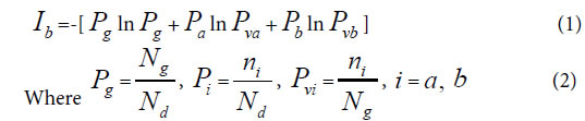
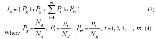
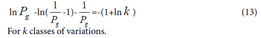
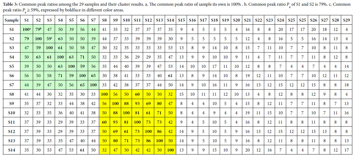
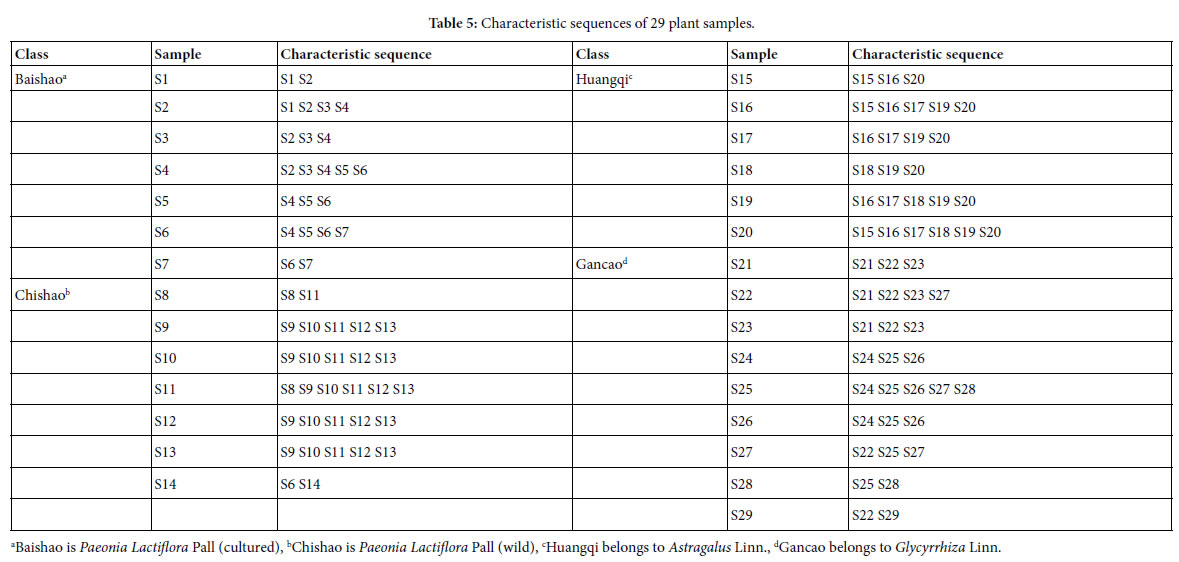
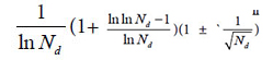
was derived from generally biological heredity and variation information equation. It can be used as a strong tool for determining close relatives.

The laws or rules of material actions are usually more universal and more rigorous than that obtained by statistics. Through actions of substances, which produce differently biological phenomena, the strictly, scientifically and universally theoretical standards for classification should be established. This progress may promote biological science to some extent in the future, and open the door towards quantitative researches on intrinsically scientific principles of biology.

Acknowledgement

I sincerely thank academician WANG yongyan of Chinese Engineering Academy for his moral encouragement and support.

References

  1. Zhao-Hai L, Hong Q, Hong Z (2008) Statistical methods in genetics. Beijing: Science Press.
  2. Zhi-Fa Y (2011) Population genetics, evolution and entropy. Beijing: Science Press.
  3. Gang-biao X (2009) Plant population genetics. Beijing: Science Press.
  4. Mendel GJ (2012) Selected works of genetics. Beijing: Peking University Press.
  5. Robert P, John C DeFries, Gerald E McClean, Peter McGuffin (2008) Behavioral Genetics (Firth edition). Shanghai: East China Normal University Press.
  6. Francisco JA, Kiger JJ Jr (1987) Modern Genetics. Changsha: Hunan Science & Technology Press.
  7. Zhi-fa YUAN, Zhi-jie CHANG, Man-cai GUO, Shi-duo SUN (2015) Genetic analysis of quantitative traits. Beijing: Science Press.
  8. Clark MS (2007) Comparative genomics. Beijing: Science Press 110-136.
  9. Thomas HM (2007) The theory of the gene. Beijing: Peking University Press.
  10. Michael RB (2009) Bioinfomatics for geneticists: A bioinformatics primer for the analysis of genetic data. Beijing: Science Press.
  11. Dale JW, Von Schantz M (2004) From genes to genomes: concepts and applications of DNA technology. Beijing: Science Press.
  12. Campbell M, Heyer JL (2005) Discovering genomics,proteomics & bioinformatics. Beijing: Science Press.
  13. Licinio J, Wang ML (2006) Pharmacogenomics. Beijing: Science Press.
  14. Baldi P, Brunak S (2003) Bioinformatics: the Machine learing approach. Beijing: CITIC Press.
  15. Song-nian HU, Qing-zhong XUE (2004) Handbook of genomic data analysis. Hangzhou: Zhejiang University Press.
  16. Yang ZHONG, Liang Z, Qiong ZHAO (2001) An introduction to bioinformatics. Beijing: Higher Education Press.
  17. David WM (2006) Bioinformatics: sequence and genome analysis (second edition). Beijing: Science Press.
  18. Pevsner J (2006) Bioinformatics and functional genomics. Beijing: Chemical Industry Press.
  19. Jones NC, Pevzner PA (2007) An introduction to bioinformatics algorthms. Beijing: Chemical Industry press.
  20. David WM (2003) Bioinformatics: sequence and genome analysis. Beijing: Higher education press.
  21. Xian -yong WANG, Zheng-hua WANG (2011) An introduction to bioinformatics- the algorithms and applications facing to high performance computing. Beijing: Tsinghua University press.
  22. Yang Z (2008) Computational molecular evolution. Shanghai: Fudan University Press.
  23. Setubol J, Meidannis J (2004) Introduction to computational molecular biology. Beijing: Science Press.
  24. De-shuang HUANG (2009) The data mining methods of gene expression profile.Beijing: Science Press.
  25. Jun WANG, Li-juan CONG, Hong-kun Z (2008) Commomly used biological data analytical software. Beijing: Science Press.
  26. Nelson P (2016) Biological physics: energy, information, life. Shanghai: Shanghai Science & Technical Publishers.
  27. Rob Phillips, Jane Kondev, Julie Theriot (2012) Physical biology of the cell. Beijing: Science press.
  28. Park S Nobel (2010) Physicochemical and Environmental plant physiology (fourth edition). Beijing: Science Press.
  29. Plonsey R, Barr RC (1992) Bioelectricity- A Quantitative approach. Shanghai: Fudan University Press.
  30. Hua-bin Z (2009) Dual index information markedly similar sequence clustering analysis on IR fingerprint spectra of extracts of Guifu Dihuang and Jingui Shenqi pills with ethanol. China Journal of Chinese Materia Medica 34: 2325-2330. [crossref]
  31. Huabin Z (2015) A systematically theoretical distinguish approach for traditional Chinese medicine with identical quality. WorldChinese Medicine 10: 1078-1082.
  32. Huabin Z (2017) Two Chinese medicine species constants and the accurate identification of Chinese medicines. bioRxiv.
  33. Huabin Z (2018) Two biological constants for accurate classification and evolution pattern analysis of Subgen.strobus and subgen. Pinus. bioRxiv.
  34. Huabin Z (2018) A theoretical approach for discriminating accurately intrinsic pattern of biological systems and recognizing three kind soybean proteomes. bioRxiv.
  35. Huabin Z (2018) Accurate identification of varieties constants and multiple pairs between 2 Chinese medicine species. WorldChinese Medicine 13: 3158-3165.
  36. Huabin Z, Yuan J, Aiqin D, Linlin S (2005) Dual-index sequence analytical method for IR fingerprint spectra of the chloroform extract of Radix Glycyrrhizae.China. Journal of Chinese Materia Medica 30: 16-20. [crossref]
  37. Hua‐Bin Z, Yuan J, Ai‐qin D, Lin‐lin S, Hassan Y (2005) Dual-Index Sequence Analytical Method for IR Fingerprint Spectra of Ethanolic Extract of Various Gylcyrrhizae’s Root Species components. Analytical Letters 38: 1167-1178.
  38. De-Xin K, Shu-shi H, Rong-shao H, Xiu-li C, Yi-Bing W et al. (2010) Comparative study on the infrared fingerprint of Abrus Cantoniensis based on the methoods of sequential analysis of Dual-index and cluster analysis. Spectroscopy and Spectral Analysis 30: 45-49. [crossref]
  39. Hong L, Chang-ri H, Hong-xia L, Yuan-feng L, Meng-xiong HE (2008) Study on IR Fingerprint Spectra of Alpinia Oxyphylla Miq. Spectroscopy and Spectral Analysis 28: 2557-2560. [crossref]
  40. Hua-bin ZOU, Zhi-feng H, Hong ZHAI, Ying-qin WEI, Fei WANG (2007) The First and Second Cluster Analysis of Dual Index Grade Sequence of IR Fingerprint Spectra of Guifudihuang Pill and Jinkuishenqi Pill Samples and Their Quality Evaluation. Spectroscopy and Spectral Analysis 27: 2432-2436. [crossref]
  41. Hua-bin ZOU, Xin-1ing Z, Hong ZHAI, Ai-qin DU (2008) Dual index grade sequence pattern recognition of extracts with ethanol of Mingmu Dihuang pills and Zhibai Dihuang pills. ChinaJournalofChineseMateriaMedica33: 1543-1549. [crossref]
  42. Gurcharan Singh (2008) Plant systematics: An integrated approach. Beijing: Chemical Industry Press.
  43. Chang-Fa ZHOU (2009) Biological evolution and taxonomical principle. Beijing: Science Press.
  44. Han-Chen Z, Hao Z, Sheng-Li PAN (2009) Pharmaceutical botany (fifths edition).Beijing: People’s Medical Publishing House.
  45. Jin-wu WANG (2011) Spermatophyte taxonomy (second edition). Beijing: Higher Education Press.
  46. Simpson MG (2012) Plant systmatics (second edition). Beijing: Science Press.
  47. Hua-bin ZOU, Jiu-rong YUAN, Ai-qin D, Lin-lin S (2005) Dual-index sequence analytical method for IR fingerprint spectra of the chloroform extract of Radix Glycyrrhizae. China Journal of Chinese Materia Medica 30: 16-20. [crossref]
  48. Hua‐Bin Zou, Guo‐Sheng Yang, Zheng‐Ran Qin, Wen‐Qiang Jiang, Ai‐Qin Du et al. (2005) Progress in quality control of herbal medicine with IR fingerprint spectra. Analytical Letters 38: 1457-1475.
  49. Tao-tao PANG, Li-ming DU (2007) Common and Variant Peak Ratios in IR Fingerprint of Ilex Kudingcha with Duel-Index Sequence Analysis. Spectroscopy and Spectral Analysis 27: 486-489. [crossref]
  50. Hua-bin Zou, Jiu-rong Y, Aiqin D, Lin-lin S, Hassan YA (2005) Dual-Index Sequence Analytical Method for IR Fingerprint Spectra of Ethanolic Extract of Various Gylcyrrhizae’s Root Species components. Analytical Letters 38: 1167-1178.
  51. Kevin Yi-LY, Yung CS, Lim CS (2007) Infrared-based protocol for the identification and categorization of ginseng and its products. Food Research International 40: 643- 652.
  52. Hong-xia L, Su-qin S, Guang-hua LV, Kelvin KC Chan (2006) Study on Angelica and its different extracts by Fourier transform infrared spectroscopy and two-dimensional correlation IR spectroscopy. Spectrochimica Acta Part A 64: 321-326. [crossref]
  53. Huabin Zou, Jiurong Y, Wang Wei (2004) Theoretical identification of common peaks in fingerprint of Chinese medicine -a W- testing and discriminatory method. World Science and Technology/Modernization of Traditional Chinese Medicine and MateriaMedica 6: 50-56.
  54. Chinese Pharmacopoeia Commission (2005) Chinese pharmacopoeia 2005 edition (I)[S], Beijing: Chemical Industry Press.

Can One Teach Old Drugs New Tricks? Reformulating to Repurpose Chloroquine and Hydroxychloroquine

Abstract

The outbreak of the novel corona virus disease, COVID-19, has presented health care professionals with the unique challenges of trying to select appropriate pharmacological treatments with little time available for drug testing. Given the development times and manufacturing requirements for new products, Value Added Medicines (repurposing – reformulation of existing drugs) could be one possibility to beat the COVID-19 outbreak. This review explores reformulation alternatives which could be progressed with chloroquine and hydroxychloroquine; two antimalarial drugs, that are being tested on a global scale as a potential therapeutic option. The key areas for improvement have been reviewed and the potential solutions to the problems and limitations of current formulations are discussed. The pharmaceutical challenges discussed are those of highly soluble drugs, needed to be given at high doses and presenting a real bitter taste challenge with significant gastrointestinal side effects that could be translated and repurposed into fit for purpose reformulations.

Introduction

COVID-19 is the infectious disease caused by the most recently discovered corona virus. This newly emerged virus and disease were unknown before the outbreak began in Wuhan, China, in December 2019. COVID-19 is now a pandemic affecting many countries globally and to date no antiviral or therapeutic has been approved for treating patients. As the number of cases continues to rise, the geographic range of the virus increases, and with the development of a vaccine being at least 12 months away, there is a growing urgency/pressure on pharmaceutical industry and regulatory agencies to expedite the development and approval of both experimental drugs and repurposing of existing therapeutics that have been already approved for human use by the health agencies. Among the landscape of therapeutics being analysed as potential repurposing candidates for COVID-19, the antimalarial and immunomodulatory drugs chloroquine (CQ) and hydroxychloroquine (HCQ), both 4-aminoquinolines, are being tested on a global scale as a potential treatment and prevention of COVID-19 [1]. Recent publications have drawn attention to the possible benefit of CQ sulphate and phosphate salts (CQ diphosphate) and HCQ for the treatment of SARS-CoV-2 infected patients [2-7]. CQ phosphate or sulphate is referenced on the World Health Organisation (WHO) Model List of Essential Medicines for the treatment of Plasmodium vivax infection (malaria) [8, 9]. In addition to their antimalarial use, both CQ and HCQ are used in continuous daily dosing for rheumatoid arthritis, systemic and discoid lupus erythematosus and psoriatic arthritis. CQ and HCQ are one of four potential treatments that WHO has included in the global SOLIDARITY Clinical Trial in 90 countries to generate the robust data needed to establish efficacy and safety in COVID- 19 treatments [10]. There are several other trials ongoing in different countries [11, 12] to name a few, a UK wide randomized, controlled trial in over 130 hospitals called Randomised Evaluation of COVID Therapy (RECOVERY) [13] is underway and in Europe its Trial of Treatments for COVID-19 in Hospitalized Adults (DisCoVeRy) [14]. In US, NYU and University of Washington has fast trackeda major clinical trial to determine role of HCQ in prevention of corona virus [15]. Both CQ and HCQ are primarily available as tablets for oral administration [16-18] and have been in clinical use for decades thus their safety profile is well established [19]. However, this oral formulation presents the following problems: swallow ability difficulties for certain patients groups such as the young, older people and patients in critical care, as well as extremely poor palatability due to the bitter taste of the drugs [20, 21]. In addition, the oral administration of CQ and HCQ frequently causes gastrointestinal side (GI) effects such as nausea and vomiting [22-24]. HCQ being reported to have better safety profile than CQ, better gastrointestinal tolerability, and less retinal toxicity [25]. This review sets the scene and explores promising reformulation alternatives which could be progressed with CQ and HCQ, for adults and children. Alternatives routes of administration are also explored to address oral administration challenges. The problems and limitations with existing formulations are discussed and the key areas for improvement are reviewed. It is important to note while CQ and HCQ are under investigation in clinical trials for use on COVID-19 patients, as of the date of this publication, none of these compounds and medications have been approved for the treatment of COVID-19. Considering it is a rapidly changing area with new conflicting outcomes coming up every day, the repurposing of CQ and HCQ for COVID-19 is still questionable as only limited evidence is available at the present time. However, this review will be useful in various scenarios 1) if the trials are successful and the use of CQ in HCQ for COVID -19 is recommended 2) if no good evidence on use of CQ and HCQ for COVID-19 is generated in time, the reformulation strategies proposed in this review will be still relevant for antimalarial and rheumatic disorders treatment 3) the approaches discussed could be translated to other Active Pharmaceuticals Ingredients (APIs) presenting similar pharmaceutical challenges.

Challenges with existing CQ and HCQ formulation for COVID-19 treatment

CQ and HCQ are both Biopharmaceutics Classification System Class 1 compound [26] and extremely bitter [20, 21]. One study found the threshold bitterness of the pure CQ to be at 40 μg/ml [27]. After oral administration both are rapidly and almost completely absorbed from the gastrointestinal tract. They have a long and variable plasma elimination half-life because of a high volume of distribution with about half the drug metabolites undergoing unmodified renal clearance. CQ has a low safety margin and is very dangerous in overdose situations or when combined with other medicines [28]. There is no firm evidence on the optimal dosing and duration treatment for CQ or HCQ, hence the range of regimens are used across trials. In general, the regimen of CQ and HCQ used is substantially more aggressive than that recommended as an antimalarial [29]. For instance, National Health Commission of the People’s Republic of China recommended dose of 500mg twice daily for seven days for oral administration in 18–65 years of infected adults [30]. In the RECOVERY Trial, the loading dose of HCQ (1860mg) is twice the normal dose for treating malaria. However, this dose has been selected based on the available data of the IC50for SARS-CoV-2 [13] Several dosing regimens are proposed based on PBPK simulation combined with known clinical exposure–response relationships [3, 31-33]. Based on PBPK model, the typical dose for HCQ for treating COVID-19 is 400 mg twice daily on the first day, followed by 200 mg twice daily for four more days [31, 33]. Although HCQ shows better safety and toxicity profiles than CQ, symptomatic effects at these high doses have not been explored in enough depth. The question remains, how will gastro-intestinal symptoms prevail, will the benefits outweigh the risks and symptomatic effects; especially for critically ill COVID-19 patients in ICU. A wide range of data from various patients groups needs to be gathered to reliably surmise this. Both CQ and HCQ are metabolised in the liver with renal excretion of some metabolites, hence should be prescribed with care in people with liver or renal failure [34-36]. Recently published Surviving Sepsis Campaign guidelines [37] on the management of critically ill patients with COVID-19 concluded that there was insufficient evidence to offer any recommendation on the routine use of these drugs in patients admitted to the intensive care unit (ICU).The ongoing trials will be able to answer whether antimalarial drugs could be effective in changing the disease course in patients with severe COVID-19—in particular, in cases requiring ICU admission. At present, the safety of CQ in the treatment of elderly patients with COVID‐19 is unclear. The rate of critical illness in this population is high and CQ could still be used as an alternative drug, although it should not be given to those elderly patients with underlying heart and other conditions [38]. Based on the published clinical guidelines and research results, Sun et.al (38) have proposed the pharmaceutical care for the elderly using CQ phosphate in the treatment of COVID‐19. This includes the administration method, dosage of CQ phosphate for elderly, adverse drug reactions and drug interactions of CQ phosphate. For elderly patients with a bodyweight of more than 50 kg, CQ phosphate 500 mg orally, bid, for 7 days is recommended. CQ and HCQ is licensed for use in children with malaria. The WHO recommended HCQ dose to treat COVID-19 infected children is 25mg/kg given over 3 days. However, this may not be optimal to treat COVID-19, as recent studies show that older infants and children may need a higher mg/kg dose to reach the similar concentrations as adults. In contrasts, it is likely that neonates and young infants will need lower doses per kg body weight. Paediatric CQ dose for COVID-19was determined by Verscheijden et.al. [39]. The study proposestotal cumulative doses: 35 mg/kg (CHQ base) for children 0-1month, 47 mg/kg for 1-6 months, 55 mg/kg for 6 months-12 years and 44 mg/kg for adolescents and adults, not to exceed 3300 mg in any patient. Currently, there is a CQ phosphate syrup [40] on the market, while no pediatric or easy to swallow formulations exist for HCQ. However, as HCQ is highly soluble compound, it is expected that manipulation of the formulation will have minimal impact on bioavailability. The European Paediatric Formulary (PaedF) Working Party at the European Directorate of the Quality of Medicines and Healthcare (EDQM) has compiled existing knowledge on pediatric formulations for active substances which are under investigation for the treatment of COVID-19 as well as known authorised medicinal products [41]. This includes the information on extemporaneous preparations of CQ and HCQ which may be suitable for treatment of pediatric patients with COVID-19. It suggests the preparation of a pediatric suspension formulation from a 200mg tablet. This instructs pharmacists to ‘strips’ the outer film coating, crush the tablet(s), and then suspend the powder in water with a flavouring agent such as Ora-plus®. In the USA, The Nationwide Children’s pharmacy [42] and Michigan Collaborative Standardization of Compounded Oral Liquids [43] have formerly investigated utilising a crushed standard 200mg tablet of HCQ in Ora-plus® to form an oral liquid suspension. The resultant suspension concentration is 25mg/ml (i.e. 800mg (32ml) of suspension). A liquid dosage form of HCQ would be applicable to both the younger and older generations to address both patient dysphagia and compliance respectively. However, Ansah EK et al. reported improved compliance in children in rural Africa who were treated with CQ tablets or segments (crushed and mixed with sugar or honey) rather than CQ syrup [44] Moreover, the preparation of this extemporaneous suspension, especially stripping or/and crushing process is cumbersome. The suspension media such as Ora-plus® is also costly and may not be easily available in some regions, and the process may result in a loss of active pharmaceutical ingredient. Batches could be prepared on a demand basis for these patient groups but it is time consuming to prepare large quantities of suspension that have a shelf life of only 30 days at 2-8°C protected from light. Besides, the same GI side effects would likely still be present with additional issues of the drug’s bitter taste [45, 46] which is unlikely to be concealed. Thus, for this suspension to obligate its benefits, taste masking seems essential [47].

Current approaches for taste masking of CQ and HCQ

Sensory based taste masking approaches in which sweeteners and flavours are added to obscure taste, have been commonly used for decades, but this approach does not work well for highly soluble, highly aversive APIs and/or for APIs with an intense lingering aftertaste, as any compounds dissolved in the saliva will interact with the taste receptors and elicit a response [48]. Alternatively, taste masking techniques are utilised to improve the palatability of formulations. The different complexation approaches that have been used to taste mask by creating ‘molecular’ barrier around the API CQ and HCQ were scoped as part of this review and are discussed below.

Ion pairing

Ion-pairing is a process that involves stoichiometric replacement of polar counter-ions (i.e., chloride, acetate, nitrate, etc.) in the drug with an ionic excipient of similar charge. Ion-pairing has been used in the pharmaceutical industry, mainly as an additional drug-delivery method, and has proven to be a very effective mean for controlled drug release and taste masking [49]. Pauli et al [47] created a prototype formulation using a Coni-Snap Sprinkle Capsule containing sodium carboxymethyl cellulose (Na-CMC) as the ion-pairing agent to investigate if the addition of ion-pairing excipients and the incorporation of a buffered system into the conventional tablet could overcome some of the bitter-taste issues of HCQ. They hypothesised that this would enable a dual use formulation: adults can swallow the capsule whole whereas children (or indeed any patients with swallowing difficulties) could be administered the capsule content in water. They compared the dissolution profile of this capsule to another prototype formulation in the same capsule but without any ion-pairing agent and concluded that both profiles were comparable to that of a commercially available tablet of HCQ. This a bridged research also presented how the ion-paring system as provided by Na-CMC, and by another ion-pairing agent; sodium citrate, buffered to pH 8, taste-masked the drug in vitro with an Astreeel ectronic (e-tongue) assay. However, no in vivo assessment has been made. Moreover, the concentrations tested with the e-tongue were not reflective of the clinical situation for adults with COVID-19 as it was to treat children with uncomplicated malaria, lupus erythematosus, and rheumatoid arthritis at much lower doses. This integrated system could still have promise with higher drug loads, but it is likely the ion-pairing agents would need to be present in much higher concentrations in order to match the 1:1 ratio used in the study. Independent of this, buffering the system alone to pH 8 seems to have a significant effect on taste-intensity so this could be a quicker avenue to explore, given the current urgent need. It would likely be time-consuming to re-test alternative amounts of ion-pairing agents and then carry out further compatibility testing alongside the other components of the formulation.

Ion – exchange resins

Taste masking by drug–resin complexation is achieved when an ionizable drug reacts with a suitable ion exchange resin to form a drug–resinate complex [48]. Ion exchange resins (IERs) are insoluble, pharmacologically inert, high molecular weight cross linked polymers with cationic and anionic functional groups. They bind to compounds that exchange mobile ions and ultimately form of a tasteless drug- resin complex or resinate [50]. Drugs are attached to the oppositely charged resin substrate, forming insoluble adsorbates or resonates through weak ionic bonding. The resinate needs to be stable in the drug formulation e.g. a suspension or a tablet formulation and the dissociation of the drug–resin complex should not occur under the salivary pH conditions (pH 6-7). However, at enteric pH conditions (pH<5), the drug should be rapidly and almost entirely released in order to prevent reduced bioavailability. This can suitably mask the unpleasant taste and odour of drugs [51]. Characterization of drug–resin complexes and taste masking of CQ phosphate by complexation using weak cation exchange resin have been described in the literature [27, 50, 52]. All studies showed some in vivo taste improvement. No taste masking study of HCQ using ion exchange resins was identified.

Simple and Supra molecular Complexation

The host-guest complexation is a common taste masking technology [53]. By embedding the drug molecule (guest) into the cavity of a host molecule, a stable complex is generated. Generally, there are 2 mechanisms for explaining the taste-masking effect of complexation. The first mechanism is that the complexing agent will hinder interactions between drug molecules and taste cells through forming the strong binding with drugs [54]. Secondly, the complexing agent may directly bind to taste cells to mask the unpleasant. Among all the complexing agents, cyclodextrin (CD) group is a typical example [53]. Derived from the starch, CDs are cyclic polymers composed of glucopyranoside units (n) that are linked by α-1,4-glycosidic bonds. With their doughnut-shaped structures, CDs are capable of fitting lipophilic drugs or lipophilic moieties of drugs inside their hydrophobic central cavities (Figure 1). The natural and well-known CDs are α- (n=6), β- (n=7), and γ- (n=8)CD, respectively (Figure 1).

JPPR-3-1-309-g001

Figure 1. Cyclodextrin structure and representation of an inclusion complex of a drug residing in the cavity formed by the cyclodextrins

There is limited literature regarding the complexation between CDs and HCQ (or CQ). Guo, Wu et al [55] built a 3D model to predict the taste masking effect of CDs in different drug-CD complexes, where the Euclidean distance using an α-Astree e-Tongue was adopted to quantitate a taste masking effect (100 being the smallest euclidean distance to show taste masking efficiency). The study concluded that taste masking of HCQ was not achieved as the Euclidian distance was only 91 and proposed that CDs were ineffective because the halogen group and the chlorobenzene of the4-aminoquinolines significantly increased their molecular size and hampered the complete encapsulation of the drugs inside CDs cavities or that the alkylamino side chain is not part of the inclusion complex, allowing the tertiary amine to participate in bitter taste. Therefore, to date, no published paper indicated that natural CDs were effective in masking the bitter taste of HCQ or CQ. Woertz et al (56)conducted an INSENT e-tongue experiment to investigate taste masking effects of CDs (α-CD, β-CD, γ-CD, hydroxypropyl-β-CD, maltodextrin as well as sulfobutyl ether-β-CD (SBE-β-CD) on quinine. All CDs failed to mask the bitter taste of quinine, except for the modified CD, SBE-β-CD. It was thought that the aliphatic ring of quinine was embedded inside the CD cavity, whereas the quinoline ring of quinine was left outside the cavity, generating an incomplete inclusion complex with CDs, thereby the quinoline part could still interact with taste receptors and cause the bad taste. The study concluded that SBE-β-CD was able to mask bitter taste of quinine because of the ionic interaction between its –SO 3 2- group and the deformed –NH 3 + group from the quinine [56] (Figure 2).

JPPR-3-1-309-g002

Figure 2. Chemical structures of Quinine, Chloroquine and Hydroxychloroquine.

Since the chemical structure of quinine is similar to CQ and HCQ (Figure 2), the potential of SBE-β-CD for taste masking offer a masking option. However, according to Ghateet al [57] the native SBE-β-CD is salty, which may impede it use for taste masking. Captisol® is an FDA approved SBE-β-CD. This enabling technology available to research and development through flexible licensing arrangements is claimed to be tastelessness in oral formulas. It is and has been used in drug products on the market [58, 59]. Remarkably, Joneset al [60] recently developed a novel CD, modified with mercaptoundecane sulfonic acids. This highly sulfonated CD could mimic heparan sulphates (HS) and kill most HS-dependent viruses, such as herpes simplex virus, respiratory syncytial virus, dengue virus, and Zika virus. The study indicated that the newly modified CD had a potential to act as a broad-spectrum antiviral agent. Although corona viruses were not tested, their discovery reveals a different choice, where the modified CD could mask bitter taste, and at the same time, kill viruses. It is to be noted that this is probably the furthest away from translation as this would require extensive studies to support the claim. Apart from CDs, the cucurbituril (CB) family is another emerging complexing strategy and believed to be promising and attractive for pharmaceutical development. CBs ares upramolecular host molecules or macrocycles consisting of 5 or more glycoluril units joint together by methylene linkages [61]. CBs can also accommodate lipophilic drug moieties inside their hydrophobic cavities [62]. Currently, there is no literature about the HCQ-CB complexation or the CQ-CB complexation. However, it was confirmed by Boraste, Chakraborty et al [63] that CB7, which connects 7glycoluril units, was able to form a stable and a complete inclusion complex with quinine at a ratio of 2:1 (CB7 to quinine). Compared with CDs, CB7 was demonstrated to not only encapsulate the small aliphatic ring of quinine, but also fit the large quinoline ring into its cavity. This process was accomplished by binding the aliphatic ring of quinine to one CB7, while the quinoline moiety of quinine entered into a second CB7 cavity, at a lower pH. Accordingly, it is hypothesized that it could be possible for CQ and HCQ to form complete inclusion complexes with CBs. In this way, interactions between bitter drugs and taste receptors could be inhibited and taste masking achieved. However, before they are ready to be used in practice or tested in humans, there are number of issues that needs to be addressed including safety (Figure 3).

JPPR-3-1-309-g003

Figure 3. Molecular structure of CB7

Crystal engineering strategies

Pharmaceutical cocrystals are molecular crystals that are multi-component crystalline substances, where one of the components is an API and the other component/components are crystalline substances that have been approved by the regulatory bodies within the jurisdiction of commercial use; these species are known as the cocrystal former or the conformer [64]. Co-crystallization is one of the emerging crystal engineering techniques for modulating pharmaceutical performance through controlling solid-state properties of APIs and expanding the access to new solid forms differing in structures. The approach relies on the self-assembly of a bitter-tasting drug and a taste-masking agent, whereby the molecules are held together by non-covalent interactions including hydrogen and halogen bonds. Co-crystallization can modify different physicochemical properties of the APIs, without any change in their activity, such as improving the solubility of poorly soluble drugs, masking the bitter taste, increasing chemical stability and decreasing hygroscopicity, enhancing manufacturability as well dissolution rate and bioavailability [65]. Investigations into the solid-state landscape of CQ and HCQ have demonstrated the advantages of complexation within the crystal lattice. They have distinct acid base properties and hydrogen bond abilities that makes them suitable to form salts and co-crystals. The only crystal form of HCQ currently on the market is in the form of the sulphate salt [66]. Whereas CQ is on the market as the stand-alone API or as the phosphate, sulphate or hydrochloride salt [67, 68]. One study demonstrated how improvements can be made to these solids using crystal engineering to enhance the physicochemical properties and pharmacological activity of these co-crystallised compounds over parent compound. CQ and HCQ are both part of the quinolone family and are closely related to quinolone. Baruah et al [69] showed that salts and cocrystal of quinoline with hydroxyaromatic carboxylic acids enhance antimalarial activities over parent compounds. However, there is no literature to support the application of crystal-engineering approach to taste masking neither of CQ or HCQ. Research into the preparation of multicomponent crystals involving other APIs containing quinoline moieties suggest that this group of compounds could either partake in salt formation or cocrystal formation to improve API taste depending on the type of cocrystal or salt former used [70-72].

Alternative routes of administration to address formulation challenges

Rectal drug delivery

To subsequently tackle at the same time the bitter taste issue and high dose administration whilst still being applicable to all patient groups with possible comorbidities/polypharmacy, the rectal route may be the most appropriate alternative administration route. Symptomatic relief would also be achieved and allow critically ill/unconscious patients in ICU to be treated equally. Suppositories are often used as drug delivery system in case of nausea or vomiting, in case of oral administration rejection due to the bad taste or in case a medication is readily decomposed in gastric fluid. Published studies showed that CQ given in suppositories with the same dose as oral formulations reached lower blood concentrations and it was slower to produce the same antimalarial effects than when administered orally [73-76]. Tjoenget al [77] performed comparative bioavailability studies of rectal and oral formulations of CQ in healthy volunteers. The study demonstrated that the relative bioavailability of CQ 500 mg suppositories varied between 10-53% compared to a tablet formulation in adults. Onyeji at al [75] demonstrated that the bioavailability of chloroquine 100 mg suppositories was 63.4 +/- 8.8% (mean +/- SEM) relative to the tablet formulation. Bruce-Chwatt et al [73] observed that only when the same dose of CQ 300mg tablets was administered rectally over a 5 days period, it was able to reach the same parasite clearance of the oral dose but more slowly. The study concluded that CQ given by mouth was better and faster absorbed compared to the rectal route. However, in all these studies, no consideration was drawn on formulation improvements that could enhance the rectal release and adsorption of CQ from the suppository. Suppository are made of relatively low-cost excipients but their manufacture can be more challenging than other common dosage forms (tablets, liquids): they may need temperature-controlled storage depending on melting point and humidity control may be required during manufacture [78]. The nature of the base and the surfactant content in the suppository composition need to be carefully chosen to obtain the optimal mechanical and drug release properties of CQ [79]. Redgon et al [80] assessed different lipophilic and hydrophilic bases for CQ phosphate. They found that the hard-fat base Witepsol H15 was the best in terms of disintegration times, a with good storage conditions, was also suitable for countries with a continental climate. Onyeji et al [81] on the other hand, studied the effect of absorption-enhancing agents, non-ionic surfactants and sodium salicylate, on the in vitro release characteristics of CQ from polyethylene glycol (1000:4000, 75:25%, w/w) suppositories. The study concluded that the incorporation of 4% Tween 20 or 25% sodium salicylate improved the invitro release of CQ from the suppository. Considering these adjuvants also have absorption-promoting properties, association of the improved in-vitro release with enhanced in vivo availability is envisaged but would require a PK study for confirmation. Thus, results of this study serve as a guide in the selection of an optimal formula regarding the type and concentrations of absorption enhancers required for optimization of CQ release and a possible enhancement of rectal absorption of the drug. Finally, Okubanjoet al [79] studied the effects of interacting variables on the mechanical and release properties of CQ phosphate suppositories. A23 factorial experiment was designed to study the effects of the type and nature of the base, the concentration of surfactant and storage conditions. The study demonstrated that the presence and concentration of surfactant was the main individual variable affecting the release properties of suppository formulations. The addition of surfactants increased the crushing strength and decreased the dissolution times of CQ suppositories. Also, the type of suppository base played a role in the modification of the mechanical and release properties. Witepsol H15, as previously highlighted by Redgon et al [80] was better than Suppocire AS2 in increasing the crushing strength and dissolution rates and decreasing the dissolution times while storage conditions had the lowest effect. In the past, the rectal administration of CQ, but not HCQ, was explored for the treatment of malaria. Evidence tends to suggest that the use of rectal formulations for the administration of CQ could be re-considered as an alternative route to the oral administration to overcome the problems associated with this route. Due to the similarity of the 2 compounds, it is speculated that HCQ could also be a good candidate to be reformulated for rectal administration. It would be interesting to explore more recent advances introduced for the release and adsorption of drugs form rectal suppositories, such as the use recto dispersible dosage forms with non-melting excipients [82] or the use of hollow-type suppositories which have been developed to enhance the adsorption of various drugs [47]. The use of rectal formulations could be particularly useful for the treatment of certain patients’ groups affected by COVID-19, such as those with swallowability difficulties, critically ill patients, unconscious or vomiting patients or pediatric patients from birth. Although speculatively, by this route, there may be the added advantages of attaining the necessary higher plasma concentrations with a lower drug dose (yet high doses of API can be delivered rectally) as it generally avoids first pass metabolism. Finally, it is suitable for APIs that are gastro-irritant and could speculatively again help with some GI side effects. Socio-cultural norms drive recommendations regarding the knowledge, attitude, preference, and behaviour of people. To implement rectal delivery of CQ and HCQ, beside positive pharmacological outcomes, it would be important to consider patients’ real barriers versus cultural, perceived barriers or lack of understanding of the potential of this mode of administration [83]. Two further routes of administration for CQ and HCQ have been recently re-proposed as alternatives to the oral drug delivery.

Pulmonary drug delivery

An aerosolized formulation of HCQ was developed and tested in early phase clinical trials by the American company APT Pharmaceuticals. For the anti-inflammatory effect of HCQ, this formulation was developed for the potential treatment of respiratory diseases such as asthma, chronic obstructive pulmonary disease (COPD), rhinitis and severe acute respiratory syndrome (SARS). The drug was delivered by using an aerosol generating system (AERx®) for pulmonary delivery developed by Aradigm Corporation of Hayward, to maximize drug delivery in a patient-friendly format. By using this targeted delivery system, the company believed that the aerosolized dosage form and the use of the pulmonary route could achieve a faster onset of action, within hours, and greater therapeutic effects than conventional oral therapy at substantially lower systemic doses [84]. However, despite the favourable results showed in a phase 1 clinical study, the subsequent phase 2a data failed on efficacy endpoints and the study was stopped. In light of the potential role that CQ and HCQ might have as potential candidate therapies for COVID-19, Klimke et Al [85] re-proposed the use of them as pulmonary aerosol in a dosage of 2-4 mg of HCQ per inhalation for reduction and prevention of severe symptoms after SARS-CoV-2 infection. They speculated that by administering the drug targeting directly the lung, a lower drug dosage would be required to reach the optimal concentrations, compared to the oral route and avoid the oral side effects. Based on their speculations, the 2 authors decided to inhaled themselves tolerability and safety of 1 mg of HCQ in 2ml sodium chloride 0.9%, b.i.d. increased to 4 mg daily over one week. The dose was deemed well tolerated, with after 4 days still the feeling of a transient bitter taste in the mouth, which lasted 2-3 hours after each dosing. However, no efficacy data are available to validate their hypothesis.

Transdermal Drug Delivery

A study by Musabayane et al [86] investigated the potential application of pectin hydrogel patch as a matrix polymer for transdermal administration of CQ with Dimethyl sulphoxide as a penetration enhancer. They tested on rats the effects of CQ via intravenous infusion and the patch applied on shaved area during the 1 h 20 min. The results (plasma profile) showed good potential for transdermal delivery of CQ. However, the loading efficiency was only 46% of the theoretical 10 g. Moreover, the dose administered (16 µg/kg) was much lower than those previously used in rats (20–25 mg/kg), and in man (300 mg/kg). Glan is Pharmaceuticals Inc. recently obtained the rights for a US provisional patent for a transdermal drug delivery system of HCQ as a potential treatment for COVID-19. They suggest that controlled transdermal delivery could provide constant drug plasma concentrations for pre-determined periods of time, potentially reducing side effects associated to the oral delivery. However, so far, it seems that only literature search and pre-formulation studies have been done in collaboration with Reformulation Research Laboratories Inc. but no related clinical information are available [87]. The development of transdermal formulations [88] of CQ and HCQ with novel strategies would need to be carefully studied in order to address some challenges such as the hydrophilic nature, the need to administer larger doses of drug, and potential skin irritation due to enhancers or other additives added to the transdermal formulation.

Conclusion

COVID-19, officially designated as severe acute respiratory syndrome-related corona virus SARS-CoV-2 currently represents a pandemic threat to global public health. Researchers are leaving no stone unturned in an effort to understand this new emergent disease and uncover existing drugs with therapeutic potential for COVID-19. CQ and HCQ, antimalarial drugs are among the existing drugs being investigated in clinical trials as a possible treatment protocol for COVID-19.The clinical evidence base is currently limited and there is hope that the ongoing clinical trials may unfold the missing evidence if these antimalarial drugs could be effective in changing the disease course in patients with COVID-19. Although it is the elderly and those with underlying health conditions find themselves most gravely affected by COVID-19, the virus does not discriminate by age. All age groups are at risk and more likely to require drugs such as CQ or HCQ in an hospital setting. A formulation which does not rely on swallowing a bitter tablet or suspension and that would suit both ambulatory and critical care settings, would be welcomed. CQ and HCQ, in currently available forms (tablet and syrup) are inundated with challenges in terms bitter taste, high dose etc. This review has outlined different reformulation approaches that could be utilised for taste masking the CQ and HCQ and alternative routes of administration to surpass the problems associated with the oral administration. We hope that teaching these old drugs new tricks may represent an opportunity to address the pharmaceutical challenges and deliver better health to patients with COVID-19 and/or even for antimalarial treatment.

References

  1. WHO. Landscape analysis of therapeutics. 2020.
  2. Gao J, Tian Z, Yang X (2020) Breakthrough: Chloroquine phosphate has shown apparent efficacy in treatment of COVID-19 associated pneumonia in clinical studies. Bioscience trends 14: 73-74. [crossref]
  3. Gautret P, Lagier J-C, Parola P, Meddeb L, et al. (2020) Hydroxychloroquine and azithromycin as a treatment of COVID-19: results of an open-label non-randomized clinical trial. International journal of antimicrobial agents. [crossref]
  4. Schlagenhauf P, Grobusch MP, Maier JD, Gautret P (2020) Repurposing antimalarials and other drugs for COVID-19. Travel Med Infect Dis. [crossref]
  5. Keyaerts E, Vijgen L, Maes P, Neyts J, Van Ranst M (2004) In vitro inhibition of severe acute respiratory syndrome coronavirus by chloroquine. Biochemical and biophysical research communications 323: 264-268. [crossref]
  6. Vincent MJ, Bergeron E, Benjannet S, Erickson BR, Rollin PE, et al. (2005) Chloroquine is a potent inhibitor of SARS coronavirus infection and spread. Virology Journal 2. [crossref]
  7. Wang M, Cao R, Zhang L, Yang X, Liu J, et al. (2020) Remdesivir and chloroquine effectively inhibit the recently emerged novel coronavirus (2019-nCoV) in vitro. Cell research. 30: 269-271. [crossref]
  8. WHO (2015) 19th WHO Model List of Essential Medicines.
  9. WHO (2020) Medical Product Alert N°4/2020.
  10. WHO (2020) “Solidarity” clinical trial for COVID-19 treatments.
  11. Overview of planned or ongoing studies of drugs for the treatment of COVID-19. 2020.
  12. ClinicalTrials.gov. (2020) OVID-19 Studies from the World Health Organization Database.
  13. Recovery Trial (2020) Randomised Evaluation of Covid-19 Therapy.
  14. Institut National de la Santé Et de la Recherche Médicale (2020) Trial of Treatments for COVID-19 in Hospitalized Adults (DisCoVeRy).
  15. ClinicalTrials.gov. (2020) Federally-funded clinical studies related to COVID-19.
  16. Agency EM (2016) List of nationally authorised medicinal products. London.
  17. BNF (2020) Chloroquine | Drug | BNF content published by NICE: NICE.
  18. BNF (2020) Hydroxychloroquine Sulfate | Drug | BNF content published by NICE: NICE.
  19. Devaux CA, Rolain J-M, Colson P, Raoult D (2020) New insights on the antiviral effects of chloroquine against coronavirus: what to expect for COVID-19?. International journal of antimicrobial agents. [crossref]
  20. Wallace DJ, Tse K, Hanrahan L, Davies R, Petri MA (2019) Hydroxychloroquine usage in US patients, their experiences of tolerability and adherence, and implications for treatment: survey results from 3127 patients with SLE conducted by the Lupus Foundation of America. Lupus Sci Med. [crossref]
  21. Weber JC, Alt M, Blaison G, Welsch M, Martin T, et al. (1996) [Changes in taste and smell caused by hydroxychloroquine]. Presse Med 25. [crossref]
  22. Braga CB, Martins AC, Cayotopa AD, Klein WW, Schlosser AR, et al. (2015) Side Effects of Chloroquine and Primaquine and Symptom Reduction in Malaria Endemic Area (Mancio Lima, Acre, Brazil). Interdiscip Perspect Infect Dis. [crossref]
  23. Deen JL, von Seidlein L, Dondorp A (2008) Therapy of uncomplicated malaria in children: a review of treatment principles, essential drugs and current recommendations. Trop Med Int Health. 13: 1111-1130. [crossref]
  24. Srinivasa A, Tosounidou S, Gordon C (2017) Increased incidence of gastrointestinal side effects in patients taking hydroxychloroquine: a brand-related issue?. The Journal of rheumatology 44. [crossref]
  25. Mc Chesney EW (1983) Animal toxicity and pharmacokinetics of hydroxychloroquine sulfate. Am J Med 75: 11-18. [crossref]
  26. Ploger GF, Hofsass MA, Dressman JB (2018) Solubility Determination of Active Pharmaceutical Ingredients Which Have Been Recently Added to the List of Essential Medicines in the Context of the Biopharmaceutics Classification System-Biowaiver. J Pharm Sci 107: 1478-1488. [crossref]
  27. Sharma T, MoonRajkumar S (2012) Formulation and Evaluation of Taste Masked Dispersible Tablets of Chloroquine Phosphate. International Research Journal of Pharmacy 3.
  28. European Medicines Agency (EMA) (2020) COVID-19: chloroquine and hydroxychloroquine only to be used in clinical trials or emergency use programmes.
  29. Wong YK, Yang J, He Y (2020) Caution and clarity required in the use of chloroquine for COVID-19. The Lancet Rheumatology 2.
  30. National Health Commission of the People’s Republic of China Interpretation of COVID-19 treatment guidelines (6th version). 2020.
  31. Cui C, Zhang M, Yao X, Tu S, Hou Z, et al. (2020) Dose selection of chloroquine phosphate for treatment of COVID-19 based on a physiologically based pharmacokinetic model. Acta Pharmaceutica Sinica B.
  32. Kapoor KM, Kapoor A (2020) Role of Chloroquine and Hydroxychloroquine in the Treatment of COVID-19 Infection- A Systematic Literature Review. medRxiv.
  33. Yao X, Ye F, Zhang M, Cui C, Huang B, et al. (2020) In Vitro Antiviral Activity and Projection of Optimized Dosing Design of Hydroxychloroquine for the Treatment of Severe Acute Respiratory Syndrome Coronavirus 2 (SARS-CoV-2). Clin Infect Dis. [crossref]
  34. Rismanbaf A, Zarei S (2020) Liver and Kidney Injuries in COVID-19 and Their Effects on Drug Therapy; a Letter to Editor. Archives of Academic Emergency Medicine 8. [crossref]
  35. Taccone FS, Gorham J, Vincent J-L (2020) Hydroxychloroquine in the management of critically ill patients with COVID-19: the need for an evidence base. The Lancet Respiratory Medicine. [crossref]
  36. Wang Y, Zhu LQ (2020) Pharmaceutical care recommendations for antiviral treatments in children with coronavirus disease. World Journal of Pediatrics 12: 1-4. [crossref]
  37. Alhazzani W, Møller MH, Arabi YM, Loeb M, Gong MN, et al. (2020) Surviving Sepsis Campaign: guidelines on the management of critically ill adults with Coronavirus Disease 2019 (COVID-19). Intensive Care Medicine. 46: 854-887 [crossref]
  38. Sun X, Li S, Li K, Hu X (2020) Pharmaceutical care of chloroquine phosphate in elderly patients with coronavirus pneumonia (COVID-19). Aging Medicine.
  39. Verscheijden LFM, van der Zanden TM, van Bussel LPM, de Hoop-Sommen M, Russel FGM, et al. (2020) Chloroquine dosing recommendations for pediatric COVID-19 supported by modeling and simulation. Clinical Pharmacology & Therapeutics. [crossref]
  40. Electronic Medicines Compendium (EMC), Malarivon Syrup. 2018.
  41. EDQM (2020) European Directorate for the Quality of Medicines and Healthcare, Products and extemporaneous preparation of paediatric formulations that may be useful in the treatment of COVID-19.
  42. Nationwide Children’s Pharmacy Services. Compound Evaluation Form for Hydroxychloroquine. 2020.
  43. Mipeds Compounds. Michigan Collaborative Standardization of Compounded Oral Liquids 2013.
  44. Ansah EK, Gyapong JO, Agyepong IA, Evans DB (2001) Improving adherence to malaria treatment for children: the use of pre-packed chloroquine tablets vs. chloroquine syrup. Trop Med Int Health 6: 496-504. [crossref]
  45. Jiménez-Alonso J, Sabio J, Carrillo-Alascio P, Jiménez-Jáimez J, Ortego-Centeno N, et al. (2004) Intolerance to hydroxychloroquine marketed in Spain (Dolquine) in patients with autoimmune conditions. Revista clinica espanola 204: 588-591. [crossref]
  46. Pauli E, Joshi H, Vasavada A, Brackett J, Towa L (2020) Evaluation of an Immediate-Release Formulation of Hydroxychloroquine Sulfate With an Interwoven Pediatric Taste-Masking System. Journal of Pharmaceutical Sciences.109: 1493-1497. [crossref]
  47. Hua S (2019) Physiological and Pharmaceutical Considerations for Rectal Drug Formulations. Frontiers in pharmacology 10. [crossref]
  48. Walsh J, Cram A, Woertz K, Breitkreutz J, Winzenburg G, et al. (2014) Playing hide and seek with poorly tasting paediatric medicines: do not forget the excipients. Adv Drug Deliv Rev 73: 14-33. [crossref]
  49. Agresti C, Tu Z, Ng C, Yang Y, Liang JF (2008) Specific interactions between diphenhydramine and α-helical poly(glutamic acid) – A new ion-pairing complex for taste masking and pH-controlled diphenhydramine release. European Journal of Pharmaceutics and Biopharmaceutics. 70: 226-233.
  50. Agarwal R, Mittal R, Singh A (2000) Studies of Ion-Exchange Resin Complex of Chloroquine Phosphate. Drug Development and Industrial Pharmacy 26: 773-776. [crossref]
  51. Sivaneswari S, Karthikeyan E, Veena D, Chandana PJ, Subhashree P, et al. (2016) Physiochemical characterization of taste masking levetiracetam ion exchange resinates in the solid state and formulation of stable liquid suspension for pediatric use. Journal of Basic and Applied Sciences 5: 126-133.
  52. Akshay B, Akshay M, Vaishali P (2014) Formulation and Evaluation of Taste Masked Oral Suspension of Chloroquine Phosphate. Journal of Drug Delivery & Therapeutics 4: 16-24.
  53. Arima H, Higashi T, Motoyama K (2012) Improvement of the bitter taste of drugs by complexation with cyclodextrins: applications, evaluations and mechanisms. Therapeutic delivery 3: 633-644. [crossref]
  54. Shah PP, Mashru RC (2010) Palatable reconstitutable dry suspension of artemether for flexible pediatric dosing using cyclodextrin inclusion complexation. Pharmaceutical development and technology 15: 276-285. [crossref]
  55. Guo Z, Wu F, Singh V, Guo T, Ren X, et al. (2017) Host-guest kinetic interactions between HP-β-cyclodextrin and drugs for prediction of bitter taste masking. Journal of pharmaceutical and biomedical analysis 140: 232-238. [crossref]
  56. Woertz K, Tissen C, Kleinebudde P, Breitkreutz J (2010) Rational development of taste masked oral liquids guided by an electronic tongue. International journal of pharmaceutics 400: 114-123. [crossref]
  57. Das O, Ghate VM, Lewis SA (2019) Utility of Sulfobutyl Ether beta-Cyclodextrin Inclusion Complexes in Drug Delivery: A Review. Indian Journal of Pharmaceutical Sciences 81: 589-600.
  58. DeDora DJ, Suhrland C, Goenka S, Mullick Chowdhury S, Lalwani G, et al. (2016) Sulfobutyl ether β‐cyclodextrin (Captisol®) and methyl β‐cyclodextrin enhance and stabilize fluorescence of aqueous indocyanine green. Journal of Biomedical Materials Research Part B: Applied Biomaterials 104: 1457-1464. [crossref]
  59. Beig A, Agbaria R, Dahan A (2015) The use of captisol (SBE7-β-CD) in oral solubility-enabling formulations: comparison to HPβCD and the solubility–permeability interplay. European Journal of Pharmaceutical Sciences 77: 73-78. [crossref]
  60. Jones ST, Cagno V, Janeček M, Ortiz D, Gasilova N, et al. (2020) Modified cyclodextrins as broad-spectrum antivirals. Science Advances 6. [crossref]
  61. Nau WM, Assaf K, Das D (2019) Applications of Cucurbiturils in Medicinal Chemistry and Chemical Biology. Frontiers in chemistry 7. [crossref]
  62. Wheate NJ, Limantoro C (2016) Cucurbit [n] urils as excipients in pharmaceutical dosage forms. Supramolecular Chemistry 28: 849-856.
  63. Boraste DR, Chakraborty G, Ray AK, Shankarling GS, Pal H (2017) pH‐Responsive Interaction of Fluorogenic Antimalarial Drug Quinine with Macrocyclic Host Cucurbit [7] uril: Modulations in Photophysical and Acid‐Base Properties. ChemistrySelect 2: 5128-5142.
  64. Berry DJ, Steed JW (2017) Pharmaceutical cocrystals, salts and multicomponent systems; intermolecular interactions and property based design. Advanced Drug Delivery Reviews 117: 3-24. [crossref]
  65. Karimi-Jafari M, Padrela L, Walker GM, Croker DM (2018) Creating Cocrystals: A Review of Pharmaceutical Cocrystal Preparation Routes and Applications. Crystal Growth & Design 18: 6370-6387.
  66. Semeniuk A, Kalinowska-Tluscik J, Nitek W, Oleksyn BJ (2008) Intermolecular interactions in crystalline hydroxychloroquine sulfate in comparison with those in selected antimalarial drugs. Journal of Chemical Crystallography 38: 333-338.
  67. Preston HS, Stewart JM (1970) The crystal structure of the antimalarial chloroquine diphosphate monohydrate. Journal of the Chemical Society D: Chemical Communications.
  68. Groom CR, Bruno IJ, Lightfoot MP, Ward SC (2016) The Cambridge Structural Database. Acta Crystallographica Section B  72: 171-179.
  69. Baruah J, Khakhlary P, Holland S, van Zyl RL (2016) Potent Anti-Malaria Salts, Co-Crystals and Derivatives of Aminoquinolines with Hydroxyaromatic Acids. Pharmaceutical Bioprocessing 4: 31-37.
  70. Ramon G, Davies K, Nassimbeni LR (2014) Structures of benzoic acids with substituted pyridines and quinolines: salt versus co-crystal formation. CrystEngComm 16: 5802-5810.
  71. Romañuk CB, Garro Linck Y, Chattah AK, Monti GA, Cuffini SL, et al. (2010) Crystallographic, thermal and spectroscopic characterization of a ciprofloxacin saccharinate polymorph. International Journal of Pharmaceutics 391: 197-202.
  72. Li J, Fu X, Li J, Kong M, Yu H, Wang J, et al. (2017) Quinine acesulfamates. Crystal Growth & Design 17: 58-66.
  73. Bruce-Chwatt LJ, Gibson FD (1959) Chloroquine per rectum for malaria in children. British medical journal 1: 894-896. [crossref]
  74. Westman L, Kamanda S, Hellgren U, Ericsson Ö, Rombo L (1994) Rectal administration of chloroquine for treatment of children with malaria. Transactions of The Royal Society of Tropical Medicine and Hygiene 88. [crossref]
  75. Onyeji CO, Osilana AO, Ogunbona FA, Akala E (1996) Chloroquine bioavailability following rectal administration in man. European journal of pharmaceutics and biopharmaceutics 42: 204-207.
  76. Antia-Obong OE, Alaribe AA, Young MU, Bassey A, Etim BV (1995) Chloroquine phosphate suppositories in the treatment of childhood malaria in Calabar, Nigeria. Current Therapeutic Research 56: 928-935.
  77. Tjoeng MM, Hogeman PHG, Kapelle H, De Ridder MLJ, Verhaar H (1991) Comparative bioavailability of rectal and oral formulations of chloroquine. Pharmaceutisch Weekblad 13:176-178. [crossref]
  78. Gerrard SE WJ, Bowers N, Salunke S, Hershenson S (2019) Innovations in Pediatric Drug Formulations and Administration Technologies for Low Resource Settings. . Pharmaceutics 11. [crossref]
  79. Okubanjo OO, Odeku OA (2009) Effect of interacting variables on the mechanical and release properties of chloroquine phosphate suppositories. Acta Pharmaceutica Sciencia 51: 281-288.
  80. Regdon JRG, Schirm I, Pittmann A, Regdon G (1995) Production technology and in vitro study of rectal suppositories containing chloroquine phosphate. Acta pharmaceutica Hungarica 65: 45-50. [crossref]
  81. Onyeji CO, Adebayo AS, Babalola CP (1999) Effects of absorption enhancers in chloroquine suppository formulations: I: In vitro release characteristics. European journal of pharmaceutical sciences 9: 131-136. [crossref]
  82. Kauss T, Langlois M-H, Guyonnet-Dupérat A, Phoeung T, Xie XY, et al. (2019) Development of rectodispersible tablets and granulate capsules for the treatment of serious neonatal sepsis in developing countries. Journal of pharmaceutical sciences 108: 2805-2813.
  83. Jannin V, Lemagnen G, Gueroult P, Larrouture D, Tuleu C (2014) Rectal route in the 21st Century to treat children. Advanced drug delivery reviews 73: 34-49. [crossref]
  84. Research Corporation Technologies, APT Completes Phase I Studies with Aerosolized Hydroxychloroquine for Asthma 2004.
  85. Klimke A, Hefner G, Will B, Voss U (2020) Hydroxychloroquine as an Aerosol Might Markedly Reduce and Even Prevent Severe Clinical Symptoms after SARS-CoV-2 Infection. Medical Hypotheses. [crossref]
  86. Musabayane C, Munjeri O, Matavire T (2003) Transdermal delivery of chloroquine by amidated pectin hydrogel matrix patch in the rat. Renal failure 25: 525-534. [crossref]
  87. Codebase Ventures Inc., Codebase to Invest in Company Developing Hydroxychloroquine Transdermal Treatment 2020.
  88. Prausnitz MR, Langer R (2008) Transdermal drug delivery. Nat Biotechnol 26: 1261-1268. [crossref]

Simplified Upper Airway Collapsibility Measurement for Uvulopalatopharyngoplasty (UPPP): Perspectives

DOI: 10.31038/OHT.2020113

Keywords

Airway volume, Obstructive sleep apnea, Pcrit, Uvulopalatopharyngoplasty.

Editorial

Obstructive sleep apnea (OSA) syndrome is a highly prevalent disease, with an estimated prevalence of approximately4%in men and 2%in women [1]. It isassociated with significant morbidity and mortality that increases with age and its prevalence peaks at approximately 55 years of age in men [2]. Therefore, effective treatment of patients with OSA is critical.Unfortunately,treatments that bypass the airway, or simply open the pharyngeal airway are either morbid or unsuccessful, respectively. Uvulopalatopharyngoplasty (UPPP), since Fujita et al[3] first described as a surgical procedure in 1981, has been developed as a surgical approach for treating adultswith OSA, with the aim of opening (dilating) the pharyngeal airway. It stillremains the most common surgical procedure performed to treatadults with OSA, with an overall success rate of approximately 40% in unselected patients [4, 5]. Therefore, OSA surgeonsdo not favor UPPPas a treatment of choice for all patients with OSA.UPPP could be effective, in appropriately selected patients, such as those with hypertrophic large tonsils, webbing of the posterior pillars, elongated and thickened uvulas, redundant pharyngeal folds and a normal tongue with a retro-displaced soft palate(“favorable” anatomic structures).OSA is characterized by upper airway collapse and/or occlusion during sleep, which mainly affects the middle pharyngeal area especially velopharyngeal and glossopharyngeal portions.Patients with OSA tend to have a more narrow middle pharyngeal space, smaller middle pharyngeal airway volume [6], that is characterized with the anatomical imbalance between the large volume of upper airway contents(i.e., tonsils and surrounding soft-tissues) andsmall volume of container (i.e., craniofacial bony structures)(Anatomical balance theory) [7] .This means if patients have a large volume of soft tissue content and/or small container volume, the residual pharyngeal air space mightresultin crowding and stuffing,which may cause airway occlusion during sleep. In applying this theory to sleep surgery, OSA surgeons often try to reduce the soft tissue contents and/or dilate the bony container surrounding the pharyngeal airway, which should increase airway volumeand enlarge the pharyngeal airway. UPPP is designed to resect large hypertrophic tonsils, removing the redundant excessive distal palatal tissue. It will dilate the airway lumenat the level ofvelopharyngeal area,which is expected to increaseupper airway volume.It is not clear, however, why UPPP does not always increaseupper airway volume as the OSA surgeon anticipates.Recently, the changes in velopharyngeal and glossopharyngeal airway morphology and volume after UPPP were examined in adult patients with OSA and bilateral large tonsils by three-dimensional computed tomography [8]. In this paper, morphology of the glossopharyngeal airway was compared before and after UPPP. In their three cases, patients’ apnea-hypopnea indices and daytime sleepiness had improved dramatically after UPPP, but interestingly enough,they found that the glossopharyngeal airway clearly dilated after UPPP, although the volume changes in the velopharyngeal and glossopharyngeal airways were negligible.

Just imagine two 250ml coca-cola or pepsi bottles, one is made of plastic and the other is made of aluminum. Consider their characteristics: is the crushability (collapsibility) of those two bottles equal? As we imagined, even if the size of two empty coca-cola or pepsicontainersis the same, the plastic bottle is crushable and aluminum can might be quite stiff.Even a plastic bottle of 500ml would be easy to be dent, whereas an aluminum can of similar or even smaller size would be hard to crush or collapse. Thus, the ultimate size of the airway impacts less on the success of surgery than the change in its mechanical properties (stiffness or collapsibility). It is worth recalling that OSA is characterized by upper airway collapse and/or occlusion during respiration and sleep,which is not static but dynamic phenomenon. Treating OSA is complicated,of course. Dilating of the upper airway might be necessary yet insufficient to open the upper airway. To be sure, dilating a small airway might be an important treatment component of therapy, but it might not be the main goal of therapy. Even if the airway becomes wide and dilated post-operatively, if it is still soft and collapsible (and easy to dent), the airway will dynamically collapse and/or occlude very easily during sleep.In a similar vein,just imagine a rubber band,whenstretched (dilated), it will become stiff or even rigid and less collapsible. It is possible that when we dilate (stretch) the patient’s pharyngeal airway with a UPPP procedure, it could become less collapsible. But anatomic factors (i.e., small airway) may not be the only reason a patient has OSA. Instead, a dynamicphenomenon (i.e., respiration and sleep),airway characteristics (i.e., not easy to dent), airway stiffness (collapsibility) must be also considered in evaluating the airway and the potential effects of surgery. It is likely that airway dilation (with a concomitant increase in volume) is not our primary surgical goal. Our goal must be to stabilize the upper airway (i.e., make it uncrushable: not easy to dent) against dynamiccollapse that threatens its patency in patients with OSA.Whereas investigators have identified both anatomic and neuromuscular control factors that increase pharyngeal collapsibility during sleep in patients with OSA [9], a physiologic basis for measuring pharyngeal collapsibility (critical pressure: Pcrit) would be useful to evaluate in patientswith OSA; the collapsibility of individuals with varying levels of pharyngeal airway obstruction during sleep could be examined before surgery; and the relationship between changes in pharyngeal collapsibility and changes in the severity of OSA before and after UPPP could be elucidated [10]. Evaluation of Pcritpre-operatively for individualpatients with OSA before surgery, might help predict success in treating a patient. Despite the potential utility of Pcrit measurement, Pcrit cannot be easily measured, especially in the outpatient clinic or in the hospital ward. Such measurements should be facilitated in the outpatient clinic or in the hospital ward for individual OSA patients before and after surgery, as if it were simply a blood pressure measurement.Recently, a simple, novel non-invasive streamlined approach for measuring Pcritwas published [11]. Additional research to extend this approach to Pcrit measurements in prospective UPPP patients is warranted.

References

  1. Young T, Palta M, Dempsey J, Skatrud J, Weber S et al. (1993)The occurrence of sleep- disordered breathing among middle-agedadu lts.N Engl J Med 328: 1230-1235. [crossref]
  2. Bixler EO, Vgontzas AN, Ten Have T, Tyson K, Kales A (1998) Effects of age on sleep apnea in men: I. Prevalence and severity.Am J Respir Crit Care Med157: 144-1488. [crossref]
  3. Fujita S, Conway W, Zorick F, Roth T (1981) Surgical correction of anatomic abnormalities in obstructive sleep apnea syndrome: uvulopalatopharyngoplasty.Otolaryngol Head Neck Surg89: 923-934. [crossref]
  4. Sher AE, Schechtman KB, Piccirillo JF.(1996) The efficacy of surgical modifications of the upper airway in adults with obstructive sleep apnea syndrome. Sleep.19: 156-177. [crossref]
  5. Senior BA, Rosenthal L, Lumley A, Gerhardstein R, Day R (2000)Efficacy of uvulopalatopharyngoplasty in unselectedpatients with mild obstructive sleep apnea.Otolaryngol Head Neck Surg123: 179-182. [crossref]
  6. Schwab RJ, Pasirstein M, Pierson R, Mackley A, Hachadoorian R et al. (2003) Identification of upperairway anatomic risk factors for obstructive sleep apnea with volumetric magnetic resonance imaging.Am J Respir Crit Care Med168: 522-530. [crossref]
  7. Watanabe T, Isono S, Tanaka A, Tanzawa H, Nishino T (2002) Contribution of body habitus and craniofacial characteristics to segmental closing pressures of the passive pharynx in patients with sleep-disordered breathing. Am J Respir Crit Care Med 165: 260-265. [crossref]
  8. Nishimura Y, Fujii N, Yamamoto T, Hamed MA, Nishimura M et al. (2016)Volumes of Velopharyngeal and Glossopharyngeal Airway Were Not Changed afterUvulopalatopharyngoplasty: Report of Three Cases.Case Rep Otolaryngol. [crossref]
  9. Patil SP, Schneider H, Schwartz AR, Smith PL (2007) Adult obstructive sleep apnea: pathophysiology and diagnosis.Chest132:325-37. [crossref]
  10. Schwartz AR, Schubert N, Rothman W, Godley F, Marsh Bet al. (1992) Effect of uvulopalatopharyngoplasty on upper airway collapsibility in obstructive sleep apnea.Am Rev Respir Dis 145: 527-532. [crossref]
  11. Nishimura Y, Arias RS, Pho H, Pham LV, Curado TF et al. (2018)A Novel Non-invasive Approach for Measuring Upper Airway Collapsibility in Mice.Front Neurol9. [crossref]