Archives

A Supplement to the Invariance Principle of the Speed of Light and the Quantum Theory on a COMPUTER-assisted Identified Ii-Key/HER-2/ neu(776-790) Hybrid poly-mimic peptide mimotopic vaccine-like chemostructure with active pharmacophore sites as a future in silico promising novel inhibitor trans-activator in Prostate Cancer Patients

Abstract

Active immunotherapy is emerging as a potential therapeutic approach for prostate cancer. First phase I trials of an Ii-Key/HER-2/neu(776–790) hybrid peptide vaccine (AE37) with recombinant granulocyte macrophage colony-stimulating factor as adjuvant in patients with HER-2/neu+prostate cancer have shown positive resutls. The primary functionalities of ChemMine Tools fall into five major application areas: data visualization, structure comparisons, similarity searching, compound clustering and prediction of chemical properties. First, users can upload compound data sets to the online Compound Workbench. Numerous utilities are provided for compound viewing, structure drawing and format interconversion. Second, pairwise structural similarities among compounds can be quantified. Third, interfaces to ultra-fast structure similarity search algorithms are available to efficiently mine the chemical space in the public domain. These include fingerprint and embedding/ indexing algorithms. Fourth, the service includes a Clustering Toolbox that integrates cheminformatic algorithms with data mining utilities to enable systematic structure and activity based analyses of custom compound sets. Fifth, physicochemical property descriptors of custom compound sets can be calculated. These descriptors are important for assessing the bioactivity profile of compounds in silico and quantitative structure—activity relationship (QSAR) analyses. ChemMine Tools is available at: http://chemmine.ucr.ed. Richard Feynman once said, “I think it is safe to say that no one understands Quantum Mechanics”. The well-known article on the Einstein-Podolsky-Rosen (EPR) paradox brought forth further doubts on the interpretation of quantum theory. Einstein’s doubt on quantum theory is a double- edged sword: experimental verification of quantum theory would contradict the hypothesis that speed of light is finite. It has been almost a century since the creation of quantum theory and special relativity, and the relevant doubts brought forward remain unresolved. We posit that the existence of discontinuity points and quantum wormholes would imply superluminal phenomenon or infinite speed of light, which provides for an important supplement to the invariance principle of the speed of light and superluminal phenomena. This can potentially resolve the inconsistency between special relativity and quantum theory applied to a Supplement of the Invariance Principle of the Speed of Light and the Quantum Theory on a COMPUTER-assisted Identified Ii-Key/HER-2/ neu(776-790) Hybrid poly-mimic peptide mimotopic vaccine-like chemostructure with active pharmacophore sites as a future in silico promising novel inhibitor trans-activator in Prostate Cancer Patients.

Keywords

Invariance Principle of the Speed of Light, Superluminal Phenomena, Uncertainty Principle, Quantum Nonlocality, Quantum Wormholes, COMPUTER-assisted, Ii-Key/HER-2/ neu(776-790), Hybrid poly-mimic, peptide mimotopic, vaccine-like ,chemostructure, active pharmacophore sites, in silico, novel inhibitor, trans-activator, Prostate Cancer Patients.

Discovery of Resistance Pathways to Fibroblast Growth Factor Receptor Inhibition in Bladder Cancer

DOI: 10.31038/CST.2017261

Abstract

Background: Aberrant fibroblast growth factor receptor (FGFR) signaling drives the growth of many bladder cancers. NVP-BGJ398 is a small molecule with potent inhibitory activity of FGFRs 1, 2, and 3, and has been shown to selectively inhibit the growth of bladder cancer cell lines that over-express FGFR3 or have oncogenic FGFR3 fusions. As with many agents targeting receptor tyrosine kinases, resistance is known to develop.

Objective: We sought to identify potential mechanisms of resistance to NVP-BGJ398 in cell culture models of bladder cancer.

Methods: RT-112 bladder cancer cell lines were derived that were resistant to growth in 3uM NVP-BGJ398. RNA-sequencing was performed on resistant and parental cell lines to identify potential resistance mechanisms and molecular experiments were carried out to test these predictions.

Results: RNA-seq demonstrated decreased expression of FGFR3 and increased expression of FGFRs 1 and 2 in resistant cell lines. Over-expression of FGFR3 in NVP-BGJ398 resistant cells decreased their proliferation. Pathway analysis of RNA-seq data also implicated PIM kinase signaling, among other pathways, as a potential mediator of resistance. Treatment of BGJ398 resistant cells with the PIM kinase inhibitor SGI-1776 reduced the growth of the cells. Conclusions: Our results suggest that altered FGFR expression and PIM kinase activity could mediate resistance to NVP-BGJ398. These pathways should be investigated in samples from patients resistant to this drug.

Keywords

NVP-BGJ398, FGFR, bladder cancer, resistance, PIM kinase

Introduction

Bladder cancer, the vast majority of which is urothelial carcinoma, is the fifth most common cancer and one of the most expensive cancers to treat in the United States due to the length of required treatment and degree of recurrence [1]. Bladder cancers are most readily divided into two major groups depending on the clinical and molecular features; non-muscle invasive and muscle invasive cancers. 70% of cases are diagnosed as non-muscle-invasive bladder cancer (NMIBC) with a favorable prognosis following transurethral resection and intravesical chemotherapy or immunotherapy with Bacillus Calmette-Guérin (BCG) [2]. However, up to 70% of these patients will experience one or more intravesical tumor recurrences, which means that cystoscopical examination is required at regular intervals to identify and remove recurrent tumors. Furthermore, 10 to 40% will eventually progress to muscle invasive bladder cancer (MIBC) and to metastatic disease. The aggressive biological behavior of MIBC coupled with limited therapeutic options results in a median survival of less than two years for patients with metastatic disease. Novel targeted treatments have the potential to inhibit the growth of recurrent NMIBC, thus reducing the burden of repeated cystectomy, and to treat MIBC, thus prolonging survival.

Fibroblast growth factors (FGF) play an important role in cellular development, wound-healing, proliferation, and angiogenesis [3]. These growth factors signal through four transmembrane glycoprotein receptors (FGFR1–4). Ligand binding leads to receptor dimerization, phosphorylation of the cytoplasmic tyrosine kinase domain and activation of downstream targets that mediate the activity of FGFs [4]. It has been recognized for some time that mutations in FGFRs, particularly FGFR3, are common in bladder cancers [5, 6]. Activating point mutations of FGFR3 are found in up to 80% of NMIBC and data from the cancer genome atlas (TCGA) suggest they can be found, along with chromosomal amplification, in 17% of MIBC as well [7]. Increased expression of wild type FGFR3 is also found in up to 40% of MIBC [8, 9]. Chromosomal translocations, including one on chromosome 4 involving FGFR3 and TACC3, have also been identified in patients [7]. These data suggest that FGFR3 is an important therapeutic target in both NMIBC and MIBC. Indeed, several studies have shown that FGFR3 inhibition has a profound inhibitory effect on some bladder cancer cell lines in preclinical models [10, 11]. Several FGFR3 inhibitors have entered clinical trials and early data is promising for several compounds [12, 13], including NVP-BGJ398 (BGJ398). In a global phase I trial, BGJ398 was found to have an acceptable adverse event profile and encouraging initial findings of efficacy in FGFR3-mutant bladder and other cancers.

BGJ398 was developed to be a highly selective FGFR inhibitor [14]. It inhibits FGFR1, FGFR2, and FGFR3 with IC50 ≤ 1 nM, FGFR3-K650E with IC50 = 4.9 nM, and FGFR4 with IC50 = 60 nM. Of over 70 other kinases tested, only VEGFR2 (0.18 uM), KIT (0.75 uM), and LYN (0.3 uM) were inhibited at submicromolar concentrations, demonstrating its high selectivity. Like the small molecule FGFR inhibitors PD173074, TKI-258, and SU5402, BGJ398 was shown to inhibit the growth of a subset of bladder cancer cell lines, including SW780, RT-112, and RT-4 cells [10, 14]. These cells have increased expression of non-point-mutated FGFR3 and do not show high- level gene amplification, and were much more sensitive to FGFR inhibition than cell lines with point mutations [10]. RT-112 cells have been shown to require FGFR3 activity for proliferation in vitro and as xenografts in mice [10, 11]. Seeking an explanation for the great sensitivity of these cell lines to FGFR3 inhibition, Williams, et al identified two novel fusions between FGFR3 and other proteins resulting from chromosomal translocations, in patient samples and cell lines, including a FGFR3-TACC3 fusion protein in RT-112 cells [15]. This protein is highly activated and transforms NIH-3T3 cells and at least partially explains the sensitivity of this cell line to BGJ398 and other FGFR3 inhibitors. The exquisite sensitivity of RT112 cells to FGFR3 inhibition makes them an ideal cell line in which to study resistance. As such, we developed RT112 lines resistant to BGJ398 and identified potential mechanisms of resistance, which may predict resistance mechanisms in humans.

Materials and Methods

Cells, culture conditions and reagents: RT-112 and HEK293 cells were purchased from the ATCC and were maintained in RPMI 1640 or DMEM supplemented with 10% FBS and antibiotics, respectively. Cell line authentication has been performed by the ATCC within the last two years. NVP-BGJ398 was provided by Novartis. Other chemicals were purchased from Sigma or Cayman Chemicals. In some experiments, cells were transfected with control or FGFR expression vectors (Harvard Plasmid Repository HsCD00327305 (FGFR1), HsCD00459716 (FGFR2), HsCD00462255 (FGFR3)) using Lipofectamine LTX & Plus (Thermofisher).

RT and qPCR: Total RNA was isolated from cells using the GeneJet RNA purification kit (Thermo Scientific). The isolated RNA was then reverse-transcribed with MMLV-reverse transcriptase (Invitrogen). Relative target-gene expression was then assessed by quantitative- PCR (qPCR) with a SYBR green detection dye (Invitrogen) and Rox reference dye (Invitrogen) on the StepOne Real Time PCR System (Applied Biosystems). Using the ΔΔCt relative quantification method, target gene readouts were normalized to RPL19 and GADPH transcript levels. Experiments are the average of biological triplicates; p values were calculated using a two-tailed Student’s t test.

RNA-seq: RNA sequencing was performed by the City of Hope Integrative Genomics core facility. cDNA synthesis and library preparation was performed using TruSeq RNA Library prep kit in accordance with the manufacturer supplied protocols. Libraries were sequenced on the Illumina Hiseq 2500 with single read 40 bp reads. The 40-bp long single-ended sequence reads were mapped to the human genome (hg19) using TopHat and the frequency of Refseq genes was counted with customized R scripts. The raw counts were then normalized using trimmed mean of M values (TMM) method
and compared using Bioconductor package “edgeR”. The average coverage for each gene was calculated using the normalized read counts from “edgeR”. Differentially regulated genes were identified using one-way ANOVA with linear contrasts to calculate p-values, and genes were only considered if the false discovery rate (FDR) was < 0.25 and the absolute value of the fold change was > 2. There were over 40.2 million reads on average with greater than 90% aligned to the human genome. Gene ontology analyses were performed using Ingenuity Pathway Analysis (Qiagen).

Cell proliferation assays: For growth curves, cells were plated at a density of approximately 20,000 cells/well in 48 well plates. The following day, medium with vehicle or drugs was added to the cells, in quadruplicate. Proliferation was determined by measuring the DNA content of the cells in each well. Every other day, the cells were fixed in 2% paraformaldehyde, followed by staining for 5min at RT with 0.2ng/mL 4’,6-diamidino-2-phenylindole (DAPI) in PBS. The cells were washed with PBS, then read on a fluorescence plate reader (FPR) using 365/439 excitation/emission wavelengths.

Results

Creation of resistant cell lines. RT-112 cells have been shown to be very sensitive to FGFR inhibition, and were used in the original selection and testing of NVP-BGJ398 [14]. We gradually increased the concentration of BGJ398 over time and selected two cell lines that readily grew in 3uM BGJ398, a concentration which significantly inhibited the growth of the parental drug (Figure 1A). While the resistant cells do not grow as rapidly as parental cells, their proliferation is still quite rapid. Interestingly, they have a different morphology than parental cells (Figure 1B). While parental cells maintain a uniformly circular shape, many BGJ398 resistant cells take on a flattened, crescent shape, with clusters looking as if they are forming a glandular structure.

RNA-seq and qPCR validation: Two separate plates of parental RT-112 cells were treated with vehicle or 3uM BGJ398 overnight, at which point RNA was harvested from these cells, as well as from the two independent BGJ398 resistant cell lines that had been growing continuously in 3uM drug. To identify potential pathways of resistance, we performed RNA-sequencing and pathway analysis. Hierarchical clustering demonstrated that similar samples clustered together and that the resistant cell lines, while not having identical patterns of transcription, clustered more closely to the vehicle treated parental cells than did the drug-treated parental cells (Figure 2A). Using cut- offs described in the methods, many significant differences were found in gene regulation among the three groups (Figure 2B). The smallest number of differences was found between drug-treated parental cells and drug-resistant cells, but these are the most informative for they likely reflect the adaptive response to drug treatment. Two of the ten transcripts most decreased in the drug resistant cells were s100A8 (34 fold) and s100A9 (15 fold). However, these genes were also decreased by BGJ398 treatment in parental cells, just significantly more so in the resistant cells; this was confirmed by qPCR (Figure 2C). Interestingly, differences in several FGF-related transcripts were also significant. FGFR1 was increased in resistant cells 5 fold, while FGFR3 was decreased 4 fold. FGFR2 was increased slightly, but not significantly in the RNA-seq data. The FGF binding protein 1 (FGFBP1), which facilitates release of FGFs from the extracellular matrix [16], was decreased 2 fold. qPCR confirmed the regulation of the FGF-related factors (Figure 2D), and for each of the receptors, showed that significant changes occurred only in the resistant cell line, not in parental cells challenged with drug overnight, suggestion a unique adaptation to growth in BGJ398.

CST2017-231_F1

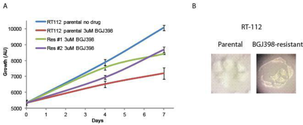
Figure 1. Development of NVP-BGJ398 resistant cell lines. RT-112 cells were grown in increasing amounts of NVP-BGJ398 until such time two, independent lines were able to grow in 3uM of drug. (A) Parental and resistant cell lines were plated in quadruplicate in 48-well plates and the indicated drugs were added a day 0. Cell density was measured at days 0, 4, and 7, and growth curves were created. (B) Pictures of parental and resistant lines demonstrating altered morphology.

CST2017-231_F2

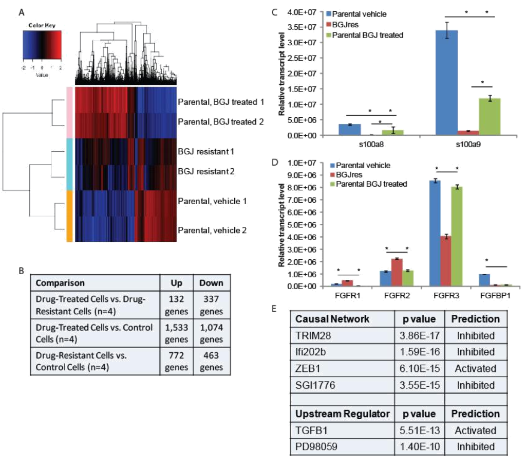
Figure 2. RNA-seq analysis: (A) Hierarchical clustering was performed on RNA-sequencing data from two independent untreated and BGJ398 treated parental RT-112 cell samples as well as two BGJ398 resistant RT-112 cell lines. (B) The numbers of significantly differentially regulated transcripts between treatment conditions is shown. (C,D) To validate RNA-sequencing results, RNA was extracted from the indicated cells and RT-qPCR was performed using primers for the indicated genes. S100A8 and S100A9 represent two of the most highly enriched transcripts in the drug resistant versus drug-treated parental cell datasets while the FGFR transcripts, which could play a direct role in mediating BGJ398 resistance, were also confirmed to be significantly different among treatment groups. (E) Ingenuity Pathway Analysis was performed to identify pathway signatures that were significantly different between drug resistant and drug-treated parental data sets. The most significantly different Causal Network and Upstream Regulator signatures are shown. (* p<0.05)

Ingenuity Pathway Analysis was used to identify pathways that were uniquely affected in the BGJ398 resistant cells (Figure 2E). Causal Network analysis suggested that TRIM28 (or KAP1) and Ifi202b networks, both of which regulate the interferon response [17, 18], were inhibited in BGJ398-resistant cells. Related to this, Upstream Regulatory analysis suggested that TGFβ signaling, which can repress the interferon response [19], was activated in resistant cells. Upstream regulator analysis also suggested that the pathway controlled by PD98059, a MEK1 inhibitor [20], was inhibited, which perhaps suggests that the MEK pathway is activated in resistant cells. Likewise, Causal Network analysis suggests that the pathway controlled by SGI1776, a PIM kinase inhibitor [21], was inhibited, which perhaps suggests that PIM kinases are activated in resistant cells. Finally, IPA Causal Network analysis also found the ZEB1 network to be activated in resistant cells. ZEB1 represses E-cadherin expression, driving epithelial mesenchymal transition (EMT) [22].

FGFR3 and PIM kinase mediate resistance: To determine if changes in FGFR levels affected the growth of RT-112 cells or their sensitivity to BGJ398, we transfected FGFR1 or FGFR2 expression plasmids into parental RT-112 cells or FGFR3 into BGJ398 resistant cells and performed growth assays (Figure 3). Transient expression of FGFR1 or FGFR2 alone did not affect the growth of parental RT-112 cells, nor did it affect their sensitivity to BGJ398. However, expression of FGFR3 in BGJ398 resistant cells caused decreased growth in the presence of BGJ398. This might imply a restoration of sensitivity to the drug in these cells.

Because the SGI1776 signal was found to be decreased in BGJ398 resistant cells, we examined what effect this inhibitor would have on the growth of parental and resistant RT-112 cells. We did not observe any significant differences in PIM transcript expression in drug treated or drug resistant cell lines (Figure 4A). However, treatment of BGJ398 resistant cells with SGI1776 significantly inhibited the growth of these cells (Figure 4B). Interestingly, a combination of BGJ398 and SGI1776 was significantly better at preventing growth of parental RT-112 cells than BGJ398 alone. SGI1776 displayed no overt toxicity at the concentration used to inhibit RT-112 cell growth as it did not inhibit the growth of HEK293 cells (Figure 4B).

CST2017-231_F3

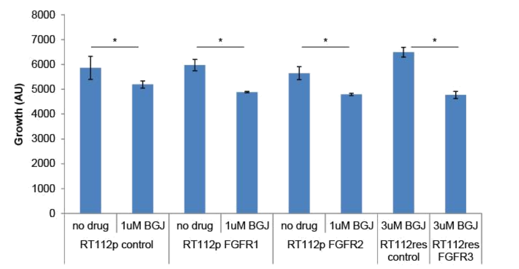
Figure 3. FGFR over-expression in RT-112 cells: Parental RT-112 cells were transiently transfected with a control plasmid or FGFR1 or FGFR2 expression plasmids while BGJ398 resistant cells were transfected with control or FGFR3 expression plasmids. Growth was measured over seven days by DAPI staining, and the relative cell number at day 7 is shown (AU = arbitrary units, * p<0.05).

CST2017-231_F4

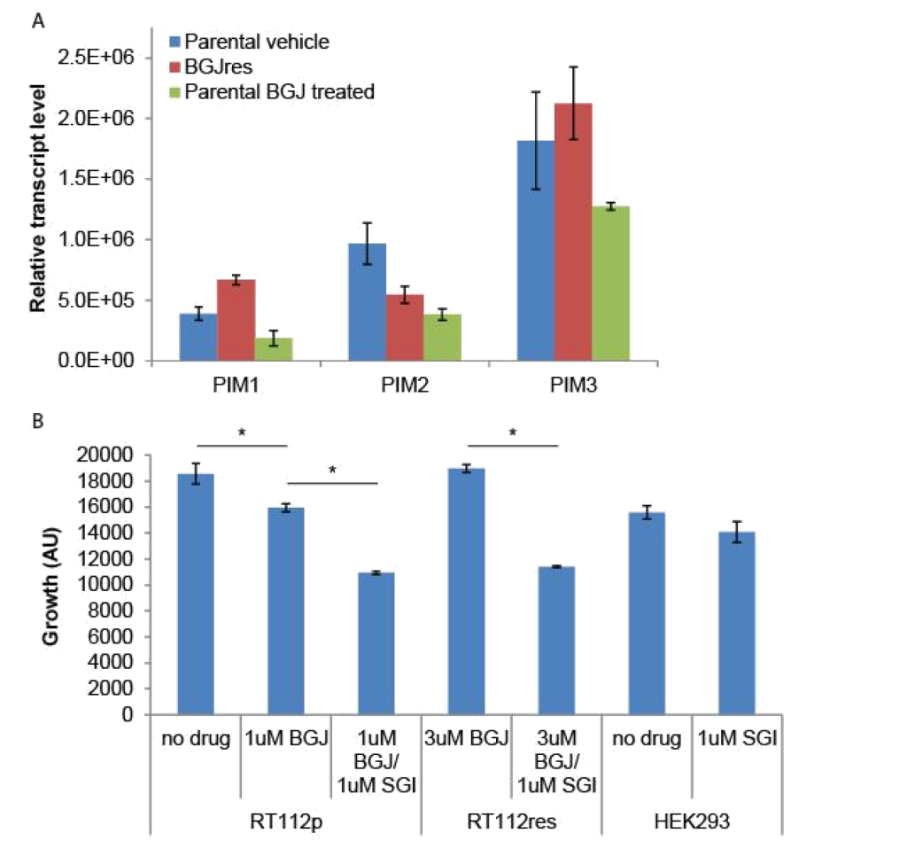
Figure 4. SGI1776 treatment of RT-112 cells: (A) RNA was extracted from the indicated cells and RT-qPCR was performed using primers for the three PIM transcripts. No significant differences were observed. (B) Parental and BGJ398 RT-112 cells as well as control HEK293 cells were treated with BGJ398 and/or SGI1776 as indicated and growth at day 7 is shown (AU = arbitrary units, * p < 0.05).

Discussion

Recently reported data from Phase I clinical trials with two FGFR- targeted agents are very encouraging [12, 13]. Furthermore, alterations in FGFR, including FGFR3 mutations, FGFR3-TACC3 translocations, and FGFR2 alterations have been associated with response to the FGFR inhibitors JNJ-42756493 and BGJ398. Other FGFR inhibitors, including LY2874455, BMS-582664, BIBF 112, and BAY1163877 are in development for bladder and other cancers with FGFR alterations [23]. FGFR-targeting agents will hopefully soon be approved for use in bladder and other cancers, but like most targeted agents, resistance is expected to develop. Using the RT-112 cell model, which was used in the original development of BGJ398, we developed two independent resistant cell lines, which grew in 3uM of drug, well above its IC50 in parental cells. Using an RNA-seq approach, we identified several pathways that potentially mediate resistance to BGJ-398. Two of the most promising are alternate FGFR usage and activation of PIM kinase signaling.

We found that FGFR1 and FGFR2 transcript levels were increased in resistant cells while FGFR3 levels were decreased. BGJ398 has nearly equal affinity for all three receptors so differential receptor affinity cannot explain resistance to BGJ398. However, FGFR1 versus FGFR3 expression on bladder cancer cells is indicative of an altered phenotype and has been shown to mediate BGJ398 sensitivity [24]. Chen, et al found that FGFR1 was expressed on bladder cancer cells that also expressed the mesenchymal markers ZEB1 and vimentin, whereas FGFR3 expression was restricted to the E-cadherin- and p63-positive epithelial subset. Sensitivity to the growth-inhibitory effects of BGJ398 was also restricted to the epithelial cells and it correlated directly with FGFR3 mRNA levels but not with the presence of activating FGFR3 mutations. In contrast, BGJ398 did not strongly inhibit proliferation but did block invasion in the mesenchymal type bladder cancer cells in vitro [24]. We observed a morphological change in our BGJ398 resistant cells that could very well be a reflection of a more mesenchymal state. Indeed, in our RNA-seq data vimentin levels were increased 2.5 fold (p = 0.003) in resistant cells, as were ZEB1 levels, although not significantly (p = 0.11). Furthermore, our pathway analysis suggested that ZEB1 signaling was activated in resistant cells. ZEB1 is a documented mediator of epithelial-mesenchymal transition and is known to be induced by FGF2 signaling [25], further supporting a role for altered FGFR expression in lineage transition and drug resistance. In line with the Chen, et al report, we saw that BGJ398 sensitivity correlated with FGFR3 levels, as over-expression re-sensitized the cells to growth inhibition by BGJ398 (Figure 3). Our data, combined with the Chen, et al report, strongly suggest that altered FGFR expression drives, or is at least reflects a lineage transition that mediates resistance to BGJ398. This is reminiscent of the lineage plasticity that mediates anti-androgen resistance in metastatic prostate cancer [26], and may represent a wider mechanism of resistance to targeted agents. Whether lineage plasticity mediates resistance to BGJ398 and other FGFR inhibitors and exactly how such plasticity develops should be further investigated.

We also found that the PIM kinase inhibitor, SGI1776, significantly inhibits the growth of BGJ398 resistant cells. Pim1 is a serine-threonine kinase which promotes early transformation, cell proliferation, and cell survival during tumorigenesis in several cancer types, including bladder cancer, where it was found to be over-expressed in invasive cancers compared to non-invasive cancers and normal tissues [27]. Another study found high levels of expression of all three PIM family members in both non-invasive and invasive urothelial carcinomas compared to normal tissue [28]. Furthermore Pim1 knock-down [27] or treatment with the PIM kinase inhibitor TP-3654 [28] reduced the growth of several bladder cancer cell lines in culture and in xenografts. Our data that demonstrated inhibition of both parental and BGJ398 resistant cell lines using a PIM kinase inhibitor, suggesting that PIM kinase likely remains a viable target in bladder cancer, even after FGFR inhibitor resistance develops.

Pathway analysis suggested other possible mechanisms of resistance, each of which bears further investigation. Upstream regulator analysis suggested that TGFβ signaling was activated in resistant cells. TGFβ has long been known to play an important role in bladder cancer, in part through its regulation of interferons [29]. Interestingly, the two most significantly inhibited Causal Networks in the IPA analysis were those controlled by TRIM28 (or KAP1) and Ifi202b (Figure 2C), both of which also regulate the interferon response [17, 18]. S100A8 and S100A9, two of the most inhibited genes in the resistant cells, are known to be inhibited by TGFβ and activated by interferons (in part through TRIM28 and Ifi202b) [30, 31], which fits well with the IPA analysis and strongly suggests that TGFβ activation and interferon suppression is important for growth of RT-112 cells in BGJ398. TGFβ and interferons play a complicated role in tumor development and progression, making their value as therapeutic targets questionable. Upstream regulator also suggested that the pathway controlled by PD98059, a specific MEK1 inhibitor, was inhibited, which perhaps suggests that the MEK pathway is activated in resistant cells. Activation of MEK1 has been previously reported in bladder cancer and PD98059 has been shown to reduce proliferation in bladder cancer cells in vitro [32]. This suggests that MEK1 inhibition might be useful in FGFR inhibitor resistant bladder cancer as well.

In a recent report, a RT-112 cell line was developed that had resistance to the FGFR inhibitor AZD4547 [33].The authors performed a synthetic lethality RNAi screen to identify kinases that, when depleted, increased the activity of AZD4547. They identified multiple members of the phosphoinositide 3-kinase (PI3K) pathway and found that the PI3K inhibitor BKM120 acted synergistically with inhibition of FGFR in multiple cancer cell lines having FGFR mutations. Synergy was attributed to PI3K-protein kinase B pathway activity resulting from epidermal growth factor receptor or Erb-B2 receptor tyrosine kinase 3 reactivation caused by FGFR inhibition. These pathways were not identified by our transcriptomic analysis. This could be due to a difference in approach, or it could suggest that the PI3K signaling pathway is more important in mediating the response to AZD4547 than it is for NVP-BGJ398. Regardless, there are likely multiple pathways that can lead to FGFR inhibitor resistance, and each of these reports supports studies in humans to determine if these, or other, mechanisms mediate resistance in actual patients.

Conclusions

Our results suggest that altered FGFR expression and PIM kinase activity could mediate resistance to NVP-BGJ398. These pathways should be investigated in samples from patients resistant to this drug.

Acknowledgments:
The authors would like to thank the Integrative Genomics core staff for assistance with this project.

Funding: Research reported in this publication included work performed in core facilities supported by the National Cancer Institute of the National Institutes of Health under award number P30CA033572. The content is solely the responsibility of the authors and does not necessarily represent the official views of the National Institutes of Health.

Author contributions: SKP helped with the design of the study, acquisition of materials, and writing of the paper. MH carried out experiments and edited the paper. JOJ managed the project, assisted with experimental design and execution, and writing of the paper

Competing interests: SKP is a consultant for Novartis. JOJ and MH have no conflicts to report.

References

  1. Siegel RL, Miller KD, Jemal A (2016) Cancer statistics, CA Cancer J Clin 66: 7–30. PubMed PMID: Medline: 26742998.
  2. Burger M, Catto JW, Dalbagni G, Grossman HB, Herr H, et (2013) Epidemiology and risk factors of urothelial bladder cancer. Eur Urol 63: 234–241. [crossref]
  3. Beenken A, Mohammadi M (2009) The FGF family: biology, pathophysiology and therapy. Nat Rev Drug Discov 8: 235–253. [crossref]
  4. Carneiro BA, Meeks JJ, Kuzel TM, Scaranti M, Abdulkadir SA, Giles FJ (2015) Emerging therapeutic targets in bladder cancer. Cancer Treat Rev 41: 170–8.
  5. Cappellen D, De Oliveira C, Ricol D, de Medina S, Bourdin J, et al. (1999) Frequent activating mutations of FGFR3 in human bladder and cervix carcinomas. Nat Genet 23: 18–20.
  6. Billerey C, Chopin D, Aubriot-Lorton MH, Ricol D, Gil Diez de Medina S, Van Rhijn B, et (2001) Frequent FGFR3 mutations in papillary non-invasive bladder (pTa) tumors. Am J Pathol 158: 1955–9.
  7. Cancer Genome Atlas Research N, Weinstein JN, Akbani R, Broom BM, Wang W, Verhaak RGW, et (2014) Comprehensive molecular characterization of urothelial bladder carcinoma. Nature 507: 315–22.
  8. Di Martino E, Tomlinson DC, Knowles MA (2012) A Decade of FGF Receptor Research in Bladder Cancer: Past, Present, and Future Adv Urol 2012: 429213. [crossref]
  9. Tomlinson DC, Baldo O, Harnden P, Knowles MA (2007) FGFR3 protein expression and its relationship to mutation status and prognostic variables in bladder cancer. J Pathol 213: 91–8.
  10. Lamont FR, Tomlinson DC, Cooper PA, Shnyder SD, Chester JD, et (2011) Small molecule FGF receptor inhibitors block FGFR-dependent urothelial carcinoma growth in vitro and in vivo. Br J Cancer 104: 75–82.
  11. Qing J, Du X, Chen Y, Chan P, Li H, et (2009) Antibody-based targeting of FGFR3 in bladder carcinoma and t(4;14)-positive multiple myeloma in mice. J Clin Invest 119: 1216–29.
  12. Nogova L, Sequist LV, Perez Garcia JM, Andre F, Delord J-P, Hidalgo M, et (2016) Evaluation of BGJ398, a Fibroblast Growth Factor Receptor 1–3 Kinase Inhibitor, in Patients With Advanced Solid Tumors Harboring Genetic Alterations in Fibroblast Growth Factor Receptors: Results of a Global Phase I, Dose-Escalation and Dose-Expansion Study. J Clin Oncol.
  13. Tabernero J, Bahleda R, Dienstmann R, Infante JR, Mita A, et (2015) Phase I Dose-Escalation Study of JNJ-42756493, an Oral Pan-Fibroblast Growth Factor Receptor Inhibitor, in Patients With Advanced Solid Tumors. J Clin Oncol 33: 3401–8.
  14. Guagnano V, Furet P, Spanka C, Bordas V, Le Douget M, et (2011) Discovery of 3-(2,6-dichloro-3,5-dimethoxy-phenyl)-1-{6-[4-(4-ethyl-piperazin-1-yl)-phenylamino]-pyrimidin-4-yl}-1-methyl-urea (NVP-BGJ398), a potent and selective inhibitor of the fibroblast growth factor receptor family of receptor tyrosine kinase. J Med Chem 54: 7066–7083. [crossref]
  15. Williams SV, Hurst CD, Knowles MA (2013) Oncogenic FGFR3 gene fusions in bladder cancer. Hum Mol Genet 22: 795–803. [crossref]
  16. Abuharbeid S, Czubayko F, Aigner A (2006) The fibroblast growth factor-binding protein FGF-BP. Int J Biochem Cell Biol 38: 1463–1468. [crossref]
  17. Min W, Ghosh S, Lengyel P (1996) The interferon-inducible p202 protein as a modulator of transcription: inhibition of NF-kappa B, c-Fos, and c-Jun activities. Mol Cell Biol 16: 359–368. [crossref]
  18. Kamitani S, Ohbayashi N, Ikeda O, Togi S, Muromoto R, et (2008) KAP1 regulates type I interferon/STAT1-mediated IRF-1 gene expression. Biochem Biophys Res Commun 370: 366–370. [crossref]
  19. Letterio JJ, Roberts AB (1998) Regulation of immune responses by TGF-beta. Annu Rev Immunol 16: 137–161. [crossref]
  20. Dudley DT, Pang L, Decker SJ, Bridges AJ, Saltiel AR (1995) A synthetic inhibitor of the mitogen-activated protein kinase Proc Natl Acad Sci U S A 92: 7686–7689. [crossref]
  21. Chen LS, Redkar S, Bearss D, Wierda WG, Gandhi V (2009) Pim kinase inhibitor, SGI-1776, induces apoptosis in chronic lymphocytic leukemia Blood 114: 4150–7.
  22. Eger A, Aigner K, Sonderegger S, Dampier B, Oehler S, et (2005) DeltaEF1 is a transcriptional repressor of E-cadherin and regulates epithelial plasticity in breast cancer cells. Oncogene 24: 2375–85.
  23. Morales-Barrera R, Suarez C, de Castro AM, Racca F, Valverde C, et (2016) Targeting fibroblast growth factor receptors and immune checkpoint inhibitors for the treatment of advanced bladder cancer: New direction and New Hope. Cancer Treat Rev 50: 208–16.
  24. Cheng T, Roth B, Choi W, Black PC, Dinney C, McConkey DJ (2013) Fibroblast growth factor receptors-1 and -3 play distinct roles in the regulation of bladder cancer growth and metastasis: implications for therapeutic tar PLoS One 8: e57284.
  25. Lau MT, So WK, Leung PC (2013) Fibroblast growth factor 2 induces E-cadherin down-regulation via PI3K/Akt/mTOR and MAPK/ERK signaling in ovarian cancer cells. PLoS One 8: e59083. [crossref]
  26. Mu P, Zhang Z, Benelli M, Karthaus WR, Hoover E, et al. (2017) SOX2 promotes lineage plasticity and antiandrogen resistance in TP53- and RB1-deficient prostate cancer. Science 355: 84–8.
  27. Guo S, Mao X, Chen J, Huang B, Jin C, et (2010) Overexpression of Pim-1 in bladder cancer. J Exp Clin Cancer Res 29: 161. [crossref]
  28. Foulks JM, Carpenter KJ, Luo B, Xu Y, Senina A, et (2014) A small-molecule inhibitor of PIM kinases as a potential treatment for urothelial carcinomas. Neoplasia 16: 403–12.
  29. Champelovier P, E Atifi M, Mantel F, Rostaing B, Simon A, et (2005) In vitro tumoral progression of human bladder carcinoma: role for TGFbeta. Eur Urol 48: 846–851. [crossref]
  30. Rahimi F, Hsu K, Endoh Y, Geczy CL (2005) FGF-2, IL-1beta and TGF-beta regulate fibroblast expression of FEBS J 272: 2811–2827. [crossref]
  31. Xu K, Geczy CL (2000) IFN-gamma and TNF regulate macrophage expression of the chemotactic S100 protein S100A8. J Immunol 164: 4916–4923. [crossref]
  32. Kumar B, Sinclair J, Khandrika L, Koul S, Wilson S, et (2009) Differential effects of MAPKs signaling on the growth of invasive bladder cancer cells. Int J Oncol 34: 1557–1564. [crossref]
  33. Wang L, Sustic T, Leite de Oliveira R, Lieftink C, Halonen P, et (2017) A Functional Genetic Screen Identifies the Phosphoinositide 3-kinase Pathway as a Determinant of Resistance to Fibroblast Growth Factor Receptor Inhibitors in FGFR Mutant Urothelial Cell Carcinoma. Eur Urol 71: 858–62.

A Survey of Quantum Lyapunov Control Methods of a novel chemo-hyperstructure as a novel drug discovery dual targeting of the p53 and NF-κB pathways for the activation of the p53 tumor suppressor pathway by an engineered P44 cyclotidomimic agonisitic mechanistic pharmacoligand

Abstract

The p53 and nuclear factor κB (NF-κB) pathways play crucial roles in human cancer development. Simultaneous targeting of both pathways is an attractive therapeutic strategy against cancer. The use of pharmacologically active short peptide sequences has prooven to be a better option in cancer therapeutics than the full-lengthprotein. It has been previously report ed one such 44-mer peptide sequence of SMAR1 (TAT-SMAR1 wild type, P44) that retains the tumor suppressor activity of the full-length protein.P44 peptide could efficiently activate p53 by mediating its phosphorylation at serine15, resulting in the activation of p21 and in effect regulating cell cycle checkpoint. In vitrophosphorylation assays with point-mutated P44-derived pep-tides suggested that serine 347 of SMAR1 was indispensable forits activity and represented the substrate motif for the proteinkinase C family of proteins. In this Research Scientific Project we generated an antitumor multi-targeted hyper-molecule that bears a pyrrolo[3,4-clomifene-diamizido-c]pyrazole scaffold and functions as an enantiomeric P44 peptide mimeto inhibitor against both the p53-MDM2 interaction and the NF-κB activation. This pharmacophjoric scaffold may be a first-in-class dual targeted enantiomeric inhibitor with dual efficacy for cancer therapy with potential synergistic effect in vitro and in vivo. Docking and molecular dynamics simulation studies further provided insights into the nature of stereoselectivity. Here, we have for the first time in silico discovered a novel survey of Quantum Lyapunov Control Methods of a novel chemo-hyperstructure as a novel drug discovery dual targeting of the p53 and NF-κB pathways for the activation of the p53 tumor suppressor pathway by an engineered P44 cyclotidomimic agonisitic mechanistic pharmacoligand.

Keywords

Survey of Quantum Lyapunov, Control Methods, discovery of novel chemo-hyperstructure, novel drug discovery, dual targeting, p53 and NF-κB pathways, p53 tumor, suppressor pathway, engineered P44, cyclotidomimic, agonisitic mechanistic, pharmacoligand.

A surface representation parallel adaptive finite element simplified spherical harmonics approximation solver for frequency domain fluorescence molecular imaging of IHMVYSK peptide-mimo based chemo-ligands comprising therapeutic vaccine-like agonistic properties as a potential novel druggable synthetic regulator for future allergic and autoimmune treatment applications

Abstract

Fluorescence molecular imaging/tomography may play an important future role in preclinical research and clinical diagnostics. Time- and frequency-domain fluorescence imaging can acquire more measurement information than the continuous wave (CW) counterpart, improving the image quality of fluorescence molecular tomography. Although diffusion approximation (DA) theory has been extensively applied in optical molecular imaging, high-order photon migration models need to be further investigated to match quantitation provided by nuclear imaging. In this paper, a frequency-domain parallel adaptive finite element solver is developed with simplified spherical harmonics (SPN) approximations. To fully evaluate the performance of the SPN approximations, a fast time-resolved tetrahedron-based Monte Carlo fluorescence simulator suitable for complex heterogeneous geometries is developed using a convolution strategy to realize the simulation of the fluorescence excitation and emission. The validation results show that high-order SPN can effectively correct the modeling errors of the diffusion equation, especially when the tissues have high absorption characteristics or when high modulation frequency measurements are used. Furthermore, the parallel adaptive mesh evolution strategy improves the modeling precision and the simulation speed significantly on a realistic digital mouse phantom. This solver is a promising platform for fluorescence molecular tomography using high-order approximations to the radiative transfer equation. A parallel adaptive finite element simplified spherical harmonics approximation solver for frequency domain fluorescence molecular imaging A surface representation designed IHMVYSK peptide-mimo based chemo-ligand comprising therapeutic vaccine-like agonistic properties as a potential novel druggable synthetic regulator for future allergic and autoimmune treatment applications.Abstract: Allergic and autoimmune diseases are forms of immune hypersensitivity that increasingly cause chronic ill health. Most current therapies treat symptoms rather than addressing underlying immunological mechanisms. The ability to modify antigen-specific pathogenic responses by therapeutic vaccination offers the prospect of targeted therapy resulting in long-term clinical improvement without nonspecific immune suppression. Examples of specific immune modulation can be found in nature and in established forms of immune desensitization. Allergic and autoimmune diseases are forms of immune hypersensitivity that increasingly cause chronic ill health. Most current therapies treat symptoms rather than addressing underlying immunological mechanisms. The ability to modify antigen-specific pathogenic responses by therapeutic vaccination offers the prospect of targeted therapy resulting in long-term clinical improvement without nonspecific immune suppression. Examples of specific immune modulation can be found in nature and in established forms of immune desensitization. Targeting pathogenic T cells using vaccines consisting of synthetic peptides representing T cell epitopes is one such strategy that is currently being evaluated with encouraging results. Future challenges in the development of therapeutic vaccines include selection of appropriate antigens and peptides, optimization of peptide dose and route of administration and identifying strategies to induce bystander suppression. Structure-based computational methods have been widely used in exploring protein-ligand interactions, including predicting the binding ligands of a given peptide based on their structural complementarity. Compared to other peptide and ligand representations, the advantages of a surface representation include reduced sensitivity to subtle changes in the pocket and ligand conformation and fast search speed. Peptidomimetics, deriving from structure-based, combinatorial or protein dissection approaches, can play a key role as hit compounds. We believe that using a surface patch approach to better understand protein-ligand interactions has the potential to significantly enhance the design of new ligands for a wide array of drug-targets using a surface representation parallel adaptive finite element simplified spherical harmonics approximation solver for frequency domain fluorescence molecular imaging of IHMVYSK peptide-mimo based chemo-ligands comprising therapeutic vaccine-like agonistic properties as a potential novel druggable synthetic regulator for future allergic and autoimmune treatment applications.

Keywords

parallel adaptive, finite element, simplified spherical harmonics, approximation solver, frequency domain, fluorescence molecular imaging, surface representation, IHMVYSK peptide-mimo, based chemo-ligand, therapeutic vaccine-like, agonistic properties, novel druggable, synthetic regulator, future allergic, autoimmune treatment applications.

A surface representation Modeling for Collapsing Cavitation Bubble near Rough Solid Wall by Mulit-Relaxation-Time Pseudopotential Lattice Boltzmann Model of a designed IHMVYSK peptide-mimo based chemo-ligand comprising therapeutic vaccine-like agonistic properties as a potential novel druggable synthetic regulator for future allergic and autoimmune treatment applications

Abstract

Cavitation bubble collapse near rough solid wall is modeled by the multi- relaxation-time (MRT) pseudopotential lattice Boltzmann (LB) model. The modified forcing scheme, which can achieve LB model’s thermodynamic consistency by tuning a parameter related with the particle interaction range, is adopted to achieve desired stability and density ratio. The bubble collapse near rough solid wall was simulated by the improved MRT pseudopotential LB model. The mechanism of bubble collapse is studied by investigating the bubble profiles, pressure field and velocity field evolution. The eroding effects of collapsing bubble are analyzed in details. It is found that the process and the effect of the interaction between bubble collapse and rough solid wall are affected seriously by the geometry of solid boundary. At the same time, we demonstrates that the MRT pseudopotential LB model is a potential tool for the investigation of the interaction mechanism between the collapsing bubble and complex geometry boundary near Rough Solid Wall by Mulit-Relaxation-Time Pseudopotential Lattice Boltzmann Model of a designed IHMVYSK peptide-mimo based chemo-ligand comprising therapeutic vaccine-like agonistic properties as a potential novel druggable synthetic regulator for future allergic and autoimmune treatment applications.

Keywords

Modeling for Collapsing, Cavitation Bubble, Rough Solid Wall, Mulit-Relaxation-Time, Pseudopotential Lattice, Boltzmann Model, surface representation, IHMVYSK peptide-mimo, chemo-ligand, therapeutic vaccine-like, agonistic properties, novel druggable, synthetic regulatorfor future allergic, autoimmune treatment applications, Cavitation Bubble, Bubble Collapse, Lattice Boltzmann Method, Pseudopotential Model, Rough Solid Wall.

Efficacy and Safety of Long Term Brentuximab Vedotin Therapy and Retreatment in an AIDS Patient Affected by Refractory/Relapsed Anaplastic Large Cell Lymphoma: Case Report and Literature Review

DOI: 10.31038/CST.2017254

Abstract

Lymphoma is the most common type of cancer in patients infected with HIV despite of the introduction of the antiretroviral therapy. Brentuximab Vedotin (BV) is an anti-CD30 antibody-drug conjugate, which has been approved for the treatment of relapsed or refractory Hodgkin lymphoma and anaplastic large cell lymphoma (ALCL). However, fewer data are available on the role of the BV in the treatment of HIV-CD30+ lymphomas and of its impact on outcomes. We describe the first case of retreatment with BV in a HIV patient with ALCL, ALK- who relapsed after a complete response to a previous BV treatment.

Key words

Anaplastic large cell lymphoma, HIV infection, Brentuximab vedotin.

Introduction

Infection sustained by Human Immunodeficiency Virus (HIV) and the subsequent impairment of the immune system represent a major risk for developing lymph proliferative disorders like non Hodgkin lymphomas (NHL) [1], also after the introduction of the Highly Active Antiretroviral Therapy (HAART).

Diffuse large B cell lymphoma (DLBCL), Burkitt lymphoma and primary lymphoma of the central nervous system are the most common histologic variants of NHL in this population, even if classical Hodgkin Lymphoma (cHL) is more frequent histology in the HAART era. Among the less frequent T-cell NHLs, peripheral T-cell lymphoma not otherwise specified (PTCL-NOS), cutaneous and systemic anaplastic large cell lymphoma (ALCL) ALK-negative seems to be the most common variants [2].

ALCL is a rare form of disease whose incidence does not exceed 12% of all cases of T-cell NHLs [3]; the expression of the CD30 antigen on the neoplastic cell surface makes it a potential target for the treatment with brentuximab vedotin (BV), an anti-CD30 humanized antibody drug conjugated. However, very few data are available regarding its efficacy and toxicity in the specific cohort of HIV patients treated with HAART.

We report a case of an ALCL, ALK- in a patient affected by acquired immuno-deficiency syndrome (AIDS) with progressive disease after the first-line chemotherapy who experienced a complete remission with BV monotherapy and obtained a second objective response to the retreatment with BV at the subsequent relapse.

Case report

A 49-years old woman was diagnosed with AIDS in November 2012, A3 according CDC categorization for HIV/AIDS. Her CD4 counts were 16 cells/μl and the HIV-RNA circulating copies were 12.540.000 IU/ml. She started HAART therapy with etravirine and tenofovir and lamivudine that resulted in a reduction of the viral load to undetectable levels and in a slightly improvement of CD4 counts with stable levels around 100 cells/μl. The HAART therapy was modified with raltegravir and etravirine and lamivudine in 2013 due to a new hospitalization for esophageal candidiasis.

In November 2014 she was admitted to our hospital for a wasting syndrome, esophageal candidiasis and lymphonodal swelling, the patient noticed also night sweats. At that moment, the CD4 counts were 123 cell/μL and the circulating HIV-RNA copies were undetectable. Based on onset of opportunistic infections and wasting syndrome the CDC classification was modified in C3. She performed a total body CT scan that showed the involvement of right cervical and supraclavicular lymphnodes, as well as mesenterial and left inguinal ones. Then, she performed a supraclavicular lymphnode biopsy which revealed a lymphomatous infiltration consistent with ALCL, ALK-. The neoplastic cells were positive for CD3, CD4+, CD30+, CD45+, Granzyme +, EMA+. The bone marrow biopsy was negative for neoplastic involvement. According to current guidelines, the patient started chemotherapy regimen with the cyclophosphamide, adriamycin, vincristine, etoposide, prednisone (CHOEP-scheme) which was continued for 4 courses.

After the fourth cycle, the disease evaluation performed with CT scan revealed the persistence of the supraclavicular lymphadenopathy and the onset of a new axillary lymphadenopathy. Despite the progression disease, the patient was not suitable candidate for more intensive second line therapy followed by autologous stem cell transplantation because of the persistence of wasting syndrome and of her body mass index which (BMI) were 15. For these reasons BV at dose of 1.8 mg/kg every 21 days was started. In May 2015, after 4 cycles of BV, a new disease evaluation with FDG-PET scan demonstrated a complete metabolic response (CMR). Considering the surprising and very fast response to the treatment and the slow improvement of the wasting syndrome, the patient continued on treatment with BV for a total of 16 courses. HAART with etravirine/lamivudine/ raltegravir was administered concurrently with the chemotherapy. She was also treated with prophylactic agents such as acyclovir for herpes simplex and zoster reactivation, fluconazole for fungal infection, sulfamethoxazole / trimethoprim for Pneumocystis Jiroveci and azithromycin for Mycobacterium Avium-Intracellulare Complex (MAC). The treatment was complicated by grade 4 asymptomatic neutropenia after the first cycle which not required dose delay or reduction. A secondary prophylaxis of neutropenia febrile with granulocyte colony stimulating factor (G-CSF) for 5 days for each cycle was started after the second course avoiding the occurrence of new side effects. Furthermore the BV therapy did not worse the antiretroviral associated neuropathy.

During this period, the patient obtained also an increase of BMI returning in a normal range. The virological assessment evaluated during the therapy showed an isolated viremic increase in March 2015 which was due to the incorrect assumption of antiretroviral drug; later, a correct adherence to the therapy guaranteed the negativity of quantitative plasmatic HIV-RNA determination assessed on April, June, November 2015 and January 2016. For the all treatment the CD4 counts did not exceed the 100/μL. At the end of treatment in January 2016, the final disease evaluation with PET scan confirmed the CMR and the patient showed a very good performance status with a BMI of 19.38.

In July 2016, after 5 months of observation, the patient presented a relapse of disease with the reappearance of the fever and axillary and supraclavicular lymphadenopathies. A total body CT scan confirmed these lymphadenopathies and revealed also the presence of pathologic abdominal lymph nodes. No bone marrow infiltration was detected by biopsy sample. Considering the good performance status of the patient at the relapse, she was considered eligible for a third line chemotherapy including the autologous stem cells transplantation. Then, she received three cycles of IGEV (Ifofosfamide, Gemcitabine, Vinorelbine) as salvage treatment. After the second cycle of IGEV a CD34-positive harvest of 3,0×10⁶ cells/kg was obtained by G-CSF plus Plerixafor. A grade 4 neutropenia occurred after every cycle despite of the primary prophylaxis of the febrile neutropenia with G-CSF. A CT scan performed after the third cycle showed an increase of the diameter of lymph nodes and documented a progressive disease. According to the recent publication of Bartlett et al. [4] we decided to treat again our patient with BV. After only two cycles of BV the fever disappeared. A only drug related grade 3 anaemia occurred after the second cycle leading to blood transfusion and to reduce the next doses at 1.2 mg/kg. The third course was administered but the patient experienced a grade 4 fatigue and a peripheral motor neuropathy which led to a therapy delay. The CT scan performed after the fourth cycle revealed a very good partial response but the treatment was complicated by a back pain due to a vertebral fracture, therefore the patient discontinued the treatment on April 2017. She continued to remain in partial response until to June 2017 when she was hospitalized in another institution for a non neutropenic fever with septic shock and despite of a broad spectrum antibiotics was started, her condition worsened and she died several day later.

Discussion

The introduction of HAART improved the outcome of HIV infected patient affected by NHL. The control of HIV replication obtained with antiretroviral therapy allowed similar treatment approach as for HIV negative patients. Indeed the current guidelines for treatment of HIV associated lymphomas recommended that patients with HIV related lymphoma should usually be treated in an identical manner to HIV-negative patients [5]. The risk of HIV infected patients for developing a NHL varied during the decades according to the introduction of more effective therapies: in 2003, Biggar et al. reported a cumulative risk of 15% for T NHL in a cohort of 302.834 AIDS patients diagnosed between 1978 and 1996, before the introduction of the HAART [6]. More recently, a population based study of Gibson et al. demonstrated that HAART therapy reduced but not removed the risk for NHL development [2]. In particular for ALCL, an historical confront in HIV patients demonstrated a reduction of the standardized incidence ratio (SIR) which passed from 9 in the period 1996-2002 to 1.8 in the period 2003-2010 with an overall SIR of 14.2 compared to the general population. Interestingly, the authors reported an increased incidence of non AIDS-defining subtypes like marginal zone lymphoma, Waldestrom macroglobulinemia and T-cell NHL. In particular, the incidence of ALCL was reported to be higher during the AIDS period rather than in the HIV seropositive status only, suggesting the determinant role of the immune system in the control of this lymphoproliferative disorder [2].

BV is approved for the treatment of CD30 positive lymphomas (cHL, ALCL), but at the present time, few data are available regarding its use in HIV infected patients, because of a possible challenge may be represented by pharmacological interaction between BV and antiretroviral therapy. Regarding ALCL, in 2012 Pro et al. published the results of a phase II trial in which BV was administered in patients with relapsed/refractory ALCL; The authors reported an overall response rate (ORR) of 86% (57% CR and 29% PR) with a median duration of response of 12.6 months, but no mention was made regarding HIV patients included in this trial. It is remarkable that in this trial the use of BV cancelled the differences between ALK positive (which traditionally show a better outcome) and ALK negative patients; moreover the median duration of response of patients in CR treated with BV without stem cell transplantation (SCT) was similar to allogeneic SCT group [7]. More recently, BV was also tested in PTCL; in a phase I trial, the addition of BV to CHOP or CHP (without vincristine in order to minimize neurological toxicity) was safety and showed a significant antitumor activity. At the present time, a phase III trial is comparing efficacy of BV plus CHP versus CHOP alone in CD30 positive T cell neoplasms (Clinical trial NCT01777152).

The exclusion of HIV infected patients from phase I-III trials of antineoplastic drugs gives few informations for clinicians who need to treat these forms of disease in this population. For this reason, there are few data about interaction of antiretroviral drugs and antineoplastic ones. If we consider the German guidelines [5], HAART should be maintained during chemotherapy and should be selected those compounds which demonstrated the lower interaction profile. Potential of interactions with increased toxicities appears to be higher with antiretroviral combinations that include strong enzyme inhibitors such as ritonavir-boosted protease inhibitors [8]. On the contrary, raltegravir undergoes glucuronidation and allows lower interaction with chemotherapy, based on these reasons our patient replaces tenofovir with raltegravir [9].

Recently, at the 57th Annual Meeting of the American Society of Hematolgy in Orlando, the AIDS Malingnancy Consortium (AMC) presented the results of the phase I portion of the first trial using BV with AVD in the upfront treatment of stage II-IV HIV-associated Hodgkin Lymphoma. Six HIV positive patients with untreated classical Hodgkin lymphoma (cHL) in advanced stage were enrolled. Five of the 6 patients had a negative PET/CT after two doses of BV and the 5 patients who completed the therapy, achieved a CR. The treatment was well tolerated and a phase II portion with 51 subjects to enroll is actively accruing in both the USA and France, in an AMC/LYSA collaboration, clinicaltrials.gov NCT01771107. The recommended Phase II dose is 1.2 mg/kg +AVD every other week [10]. Until major data from prospective clinical trial are available for the safety and efficacy of BV in HIV related lymphoma,only case reports and case series remain an important source of informations. We have found only two case reports described by Gandhi et al. They reported a first patient who had a relapsed ALCL after a previous diagnosis of cHL and a second one who had a relapsed advanced cHL. Both patients were treated with BV and experienced a rapid and complete remission with minimal toxicity, they remained in durable remission until the time of the publication in 2013 [11].

The safety profile of BV is now well described and it is mainly represented by neurological, haematological and pancreatic toxicity. Neuropathy remains a major problem also in patients treated with HIV infection and it may be caused both from antiretroviral drugs and HIV infection. In our patient during all the first treatment period, we did not observe any neurological as well as pancreatic toxicity. Regarding hematological toxicity, a neutropenia G4 was identified after the first cycle with BV but the use of secondary prophylaxis with G-CSF bypassed this potential side effect for all the further cycles of BV. We emphasize this, because the previous experience with chemotherapy and antiretroviral drugs demonstrated a higher incidence of haematological toxicity in HIV infected patients who received both antiretroviral and chemotherapy.

Regarding infectious complications, during the first treatment we did not identify any adverse events, although CD4 counts during all the treatment period remained around 100 cells/μl, the quantitative HIV circulating RNA was always negative during the same period.

We demonstrated also that the retreatment with BV in patients with relapsed ALCL has still antitumor activity. Bartlett et al demonstrated in an open-label multicentre study, the antitumor activity of the retreatment with BV in 68% of relapsed HL or ALCL previously treated with BV [4]. The estimated median time for responding patients was 9.5 months. The rates of AEs were generally very similar to those reported in the pivotal phase 2 trials [7]. However peripheral motor neuropathy was seen higher in the retreatment than in the pivotal study. In our patient, we obtain a second very good partial response with the disappearance of the systemic symptoms and the reduction of the lymphadenopathies but unfortunately the retreatment led to more severe adverse events than in the previously primary treatment. We can explain these severe adverse events with the HIV infection status and the combination HAART and immunotherapy that could be more toxic.

Conclusions

To our knowledge this is the first case report with a patient with relapsed HIV associated ALCL retreated with BV. The progress in clinical and pharmacological research revolutionized the outcome of HIV patients with lymphomas but the toxicity remains a major problem so far. The introduction of novel less toxic drugs, as BV in HIV patients should be a future challenge in terms of evaluation of the efficacy and feasibility therefore new prospective clinical trials are expected with this population. Our experience demonstrated both efficacy and safety of BV in an HIV selected patient and that an early retreatment, even if in advanced progressive disease, seems to be associated with a clinical objective response. However, during BV retreatment these patients should be followed very closely for peripheral motor and sensorial neuropathy and for infectious complications. The confirmations of our data in major cohorts of patients are needed for overpass traditional concern about treatment of HIV infected patients.

References

  1. Centers for Disease Control and Prevention (1987) Revision of the CDC surveillance case definition for acquired immunodeficiency Council of State and Territorial Epidemiologists; AIDS Program, Center for Infectious Diseases. MMWR Morb Mortal Wkly Rep. 36: 1S–15S.
  2. Gibson TM, Morton LM, Shiels MS, Clarke CA, Engels EA (2014) Risk of non- Hodgkin lymphoma subtypes in HIV-infected people during the HAART era: a population-based study. AIDS 28: 2313–2318. [crossref]
  3. William BM, Armitage JO (2013) International analysis of the frequency and outcomes of NK/T-cell Best Pract Res Clin Haematol 26: 23–32. [crossref]
  4. Bartlett, et al. (2014) Retreatment with brentuximab vedotin in patients with CD30- positive hematologic Malignancies. Journal of Hematology & Oncology 7: 24
  5. Hentrich M, et (2014) Therapy of HIV-associated lymphoma recommendations of the oncology working group of the German Study Group of Physician in Private Practice Treaning HIV-Infected Patients (DAGN&#0x00C4;), in cooperation with the GermanAIDS Society (DAIG). Ann Hematol 93: 913–21.
  6. Biggar RJ, Engels EA, Frisch M, Goedert JJ (2001) AIDS Cancer Match Registry Study Group, Risk of T-cell lymphomas in persons with J Acquir Immune Defic Syndr 26: 371-376. [crossref]
  7. Pro B, et (2012) Brentuximab vedotin (SGN-35) in patients with relapsed or refractory systemic anaplastic large-cell lymphoma: results of a phase II study. J Clin Oncol 30: 2190–6.
  8. Ezzat HM, et al. (2012) Incidence, predictors and significance of severe toxicity in patients with human immunodeficiency virus-associated Hodgkin Leuk Lymphoma 53: 2390–2396
  9. Kassahun K, et (2007) Metabolism and disposition in humans of raltegravir (MK-0518), an anti-AIDS drug targeting the human immunodeficiency virus 1 integrase enzyme. Drug Metab Dispos 35: 1657–63.
  10. Rubistein PG, et al. (2015) AMC-085: A Pilot Trial of AVD and Brentuximab Vedotin in the Upfront Treatment of Stage II-IV HIV-Associated Hodgkin Lymphoma. A Trial of the AIDS Malignancy Consortium. Abstr at 57th ASH Meeting, Blood 126: 1526
  11. Gandhi M, Petrich A (2014) Brentuximab vedotin in patients with relapsed HIV- related lymphoma. J Natl Compr Canc Netw 12: 16–19. [crossref]

Primary Cardiac Hemangioendothelioma in the Adult and Cardiac Involvement in Patient with Non-Cardiac Hemangioendothelioma. Review of the Literature

DOI: 10.31038/CST.2017253

Abstract

Background: Cardiac hemangioendothelioma (CHE) in the adult and cardiac involvement in adult patient with non cardiac-hemangioendothelioma (NC-HE) were rare and uncommon in daily clinical experience.

Methods: We researched in PubMed, Web of Science, Scopus, Cochrane library, and Medline for identifying relevant studies, case series, review and case reports.

Results: We found 33 cases of CHE in the adult and 7 cases of cardiac involvement in NC-HE patients. According to scientific data, the most of CHE patients were female (52%), histological type epithelioid (64%), with main localization in right atrium (41%), and the main clinical diagnosis was incidentally discovered on echocardiogram. The main therapeutic choice was surgical. Otherwise the cardiac metastasis in NC-HE patients (7 cases reported) were most frequent in the hepatic hemangioendothelioma primary localization (57%) than others (spleen, bone, limb, intracranial). The most of patients were female (85.7%) and the main clinical presentation were heart failure (85%).

Conclusion: CHE and cardiac metastasis of NC-HE patients in the adult were rare and unusual neoplasms. The early diagnosis and surgical treatment in each case demonstrated an increase of patients’ survival and quality of life. We explained the scientific data to increase physician knowledge about this rare adulthood vascular neoplasm.

Keywords

Hemangioendothelioma; Primary Cardiac Tumor; Metastatic cardiac tumor; cardiac tumor; cardiac neoplasm; cardiac metastasis.

Introduction

Historical background: Hemangioendothelioma (HE) was described for the first time by Mallori in the 1908 and it was defined as a vascular neoplasms characterized by intermediate features between hemangiomas and angiosarcomas [1, 2]. With the technological evolutions, Stout and colleagues [3] studied firstly in deep the microscopic appearances in the 1943 and explained the likeness of HE to the blood vessels. In the 1967, Fernholz edited one of the first article on “Contribution of History of HE”. Only in the 1988, Enzinger and colleague defined the HE features in intermediate, borderline, or low grade malignancy.

Classification and Clinical presentation: HE was a vascular neoplasm with typical prevalent proliferation of endothelial cells and is was distinguished according to its histological characteristics as [2, 4]:

  • papillary intralymphatic,
  • retiform hemangioendothelioma,
  • kaposiform hemangioendothelioma,
  • epithelioid hemangioendothelioma,
  • pseudomyogenic hemangioendothelioma
  • and composite hemangioendothelioma

HE was characterised by suggestive histological feature as intracytoplasmic vacuoles, nuclear cytoplasmic inclusion, myxoid stroma, hyaline stroma and chondroid stroma (Figures 1 a and b). The most of patient had expression of vascular markers (CD 31, CD 34, ERG, FL1I) and cytokeratins (CAM 5.2, CK7, CK 18) [5].

Figure 1. Kaposiform hemangioendothelioma of the right atrium associated to KAsabach- Merritt syndrome: a) image of the vascular neoplasm with epithelial membrane antigen (EMA) antibodies and b) eosin and hematoxylin view.

HE involved mainly liver, lung, skin, soft tissue, bone and spleen and occurring firstly in infancy and early childhood, in the adult it was very uncommon [2].

Each of the histological types of HE had own typical presentations and patterns, such as association with lymphatic vessel proliferation as well as Kasabach-Merritt syndrome (KMS) [6-9]. KMS included thrombocytopenia, microangiopathic hemolytic anemia and mild consumptive coagulopathy, and developed often in KHE, Kaposiform lymphatic anomaly (KLA) and tufted angioma (TA) [7, 8].

HE would have different grades of aggression like:

  • finite
  • low aggression
  • mild aggression
  • moderate aggression
  • severe aggression, like Kaposiform HE
    • Local to the adjoining structures
    • Metastatic

Singularly, epithelioid HE was defined in two histological grade according to its aggressiveness: classic (low-grade) and malignant (intermediate-grade) [5-55].

The progression of HE was unpredictable: sometimes it grew slowly and sometimes the tumour was more active and spread quickly [2]. The characteristics of the patients, the different clinical and spreading of HE influenced the therapeutic choices: surgical, medical or palliative.

Finally, also the mortality depended to histological type and grade of aggression and clinical correlations.

Methods

We researched english article on PubMed, Web of Science, Scopus, Cochrane library, and Medline for identifying relevant english studies, reviews, case series and case reports in the adult with the following keywords: 1) cardiac hemangioendothelioma,

2) primary cardiac hemangioendothelioma, 3) cardiac metastasis in hemangioendothelioma, 4) Cardiac Involvement in Hemangioendothelioma in the adult, and 5) Hemangioendothelioma/ Heart/ Adult. We found 272 articles with those criteria (Figure 2) and we selected and chosen 33 case reports and 7 reviews that satisfied our research criteria (Figure 2).

Figure 2. Description of scientific literature review process: first step we had researched all article in the literature

Results

According to our scientific research, the primary cardiac hemangioendothelioma was 33 reported cases. The patient age range was from 19 to 77 year-old, the average age was 45.1 ± 16.5 year-old (Table 1). The most of patients were female (n=17, 52%, Figure 3). The most common primary cardiac localization was right atrium (n=14, 41%) then the cardiac valve (n=5, 15%), left atrium ( n=4, 12%), right ventricle ( n=3, 9%), coronaric sinus (n=2, 6%), superior vena cava/ right atrium (n=2, 6%), right appendage (n=1, 3%), left ventricle (n=1, 3%), and not defined (n=2, 6%) (Figure 4). The histopathological feature of the HE in the case reported was predominantly ephiteloid (n=21, 66%), kaposiform (n=1, 3%) and defined malignant ( n=3, 9%) and not defined (n=8, 24%) (Figure 4). The clinical presentations of these patients were characterised by incidentally discovered on echocardiogram (n=11, 34.4%), dyspnoea (n=2, 6.3%), chest pain (n=4, 12.5%), alveolar hemorrhage (n=1, 3.1%), cardiac tamponade (n=3, 9.4%), incidentally discovered an heart murmur (n=3, 9.4%), arrhythmia (n=1, 3.1%), cerebrovascular event like stroke (n=1, 3.1%), widened superior mediastinum noticed on a routine chest radiograph (n=1, 3.1%), thromboembolism (n=1, 3.1%), and incidentally discovered by autopsy (n=1, 3.1%) (Figures 5 and 6). The most of patients were CD31 and CD34 positive (30.3%) and CD 99 positive (18.1%). The main therapy were the surgery (n=21, 65.6%). In the case of cardiac metastasis of hemangioendothelioma (seven reported cases in literature), the age of patients were 61.7± 12.7 year-old and they were female (85.7%). The first localization of the tumour was indicated in the Table 2. In all the cases, the patient suffered of heart failure and the most of them were undergone to surgery.

Figure 3

Figure 3. The percentage of cardiac hemangioendothelioma localization in the reported literature cases.

Figure 4

Figure 4. The percentage of cardiac hemangioendothelioma histological type in the reported literature cases.

Figure 5

Figure 5. Type of hemangioendothelioma

Figure 6

Figure 6. The clinical presentation of cardiac hemangioendothelioma in the reported literature cases were absolutely unpredictable in the adult with a major incidence of chest pain or dyspnea but wide variability in the clinical first manifestation due to patient’s health background, histological type of hemangioendothelioma and grade of aggression.

Discussion

The cardiac hemangioendothelioma was predominantly epithelioid type (66% of the cases), with a peculiar tumour site in right atrium (40% of the cases). The most of these patients were female and they had good outcome (follow up positive after 6 months in the 46.9%). In particularly, the patients undergone to aggressive and rapid surgery, were better outcome than delay diagnosis or late surgical procedure [15-45]. As reported by scientific literature, cardiac malignant tumours were rare and uncommon, especially metastatic cardiac involvement [53, 54]. Cardiac hemangioendothelioma were considered an atypical unexpected malignant cardiac tumours with high local aggressiveness and metastasizing potential [30-32, 34, 36-37].

On the other hand, we had researched the reported articled of cardiac metastasis in patient with non cardiac hemangioendothelioma reported in Table 2 [48-52]. Bisesi and colleagues [14], had already firstly documented the possibility of cardiac involvement in multifocal epithelioid hemangioendothelioma in 1996, hence we had studied in deep the scientific literature to look for other cases like this and to understand the clinical presentation and evolution of the cardiac metastasis of a hemangioendothelioma.

In our research, we had found 6 cases of cardiac metastasis of hemangioendothelioma. All of the cases were in female (100%) with a primary HE in liver (50%), one ovarian HE, one bone HE and the last intracranial HE. The most of them showed heart failure and worsening hemodynamic parameters in association with coagulative disorders and hence end–stage of multi-organ failure. The prognosis of them was unfavourable specially in the presence of coagulative disorders.

Conforming in Table 2 and our review of the literature, cardiac metastasis in patients with non-cardiac hemangioendothelioma were out of ordinary and exceptional but remarkable for the prognosis of the patients [14, 48-52].

Table 1. Cases of cardiac hemangioendothelioma in the adult reported in scientific literature. M: male, F: female, ND: non defined, RA: right atrium, MV: mitral valve, LA: left atrium, RV: right ventricle, LV: left ventricle, PV: pulmonary valve, AV: aortic valve, TV: tricuspid valve, CS: coronaric sinus, SCV: superior vena cava, F-up: follow up.

Reference Year Age Sex Site HE Type Presentation Therapy Follow up
1 Blanchard et al15 1952 29 M Right
Appendage
ND Chest pain ND ND
2 Crenshaw et al16 1959 36 F RA ND Chest pain ND ND
3 Allaire et al17 1964 ND ND ND ND ND ND ND
4 Hayward et al18 1979 49 F MV Epithelioid Diastolic murmur typical of mitral stenosis Surgical Alive after 4 years
5 Di Biasi et al20 1988 ND M RV Epithelioid Incidentally discovered on echocardiogram Surgical ND
6 Singal et al19 1987 19 F LA Epithelioid Heart murmur, clubbed finger Resection Dead
7 Gegenbach et al21 1991 ND ND LV Epithelioid Incidentally discovered on echocardiogram ND Alive after 4 months
8 Montes Orbe et al 1991 56 F PV Epithelioid Thromboembolism Surgical and
medical
ND
9 Marchiano et al22 1993 71 F RA Epithelioid Palpitation, dizziness, low grade fever Surgical
resection
Alive after surgery
10 Billé-Turc et al23 1993 59 M AV Epithelioid Stroke Surgical Alive (F-up 1
year)
11 Di Biasi et al24 1995 35 M RV Epithelioid Incidentally discovered on echocardiogram Surgical Alive (F-up 1
year)
12 Ichikawa et al25 1997 32 F CS Malignant Incidentally discovered on echocardiogram Surgical Alive
13 Hongquan et al26 1998 42 F RA ND Incidentally discovered on echocardiogram Surgical Alive
14 Yoshida et al27 1999 ND M RA Malignant Cardiac tamponade Surgical Dead
15 Di Biasi et al24 1995 35 M RV ND Dyspnoea
16 Kamiyoshihara M28 2001 32 M ND Malignant ND Surgical
Excision
Dead after 41 months
17 Agaimy et al29 2002 68 M RV/TV Epithelioid Incidentally discovered antopsy ND Dead
18 Kitamura et al30 2005 36 F RA Epithelioid Cardiac tamponade Surgical
resection
Alive (F-up 3
years)
19 Vall Bernal et al31 2005 69 F MV Epithelioid Edema, exertional dyspnea Surgical
resection
Alive (F-up 6
months)
20 Wang et al32 2006 36 F RA ND Dyspnoea ND ND
21 Moulai et al33 2006 53 M CS Epithelioid Incidentally discovered on echocardiogram Heart transplant Alive (F-up 10
years)
22 Lisy et al34 2006 61 M LA Epithelioid Incidentally discovered on echocardiogram Surgical
resection
ND
23 Safirstein et al35 2007 51 F RA Epithelioid Incidentally discovered on echocardiogram Surgical
resection
Alive (F-up 8
years)
24 Zhang et al36 2008 66 F LA Epithelioid Incidentally discovered on echocardiogram Heart transplantation Alive
25 Messias et al37 2008 21 F LA Epithelioid Chest pain Surgical
resection
Alive
26 Kahlout M et al38 2009 44 F RA Epithelioid Cardiac tamponade ND ND
27 Guray et al39 2010 25 M RA ND Heart failure Medical ND
27 Lahon et al40 2012 29 F SVC Epithelioid widened superior mediastinum noticed on a routine chest radiograph Surgical
resection
Alive
28 Sugimoto T et al41 2013 77 F RA Epithelioid Incidentally discovered on echocardiogram ND ND
29 Allain et al42 2014 ND ND SVC/RA Epithelioid ND ND ND
30 Ellouze et al43 2015 53 M RA Epithelioid Incidentally discovered on echocardiogram Surgical
resection
Alive
31 Gaytán-Cortés et al44 2016 23 M RA ND chest pain, intermittent paroxysmal nocturnal dyspnea and malaise Surgical
resection
Alive
32 Lappa et al45
(article in press)
2017 36 F RA Kaposiform Alveolar Hemorrhage Surgical and
drug supports
Dead

Table 2. Cases of cardiac metastasis in adult patients with non-cardiac primary hemangioendothelioma, reported in scientific literature. M: male, F: female, ND: non defined, MTs: metastasis; RA: right atrium; HE; hemangioendothelioma; IL: interleuchin; CT: chemotherapy; Pt: patient.

Principal Reported Case of metastatic cardiac hemangioendothelioma
Author Age Pt Gender Primary Tumor Complications Therapy
Myauchi et al (1987)46 ND F Ovarian Hemangiomatosis MTS pericardium and RA Medical
Dubois et al (1987)47 64 F hepato-splenoganglionic hemangioendothelioma MTs at the heart à heart failure Surgical and medical
Hurley et al (1994)48 ND F Intracranial epithelioid HE Seizures, Liver and heart multifocal
locliation
Aggressive surgical treatment
Bhutto et al (1995)49 ND M hepatic epithelioid hemangioendothelioma Heart MTs irradiation, IL-2 injection, and/or
surgery
Bellmunt et al (1999)50 48 F hepatic epithelioid hemangioendothelioma Heart metastasis
Portal hypertension
CT à adriamycin
Ilasi et al (1999)51 ND F HE of the bone Heart failure
MTs in RA
Chemotherapy
Hsu CY (2014)52 73 F hepatic epithelioid hemangioendothelioma Heart failure embolization of the abnormal neoangiogenic vessels

Conclusion

Our literature review would like to be a resume of the cardiac hemangioendothelioma and cardiac involvement in patients with non- cardiac hemangioendothelioma in daily clinical experience. In line with our analysis, the cardiac-HE was vascular aggressive neoplasm with variable histopathological pattern and outcome, principally in female patient and right atrium. Its outcome would be influenced to the early diagnosis and prompt surgical and medical treatments. On the contrary, the cardiac involvement in patients with non-cardiac hemangioendothelioma was weird and life-threatening: only seven cases described and with worsening evolution due to coagulative disorders, heart failure and end-stage multi-organ disease. In case of suspicion of CHE or cardiac involvement of NC-HE were immediately undergone to appropriate diagnostic exams (lab test, echocardiogram, computed tomography, and so on) and rapid surgical and medical therapies in order to avoid the life-threatening evolution of these malignant pathologies.

Acknowledgements: A special thanks go to Dr. Rosalba Grillo and her collaboration in the production of histological images.

Funding: none

Conflicts of interest: All the authors declare non conflict of interest.

References

  1. Mallory FB (1908) The Results Of The Application Of Special Histological Methods To The Study Of T J Exp Med 10: 575–593. [crossref]
  2. Requena L, Kutzner H (2013) Semin Diagn Pathol.
  3. Stout AP (1943) Hemangio-Endothelioma: a Tumor of Blood Vessels Featuring Vascular Endothelial Ann Surg 118: 445–464.
  4. Fernández Y, Bernabeu-Wittel M, García-Morillo JS, et (2009) Kaposiform hemangioendothelioma. Eur J Intern Med.
  5. Anderson T, Zhang L, Hameed M, Rusch V, Travis WD, Antonescu CR (2015) Thoracic epithelioid malignant vascular tumors: a clinicopathologic study of 52 cases with emphasis on pathologic grading and molecular studies of WWTR1- CAMTA1 Am J Surg Pathol 39: 132–139.
  6. Larsen EC, Zinkham WH, Eggleston JC, Zitelli BJ (1987) Kasabach-Merritt syndrome: therapeutic considerations. Pediatrics 79: 971–980. [crossref]
  7. Maguiness S, Guenther L (2002) Kasabach-Merritt syndrome. J Cutan Med Surg.
  8. Kelly M (2010) Kasabach-Merritt Pediatr Clin North Am
  9. Haahr V, Jacobsen E, Bendix K, Nielsen JL, Peterslund NA (1994) [Kasabach–Merritt syndrome]. Ugeskr Laeger 156: 6011–6014. [crossref]
  10. Messias P, Bernardo J, Antunes MJ (2008) Primary left atrial haemangioendothelioma. Interact Cardiovasc Thorac Surg 7: 945–946. [crossref]
  11. Safirstein J, Aksenov S, Smith F (2007) Cardiac epithelioid hemangioendothelioma with 8-year follow-up. Cardiovasc Pathol.
  12. Gaytan-Cortes FC, Arteaga-Adame J, Careaga-Reyna G, Lezama-Urtecho C, Alvarez-Sanchez L (2016) [Primary cardiac hemangioendothelioma: early diagnosis and surgical resection]. Rev Med Inst Mex Seguro
  13. Toursarkissian B, O’Connor WN, Dillon ML (1990) Mediastinal epithelioid hemangioendothelioma. Ann Thorac Surg.
  14. Bisesi MA, Broderick LS, Smith JA (1996) MR demonstration of right atrial involvement in multifocal epithelioid AJR Am J Roentgenol 167: 953–954. [crossref]
  15. BLANCHARD AJ, HETHRINGTON H (1952) Malignant haemangioendothelioma of heart. Can Med Assoc J 66: 147–150. [crossref]
  16. CRENSHAW JF, DOWLING EA, CRESSWELL WF Jr (1959) Primary hemangio-
    endothelioma of the heart. Ann Intern Med 50: 1289–1298. [crossref]
  17. Allaire FJ, Grimm CA, Taylor LM, et (1964) Primary Hemangioendothelioma of the heart. Report of a case treated with irradiation and cyclophosphamide. Rocky Mt Med J 61: 34–37.
  18. Hayward RH, Korompai FL (1979) “Endothelioma of the Mitral Valve,” The Annals of Thoracic Surgery 28: 87–89.
  19. Singal KK, Alagaratnam DM, Brundage B, Ferlinz J, Ghou P (1987) Intracardiac mass in a young woman with a history of brain tumor. Chest 92: 337–341. [crossref]
  20. Di Biasi P, Santoli E, Santoli C (1988) Primary tumors of the right ventricle: apropos of a case of hemangioendothelioma treated surgically. G Ital Cardiol 18: 1025–7
  21. Gengenbach S, Ridker PM (1991) Left ventricular hemangioma in Kasabach- Merritt syndrome. Am Heart J 121: 202–203. [crossref]
  22. Marchiano D, Fisher F, Hofstetter S (1993) Epithelioid hemangioendothelioma of the heart with distant metastases. A case report and literature review. J Cardiovasc Surg (Torino) 34: 529–33.
  23. Bille’-turk F, Padovani R, Rosario R, et (1993) “Hemangio- endothelioma of the Aortic Valve Revealed by Transient Ischemic Episodes,” La Presse Médicale.
  24. Di Biasi P, Scrofani R, Santoli C (1995) Cardiac Ann Thorac Surg 59: 792–3.
  25. Ichikawa H, Kaneko T, Obayashi T, et (1997) Surgical treatment of malignant hemangioendothelioma originated from the right atrium: a case report. Kyobo Geka 50: 67–70.
  26. Hongquan Y, Hua R, Quancai C, Qi M, Xiaocheng L, et (1998) Cardiac hemangioendothelioma. J Cardiovasc Surg (Torino) 39: 655–658. [crossref]
  27. Yoshida A, Kanda T, Sakamoto H, et (1999) Sudden death with malignant hemangioendothelioma originating in the pericardium–a case report. Angiology 50: 607–11.
  28. Kamiyoshihara M, Ishikawa S, Morishita Y (2001) Sudden death due to rupture of an omental metastatic tumor arising from cardiac A case report. J Cardiovasc Surg (Torino) 42: 495–7.
  29. Agaimy A, Kaiser A, Wünsch PH (2002) Epithelioid hemangioendothelioma of the heart in association with myelodysplastic syndrome. Z Kardiol 91: 352–6.
  30. Kitamura K, Okabayashi H, Hanyu M, et (2005) Successful Enucleation of a Gi- ant Cardiac Hemangioendothelioma Showing an Unusual Proliferation Pattern. The Journal of Thoracic and Cardiovascular Surgery 130: 1199–1201.
  31. Val-Bernal JF, Gracia-Alberdi E, Gutierrez JA and Garijo MF (2005) Incidental in Vivo of an Epithelioid Heman-gioendothelioma of the Mitral V Pathology Interna-tional 55: 644–648.
  32. Wang LF, Liu M, Zhu H, Han W, Hu CY, et (2006) Primary cardiac hemangioendothelioma: a case report. Chin Med J (Engl) 119: 966–968. [crossref]
  33. Moulai N, Chavanon O, Guillou L, Noirclerc M, Blin D, et (2006) “Atypical Primary Epithelioid He- mangioendothelioma of the Heart,” Journal of Thoracic Oncology 2: 188–189.
  34. Lisy M, Beierlein W, Muller H, Bultmann B and Ziemer G (2007) “Left Atrial Epithelioid Hemangioendothelioma,” The Journal of Thoracic and Cardiovascular Surgery 133: 803–804.
  35. Safirstein J, Aksenov S, Smith F (2007) Cardiac epithelioid hemangioendothelioma with 8-year follow-up. Cardiovasc Pathol 16: 183–186. [crossref]
  36. Zhang PJ, Brooks JS, Goldblum JR, Yoder B, et (2008) Primary cardiac sarcomas: a clinicopathologic analysis of a series with follow-up information in 17 patients and emphasis on long-term survival. Hum Pathol 39: 1385–95.
  37. Messias P, Bernardo J, Antunes MJ (2008) Primary left atrial haemangioendothelioma. Interact Cardiovasc Thorac Surg 7: 945–946. [crossref]
  38. M Kahlout, A Al-Mulla, A Chaikhouni, et al. (2009) Unusual presentation of a rare tumor: Cardiac epithelioid hemangioepithelioma presenting as cardiac Heart Views 10: 132–5.
  39. Güray Y, Demirkan B, Güray U, BoyaciA(2010) Right atrial hemangioendothelioma: a three-dimensional echocardiographic Anadolu Kardiyol Derg 10: E7–8.
  40. Lahon B, Fabre D, De Montpreville V and Dartevelle P (2010) Epithelioid haemangioendothelioma of the superior vena Interact Cardiovasc Thorac Surg 15: 186–7.
  41. Sugimoto T, Yamamoto K and S (2013) Yoshi A Primary Epithelioid Hemangioendothelioma of the Right Atrium: Report of a Case and Literature Review. Open Journal of Thoracic Surgery 3: 63–67.
  42. Allain G, Hajj-Chahine J, Lacroix C, et (2014) Surgical management of an epithelioid hemangioendothelioma of the superior vena cava protruding into the right atrium. J Card Surg 29: 779–81.
  43. Ellouze M, Dami M, Beaulieu Y, et (2015) Resection of a right atrial epithelioid hemangioendothelioma. Cardiovasc Pathol 24: 401–4.
  44. Gaytán-Cortés FC, Arteaga-Adame J, Careaga-Reyna G, et (2016) Primary cardiac hemangioendothelioma: early diagnosis and surgical resection. Rev Med Inst Mex Seguro Soc 54: 392–6.
  45. Angela Lappa, Marzia Cottini, Silvia Donfrancesco, et (2017) Primary cardiac kaposiform hemangioendothelioma: the rare adult-onset. Review of the literature. EC Anesthesia.
  46. Miyauchi J, Mukai M, Yamazaki K, Kiso I, Higashi S, Hori S (1987) Bilateral ovarian hemangiomas associated with diffuse hemangioendotheliomatosis: a case Acta Pathol Jpn 37: 1347–55.
  47. Dubois A, Eledjam JJ, Deixonne B, et (1987) Lymph node-hepatosplenic hemangioma in an adult with consumption coagulopathy and fatal cardiac insufficiency. Ann Gastroenterol Hepatol (Paris) 23: 363–6.
  48. Hurley TR, Whisler WW, Clasen RA, Smith MC, Bleck TP, et (1994) Recurrent intracranial epithelioid hemangioendothelioma associated with multicentric disease of liver and heart: case report. Neurosurgery 35: 148–51.
  49. Bhutto AM, Uehara K, Takamiyagi A, Hagiwara K, Nonaka S (1995) Cutaneous malignant hemangioendothelioma: clinical and histopathological observations of nine patients and a review of the J Dermatol 22: 253–61.
  50. Bellmunt J, Allende E, Navarro M, Morales S, Sans M, et (1989) Epithelioid hemangioendothelioma of the liver with myocardial metastases. Jpn J Clin Oncol 19: 153–158. [crossref]
  51. Ilasi JA, Smilari TF, Kolitz J, Zanzi I, Hajdu SI (1999) Malignant hemangioendothelioma presenting as multifocal intraskeletal lesions during pregnancy. A case J Reprod Med 44: 49–52.
  52. Hsu CY, Liu YC, Li CP, Huang PH, Lin CH, Chao Y (2014) Malignant hepatic epithelioid hemangioendothelioma with high-output heart failure: successful management of heart failure with transcatheter arterial Asia Pac J Clin Oncol 10: e118–21.
  53. Salcedo EE, Cohen GI, White RD, Davison MB (1992) Cardiac tumors: diagnosis and management. Curr Probl Cardiol 17: 73–137. [crossref]
  54. Travis WD, Brambilla E, Mu¨ller-Hermelink (2004) World Health Organisation classification of Pathology and genetics of tumours of the lung, pleura, thymus and heart. Lyon, France: IARC Press.
  55. Deyrup AT, Tighiouart M, Montag AG, Weiss SW (2008) Epithelioid hemangioendothelioma of soft tissue: a proposal for risk stratification based on 49 Am J Surg Pathol 32: 924–927.

Epigenetics in Cancer: A Review

DOI: 10.31038/CST.2017252

Abstract

A normal cell has several processes that balance the process of self-renewal and differentiation. Multiple influences on gene expression orchestrate an orderly process resulting in normal tissue growth and function. Heritable changes in gene expression of a cell are called epigenetic changes. Epigenetics is a relatively new field in cancer studies and is increasingly being recognized to play a signification role in cancer formation and progression. Several epigenetic processes have been discovered ranging from DNA methylation to non-coding RNA. Epigenetic changes in a cancer cell are intriguing as they lend themselves to being targeted and provide avenues for cancer therapeutics.

Keywords

Epigenetics; cancer; chromatin; histone; noncoding RNA

Introduction

Epigenetics is the study of phenotypical changes occurring in a cell, with no change in the DNA sequence. The epigenetic creation of a new phenotype or state happens through specific mechanisms that affect gene expression in a stable, heritable manner. To further understand what the true meaning of epigenetics is and what it entails, a comparison to genetics is helpful. Genetics is the study of heredity and genes. Heredity is the process of passing on certain characteristics and traits through DNA from one generation to the next. In this case, phenotypes – or the traits and characteristics – are dependent upon the DNA sequence – or genotypes – an organism gets from the previous generation. Epigenetics is similar, in that it encompasses a study of phenotypes, but different, in that the phenotypes come from a heritable manipulation of the expression of DNA, not the DNA itself. This is a relatively new field that links heredity and developmental changes allowing for a deeper understanding of the ongoing changes occurring in organisms that cannot be accounted for by the DNA sequence. Figure 1 explains the difference between genetic and epigenetic changes affecting the phenotype.

CST 2017-204_fig_1

Figure 1. Genetic versus Epigenetic changes affecting phenotype

Methods and Materials

A descriptive review of the various aspects of epigenetics in the context of cancer was compiled using published literature sources. This is a vast subject and our review is a concise description of basic aspects of the role of epigenetic processes in cancer.

History of Epigenetics and its role in cancer

Epigenetics is a relatively new field of genetics, with the first glimpse into it occurring in 1878, when Walther Flemming began researching organisms and their phenotypes. Flemming discovered chromosomes and the process of mitosis during cell division. He focused on cytogenetics, the study of inheriting genes and traits based on the structure of chromosomes [1]. This, combined with Gregor Mendel’s work in genetics formed a foundation for understanding of genotypes and phenotypes. At this point, many researchers started to look at how, besides genetic makeup, there was another layer (epi) to the traits and characteristics an organism has. In the 1940’s, Conrad Waddington used the term “epigenetic landscape” to represent the layer of processes that occur within cells to change how DNA is expressed to decide the fate of cellular phenotype without affecting the genotype. This process is famously visualized as a ball rolling down one of various trajectories in an undulating landscape, called Waddinton’s Classical Epigenetic Landscape [2]. Interestingly enough, researchers began comparing epigenetics to the Lamarckian evolution. Lamarck’s theory looks at how acquired traits can be passed down to upcoming generations, while epigenetics examines how “acquired traits”, or traits that occur from changes in DNA expression take place, and how they can be passed down to subsequent generations, a process referred to as ‘soft inheritance’. This may be due to heritable changes in the chromatin, such as DNA methylation, occurring as a result of environmental influences [3]. In 1983, epigenetics in relation to cancer began to call more attention, as progressively lower levels of DNA methylation was discovered to correlate significantly with progression of normal tissue to cancerous tissue and metastatic disease [4]. A few years later, portions of the DNA with clusters of cytosine and guanine nucleotides called as the CpG cluster, was shown to, when highly methylated, indicate inactive promoters. Hypermethylation of certain tumor suppressor genes was discovered suggesting a very strong link between epigenetic mechanisms and cancer [5, 6]. Enormous strides in both research and clinical trials in the field of cancer epigenetics have led to the development of many epigenetic therapies to target cancer.

Epigenetic Mechanisms Linked to Cancer

Several different epigenetic mechanisms are linked to cancer. Some prominent ones are DNA methylation, histone modification and chromatin remodeling. All of these mechanisms are present in healthy organisms, and play a prominent role in growth and development as well as adaptation to the environment.

The influence of any one of these can result in many potential health problems ranging from increased risk of obesity to cancer. For example, during the Dutch Famine of 1944-1945, pregnant mothers who were not getting enough nutrition incurred less DNA methylation of the insulin-like growth factor II (IGF2) gene promoter in their early phase embryos. This prenatal exposure of embryos persisted into adulthood as a heritable epigenetic change [7]. It is important to understand that epigenetic factors can work in numerous ways, with cancer causing factors being a pivotal research topic. Environmental influences, such as eating certain kinds of foods and exposure to carcinogens can contribute to epigenetic factors causing cancer. Unlike genetic changes, epigenetic processes are targetable and can be reversed, thus offering avenues for prevention and control of cancers.

DNA Methylation and cancer

The most well-known epigenetic mechanism is DNA methylation. This occurs when a methyl group (CH3) from S-adenosylmethionine is added to a cytosine nucleotide by DNA methyltransferase (DNMT) enzymes [8]. When a section is methylated it is typically turned “off ”, or sent to a section of the major groove in the DNA that makes it hard for transcriptional factors to bind promoters and transcribe the gene. DNA hypermethylation is a process restricted to portions of DNA that are rich in CpG dinucleotides and are part of promoters of genes [9].

Consequently, many genes are hidden from transcription factors, and therefore not expressed. Repressing genes can result in cancer due to the number of tumor suppressor genes located in hypermethylated regions. Not only are tumor suppressor genes repressed, but also, other important genes that repair DNA and regulate the cell cycle are hypermethylated, causing cancers to both form and metastasize very quickly. The table below (Table 1) gives an overview of some of the pathways and genes affected by DNA hypermethylation and their relationship to various cancers.

Amongst the most prominent and earliest described epigenetic processes leading to cancer is low level of DNA methylation also called as DNA hypomethylation. The demethylation of repetitive DNA sequences, coding regions and introns, and gene poor areas are noted to be hallmarks of carcinogenesis. These processes result in chromosomal instability, reactivation of transposable elements and loss of imprinting. It leads to mitotic recombination leading to deletions and translocations as well as chromosomal rearrangements [10]. It can lead to reactivation of repetitive DNA sequences called retrotransposons an example of which is Long Interspersed Nuclear Element-1 (LINE1). LINE-1 are implicated in carcinogenesis of epithelial tumors [11]. These are genes that comprise around 17% of the human DNA are also called “jumping genes”. They propagate themselves throughout the genome via RNA transcription and reverse transcription back into the genome and are thus called retrotransposons. DNA methylation keeps these jumping genes in check in normal cells. Hypomethylation leads to activation of these transposable genes. By inserting themselves in proximity to key genes they lead to activation of oncogenic signaling pathways [12].

Table 1. Pathways affected by DNA hypermethylation in cancer

Pathway Affected Gene Cancer Reference
DNA repair pathway BRCA1 Breast , ovarian [15]
MLH1 GI (colon and gastric), endometrial, ovarian [16]
MGMT Lung, brain [17, 18]
Cell cycle checkpoint CDKN2A Colorectal [19]
p15 Bladder [20]
RASSF1A Breast, lung [21]
Cytoproliferative and metastasis pathways Ecadherin
Ovarian,
breast [22]
APC Colorectal [23]

Research suggests that hypomethylation results in activation of oncogenes, allowing cancers to form and spread rapidly. An example of this is prostate cancer, where the promoter for gene LINE1 is found to be significantly hypomethylated. This epigenetic process marks the progress of prostate cancer from localized disease to metastatic disease [13]. Global hypomethylation can affect certain tissues in the body that then go on to affect how LINE1 is transcribed. In fact, hypomethylation has been shown to result in a specific biologic signature, which marks invasiveness, and is common to breast cancer, prostate cancer and liver cancer. It utilizes major pathways such as TGF-β (transforming growth factor-beta) and ERBB2 triggered pathways [14-23].

Histone Modifications and cancer

While DNA methylation is a huge component of epigenetics; there are many other mechanisms at play. One such mechanism is chromatin remodeling which is responsible for many cancers, diseases, and conditions. To understand chromatin remodeling and cancer, it is first important to look at histones and how they affect gene expression. Histones are alkaline proteins that DNA wraps around to form nucleosomes. Nucleosomes are then tightly coiled to form chromatin, and subsequently chromatids. Naturally, because they are in such close contact with the DNA, histones play a key role in gene expression regulation. The N-terminal tail of histones undergoes posttranslational modifications (PTMs), thereby affecting the accessibility of DNA to RNA polymerase. The N-terminal tail can be changed by methylation, acetylation, ubiquitylation, and phosphorylation [24, 25]. Histone acyteltransferases (HAT’s), histone methyl transferases (HMT’s), Histone deacetylases (HDACs) and histone demethylases (KDMs) are some histone modifying enzymes performing these PTMs. An acetylated histone is comparatively looser than a normal histone, allowing for many transcription sites to become available to RNA polymerases. Increases in gene activation follow this process. Histone deacytelases (HDAC’s) support the removal of acetyl groups from the lysine amino acid in histones, allowing the DNA to tightly wrap around the histone. This results in gene silencing, as many promoter regions are hidden and cannot be transcribed. Obviously, any significant changes or mistakes in PTM’s can easily cause repression of important genes and activation of situationally harmful genes. For example, histone deacetylases act to repress the transcription of tumor suppressor genes.

Additionally, histone variants and histone chaperones are other players in epigenetic processes affecting gene expression. Histone modification processes are important for normal cellular development but can be hijacked by cancer for oncogenic signaling. This may be due to an altered histone code leading to aberrant gene expression or mutations in enzymes regulating histone modifications[26-30].

Examples of histone-mediated processes involved in cancer are presented in Table 2.

Table 2. Histone modifications in cancer

Histone modification Cancer Reference
Histone variant H3.3 mutations Glioblastoma [27]
SETD2 and KDM5C inactivating mutations Renal Cell Carcinoma [28]
Downregulation of H4K16ac and
H4K20me3
Colorectal cancer and leukemia [29]
H3K27me3 inactivates tumor suppressor genes Prostate Cancer and breast cancer [30]

Chromatin Remodeling and Cancer

Chromatin remodeling is a dynamic process that facilitates the remodeling of nucleosomes. Chromatin modification plays an essential role in DNA replication and transcription. Tightly packaged chromatin is called heterochromatin, while loosely packaged chromatin is called euchromatin. There are four well described chromatin remodeling complexes: SWI/SNF, ISWI, INO80 and NuRD/Mi-2. By means of some key characteristic features, these remodeling complexes are able to interact with the nucleosome core, use energy from ATP, bind histones and provide grounds for biochemical alteration and protein-protein interactions [31]. For example, the switch/sucrose non-fermentable chromatin remodeling complexes (SWI/SNF) are responsible for transcription control – as well as a multitude of other tasks such as DNA repair and chromosome segregation [32]. Mutations in subunits of the SWI/SNF complex are known to occur in cancer. Malignant rhabdoid tumors are the result of the loss of INI1 subunit of the SWI/SNF complex [33].

Noncoding RNAs and Cancer

Noncoding RNAs are RNAs that are not translated into protein but play a role in gene expression and translation by various mechanims both in the normal cell and cancer. Long non-coding RNAs (lncRNAs) are non-coding regulatory sequences that have a role to play in gene expression. These are implicated in tumorigenesis by various mechanisms. They may remodel chromatin, act as transcriptional co-activators or repressors, inhibit protein, post transcriptional modifications or serve as decoy elements. Single nucleotide polymorphisms (SNPs) in lncRNAs are implicated in the heritability of several cancers including breast, thyroid, prostate and ovarian [34].

Micro-RNAs (miRNA) are short non-coding RNAs that regulate translation of transcribed genes and hence influence the phenotype of a cell. miRNA mediated pathways are implicated in several cancers including glioblastoma [35].

Table 3 summarizes the various epigenetic processes presented in this review.

Table 3. Epigenetic processes implicated in cancer

Process: Function:
DNA  Methylation Used to control gene expression by methylated specific parts of  DNA,  therefore  turning  those  parts “on” or “off ”
Histone Modifications A post transitional modifications made to histones that change which aspects of the DNA  are available to be promoter regions
Chromatin Remodeling Packaging or loosening up chromatin, allowing DNA to  be  easily  read,  or  tightening  the  chromatin   so that DNA  is more difficult to be read. Both result in changes to promoter regions.
Non – coding RNA A RNA that is not translated into a protein, and instead regulations gene expression and translation

General Epigenetic Therapies

Several epigenetic therapies have been developed and approved for use in various malignancies. Several novel epigenetic therapies are in the process of preclinical and clinical development. DNMT inhibitors are the first epigenetic therapy approved for clinical use for their therapeutic role in myelodysplatic syndrome and acute myeloid leukemia,

HDAC inhibitors are approved for use in T cell lymphoma and multiple myeloma.

Several other epigenetic targets have been identified and their inhibitors are in preclinical and clinical development. These include bromodomain, EZH2 (enhancer of zeste homolog 2) and DOT1( disrupter of telomere silencing protein 1) inhibitors [36].

Conclusion

The role of epigenetics in cancer is prominent and clear. This relationship is apparent through the multiple epigenetic mechanisms implicated in cancer: DNA methylation, histone modifications, chromatin remodeling, and non-coding RNA. All of these mechanisms have been proven to significantly impact the epigenome, without impacting the DNA itself. DNA methylation can occur in a surplus or a deficit, both of which can consequently create and promote cancer within the body. Histone modifications alter the space in which DNA resides, therefore making it harder or easier for that

DNA to be transcribed. Chromatin remodeling affects the degree to which specific sites of DNA are bound, which decides to what extent certain genes are expressed. Long non-coding RNA is a more recently discovered major epigenetic mechanism regulating gene expression. While epigenetic mechanisms can result in cancer formation, research underway holds promise of using the same mechanisms to combat cancer.

Acknowledgements: The authors would like to thank Dr. Sunil Sharma and the Center for Investigational Therapeutics at Huntsman Cancer Institute for their support and providing the opportunity to learn.

Funding information: Support for the publication of this manuscript was provided by the Huntsman Cancer Institute/Huntsman Cancer Foundation.

Conflict of Interest: The authors have no competing interests with regard to this review.

References

  1. Paweletz N (2001) Walther Flemming: pioneer of mitosis research. Nat Rev MolCell Biol 2: 72–75. [crossref]
  2. Goldberg AD, Allis CD, Bernstein E (2007) Epigenetics: a landscape takes shape. Cell 128: 635–638. [crossref]
  3. Jablonka E and MJ Lamb (2008) Soft inheritance: challenging the modern synthesis. Genetics and Molecular Biology 31: 389–395.
  4. Gama-Sosa MA, Slagel VA, Trewyn RW, Oxenhandler R, Kuo KC, et al. (1983) The 5-methylcytosine content of DNA from human tumors. Nucleic Acids Res 11: 6883-6894. [crossref]
  5. Baylin SB, Höppener JW, de Bustros A, Steenbergh PH, Lips CJ, et al. (1986) DNA methylation patterns of the calcitonin gene in human lung cancers and lymphomas. Cancer Res 46: 2917–2922. [crossref]
  6. Feinberg AP, Tycko B (2004) The history of cancer epigenetics. Nat Rev Cancer 4: 143–153. [crossref]
  7. Heijmans BT, et al. (2008) Persistent epigenetic differences associated with prenatal exposure to famine in humans. Proceedings of the National Academy of Sciences 105: 17046–17049.
  8. Das PM, Singal R (2004) DNA methylation and cancer. J Clin Oncol 22: 4632–4642. [crossref]
  9. Kulis M, Esteller M (2010) DNA methylation and cancer. Adv Genet 70: 27–56. [crossref]
  10. Feinberg AP and Vogelstein B (1983) Hypomethylation distinguishes genes of some human cancers from their normal counterparts. Nature 301: 89–92.
  11. Carreira PE, Richardson SR, Faulkner GJ (2014) L1 retrotransposons, cancer stem cells and oncogenesis. FEBS J 281: 63–73. [crossref]
  12. Xiao-Jie L, et al. (2016) LINE-1 in cancer: multifaceted functions and potential clinical implications. Genet Med 18: 431–439.
  13. Yegnasubramanian S, et al. (2008) DNA Hypomethylation Arises Later in Prostate Cancer Progression than CpG Island Hypermethylation and Contributes to Metastatic Tumor Heterogeneity. Cancer Research 68: 8954–8967.
  14. Cheishvili D, et al. (2015) A common promoter hypomethylation signature in invasive breast, liver and prostate cancer cell lines reveals novel targets involved in cancer invasiveness. Oncotarget 6: 33253–33268.
  15. Hansmann T, et al. (2012) Constitutive promoter methylation of BRCA1 and RAD51C in patients with familial ovarian cancer and early-onset sporadic breast cancer. Human Molecular Genetics 21: 4669–4679.
  16. Richman S (2015) Deficient mismatch repair: Read all about it (Review). Int J Oncol 47: 1189–1202. [crossref]
  17. Gömöri É, et al. (2012) Concurrent hypermethylation of DNMT, MGMT and EGFR genes in progression of gliomas. Diagnostic Pathology 7: 8–8.
  18. Gu C, Lu J, Cui T, Lu C, Shi H, et al. (2013) Association between MGMT promoter methylation and non-small cell lung cancer: a meta-analysis. PLoS One 8: e72633. [crossref]
  19. Shima K, et al. (2011) Prognostic Significance of CDKN2A (p16) Promoter Methylation and Loss of Expression in 902 Colorectal Cancers: Cohort Study and
    Literature Review. International journal of cancer. Journal international du cancer 128: 1080–1094.
  20. Bilgrami SM, et al. (2014) Promoter hypermethylation of tumor suppressor genes correlates with tumor grade and invasiveness in patients with urothelial bladder cancer. SpringerPlus 3: 178.
  21. Grawenda AM, E O’Neill (2015) Clinical utility of RASSF1A methylation in human malignancies. British Journal of Cancer 113: 372–381.
  22. Rodriguez FJ, Lewis-Tuffin LJ, Anastasiadis PZ (2012) E-cadherin’s dark side: possible role in tumor progression. Biochimica et Biophysica Acta 1826: 23–31.
  23. Liang TJ, Wang HX2, Zheng YY3, Cao YQ4, Wu X5, et al. (2017) APC hypermethylation for early diagnosis of colorectal cancer: a meta-analysis and literature review. Oncotarget 8: 46468–46479. [crossref]
  24. Morales V, Richard-Foy H (2000) Role of histone N-terminal tails and their acetylation in nucleosome dynamics. Mol Cell Biol 20: 7230–7237. [crossref]
  25. Venkatesh S, Workman JL (2015) Histone exchange, chromatin structure and the regulation of transcription. Nat Rev Mol Cell Biol 16: 178–189. [crossref]
  26. Hattori N and Ushijima T (2014) Compendium of aberrant DNA methylation and histone modifications in cancer. Biochemical and Biophysical Research Communications 455: 3–9.
  27. Schwartzentruber J, et al. (2012) Driver mutations in histone H3.3 and chromatin remodelling genes in paediatric glioblastoma. Nature 482: 226–231.
  28. Dalgliesh GL, et al. (2010) Systematic sequencing of renal carcinoma reveals inactivation of histone modifying genes. Nature 463: 360–363.
  29. Fraga MF, Ballestar E, Villar-Garea A, Boix-Chornet M, Espada J, et al. (2005) Loss of acetylation at Lys16 and trimethylation at Lys20 of histone H4 is a common hallmark of human cancer. Nat Genet 37: 391–400. [crossref]
  30. Kondo Y, Shen L, Cheng AS, Ahmed S, Boumber Y, et al. (2008) Gene silencing in cancer by histone H3 lysine 27 trimethylation independent of promoter DNA methylation. Nat Genet 40: 741–750. [crossref]
  31. Nair SS, Kumar R (2012) Chromatin remodeling in cancer: a gateway to regulate gene transcription. Mol Oncol 6: 611–619. [crossref]
  32. Euskirchen GM, et al. (2011) Diverse roles and interactions of the SWI/SNF chromatin remodeling complex revealed using global approaches. PLoS Genet 7: e1002008.
  33. Kordes U, Gesk S, Frühwald MC, Graf N, Leuschner I, et al. (2010) Clinical and molecular features in patients with atypical teratoid rhabdoid tumor or malignant rhabdoid tumor. Genes Chromosomes Cancer 49: 176–181. [crossref]
  34. Cheetham SW, Gruhl F, Mattick JS, Dinger ME (2013) Long noncoding RNAs and the genetics of cancer. Br J Cancer 108: 2419–2425. [crossref]
  35. Katsushima K, Kondo Y (2014) Non-coding RNAs as epigenetic regulator of glioma stem-like cell differentiation. Front Genet 5: 14. [crossref]
  36. Jones PA, Issa JP, Baylin S (2016) Targeting the cancer epigenome for therapy. Nat Rev Genet 17: 630–641. [crossref]

CC-motif Chemokine Ligand 21: immune targeting, head and neck oncology, and future prospective

DOI: 10.31038/CST.2017251

Abstract

Investigations in immunomodulating therapies for cancer treatment over the past 20 years have flourished. Given the complex tumor microenvironment and differential signaling pathways a wide variety of potential targeting mechanisms have come to attention. Herein we review the immunomodulating potential of Chemokine 21 in pre-clinical and clinical studies, as well as examine the novel multi-faceted immune-based treatments in advanced head and neck cancer.

Introduction

Head and neck squamous cell carcinoma (HNSCC) is the sixth most common cancer in the world, accounting for more than 300,000 deaths annually [1]. Despite advances in surgical techniques and chemo-/radiation treatment strategies, patients with advanced T3 or T4 HNSCC continue to demonstrate 20%-30% survival with failure of primary surgical or medical (radiation and chemotherapy) management. Salvage therapies, in recalcitrant or recurrent disease, are limited by systemic toxicity or operative morbidity owing to nearby vital structures (carotid artery, skull base). Immunotherapy and modulation in HNSCC is a promising venture given recent advances in cancer biology and investigations in the tumor microenvironment.

Historically studied in the contexts of inflammation and immune response, chemokines have increasingly been studied in the context of tumor proliferation [2, 3]. Chemokines are a family of small chemotactic cytokines involved in cellular signaling and inflammation. Through interactions within a subset of G-protein- coupled transmembrane receptors (GPCRs), secreted cellular chemokines regulate cellular homing, activation, and recruitment of a variety of diverse leukocytes and activated adhesion molecules [2, 4-6]. In the tumor microenvironment, responses to specific chemokines result in the migration of different immune cell subsets and regulate tumor immune responses; these are both pro- and anti- inflammatory in nature. Chemokines, have been directly associated with cancer immunity, proliferation, and metastases [7,8]. In this review, we address the role of Chemokine 21 (CCL-21) in tumor progression, its role as an immunotherapy platform, and the potential within head and neck oncology.

CCL-21 chemotaxis and lymphocyte recruitment

Divided into four subfamilies, chemokine nomenclature is related to the location of their first N-terminal cysteine (C) residues; classified as: C-, CC-, CXC-, and CX3C-chemokines [9]. CCL-21 (also known as Thymus-derived chemokine 4, 6Ckine, or Exodus-2) is a CC chemokine expressed in high endothelial post-capillary venules, T-cell stromal zones of the spleen, Peyer’s patches, and afferent peripheral lymph nodes that strongly attracts naïve T-cells and mature dendritic cells (DC) including NK T-cells [10-13]. The effects of CCL-21 on chemotaxis have been demonstrated in neutralization studies, where the homing of T-cells and DCs has been shown to be significantly reduced [14, 15]. Acting through the GPCR CCR7, a convergence of the immune response elements to sites of CCL-21 production have been demonstrated to co-stimulate the expansion of CD4+ and CD8+ T cells and induce Th1 polarization [16]. Additionally, chemokine bound DCs form T cell adhesions resulting in a hyper-responsive T-cell on subsequent exposure to CCL-21 and antigen-presenting cells [17].

Role in cancer immunotherapy

The cancer immunosuppressant microenvironment results in ineffective antigen processing and presentation; resulting in poor host response. The CCL-21/CCR-7 axis has been studied in efforts to create a more immunogenic environment through chemotaxis of DCs, NK cells, and lymphocytes. By recruiting host antigen presenting cells (APC) for tumor antigen presentation, T cells within lymphoid organs have the ability to prime anti-tumor specific activity. Of note, while CCL-21 aids in the polarization of Th1 lymphocytes, the secondary lymphoid chemokine has also been shown to have minimal effect on the proliferation of suppressor cell population; CD4+CD25+ T-regulatory cells are hypo responsive to CCL-21 induced migration and unresponsive to CCL-21 co-stimulation [16]. High levels of T-regulatory cells within the tumor microenvironment has demonstrated poor prognosis in many cancers, dependent on the type and location of neoplasm [18].

Pre-clinical animal models

The ability to process and present a high ratio of activated tumor-antigen-APCs has long been the goal of immunotherapy in the immunosuppressive cancer microenvironment. The first established model for the creation of a CCL-21 chemotactic gradient in restoring tumor-antigen presentation was undertaken by Sharma and Dubinett [19]. In an immune competent murine lung cancer model, intratumoral injection of CCL21 induced infiltration of CD4+ and CD8+ T cells and DC in both tumor and draining lymph nodes. Additionally, a potent antitumor response was observed as complete tumor eradication was found in 40% of treated mice [19]. Furthermore, the tumor microenvironment displayed a concomitant decrease in immunosuppressive molecules such as PGE-2 and TGF-ß [19]. When introduced to the afferent axillary lymph nodes, CCL-21 injections resulted in a marked infiltration of lymphocytes and DCs into the lung tumor microenvironment, associated with a significant reduction in tumor burden [20]. The anti-tumor efficacy of CCL-21 has further been confirmed in a variety of transgenic lung, ovarian, melanoma, and liver cancer models [21-25]. Despite the promising results, in vivo studies have utilized specific cancer cell lines, with greater success observed in low-malignant weakly metastatic clones as compared to highly-metastatic clones [26]. Further research on the efficacy within specific tumor phenotypes, locations, timing, and dosing of CCL-21 continues to be underway.

Antigen presenting cells as a platform

APCs are fundamental in the activation of specific immunity and have been investigated as adjuvants to cancer immunotherapy to stimulate tumor-specific antigen presentation for promotion of T cell activation and anti-cancer immunity [27, 28]. Prior investigations have generated DCs with enhanced immune stimulatory and T-cell activity through pulsed electroporation of antigens or viral transduction of cytokines [29-33].

A platform to transduce DCs with CCL-21 (DC-CCL21) through an adenovirus vector has also recently been developed demonstrating viable in vitro chemotaxis of activated lymphocytes [34, 35]. The anti- tumor efficacy of DC-CCL21 has been studied in the murine lung and melanoma cancer models. These studies demonstrated a significant reduction in melanoma tumor growth and complete eradication of lung tumor burden in 60% of mice with lung cancer [32, 34]. Clinical trials are currently underway in late stage non-small cell lung cancer patients based on the promising results of this preclinical data [36].

Head and neck oncology

Recurrent and advanced HNSCC has been extraordinarily challenging to treat, with stagnant survival and cure rates over the past 20 years [37]. Depending on the tumor site, reoccurrence rates may range from 25%-50%, and the incidence of subsequent reoccurrences similarly fall within this broad range [38-40]. Morbidity from current salvage treatment strategies are unfortunately common and include chronic pain, respiratory distress, and dysphagia oftentimes resulting in tracheotomy, or, gastrostomy tube dependence [41]. Despite cancer outcomes, cosmetic and functional deficits may negatively impact the head and neck cancer patient’s quality of life [42-44]. Limited surgically by nearby structures and systemically by toxic effects, advancements in locoregional therapies in the tumor microenvironment are strongly desired.

Recent advances in material science, have developed biocompatible, functional, three dimensional polymer systems for molecular and cellular delivery in cancer care [45]. A novel implantable polymer- based system for the delivery of chemokines to interact directly with tumor cells, has the potential to integrate with a wide range of anti- cancer treatments. Recently, a biodegradable poly-ε-caprolactone (PCL), polylactide-co-glycolide (PLG), co-polymer seeded with DC- CCL21 and/or cisplatin has been tested in an animal model resembling unresectable head and neck squamous cell carcinoma (HNSCC) [46, 47]. Tumor cells from the squamous cell carcinoma (SCCA) VII/ SF were injected, grown, and debulked in the flanks of C3H/HeJ mice. The animals then underwent debulking surgery to replicate an unresectable cancer setting. The flexible seeded PCL/PLCL polymer was then applied to contour the cancer tissue bed. In the first study, the cisplatin-polymer group effectively reduced tumor volume by over 16-fold when compared to control polymer with intratumoral cisplatin injection groups [47]. When combined with radiation, the cisplastin-seeded polymer group enhanced the efficacy of radiation therapy by tumor volume reductions of 53% when compared to surgery and radiation alonel [47]. Given the well-established literature in the role of cytokines in tumor regression, the HNSCC murine polymer model was then seeded with DC-CCL21 using a fibrin gel delivery mechanism. After implantation to the partially resected tumor, DC-CCL21 secreting polymer significantly reduced SCC VII/ SF tumors by 41% as compared to control groups [46]. Additionally, the DC-CCL21 polymer resulted in increased CD4+ and CD11+ DCs as well as a marked decrease in T-regulatory cells within the tumor microenvironment.

Current studies are underway to assess the anti-tumor efficacy of DC-CCL21 with combination of cisplatin and radiation therapy in the HNSCC murine model. Additionally, the promising findings of APC and T cell recruitment provide the rationale for combination with immune checkpoint blockade therapy to enhance the frequency of activated T cells in the tumor micro-environment for improved patient outcome.

Conclusion

Research in the field of immunology has evolved tremendously as the complex immunosuppressive tumor microenvironment continues to unravel. The role of CCL-21 is critical in promoting DC homing and T-lymphocyte activation. Tumor antigen presentation is significantly amplified both locally and peripherally following introduction of CCL-21. Head and neck cancer therapies, previously limited by morbidity,
may now have an alternative for locoregional control in unresectable tumors. A multi-faceted approach to innovative cancer treatment is necessary in order to maximize the immunogenic potential for tumor cell death. As described, it is clear that CCL-21 is a potent chemokine in eliciting an immune response with substantial evidence supporting its use in future oncologic therapies. Collaborations with materials scientists, immunologists, and physicians can not be underestimated in the ongoing pursuit for cancer cure.

References

  1. Ferlay J, Shin HR, Bray F, Forman D, Mathers C, et al. (2010) Estimates of worldwide burden of cancer in 2008: GLOBOCAN 2008. Int J Cancer 127: 2893–2917. [crossref]
  2. Zlotnik A, Yoshie O (2000) Chemokines Immunity 12: 121–127.
  3. Nagarsheth N, Wicha MS, et al. (2017) Chemokines in the cancer microenvironment and their relevance in cancer immunotherapy. Nat Rev Immunol 17: 559–572. [crossref]
  4. Luster AD, Chemokines — Chemotactic Cytokines That Mediate Inflammation. Epstein FH, ed. N Engl J Med 338: 436–445.
  5. Stoolman LM (1989) Adhesion molecules controlling lymphocyte migration. Cell 56: 907–910. [crossref]
  6. Elices MJ, Osborn L, Takada Y, et al. (1990) VCAM-1 on activated endothelium interacts with the leukocyte integrin VLA-4 at a site distinct from the VLA-4/ Fibronectin binding site. Cell 60: 577–584.
  7. Zou W, Chen L (2008) Inhibitory B7-family molecules in the tumour microenvironment. Nat Rev Immunol 8: 467–477. [crossref]
  8. Zou W (2006) Regulatory T cells, tumour immunity and immunotherapy. Nat Rev Immunol 6: 295–307. [crossref]
  9. Zou W (2005) Immunosuppressive networks in the tumour environment and their therapeutic relevance. Nat Rev Cancer 5: 263–274.
  10. Willimann K, Legler DF, Loetscher M, et al. (1998) The chemokine SLC is expressed in T cell areas of lymph nodes and mucosal lymphoid tissues and attracts activated T cells via CCR7. Eur J Immunol 28: 2025–2034.
  11. Nagira M, Imai T, Hieshima K, et al. (1997) Molecular cloning of a novel human CC chemokine secondary lymphoid-tissue chemokine that is a potent chemoattractant for lymphocytes and mapped to chromosome 9p13. J Biol Chem 272: 19518–19524.
  12. Hromas R, Kim CH, Klemsz M, et al. (1997) Isolation and characterization of Exodus-2, a novel C-C chemokine with a unique 37-amino acid carboxyl-terminal extension. J Immunol 159: 2554–2558.
  13. Hedrick JA, Zlotnik A (1997) Identification and characterization of a novel beta chemokine containing six conserved cysteines. J Immunol 159: 1589–1593.
  14. Cyster JG (1999) Chemokines and the homing of dendritic cells to the T cell areas of lymphoid organs. J Exp Med 189: 447–450.
  15. Gunn MD, Kyuwa S, Tam C, et al. (2010) Mice lacking expression of secondary lymphoid organ chemokine have defects in lymphocyte homing and dendritic cell localization. J Exp Med 189: 451–460.
  16. Flanagan K, Moroziewicz D, Kwak H, Hörig H, Kaufman HL (2004) The lymphoid chemokine CCL21 costimulates naive T cell expansion and Th1 polarization of non-regulatory CD4+ T cells. Cell Immunol 231: 75–84. [crossref]
  17. Friedman RS, Jacobelli J, Krummel MF (2006) Surface-bound chemokines capture and prime T cells for synapse formation. Nat Immunol 7: 1101–1108. [crossref]
  18. Oleinika K, Nibbs RJ, Graham GJ, Fraser AR (2013) Suppression, subversion and escape: the role of regulatory T cells in cancer progression. Clin Exp Immunol 171: 36–45.
  19. Sharma S, Stolina M, Luo J, et al. (2000) Secondary Lymphoid Tissue Chemokine Mediates T Cell-Dependent Antitumor Responses In Vivo. J Immunol 164: 4558–4563.
  20. Sharma S, Stolina M, Zhu L, et al. (2001) Secondary lymphoid organ chemokine reduces pulmonary tumor burden in spontaneous murine bronchoalveolar cell carcinoma. Cancer Res 61: 6406–6412.
  21. Kar UK, Srivastava MK, Andersson A, Baratelli F, Huang M, et al. (2011) Novel CCL21-vault nanocapsule intratumoral delivery inhibits lung cancer growth. PLoS One 6: e18758. [crossref]
  22. Nomura T, Hasegawa H, Kohno M, Sasaki M, Fujita S (2001) Enhancement of anti-tumor immunity by tumor cells transfected with the secondary lymphoid tissue chemokine EBI-1-ligand chemokine and stromal cell-derived factor-1alpha chemokine genes. Int J cancer 91: 597–606.
  23. Novak L, Igoucheva O, Cho S, Alexeev V (2007) Characterization of the CCL21- mediated melanoma-specific immune responses and in situ melanoma eradication. Mol Cancer Ther 6: 1755–1764.
  24. Briles EB, Kornfeld S (1978) Isolation and metastatic properties of detachment variants of B16 melanoma cells. J Natl Cancer Inst 60: 1217–1222.
  25. Liang C, Zhong C, Sun R, et al. (2007) Local expression of secondary lymphoid tissue chemokine delivered by adeno-associated virus within the tumor bed stimulates strong anti-liver tumor immunity. J Virol 81: 9502–9511.
  26. Shields JD, Kourtis IC, Tomei AA, Roberts JM, Swartz MA (2010) Induction of lymphoidlike stroma and immune escape by tumors that express the chemokine CCL21. Science 328: 749–752. [crossref]
  27. Steinman RM (1991) The dendritic cell system and its role in immunogenicity. Annu Rev Immunol 9: 271–296. [crossref]
  28. Steinman RM (1996) Dendritic cells and immune-based therapies. Exp Hematol 24: 859–862.
  29. Wilgenhof S, Van Nuffel AMT, Benteyn D, et al. A phase IB study on intravenous synthetic mRNA electroporated dendritic cell immunotherapy in pretreated advanced melanoma patients. Ann Oncol Off J Eur Soc Med Oncol 24: 2686–2693.
  30. Van Nuffel AMT, Benteyn D, Wilgenhof S, et al. (2012) Dendritic cells loaded with mRNA encoding full-length tumor antigens prime CD4+ and CD8+ T cells in melanoma patients. Mol Ther 20: 1063–1074.
  31. Miller PW, Sharma S, Stolina M, et al. (2000) Intratumoral administration of adenoviral interleukin 7 gene-modified dendritic cells augments specific antitumor immunity and achieves tumor eradication. Hum Gene Ther 11: 53–65.
  32. Yang S, Hillinger S, Riedl K, et al. (2004) Intratumoral Administration of Dendritic Cells Overexpressing CCL21 Generates Systemic Antitumor Responses and Confers Tumor Immunity Intratumoral Administration of Dendritic Cells Overexpressing CCL21 Generates Systemic Antitumor Responses and Confers Tu 10: 2891–2901.
  33. Sharma S, Batra RK, Yang SC, et al. (2003) Interleukin-7 gene-modified dendritic cells reduce pulmonary tumor burden in spontaneous murine bronchoalveolar cell carcinoma. Hum Gene Ther 14: 1511–1524.
  34. Kirk CJ, Hartigan-O’Connor D, Nickoloff BJ, et al. (2001) T cell-dependent antitumor immunity mediated by secondary lymphoid tissue chemokine: augmentation of dendritic cell-based immunotherapy. Cancer Res 61: 2062–2070.
  35. Riedl K, Baratelli F, Batra RK, et al. (2003) Overexpression of CCL-21/secondary lymphoid tissue chemokine in human dendritic cells augments chemotactic activities for lymphocytes and antigen presenting cells. Mol Cancer 2: 35.
  36. Sharma S, Zhu L, Srivastava MK, et al. (2013) CCL21 Chemokine Therapy for Lung Cancer. Int Trends Immun 1: 10–15.
  37. Goodwin WJ (2000) Salvage surgery for patients with recurrent squamous cell carcinoma of the upper aerodigestive tract: when do the ends justify the means? Laryngoscope 110(3 Pt 2 Suppl 93): 1–18.
  38. Bernier J, Domenge C, Ozsahin M, et al. (2004) Postoperative irradiation with or without concomitant chemotherapy for locally advanced head and neck cancer. N Engl J Med 350: 1945–1952.
  39. Cooper JS, Pajak TF, Forastiere AA, et al. (2004) Postoperative concurrent radiotherapy and chemotherapy for high-risk squamous-cell carcinoma of the head and neck. N Engl J Med 350: 1937–1944.
  40. Agra IM, Carvalho AL, Ulbrich FS, de Campos OD, Martins EP, et al. (2006) Prognostic factors in salvage surgery for recurrent oral and oropharyngeal cancer. Head Neck 28: 107–113. [crossref]
  41. Argiris A, Karamouzis MV, Raben D, Ferris RL (2008) Head and neck cancer. Lancet 371: 1695–1709. [crossref]
  42. Ho AS, Kraus DH, Ganly I, Lee NY, Shah JP, et al. (2014) Decision making in the management of recurrent head and neck cancer. Head Neck 36: 144–151. [crossref]
  43. Rathod S, Livergant J, Klein J, Witterick I, Ringash J (2015) A systematic review of quality of life in head and neck cancer treated with surgery with or without adjuvant treatment. Oral Oncol 51: 888–900.
  44. Ko C, Citrin D (2009) Radiotherapy for the management of locally advanced squamous cell carcinoma of the head and neck. Oral Dis 15: 121–132. [crossref]
  45. Shikani AH, Domb AJ (2000) Polymer chemotherapy for head and neck cancer. Laryngoscope 110: 907–917. [crossref]
  46. Lin Y, Luo J, Zhu WE, et al. (2014) A Cytokine-Delivering Polymer Is Effective in Reducing Tumor Burden in a Head and Neck Squamous Cell Carcinoma Murine Model. Otolaryngol Neck Surg 151: 447–453.
  47. Lau OD (2012) A Novel Modular Polymer Platform for the Treatment of Head and Neck Squamous Cell Carcinoma in an Animal ModelNovel Modular Polymer Platform. Arch Otolaryngol Neck Surg 138: 412.

Effect of the Stage of Sexual Cycle, Harvesting Technique and Season on Follicular Dynamics and Oocyte Quality of Zebu Cattle under Sudano-Sahelian Climate

DOI: 10.31038/IJVB.2017111

Abstract

This study was carried out at the Ngaoundere Slaughterhouse (Adamawa region, Cameroon) on 353 normal cyclic zebu cows to investigate the effects of the stage of sexual cycle, harvesting technique and season on follicular dynamics and quality of cumulus-oocyte complexes (COCs). The stages of the sexual cycle were divided into estrus, metestrus, diestrus and proestrus. The oocytes were retrieved using the following techniques: slicing, aspiration, puncture, aspiration followed by slicing and puncture followed by slicing and classified into grades I, II, III and IV. The results indicated that the follicular population was 27.26±1.95 follicles per cow. Oocytes recovery rate was 10.14±8.37 per cow. The oocyte quality index and oocytes grade I and II acceptable for in vitro embryo production (IVEP) were 2.35 and 58.28%, respectively. The stage of sexual cycle of cows has a significant effect (P < 0.05) on the large follicles (> 8 mm) but has no effect on the yield and oocytes quality. The rainy season offers a higher number of follicles and oocyte quality than the dry season. Slicing technique allows the best yield of oocyte acceptable for IVEP. The ascending hierarchical classification revealed that, to obtain the best rate of oocyte yield and quality, they should be retrieved in Gudali from the Vina by slicing or aspiration in the rainy season at estrus or metestrus stage, lower than 6 years old with a live weight of 368.53 kg, body condition score of 3 and ovaries weight > 5g with medium follicles (3 to 8 mm).

Key words

follicular population, harvesting techniques, oocyte quality, season, stage of sexual cycle.

Introduction

The Adamawa region is an excellent pole for livestock (Bos indicus) in Cameroon. With more than 10% of the total bovine livestock [1], this region also represents the favorable area for convergence of the majority of bovine breeds present in the three northern region (Far North, Adamawa and North) and most of the cows are slaughtered at the municipal slaughterhouse of Ngaoundere. Despite the diversity of breeds, animal productivity remain low and is not enough to cover the demand of the population. For several years, the results of artificial insemination (AI) in sub-Saharan Africa and in Cameroon remains low, less than 30% [2]. The problems could be either zootechnical, nutritional, sanitary or poor control of reproductive pathologies [3]. Assisted Reproductive Technologies (ARTs) such as AI, IVEP and multiple ovulation and embryo transfer (MOET) have been used to increase reproductive efficiency and accelerate genetic gain [4]. An intense interest exists internationally in investigating in vitro procedures for the maturation, fertilization, and culture of immature oocytes from domestic mammalian species. In cattle, IVEP is established as a commercial enterprise that includes the salvage of genetics from infertile females, as well as large-scale embryo production from slaughterhouse material [5]. The method of IVEP of embryos involves several steps: in vitro maturation of oocytes (IVM), in vitro fertilization of oocytes (IVF) with capacitated sperm and in vitro culture (IVC) of embryos up to blastocyst stage.

Collection of good-quality oocytes is the initial step for IVEP. The ovaries obtained at the slaughterhouse constitute the best source of affordable oocytes for large scale production of bovine embryos through IVM and IVF [6]. The follicular population and oocyte quality of local breeds depends on several factors such age, weight, body score condition, physiological status of the female, nutritional and pathological status [3, 7]. What about the stage of the sexual cycle, the harvesting technique and the season in zebu (Bos indicus) rearing in tropical environment? It is in this context that this study was carried out with the main objective to evaluate the effects of stage of sexual cycle, collection technique and season on the follicular dynamic and oocyte quality in zebu cows. Specific objectives were to characterize the slaughtered zebu cows, determine the follicular population and oocyte quality and study the correlated effects between the characteristics of the cows, the stage of the sexual cycle, the harvesting technique and season on the follicular population and oocyte quality.

Materials and Methods

Study area and animals characteristic

The study was carried out in Ngaoundere, Adamawa region of Cameroon (Latitude 7°19′39″N and Longitude13°35′04″E), characterized by a sudano-sahelian climate. Average annual precipitations oscillate between 900 and 1500 mm with a short dry season of 4-5 months (November to March) and long rainy season of 7 to 8 months (April to October). The samples were collected at the Ngaoundere municipal slaughterhouse from August 2016 to June 2017 and analyzed in the Veterinary Research Laboratory of IRAD Wakwa.

353 cyclic local zebus of different breeds [Gudali (158), Djafoun (77), Akou (112) and Bokolo (6)] were randomly selected for this study. Cows were originated from the Mayo Rey (52.69%) and Vina (47.31%) division. The mean live weight was estimated from thoracic circumference (THC) as follows: 124.69 – 3.171 x THC + 0.0276 x THC² [8]. Body condition score (BCS) and age of cows have been determined as described by [9, 10] Natumyana et al. and Moussa Garba et al. respectively.

Determination of follicular population and stage of sexual cycle

Ovaries (706) were obtained within 2 h from the Ngaoundere municipal slaughterhouse (NMSH) and were transported to the laboratory in a thermos flask containing sterile warm (34-36°C) physiological normal saline solution (0.9%) supplemented with antibiotics (0.5 mg/ml penicillin-streptomycin sulfate). All ovaries were cleared off the attached tissue and mesovarium (trimming) and weighed using an electronic scale, Mettler PC 2000. The trimmed ovaries were subject to washings (5-6 times) with warm saline fortified with antibiotics and transferred into the laminar flow. The apparent follicles on each ovary were measured using an electronic Stainless Hardened caliper then counted and classified according to their diameter into small (<3 mm), medium (3-8 mm) and large (>8 mm) follicles [9]. Then the size and colour of corpus luteum present on the ovary were noted and permitted to distinguish four stages of the sexual cycle (proestrus, estrus, metestrus and diestrus) as described by Nguyen-Kien and Hanzen [11,12].

Oocytes recovery

Oocytes were harvested by five techniques in a graded plastic Petri dish containing oocyte collection medium (Dulbecco’s phosphate-buffered saline): Aspiration [13] slicing [14] puncture aspiration+slicing [15] and puncture+slicing with an 18 Gauge needle. The collected oocytes were finally graded as excellent (I), good (II), fair (III), and poor (IV) quality under the stereo microscope (×10) depending on their cumulus investment and cytoplasmic distribution [16]. The overall quality was calculated as an index using the formula [(G I x 1 + G II x 2 + G III x 3 + G IV x 4) / Total number of oocytes recovered] as described by Duygu et al. [17]. Index values that approache one reflected good quality oocytes.

Correlated effects of stage of sexual cycle, collection techniques, season and cow characteristics (origin, breed, body score condition, age, average ovary weight) on follicular population and oocyte quality

The correlation was realized by multiple correspondence analysis and 11 variables were retained: nine were nominal actives (origin, breed, age, BCS, mean weight of ovaries, stage of sexual cycle, season, collection technique, follicles) with two to five modalities and two were continuous illustrative variables (oocyte quality and cow weight). After the selection of these variables and their coding, the following analysis was carried out: multiple correspondence analysis, description of factorial axes, hierarchical ascending classification, tree break, class description and characterization of typology classes as recommended by Lebart et al. [18].

Statistical analysis

The collected data were analyzed using the Statfgraphic software plus version 5.0. The Wilcoxon and Kruskal-Wallis tests were used to compare different group. All the data were represented in mean ± SEM (Standard Error of the Mean) and percentage at 5% level of confidence.

Results

Characterization of slaughtered cows

The BCS and age (min- max) of animals were 2.80±0.04 (2-4) and 6.56±2.34 years (3-5), respectively. The mean weight of the ovaries was 1.91±0.07 g with 1.91±0.07g and 1.80±0.07 g for the right and left ovaries, respectively. The mean weight (MW) of ovaries in % (number) regrouped in classes [(I < 3g), (3 ≤ II ≤ 5g) and (III > 5g)] were 29.75 (105), 49.29 (174) and 20.96 (74), respectively. The weight of cows significantly increases (p < 0.05) with BCS. The right ovaries were heavier than the left one whenever the cow has a BCS ≥ 3, aged more than 5 years and in the rainy season (Table 1).

Table 1. Effects of origin, breed, BCS, age and season on weight of cows and ovaries (mean ± SEM)

Factors   N Mean weight of cows (Kg) Left ovary Weight (g) Right ovary Weight (g) Ovary weight (g)
Origin of cows Mayo Rey 186 362.11±3.98a 1.72±0.10a 1.91±0.11 1.82±0.05a
  Vina 167 367.45±4.20a 1.89±0.11 b 2.05±0.11 2.01±0.05 b
  p-value   0.28 0.02 0.06 0.01
Breed Gudali 158 368.53±4.31a 1.89±0.06a 2.03±0.06a 2.0±0.06a
  Djafoun 77 365.76±6.17a 1.77±0.08a 196±0.08a 1.83±0.08a
  Akou 112 360.63±5.12a 1.70±0.07a 1.91±0.07a 1.84±0.07a
  Bokolo 6 322.39±22.10a 1.67±0.29a 2.0±0.30a 2.0±0.30a
  p-value   0.11 0.21 0.61 0.19
BCS Thin [1-2] 70 342.67±6.38a 1.54±0.08a 1.76±0.09a 1.66±0.08a
  Good [3] 282 370.02±3.18 b 1.87±0.04 b 2.0 ±0.04 b 1.98±0.04 b
  Fat [4-5] 1  383.26±53.34 a, b 1.0±0.70 a, b 2.0±0.73 a, b 2.0±0.7 a, b
  p-value   0.00 0.00 0.02 0.00
Age (Years) [3-5] 161 361.77±4.29a 1.65±0.06a 1.8 ±0.06a 1.76±0.05a
  [6-9] 147 368.1±4.48a 1.96±0.06 b 2.06±0.06 b 2.03±0.06 b
  [10-15] 45 363.59±8.11a  1.82±0.10 a, b 2.18±0.11 b 2.07±0.10 b
  p-value   0.56 0.00 0.01 0.00
Season Rainy 162 369.43±4.26a 1.93±0.11a 2.07±011a 2.03±0.06a
  Dry 191 360.57±3.92a 1.69±0.1b  1.90±0.10 b 1.81±0.05 b
  p-value   0.05 0.00 0.03 0.00

a,b,c : In each column different letters indicated significant difference between group (p<0.05). N=number of cows. SE=standard error

Follicular population

Of the 706 ovaries collected, 9623 follicles were counted. The average total numbers of follicles per cow was 27.26±1.95. Small (< 3mm), medium (3 – 8 mm) and large (> 8 mm) follicles were 17.6±1.67 (64.56%), 9.18±0.88 (33.69%) and 0.48±0.08 (1.75%) per cow, respectively. Only the season of the year has a significant effect (P< 0.05) on the medium and large follicles as well as on the total follicular population. Follicles were more abundant in rainy than dry season (Table 2).

Table 2. Population and follicular size following the stage of the sexual cycle and season (mean±SEM)

N

Number of follicles

Average number of follicles

Factors   Small (<3mm) Medium (3-8mm)  Large  (>8mm)  
Stage of the sexual cycle Metestrus 9 11.33±5.29a 12.00±2.79 a  1.22±0.25 a, b 24.56±6.21a
  Diestrus 164 18.14±1.24a 10.26±0.65 a 0.37±0.06 c 28.77±1.45a
  Proestrus 148 18.49±1.30a 8.24±0.69 a  0.36±0.06 c, d 27.09±1.53 a
  Estrus 32 12.5±2.81 a 8.24 ±0.69 a  1.34±0.13a 21.09±3.29 a
  p-value   0.14 0.27 0.00 0.21
Season Rainy 162 17.78±2.72a 11.34±1.42a 0.67±0.15a 29.8±3.21a
  Dry 191 17.45±2.07 b 7.36±1.04 b 0.31±0.08 b 25.11±2.35 b
  p-value   0.65 0.00 0.00 0.03

a,b,c,d : In each column different letters indicated significant difference between group (p<0.05). N=number.

Oocyte quality

From 706 ovaries, 3581 oocytes were harvested. The mean oocyte yield per cow was 10.14±8.37. The oocytes quality grade I, II, III and IV were 2.93±3.07 (28.85%), 2.98±3.08 (29.41%), 1.95±2.32 (19.24%) and 2.28±2.53 (22.51%), respectively (Figure 1). The number of cultivable oocyte for IVEP (G I and II) was 9.00±5.52 (58.28%). Oocyte index was 2.35. Slicing enables to collect a higher number of oocyte especially cultivable oocyte as compared to other techniques (P=0.00). The oocyte yield and quality, grade I and II were higher (P=0.00) in the rainy than dry season (Table 3).

IJVB2017-106-JustinKouamoAfrica_Figures-1

Figure 1. oocyte quality: (a) grade I ; (b) grade II ; (c) grade III ; (d) grade IV.

Table 3. Effect on the stage of sexual cycle, harvesting techniques and season on the yield and oocyte quality (Mean ± SEM)

Oocyte grades
Factors   N Average number of oocytes I II III IV Selected oocytes for IVEP. I and II (%)
Stage of the sexual cycle Metestrus 9 12.56±2.79 a 3.00±1.03a 3.67±1.03a 2.89±0.77a 3.00±0.84a 6.67±1.85 a (53.1)
  Diestrus 164 10.47±0.65a 2.77±0.24a 3.08±0.24a 2.07±0.18a 2.50±0.20a 5.85±0.43a (56.14)
  Proestrus 148 10.16±0.69 a 3.18±0.25a 2.94±0.25a 1.93±0.19a 2.12±0.21a 6.11±0.46a(60.17)
  Estrus 32 7.94±1.48a 2.53 ±0.54a 2.50±0.55a 1.19±0.41a 1.79±0.45a 5.03±0.98a (63.39)
  p-value   0.42 0.24 0.57 0.10 0.43 0.55
Harvesting technique Slicing 83 15.61±0.83a 4.67±0.31a 4.80±0.32a 2.88±0.24a 3.27±0.27a 9.47±0.55a (60.65)
  Puncture 85 7.28±0.82 b, e 2.18±0.30 b 2.19±0.31 e,b 1.3 ±0.24 b, d 1.67±0.26 b, d 4.31±0.54 b (59.13)
  Aspiration 81 5.77±0.84 c, b 1.23±0.31 c 1.77±0.32 c, b 1.32±0.25 c, b 1.44±0.27 c, b 3.00±0.56 c, b (52.03)
  Aspiration + Slicing 80 11.94±0.84 d 3.49±0.31 d 3.13±0.32 d 2.48±0.25a 2.85±0.27 a, e 6.61±0.56 d (55.39)
  Puncture + Slicing 24 10.17±1.53 e, d  3.58±0.57 a, d  3.17±0.59 e, d 1.42±046 d, c  2.00±0.50 d, c, e 6.75±1.02 e, d (66.39)
  P-value   0.00 0,00 0,00 0.00 0.00 0.00
Season Rainy 162 11.91±1.41a 3.01±0.44 a 3.34±0.48a 2.54±0.43a 3.01±0,46a 6.35±0.82a(53.34)
  Dry 191 8.65±1.05 b 2.85±0.46a 2.68±0.44 b 1.45±0.24 b 1.66±0.27 b 5.53±0.81b (53.98)
  p-value   0.00 0.19 0.00 0.00 0.00 0.03

 a, b, c, d, e : In each column different letters indicated significant difference between group (p<0.05). N=number.

Correlated effects on the characteristics of cows (origin, race, BMI, average weight of ovaries), state of sexual cycle, harvesting technics and season on the follicular population and oocyte quality

From the hierarchical ascendant correspondence, two classes were identified as shown in Figure 2 following the factorial axe 1 and 2. Class 1 was constituted of Djafoun (38.59%), Akou (58.15%) and Bokolo  breeds from the Mayo Rey (99.46%); BCS 2- 4 ; 6-9 and 10-15 years old; mean weight of ovaries < 3g and 3-5g; sexual cycle in the stage of diestrus and proestrus; collection technique by puncture, aspiration-slicing, puncture-slicing; dry season (60.87%); diameter of follicles < 3 mm and > 8mm. Class 2 was constituted of cows from Vina (98.24%); Gudali breed (92.94%); BCS 3 (85.29%); 3-5 years old; average weight of ovary >5g; sexual cycle in the stage of estrus and metestrus; harvesting technique by slicing (32.94%) and aspiration; rainy season (53.53%) ; mean diameter of follicles 3-8 mm.

IJVB2017-106-JustinKouamoAfrica_Figures-2

Figure 2. Distribution of the two classes representing the individuals in a factorial plan 1 and 2.

The class 2 (n=170; 48.02%) presents a better yield and oocyte quality than class 1 (n=183; 51.98%) as illustrated in Table 4.

Table 4. Variation of the total follicles, yield and oocyte quality per cow the two classes (Mean ± SEM)

Factors N Mean weight of cows (Kg)      Oocytes grades Selected oocytes for IVEP. I and II (%)
Average number of oocytes I II III IV
Average number of follicles
Classe I 183 349.59±11.13 29.00±19.00 8.50±7.00 2.41±2.66 2.46±2.64 1.66±2.13 1.97±2.32 4.87±5.30 (57.3)
Classe II 170 368.53±4.31 26.00±18.00 11.91±9.00 3.48±3.37 3.54±3.42 2.27±2.47 2.62±2.67 7.02±6.79 (59.00)
P – value   0.10 0.12 0.75 0.52 0.61 0.92 0.68 0.17

Discussion

Follicular population

The average follicular population counted on the surface of ovaries was similar to those obtained by Carvalho, et al. [19] on Nellore Zebus and inferior in Moroccan zebus [20]. The high number of small follicles is due to the high numbers of reserve follicles recruited to evolve until one arrives the stage of dominant follicle. Cows of class 1 have mostly small (<3mm) and large (>8mm) follicles whereas those of class 2 have more medium (3-8mm) follicles. The differences might be due to several factors which constitutes each class like the stage of sexual cycle. In this study, many follicles were observed during diestrus and proestrus (class1) than metestrus and estrus (class 2). A high number of punctured follicles was reported at the onset of the cycle (4.9) than the middle (3.4) or the end (3.9) [12]. In estrus stage, the follicles are large in size and only one becomes dominant (12-13mm) while the others are atresia [21]. This might explain the fact that more large follicles were observed in estrus stage in this study. The higher number of follicles accounted on the ovary in the rainy season was reported by Fassi Fihiri [19] and Amer and Moose [22] who indicated that ovarian activity is better in the rainy season (class 2). The reproductive performance in the dry season is low and affects the follicular growth due to thermic stress [23, 24]. Barati et al and Al-katanani et al. explained that heat increase the number of small follicles as we observed in class 1 of this study and transferable embryos are in bad quality [25]. Armstrong [26] reported that fertility of all species reduce with age. This is in accordance with this study where the number of follicles reduce with age. Older cows in the dry season are lighter in weight due to the scarcity of forage and their ovaries are less heavy (<5g) as in the class 1 [27].

Oocyte quality

The oocyte yield and quality are similar to those obtained by Mutha and Uma [28] and superior to that reported by several authors [9, 26, 29]. The difference might be due to the harvesting techniques and other factors (breed, age, BCS, weight of ovary and the origin of the cow). Different factors between class 1 and 2 explained the variation of the oocyte production. The number of oocytes graded I, II, III and IV as well as cultivated oocytes (I and II) reduces in class 1 with respect to the global population compared to class 2. The best quality of oocyte is obtained from follicles with average size and there is a linear growth relationship between the diameter of oocytes and the size of ovarian follicle [30]. Acceptable oocytes for IVM (G I and II) are in the interval of 30 to 60 % oocytes as reported by Natumyana et al. [9]. Considering the multifactorial analysis, metestrus and estrus (class 2) offers a better yield and quality oocytes than proestrus and diestrus (class 1). Hanzen [11] reported that the level of oocytes recovered (50 and 53 %) is the same at all the stages of the sexual cycle. Meanwhile Mutha and Uma [28] showed that the oocyte yield and the quantity of cultivable oocytes are greater when the cows are in the follicular (proestrus and estrus) than (metestrus and diestrus) luteal stage. At the onset of the sexual cycle of the cow, numerous reserve follicles are continuously recruited to develop to the stage of De Graafian follicles. Thus, estrus and metestrus constitute the favourable period of the sexual cycle for the emergence of quality oocytes.

The oocyte yield and quality as well as the cultivable oocytes (I and II) are higher using the slicing technique compared to others (class 2). These results are similar to those obtained by several authors Das and Santra Mutha and Uma, Bohlooli et al.,) [31, 28, 15]. The slicing technique collects free oocytes at the surface as well as the cortex from ovaries. Moreover, certain oocytes lost by the aspiration and puncture methods can be recovered by slicing and could increase the quantity of retrieved oocytes [15]. The low yield of oocyte collected by aspiration may be due to the method which retrieves only the superficial follicles and to the pressure applied by aspiration as well as the size of the needles [32]. In an experiment in which recovered oocytes were cultured to blastocysts, Wikland et al. [33] showed that the percentage of recovered oocytes that became blastocysts decreased as pressure increased. Fry et al. [34] aspirated 5827 follicles from 720 ovaries with 17 and 20-gauge needles. More than 5000 of these follicles were 2–4 mm in diameter. The rest were 5–15 mm. With 17-gauge needles, 56 % of the follicles yielded oocytes, but with 20-gauge needles, recovery dropped to 45 %. The highest recovery also occurred with the highest aspiration pressures. The pressures evaluated ranged from 25 to 100 mm Hg. Recovery was 46 % at 25 mm Hg and 59 % at 100 mm Hg. They concluded that the optimal pressure to maximize recovery of bovine COCs was 55 mm Hg with a 17-gauge needle and 77 mm Hg for a 20-gauge needle. As aspiration pressures increased, the recovered oocytes were increasingly denuded of cumulus cells [35]. This loss of cumulus cells also occurred at lower aspiration machine pressures using larger diameter needles compared to smaller diameter needles, suggesting that needle gauge and pressure interdependently contributed to outcome. Dominguez [36] and Fassi Fihri [7] noted a significant difference in terms of oocyte yield in the European and zebu breeds as well as crossbreeds. Gudali breeds from the Vina division, of at least 6 years old during estrus and metestrus associated with other factors like slicing or aspiration techniques in the rainy season offers a better oocyte yield (class 2). This finding confirms the adaptability of the Gudali breed in its natural environment (class 2). Moreover, Natumanya et al. [9] reported that oocyte yield and quality are reduced in tropical zebus aged more than 10 years, thin (BCS 1 and 2) and having small ovaries (< 5g) (class 1).

Ovarian activity was best in the rainy season [22]. The rainy season allows emergency of medium follicles (class 2). The slicing technique, when practiced in the rainy season (class 2), optimises the oocyte yield [36, 22]. Kubovicova et al. [47] obtained a high oocyte yield when the cows had an average BCS (class 2). They confirmed that the nutritional status of the animal had an influence on the ovarian function in zebu cows, likewise the season of the year [37]. In fact, there is abundant and quality pasture in rainy season resulting in well fed cows. The metabolic and hormonal changes between the rainy and dry season affect follicular dynamics and oocyte development. During oogenesis and folliculogenesis, several dynamic processes that are regulated by endocrine, paracrine and autocrine signals have been shown to be linked with energetic status and the quality of oocyte depends on the nutritional condition under which the follicle began its development [38]. Moreover, ooocyte growth and maturation are believed to be particularly sensitive to changes in nutritional, chemical, and endocrine environments.

This study indicates that the oocyte yield and quality are high when they are retrieved in class 2 conditions. To improve the IVEP, it is recommended to harvest the oocytes in zebu cattle by slicing in the rainy season at estrus or metestrus.

Acknowledgements: The authors acknowledge contribution of NMSH.

References

  1. MINEPIA (Ministry of Livestock, Fisheries and Animal Husbandry) (2009). Schéma directeur pour le développement des filières de l’élevage au Cameroun, volume II: cartographie des filières. 82.
  2. Kouamo J, Sow A Leye A, Sawadogo GJ and Ouedraogo GA (2009) Amélioration des performances de production et de reproduction des bovins par l’utilisation de l’insémination artificielle en Afrique Sub-saharienne et au Sénégal en particulier: état des lieux et perspectives. RASPA 7: 139–148.
  3. Kouamo J, Meyoufey B and Zoli AP (2016) Pathological study of female reproductive organs of local zebus in Adamawa region. Bull Anim Hlth Prod Afr 64: 119–128.
  4. Manik RS, Singla SK and Palta P (2003) Collection of oocytes through transvaginal ultrasound-guided aspiration of follicles in an Indian breed of cattle. Anim Reprod Sci 76: 155–161.
  5. Abraham MC, Gustafsson H, Ruete A, Brandt YC (2012) Breed influences on in vitro development of abattoir-derived bovine oocytes. Acta Vet Scan 54: 1–6.
  6. Agrawal KP, Sharma T, Sexana C and Sharma N (1995) Chronology of first meiotic events of caprine oocytes matured in vitro. Indian J Anim Sci 65: 285–288.
  7. Fassi Fihri AH, Lakhdissi L, Derqaoui Kh, Hajji M, Naciri A and Goumari (2005) Genetic and non-genetic effects on the number of ovarian follicles and oocyte yield and quality in the bovine local (Oulmes Zaer), exotic breeds and their crosses in Morocco. African J Biotech 4: 9–13.
  8. Njoya A, Bouchel D, Ngo Ntama AC, Moussa CA, Martrenchar A and Leteneur L (1997). Systèmes d’élevage et productivité des bovins en milieu paysan au nord-Cameroun. World Anim Review 89: 12–23.
  9. Natumanya R, Owiny D and Kugonza R (2008) The potential of Ankole cattle abattoir ovaries for in vitro embryo production. African J Anim Biomed Sci 3: 1–5.
  10. Moussa Garba MH, Marichatou M, Issa ML, Abdoul Aziz C and Hanzen C (2013) Tractus génital des vaches zébus (Bos indicus) au Niger Rev Elev Med Vet Pays Trop 66: 137–142.
  11. Hanzen C (2009) La production d’embryons in vitro. 21: 3.
  12. Satrapa RA, Nabhan T, Silva CF, Simoens RAL, Razza EM, et al. (2010). In?uence of sire breed (Bos indicus versus Bos taurus) and interval from slaughter to oocyte aspiration on heat stress tolerance of in vitro-produced bovine embryos. Theriogenology 76: 1162–1167.
  13. Nguyen-Kien C and Hanzen C (2009) Ante mortem and post mortem examination of the genital tract of dairy cows in South Vietnam Rev Elev Med Vet Pays Trop 66: 85–90.
  14. Wang ZG, Song-Dong Y and Zi-Rong X (2007) Effects of collection methods on recovery efficiency, maturation rate and subsequent embryonic developmental competence of oocytes in holstein cow. Asian-Aust J Anim Sci 20: 496–500.
  15. Bohlooli BS and Cedden F (2015) Effect of Different Harvesting Techniques on the Recovery and Quality of Bovine Cumulus Oocyte Complexes. Iranian J Applied Anim Sci 5: 741–74.
  16. Alves BG, Alves KA, Lucio AC, Martins, MC and Silvas TH (2014) Ovarian activity and oocyte quality associated with the biochemical profil of serum and follicular fluid from girolando dairy cows postpartum. Anim Reprod Sci 146: 89–236.
  17. Duygu BA, Muhammed KB, Dogan N and Hander G (2013) Effect of the stage of oestrus cycle on follicular population oocyte yield and quality, and biochemical composition of serum and follicular fluid in Anatolian water buffalo. Anim Repro Sci 137: 8–114.
  18. Lebart L, Morineau A and Piron M (2000) Statistique exploratoire multidimensionnelle. DUNOD 3e éd 439.
  19. Carvalho JB, Carvalho NA, Reis EL, Nichi M, Souza AH and Baruselli PS (2008) Effect of early luteolysis in progesterone-based timed ai protocols in bos indicus, bos indicus x bos taurus, and bos taurus heifers. Theriogenology 69: 167–175
  20. Fassi Fihri A (2006) Collecte et maturation des ovocytes bovins: Effet de l’état nutritionnel sur le rendement et la qualité des ovocytes. Thèse de Doctorat d’Etat Es-Sciences Biologiques 163.
  21. Fieni F, Tainturuer D, Bruyas JF and Battut I (1998). Examen échotomographique des ovaires chez la vache. Journées nationales des GTV 411–415.
  22. Amer H and Moose A (2008) Relationship between season of the year, culture medium and in vitro oocyte competence in dromedary camels. Bulg J Vet Med 11: 195–204.
  23. Barati F, Niasari-naslaji A, Bolourchi A, Sarhaddi F, Razavi K, et al. (2006) Superovulatory response of Sistani cattle to three different doses of FSH during winter and summer. Theriogenology 66: 1149–1155.
  24. Al-Katanani YM1, Paula-Lopes FF, Hansen PJ (2002) Effect of season and exposure to heat stress on oocyte competence in Holstein cows. J Dairy Sci 85: 390–396. [crossref
  25. Taneja M, Totey SM and Ali A (1995). Seasonal variation in follicular dynamics of superovulated Indian water buffalo. Theriogenology 43: 451–464.
  26.  Armstrong DT (2001) Effects of maternal age on oocyte developmental competence. Theriogenology 55: 1303–1322. [crossref]
  27. Nandi S, Chauhan MS and Palta P (2001) Effect of environmental temperature on quality and developmental competence in vitro of buffalo oocytes. Vet Record 148: 278–279.
  28. Mutha RM and Uma MY (2012) Efficacy of different harvesting techniques on oocyte retrieval from buffalo ovaries. Buffalo Bulletin 31: 4.
  29. Zheng-Guang W, Song-Dong Yu and Zi-Rong X (2007) Effects of Collection Methods on Recovery Efficiency, Maturation Rate and Subsequent Embryonic Developmental Competence of Oocytes in Holstein Cow. Asian-Aust. J Anim Sci 20: 496–500.
  30. Yadav EN, Kharche SD, Goel AK, Jindal SK and Johri DK (2007) Comparative efficacy of different techniques for oocytes recovery from pre pubertal goat ovaries. Indian J Anim Sci 77: 988–990.
  31. Das SK and Santra A (2008) Comparative the efficiency of different oocyte recovery methods from cattle ovaries. Indian J Anim Sci 78: 277–278.
  32. Maina VA, Muktar A and Sabo YG (2008). Effects of Body Condition Score on ovarian activity of Bos indicus (Zebu) Cow. Asian J Sci Res 1: 421–428.
  33. Wikland M, Blad S, Bungum L, Hillensjö T, Karlström PO (2011) A randomized controlled study comparing pain experience between a newly designed needle with a thin tip and a standard needle for oocyte aspiration. Hum Reprod 26: 1377–83.
  34.  Fry RC, Niall EM, Simpson TL, Squires TJ, Reynolds J (1997) The collection of oocytes from bovine ovaries. Theriogenology 47: 977–987. [crossref]
  35. Bols PE, Ysebaert MT, Van Soom A and de Kruif A (1997). Effects of needle tip bevel and aspiration procedure on the morphology and developmental capacity of bovine compact cumulus oocyte complexes. Theriogenology 47: 1221–36.
  36. Davachi DN, Shahneh Zare A, Kohram H, Zhandi M, Dashti S, et al. (2014). In Vitro Ovine Embryo Production: the Study of Seasonal and Oocyte Recovery Method Effects. Iran Red Crescent Med J 16 (9): e20749.
  37. Ali S, Ahmad N, Akhtar N, Zia-ur R and Sarwar M (2007) Effect of season and age on the ovarian size and activity of one-humped camel (Camelus dromedarius). Asian-Aus J Anim Sci 20: 1361–1366.
  38. Monniaux D, Caraty A, Clement F, Dalbiès-Tran R, Dupont J, et al. (2009) Développement folliculaire ovarien et ovulation chez les mammifères. Inra Prod Anim 22: 59– 76.