Archives

Surrogate Markers of Liver Fibrosis in Primary Sclerosing Cholangitis (PSC)

DOI: 10.31038/IMROJ.2017231

Introduction

Primary Sclerosing Cholangitis (PSC) is a chronic inflammatory cholangiopathy that results in fibrotic strictures and dilations of the intra- and extrahepatic bile ducts. The pathogenesis of PSC has not been fully elucidated, the disease is uncommon, occurs predominantly in young males and has a strong association with Inflammatory Bowel Disease (IBD). There are significant variation in clinical course of PSC associated with age at diagnosis, sex, and ductal and IBD subtypes [1]. There is no medical treatment of proven benefit on survival; most liver-related morbidity and mortality is the results of portal hypertension and chronic liver failure. However, the course of PSC is highly variable, and so far no prognostic markers have been shown to predict outcomes in asymptomatic, early-stage patients.

The prognosis of chronic cholestatic liver disease depends at least in part on the extent of fibrosis in the liver parenchyma [2]. Semi-quantitative evaluation of nodular size and fibrotic septal width in respect to hepatic venous pressure gradient (HVPG) were proposed by Laennec based on the original histological description of the cirrhosis [3-6]. HVPG is the gold standard to estimate the severity of portal hypertension in liver cirrhosis. It correlates with structural and functional changes in liver parenchyma and gives valuable prognostic information to stratify the mortality risk [7]. Liver cirrhosis should be regarded as a multistage liver disease [8]; it can be accurately sub-classified using quantification of fibrosis with collagen proportionate area (CPA) as the predictor of clinical decompensation [9].

Liver biopsy remains the “gold standard” in evaluation of necroinflammation activity and fibrosis of the liver parenchyma. However, it has limitations due to invasiveness, small tissue samples, patchy distribution of fibrotic areas in parenchyma and inter- and intra-observer error. Moreover, liver biopsy is not appropriate to regularly monitor fibrosis progression or response to treatment [10]. Thus, ultrasound-based shear wave elastography methods enabling liver stiffness measurements (LSM) have been implemented for noninvasive evaluation of fibrosis of the liver, with biopsy reserved for uncertain cases.

The various elastography methods differ with respect to what they do with these displacement data to create an elastogram or elasticity measurement. There are three options for the property to be displayed:

1. Display of displacement without further processing, as in acoustic radiation force impulse (ARFI) imaging. Tissue displacement is associated with shear deformation. The greater the force, the greater the displacement, but stiff tissues are displaced less than soft tissues. ARFI remains the proprietary imaging technology Siemens Virtual Touch™, and it is not used for assessment of diffuse liver conditions.

2. Display of tissue strain or strain rate, calculated from the spatial gradient of displacement or velocity,

3. Display of shear wave speed, calculated by using the time varying displacement data to measure the arrival time of a shear wave at various locations. All such methods are grouped under the heading shear wave elastography (SWE), and include transient elastography (TE), point shear wave elastography (pSWE) and multidimensional shear wave elastography (2D‑SWE and 3D-SWE).

Shear wave elastography (SWE) is a method that use shear wave speed and includes:

1.Transient elastography (TE, FibroScan, Echosens, France): shear wave elastometry by measurement of the speed of a shear wave that has been generated using a surface impulse,

2.Point shear wave elastography (pSWE): shear wave elastometry at a location by measurement of the speed of a shear wave generated using acoustic radiation force,

3.Multidimensional shear wave elastography (2D-SWE, 3DSWE): quantitative SWE imaging (and elastometry) by measuring the speed of shear waves generated using acoustic radiation force.

The major potential confounding factors (liver inflammation indicated by AST and/or ALT elevation >5 times the normal limits, obstructive cholestasis, liver congestion, acute hepatitis and infiltrative liver diseases) should be excluded before performing LSM with SWE, in order to avoid overestimation of liver fibrosis [11].

Ultrasound-based methods

In chronic liver disease LSM accurately reflects liver fibrosis, which is the major component of increased intrahepatic vascular resistance leading to portal hypertension. LSM improves the noninvasive risk stratification of patients with compensated advanced chronic liver disease as a possible surrogate for portal hypertension [12]. More than 90% of patients with an LSM > 20-25 kPa ( evaluated by transient elastography ) will have clinically significant portal hypertension. In advanced chronic liver disease of non-cholestatic aetiology, endoscopy can be safety avoided by using LSM and platelet count in combination: LSM of < 20 kPa and PLT > 150 g/L pointed to < 5% risk of esophageal varices needing treatment [12].

Transient elastography (TE) (FibroScan, Echosens, France) is currently the most widely used technique, validated in chronic hepatitis C [13], in primary biliary cholangitis (PBC) [14, 15] and primary sclerosing cholangitis [16]. TE measures the speed of propagation of an elastic shear wave in the liver, and the harder the tissue, the faster the shear, which is measured in kilopascals (kPa). The examination is performed on the right lobe of the liver, and the measurement depth trough intercostal space is 25-65 mm using standard M-probe, and 35-75 mm with XL-probe (Figure 1). Liver stiffness measurement based on TE has been shown to correlate with histological fibrosis stage and severity of portal hypertension [17, 18]. TE seems to be a predictor of clinical outcomes in relationship to liver-related complications and mortality [19, 20]. Additionally, TE is able to predict clinically significant portal hypertension in patients with compensated chronic liver disease or cirrhosis [21]. However, early compensated liver cirrhosis can be overlooked in up to 30% of patients and transient elastography seems to be better at excluding advanced fibrosis rather than confirming liver cirrhosis. Fibrosis stage F > 2 is diagnosed with 84-87% accuracy, and F> 3 with 88-89%. Diagnostic accuracy is excellent – 93-96% for the diagnosis of liver cirrhosis, with sensitivity and specificity of 70-79%, 78-84% for F > 2 and 83-87% and 89-95% for the diagnosis of F = 4. Cut-offs were in the range of 7.3-7.9 kPa for F > 2, and 13.0-15.6 kPa for the diagnosis of liver cirrhosis.

IMROJ 2017-208 Figure1A

IMROJ 2017-208 Figure1B

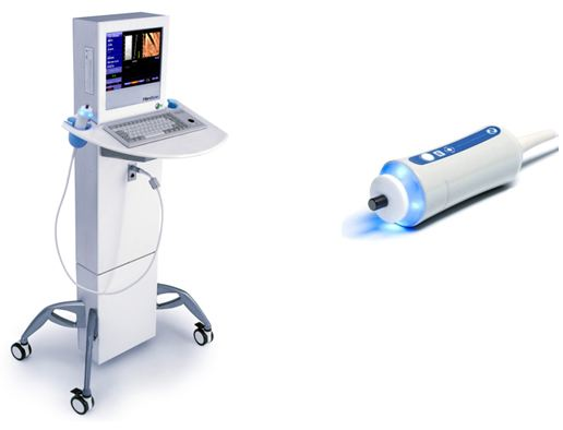
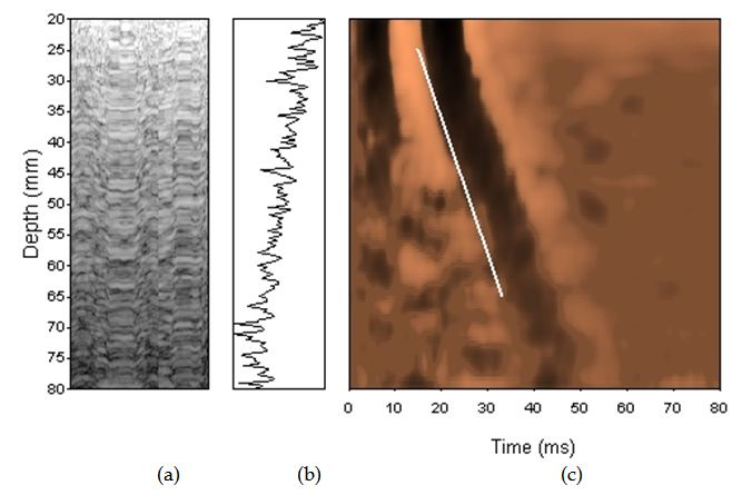
Figure 1. Transient elastrography

In the newest study of Krawczyk et al. TE correlated with Laennec stages of fibrosis, collagen contents and with diameter of thickest septa in explanted livers in PSC patients. In multivariate model liver fibrosis according to either Leannec score or collagen contents was significantly associated with TE. PSC cirrhotics patients had increased liver stiffness and the TE cut-off of 13.7 kPa showed the best predictive value (AUC=0.90, 95%CI 0.80–1.00, P<0.0001) for detecting liver cirrhosis [57].

The measurement failure rate is low (5-10%) with obesity (BMI > 30 kg/m2), ascites, congestive heart failure, postprandial time and the presence of narrow intercostal space considered to be limiting factors. However, obstructive cholestasis also influenced the results of TE [22].

Newer elastography methods based on the measurements of shear wave velocity include point share wave elastography (pSWE) and two-dimensional SWE (2D- SWE). SWE is usually integrated into conventional ultrasonography system (Figure 2). The region of interest (ROI) can be positioned under brightness-modulation (B-mode), and a single acoustic impulse is used to induce a share wave within a ROI of 1.0 x 0.5 cm or 2 x 2 cm in 2D SWE. The examination should be performed at least 1 cm below the liver capsule on the right lobe, and can be displayed in m/s and/or kPa. ROI can be positioned manually in different depths of the liver. However, there are no clear interpretation of point SWE and 2D SWE recommended to date.

IMROJ 2017-208 Figure2A

IMROJ 2017-208 Figure2B

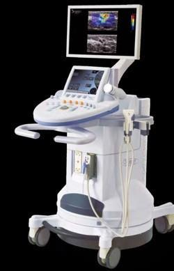
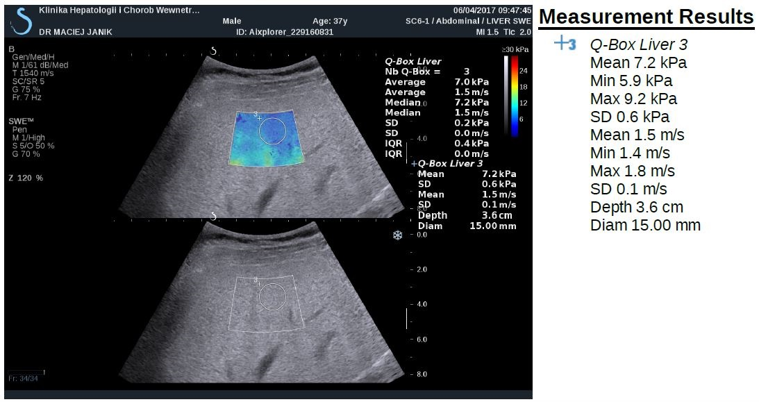
Figure 2. Shear-wave elastography

The probability of correctly diagnosing EV following a positive measurement did not exceed 70% [21]. Thus, LSM-spleen diameter to platelet ratio score and simplified combination of LSM and platelet count were also assessed with good results of ruling out varices needing treatment [23, 24]. LSM can be also used to predict clinical decompensation in the patients with compensated cirrhosis of the liver. On the other hand, spleen undergoes parenchymal modeling in patients with portal hypertension, and spleen stiffness measurement (SSM) is closely associated with portal hypertension, its severity and complications [25]. SSM is promising parameter for use in predicting the presence and size of EV [12]. Validated cut-off values in PSC are not available yet.

Magnetic-resonance based method

With magnetic resonance elastography (MRE) mechanical shear waves are sent into the tissue and displayed as elastograms using phase-contrast image sequences. MRE can examine the very large areas of the right lobe of liver. The limitation of MRE are obesity, claustrophobia and iron overload. Recently, in the study of Wang et al. the performance of MRE was significantly better than laboratory tests for detection of advanced fibrosis, and cirrhosis and better than conventional MRI for diagnosis of cirrhosis in patients with autoimmune hepatitis [26]. In a retrospective review of 266 PSC patients to examine whether liver stiffness (LS) was associated with the primary endpoint of hepatic decompensation (ascites, variceal hemorrhage and hepatic encephalopathy), MRE was able to detect cirrhosis with high specificity and LS obtained by MRE was predictive of hepatic decompensation in PSC patients in Eaton et al study. Liver stiffness of 4.93 kPa was the optimal point to detected F4 fibrosis, with sensitivity 1.00 (95% confidence interval (CI), 0.40-1.00) and specificity of 0.94 (95%CI, 0.68-1.00). LS was associated with the development of decompensated liver disease (Hazard ratio, 1.55; 95%CI, 1.41-1.70). The optimal LS thresholds that stratified patients at a low, medium and high risk for hepatic decompensation were <4.5, 4.5-6.0 and >6.0 kPa, respectively [27]. However, MRE seems to be promising modality for detection of advanced fibrosis and liver cirrhosis, with superior diagnostic accuracy compared to laboratory assessment and MRI, but not precirrhotic stages of chronic liver diseases. On the other hand, MRE is very expensive and time-consuming.

Serum biomarkers

Prospective studies demonstrated that single markers e. g., α2-macroglobulin [28], procollagen III N-peptide [29], apolipoprotein A1 [28], haptoglobin [30], hyaluronic acid [31], metalloproteinases [32] allow discrimination between advanced and absent fibrosis.

The enhanced liver fibrosis (ELF) test is a promising panel, incorporating three direct serum markers of fibrosis in an algorithm: hyaluronic acid, tissue inhibitor of metalloproteinases-1 (TIMP-1), and amino-terminal pro-peptide of type III pro-collagen (PIIINP) [33]. The ELF test accurately predicted significant liver fibrosis and furthermore predicted clinical outcome in several independent populations and in patients with various aetiologies of chronic liver disease [34] as well as with PSC. The ELF test consistently predicted liver transplant-free survival in PSC patients independently of other risk factors or risk scores [35]. The ELF test distinguished between mild and severe disease defined by clinical outcome (transplantation or death) with an area under the curve of 0.81 (95% confidence interval [CI] 0.73-0.87) and optimal cutoff of 10.6 (sensitivity 70.2%, specificity 79.1%). In multivariate Cox regression analysis ELF score was associated with transplant-free survival independently of the Mayo risk score. The ELF test correlated also with ultrasound elastography in separate assessments [35]. In a large multicenter cohort, EFL test predicts prognosis in PSC and may be used for risk stratification in clinical follow up; optimally together with clinical prognostic scores may add incremental prognostic value [36].

Placental growth factor (PLGF), growth differentiation factor-15 (GDF-15) and hepatic growth factor (HGF) are involved in hepatic fibrogenesis. The panel of these three serum markers was useful for the detection of patients with advanced fibrosis and the risks described by the combinations of these markers were independent from other classical fibrosis risk factors. The set of markers may be a useful tool to monitor patients with chronic liver diseases during and after therapy [37] .

Inflammatory protein, i.e. IL-8 in bile and serum was an important indicator of disease severity and prognosis in patients with primary sclerosing cholangitis, and associated with transplant-free survival in multivariable analyses independently of age and disease duration, indicating an independent influence on PSC progression [38]. This is also in line with the results of the study of Buck et al [39]. Hepatic venous pressure gradient (HVPG) can reflect progression of disease in the precirrhosis stage. Portal hypertension is pathogenically related to liver injury and fibrosis [40] and that in turn these are associated with the activation of inflammatory pathways [41]. The novel inflammatory serum biomarkers (e.g. Il-1, Fas-R, VCAM, CD163) were significantly correlated with HVPG in patients with compensated cirrhosis in this study.

Autotaxin (ATX), which is involved in the synthesis of lysophosphatidic acid, is not only associated with pruritus but also indicates impairment of other health-related quality of life (HRQoL) aspects, liver dysfunction, and can serve as a predictor of survival [42]. Impairment of HRQoL might be also associated with vitamin D receptor (VDR) gene polymorphisms (rs1544410-BsmI; rs7975232-ApaI). ApaI polymorphisms in VDR may exert an effect on disease-related symptoms and quality of life in the study of 275 patients with PSC [43].

However, none of the proposed markers or panels have gained as much acceptance as the invasive approach [44]. This may be due to relatively high costs of marker measurements, and low sensitivity to discriminate between fibrotic, cirrhotic or steatotic liver lesions. As a result, no scores based on serum levels of hepatic fibrosis markers are actually regarded as definite methods upon which therapeutic decisions can be based. It might be that the combination of markers reflects the presence of significant liver fibrosis detected by elastography and histology and may also identify patients at risk presenting with low elastography values as proofed by Krawczyk M, et al. [37].

Simple laboratory tests

Laboratory-based methods for staging liver fibrosis include the FibroTest® [45], the serum aspartate aminotransferase/platelet ratio index (APRI) [46], the Fibrosis 4 (FIB-4) test [47], and the enhanced liver fibrosis test [48]. AST/ALT ratio [49] can also allow to discriminate between advanced and absent fibrosis.

However, these tests may detect cirrhosis, but their ability to reflect the stages of fibrosis in AIH is uncertain [50-54]. The result of the recent study of Anastasiou et al. showed that TE, NAFLD fibrosis score and FibroQ might help in evaluation of liver fibrosis in AIH, but without differentiating mild form from advanced stages of fibrosis in autoimmune hepatitis [55].

In the study of Krawczyk et al. TE correlated with Laennec stages of fibrosis, and with serum indices of liver injury, namely AST, bilirubin as well as FIB-4 and APRI scores in patients with PSC [57].

Conclusion

Primary sclerosing cholangitis (PSC) is a progressive biliary disease lacking medical treatment with currently no established tools to predict prognosis in the individual patient. The lack of biomarkers for risk stratification is an important obstacle to the development of therapy.

Liver fibrosis seems to be the strongest predictor of liver stiffness assessed with TE. TE correlates with liver fibrosis, markers of liver injury and portal hypertension in patients with PSC. It might be that TE is a reliable tool for non-invasive monitoring of PSC. It seems also that the combination of serum profibrotic biomarkers with evaluation of liver fibrosis with elastography may improve the non-invasive diagnostic utility for clinically significant fibrosis [56]. However, still the Enhanced Liver Fibrosis (ELF®) test and Mayo risk score proved to be stronger predictors of transplant-free survival in PSC [38].

Conflict of interest: Nothing to declare

References

  • Weismuller TJ, Trivedi PJ, Bergquist A, Imam M, Lenzen H, Ponsioen CY, et al. (2017) Patient Age, Sex, and Inflammatory Bowel Disease Phenotype Associate With Course of Primary Sclerosing Cholangitis. Gastroenterology 152:1975-84 e8.
  • Dickson ER, Murtaugh PA, Wiesner RH, Grambsch PM, Fleming TR, Ludwig J, et al. (1992) Primary sclerosing cholangitis: refinement and validation of survival models. Gastroenterology 103:1893-901.
  • Kim G, Lee SS, Baik SK, Cho YZ, Kim MY, Kwon SO, et al. (2016) The need for histological subclassification of cirrhosis: a systematic review and meta-analysis. Liver Int 36:847-55.
  • Kim SU1, Oh HJ, Wanless IR, Lee S, Han KH, et al. (2012) The Laennec staging system for histological sub-classification of cirrhosis is useful for stratification of prognosis in patients with liver cirrhosis. J Hepatol 57: 556-563. [crossref]
  • Rastogi A, Maiwall R, Bihari C, Ahuja A, Kumar A, Singh T, et al. (2013) Cirrhosis histology and Laennec staging system correlate with high portal pressure. Histopathology 62:731-41.
  • Wang W, Li J, Pan R, A S, Liao C (2015) Association of the Laennec staging system with degree of cirrhosis, clinical stage and liver function. Hepatol Int 9:621-6.
  • La Mura V, Nicolini A, Tosetti G, Primignani M (2015) Cirrhosis and portal hypertension: The importance of risk stratification, the role of hepatic venous pressure gradient measurement. World J Hepatol 7:688-95.
  • Garcia-Tsao G, Friedman S, Iredale J, Pinzani M (2010) Now there are many (stages) where before there was one: In search of a pathophysiological classification of cirrhosis. Hepatology 51:1445-9.
  • Tsochatzis E, Bruno S, Isgro G, Hall A, Theocharidou E, Manousou P, et al. (2014) Collagen proportionate area is superior to other histological methods for sub-classifying cirrhosis and determining prognosis. J Hepatol 60:948-54.
  • Rockey DC, Caldwell SH, Goodman ZD, Nelson RC, Smith AD; American Association for the Study of Liver Diseases (2009) Liver biopsy. Hepatology 49: 1017-1044. [crossref]
  • Dietrich CF, Bamber J, Berzigotti A, Bota S, Cantisani V, Castera L, et al. (2017) EFSUMB Guidelines and Recommendations on the Clinical Use of Liver Ultrasound Elastography, Update 2017 (Long Version). Ultraschall Med.
  • Berzigotti A (2017) Non-invasive evaluation of portal hypertension using ultrasound elastography. J Hepatol 67: 399-411. [crossref]
  • Ziol M, Handra-Luca A, Kettaneh A, Christidis C, Mal F, et al. (2005) Noninvasive assessment of liver fibrosis by measurement of stiffness in patients with chronic hepatitis C. Hepatology 41: 48-54. [crossref]
  • Corpechot C, Carrat F, Poujol-Robert A, Gaouar F, Wendum D, Chazouilleres O, et al. (2012) Noninvasive elastography-based assessment of liver fibrosis progression and prognosis in primary biliary cirrhosis. Hepatology 56:198-208.
  • Corpechot C1, El Naggar A, Poujol-Robert A, Ziol M, Wendum D, et al. (2006) Assessment of biliary fibrosis by transient elastography in patients with PBC and PSC. Hepatology 43: 1118-1124. [crossref]
  • Corpechot C, Gaouar F, El Naggar A, Kemgang A, Wendum D, Poupon R, et al. (2014) Baseline values and changes in liver stiffness measured by transient elastography are associated with severity of fibrosis and outcomes of patients with primary sclerosing cholangitis. Gastroenterology 146:970-9; quiz e15-6.
  • Berzigotti A, Seijo S, Arena U, Abraldes JG, Vizzutti F, Garcia-Pagan JC, et al. (2013) Elastography, spleen size, and platelet count identify portal hypertension in patients with compensated cirrhosis. Gastroenterology 144:102-11 e1.
  • Talwalkar JA, Kurtz DM, Schoenleber SJ, West CP, Montori VM (2007) Ultrasound-based transient elastography for the detection of hepatic fibrosis: systematic review and meta-analysis. Clin Gastroenterol Hepatol 5:1214-20.
  • Vergniol J, Foucher J, Terrebonne E, Bernard PH, le Bail B, et al. (2011) Noninvasive tests for fibrosis and liver stiffness predict 5-year outcomes of patients with chronic hepatitis C. Gastroenterology 140: 1970-1979. [crossref]
  • Singh S, Fujii LL, Murad MH, Wang Z, Asrani SK, Ehman RL, et al. (2013) Liver stiffness is associated with risk of decompensation, liver cancer, and death in patients with chronic liver diseases: a systematic review and meta-analysis. Clin Gastroenterol Hepatol 11:1573-84 e1-2; quiz e88-9.
  • Shi KQ, Fan YC, Pan ZZ, Lin XF, Liu WY, Chen YP, et al. (2013) Transient elastography: a meta-analysis of diagnostic accuracy in evaluation of portal hypertension in chronic liver disease. Liver Int 33:62-71.
  • Millonig G, Reimann FM, Friedrich S, Fonouni H, Mehrabi A, Buchler MW, et al. (2008) Extrahepatic cholestasis increases liver stiffness (FibroScan) irrespective of fibrosis. Hepatology 48:1718-23.
  • Abraldes JG, Bureau C, Stefanescu H, Augustin S, Ney M, Blasco H, et al. (2016) Noninvasive tools and risk of clinically significant portal hypertension and varices in compensated cirrhosis: The “Anticipate” study. Hepatology 64:2173-84.
  • Augustin S, Millan L, Gonzalez A, Martell M, Gelabert A, Segarra A, et al. (2014) Detection of early portal hypertension with routine data and liver stiffness in patients with asymptomatic liver disease: a prospective study. J Hepatol 60:561-9.
  • Colecchia A, Montrone L, Scaioli E, Bacchi-Reggiani ML, Colli A, Casazza G, et al. (2012) Measurement of spleen stiffness to evaluate portal hypertension and the presence of esophageal varices in patients with HCV-related cirrhosis. Gastroenterology 143:646-54.
  • Wang J, Malik N, Yin M, Smyrk TC, Czaja AJ, Ehman RL, et al. (2017) Magnetic resonance elastography is accurate in detecting advanced fibrosis in autoimmune hepatitis. World J Gastroenterol 23:859-68.
  • Eaton JE, Dzyubak B, Venkatesh SK, Smyrk TC, Gores GJ, Ehman RL, et al. (2016) Performance of magnetic resonance elastography in primary sclerosing cholangitis. J Gastroenterol Hepatol 31:1184-90.
  • Imbert-Bismut F, Ratziu V, Pieroni L, Charlotte F, Benhamou Y, Poynard T, et al. (2001) Biochemical markers of liver fibrosis in patients with hepatitis C virus infection: a prospective study. Lancet 357:1069-75.
  • van Zanten RA, van Leeuwen RE, Wilson JH (1988) Serum procollagen III N-terminal peptide and laminin P1 fragment concentrations in alcoholic liver disease and primary biliary cirrhosis. Clin Chim Acta 177:141-6.
  • Adams LA, Bulsara M, Rossi E, DeBoer B, Speers D, George J, et al. (2005) Hepascore: an accurate validated predictor of liver fibrosis in chronic hepatitis C infection. Clin Chem 51:1867-73.
  • Wong VS, Hughes V, Trull A, Wight DG, Petrik J, Alexander GJ (1998) Serum hyaluronic acid is a useful marker of liver fibrosis in chronic hepatitis C virus infection. J Viral Hepat 5:187-92.
  • Walsh KM, Timms P, Campbell S, MacSween RN, Morris AJ (1999) Plasma levels of matrix metalloproteinase-2 (MMP-2) and tissue inhibitors of metalloproteinases -1 and -2 (TIMP-1 and TIMP-2) as noninvasive markers of liver disease in chronic hepatitis C: comparison using ROC analysis. Dig Dis Sci 44:624-30.
  • Xie Q, Zhou X, Huang P, Wei J, Wang W, et al. (2014) The performance of enhanced liver fibrosis (ELF) test for the staging of liver fibrosis: a meta-analysis. PLoS One 9: e92772. [crossref]
  • Parkes J, Roderick P, Harris S, Day C, Mutimer D, et al. (2010) Enhanced liver fibrosis test can predict clinical outcomes in patients with chronic liver disease. Gut 59: 1245-1251. [crossref]
  • Vesterhus M, Hov JR, Holm A, Schrumpf E, Nygard S, Godang K, et al. (2015) Enhanced liver fibrosis score predicts transplant-free survival in primary sclerosing cholangitis. Hepatology 62:188-97.
  • de Vries EM, Farkkila M, Milkiewicz P, Hov JR, Eksteen B, Thorburn D, et al. (2017) Enhanced liver fibrosis test predicts transplant-free survival in primary sclerosing cholangitis, a multi-centre study. Liver Int.
  • Krawczyk M, Zimmermann S, Hess G, Holz R, Dauer M, Raedle J, et al. (2017) Panel of three novel serum markers predicts liver stiffness and fibrosis stages in patients with chronic liver disease. PLoS One 12:e0173506.
  • Vesterhus M, Holm A, Hov JR, Nygard S, Schrumpf E, Melum E, et al. (2017) Novel serum and bile protein markers predict primary sclerosing cholangitis disease severity and prognosis. J Hepatol 66:1214-22.
  • Buck M, Garcia-Tsao G, Groszmann RJ, Stalling C, Grace ND, Burroughs AK, et al. (2014) Novel inflammatory biomarkers of portal pressure in compensated cirrhosis patients. Hepatology 59:1052-9.
  • Bellot P, Garcia-Pagan JC, Frances R, Abraldes JG, Navasa M, Perez-Mateo M, et al. (2010) Bacterial DNA translocation is associated with systemic circulatory abnormalities and intrahepatic endothelial dysfunction in patients with cirrhosis. Hepatology 52:2044-52.
  • Frances R, Rodriguez E, Munoz C, Zapater P, De la ML, Ndongo M, et al. (2005) Intracellular cytokine expression in peritoneal monocyte/macrophages obtained from patients with cirrhosis and presence of bacterial DNA. Eur J Gastroenterol Hepatol 17:45-51.
  • Wunsch E, Krawczyk M, Milkiewicz M, Trottier J, et al. (2016) Serum Autotaxin is a Marker of the Severity of Liver Injury and Overall Survival in Patients with Cholestatic Liver Diseases. Sci Rep 6: 30847. [crossref]
  • Kempinska-Podhorodecka A, Milkiewicz M, Jablonski D, Milkiewicz P, Wunsch E (2017) ApaI polymorphism of vitamin D receptor affects health-related quality of life in patients with primary sclerosing cholangitis. PLoS One 12:e0176264.
  • Poynard T (2011) First-line assessment of patients with chronic liver disease with non-invasive techniques and without recourse to liver biopsy. J Hepatol 54:586-7.
  • Poynard T, Imbert-Bismut F, Ratziu V, Chevret S, Jardel C, Moussalli J, et al. (2002) Biochemical markers of liver fibrosis in patients infected by hepatitis C virus: longitudinal validation in a randomized trial. J Viral Hepat 9:128-33.
  • Angulo P, Bugianesi E, Bjornsson ES, Charatcharoenwitthaya P, Mills PR, Barrera F, et al. (2013) Simple noninvasive systems predict long-term outcomes of patients with nonalcoholic fatty liver disease. Gastroenterology 145:782-9 e4.
  • Shah AG, Lydecker A, Murray K, Tetri BN, Contos MJ, et al. (2009) Comparison of noninvasive markers of fibrosis in patients with nonalcoholic fatty liver disease. Clin Gastroenterol Hepatol 7: 1104-1112. [crossref]
  • Parkes J, Guha IN, Roderick P, Harris S, Cross R, Manos MM, et al. (2011) Enhanced Liver Fibrosis (ELF) test accurately identifies liver fibrosis in patients with chronic hepatitis C. J Viral Hepat 18:23-31.
  • Sheth SG, Flamm SL, Gordon FD, Chopra S (1998) AST/ALT ratio predicts cirrhosis in patients with chronic hepatitis C virus infection. Am J Gastroenterol 93: 44-48. [crossref]
  • Gutkowski K, Hartleb M, Kacperek-Hartleb T, Kajor M, Mazur W, Zych W, et al. (2013) Laboratory-based scoring system for prediction of hepatic inflammatory activity in patients with autoimmune hepatitis. Liver Int 33:1370-7.
  • Abdo AA (2006) Clinical presentation, response to therapy, and predictors of fibrosis in patients with autoimmune hepatitis in Saudi Arabia. Saudi J Gastroenterol 12: 73-76. [crossref]
  • Abdollahi M, Pouri A, Ghojazadeh M, Estakhri R, Somi M (2015) Non-invasive serum fibrosis markers: A study in chronic hepatitis. Bioimpacts 5: 17-23. [crossref]
  • Anastasiou J, Alisa A, Virtue S, Portmann B, Murray-Lyon I, Williams R (2010) Noninvasive markers of fibrosis and inflammation in clinical practice: prospective comparison with liver biopsy. Eur J Gastroenterol Hepatol 22:474-80.
  • Luth S, Herkel J, Kanzler S, Frenzel C, Galle PR, Dienes HP, et al. (2008) Serologic markers compared with liver biopsy for monitoring disease activity in autoimmune hepatitis. J Clin Gastroenterol 42:926-30.
  • O EA, Buchter M, H AB, Korth J, Canbay A, Gerken G, et al. (2016) Performance and Utility of Transient Elastography and Non-Invasive Markers of Liver Fiibrosis in Patients with Autoimmune Hepatitis: A Single Centre Experience. Hepat Mon 16:e40737.
  • Friedrich-Rust M, Poynard T, Castera L (2016) Critical comparison of elastography methods to assess chronic liver disease. Nat Rev Gastroenterol Hepatol 13: 402-411. [crossref]
  • Marcin Krawczyk, Joanna Ligocka, Mariusz Ligocki, Joanna Raszeja-Wyszomirska, Małgorzata Milkiewicz, et al. (2017) Does transient elastography correlate with liver fibrosis in patients with PSC? Laennec score-based analysis of explanted livers. DOI: 10.1080/00365521.2017.1370009

Additive Effect of Oral Insulin and an Oral GLP-1 Agonist on Postprandial Glucose Excursions in a Porcine Model

DOI: 10.31038/EDMJ.2017131

Abstract

Combination therapy of insulin and a GLP-1 agonist injections is an attractive therapeutic approach in patients with type 2 diabetes mellitus, offering robust glycemic control, a reduced risk of hypoglycemia and weight gain and complementary mechanisms of action. Available combinations of basal insulin preparations and GLP-1 agonists have been shown to be effective and well tolerated in clinical trials. An oral alternative for such combination therapy may improve patient comfort and adherence while potentially offering the added value of liver directed insulin. The aim of this study was to investigate the concomitant pre-prandial delivery of enteric-coated oral insulin (ORMD-0801) and oral exenatide (ORMD-0901) capsules in pigs. The capsules were delivered directly to the duodenum under endoscopic guidance and blood glucose concentrations were monitored over the ensuing three hours. Preprandial delivery of ORMD-0901 or ORMD-0801 fully prevented a post-meal glycemic excursion (p=0.002 and p=0.086 respectively). When given together, a sharp decline >50% of mean baseline values in glycemia was observed, suggesting an additive effect. Peak glycemia was 5.2-fold lower than mean peak values measured in control animals at ~75 min after feeding (p<0.0001). We conclude that co-administration of oral insulin and oral exenatide compared with its components was more effective in controlling postprandial glycemia. The robust additive effect suggests a potential for complementary and non-attenuating mechanisms of action of this oral combination.

Keywords

oral insulin, oral exenatide, ORMD-0801, ORMD-0901, combination therapy, T2DM

Introduction

Type 2 diabetes is characterized by a relative insulin deficiency in combination with hyperglucagonemia, insulin resistance and often obesity [1-3]. A common therapeutic approach to address these metabolic defects combines basal insulin analogs with glucagon-like peptide-1 (GLP-1), concomitantly providing an insulin replacement with the added and complementary benefit of incretin-mediated increases in post-prandial insulin, inhibition of glucagon secretion and weight loss. [2, 4-6]. Several such combinations are currently in different stages of clinical development, including the combination of insulin degludec and the GLP-1 agonist liraglutide (marketed under the brand name Xultophy), [7-9] and the GLP-1 agonist lixisenatide combined with insulin glargine (LixiLan) [10].

Both GLP-1 analogs and insulin are protein-based drugs, currently marketed as injectable preparations only. An oral preparation, whereby the drug is absorbed through the gastrointestinal tract (GIT) and directly into the porto-hepatic venous system, may be better received by patients and perhaps offer a more physiologic mechanism of action, mimicking the hepatic first-pass effect of endogenously secreted insulin and reducing the relative hyperinsulinemia associated with subcutaneous injections. We have previously reported on a specially designed base formulation of this nature that has been shown to enhance GIT absorption of the active proteins and protect them from hydrolysis in the gut [11]. The basis of the enabling technology lies in protein encapsulation, which protects the proteins during transit through the harsh acidic gastric environment and enables them to reach the small intestines, where the capsule is disintegrated in a pH-dependent manner. The plant-derived protease inhibitors (PIs) incorporated into the formulation, provide for further protection from pancreatic and brush border proteases in the small intestines. The approach mimics that observed in neonates, where PIs are naturally incorporated in the maternal colostrum to facilitate transport of critical macromolecules, such as immunoglobulins, growth factors and antigens across the intestinal epithelial barrier [12]. We present here the results of a pre-clinical factorial study examining the post-prandial pharmacodynamic effect in pigs of co-administration of oral human insulin (ORMD-0801) and oral Exenatide, a short-acting GLP-1 agonist (ORMD-0901), as compared to no treatment and the individual agents alone.

Methods and Materials

This study was approved by the Israeli Council of Animal Experimentation, Ministry of Health. Three fasting female, commercial pigs (Ibelin Farm, Israel; age: 3-4 months-old; weight 25-30 kg) were treated in up to eight sessions each, with a minimum two-day washout period between treatments. Animals were housed individually in concrete pens, in a temperature-controlled room (18–24 °C), on a 12:12 light-dark cycle (light hours [07:00 – 19:00]), with olfactory contact between animals. The pigs had free access to water by low pressure drinking nipples. Fasting animals were anesthetized with isoflurane (2L O2 per minute and 5% isoflurane), intubated and mechanically ventilated. Pigs were positioned on their left side while enteric-coated capsules were administered directly to the duodenum under endoscopic guidance. On test days when both ORMD-0801 and ORMD-0901 were administered, ORMD-0901 (150 mg) was delivered first, followed by the ORMD-0801 (8 mg) capsule within 2-10 minutes. The pigs were fed Denkavit powdered milk for pigs (10 g/kg body weight) 30 min after drug administration and after recovering from anesthesia (10-15 minutes). Blood glucose was monitored via blood samples periodically drawn from a central line catheter over the 240-min post-dosing period. Piglets were intravenously treated with gentamycin (100 mg/10 kg) after every experiment day, to avoid infection. In cases where glucose concentrations dropped below 30 mg/dL, pigs were administered pig chowder and glucose concentrations were monitored for 30 minutes thereafter. Upon completion of the set of experiments, animals were euthanized with intravenously administered pentobarbital 1ml/1.5kg body weight.

Results

When preprandially treated with ORMD-0901 alone or ORMD-0801 alone, blood glucose excursions were significantly curbed, as compared to untreated meals (Figure 1). When ORMD-0901 and ORMD-0801 were administered together, a robust drop in blood glucose which was significantly greater than following treatment with each drug alone, was observed (Figure 1; p-value < 0.001). Blood glucose concentrations dropped to hypoglycemic levels (~20mg/dL) which were 5.2-fold lower than mean peak values measured in control meals at ~75 min after feeding (p<0.0001) and remained at a low of >50% below baseline values until the end of the 180-minute monitoring session.

Figure 1. Blood glucose profiles following concomitant delivery of oral exenatide and oral insulin to pigs.

Figure 1. Blood glucose profiles following concomitant delivery of oral exenatide and oral insulin to pigs.

Fasting, commercial pigs were treated with ORMD-0901 (150 mg exenatide; squares), ORMD-0801 (8 mg insulin; triangles) or both ORMD-0901 and ORMD-0801 (circles) capsules, 30 minutes before caloric intake. Blood samples (1 mL) were periodically drawn throughout the 180-minute observation period to determine glucose concentrations.

Discussion

This pre-clinical factorial study assessed the feasibility of simultaneous oral delivery of two hormones with complimentary modes of action [13]. Oral co-administration of human insulin and exenatide, provided a robust antihyperglycemic effect, curbing postprandial glucose excursions. The extent of the combined effect suggests an additive effect, resulting in a glycemic drop to a significantly hypoglycemic range. In the proposed combination regimen, insulin deficiency is supplemented by exogenous insulin administration, which is expected to increase glucose utilization and retard hepatic glucose production through direct actions in muscle, adipose tissue and the liver. At the same time, the GLP-1 analog was integrated to stimulate endogenous glucose-responsive insulin secretion, inhibit glucagon secretion and slow gastric emptying [2]. In addition, the GLP-1 component has been reported to induce satiety, lead to decreased food intake and eventually reduce body weight [14]. Moreover, owing to the glucose-dependent insulinotropic effect of GLP-1 and because glucagon-like peptide-1 receptor agonists (GLP-1RAs) preserve or even augment the glucagon response to hypoglycemia [2,15] the GLP-1RA-insulin combination is likely to allow for a reduction in insulin doses.

The importance of the hepatoportal system and liver, as they relate to insulin, is well known, [16] but existence of a hepato-preferential effect of incretin hormones is less clear at this time. As with insulin, there is a hepato-systemic GLP-1 gradient in the portal circulation, with several fold higher GLP-1 concentrations than in the systemic circulation [17]. Accumulating data suggest a role for GLP-1 in the liver and portal system in glucose and fat metabolism, as well as in satiety [18]. Among its known hepatic effects, GLP-1 stimulates glycogen synthesis in isolated rat hepatocytes, [19] and suppresses hepatic glucose production independently of plasma insulin, glucagon and glucose levels [20]. In both animal models [21] and humans [22] treatment with exendin-4 or liraglutide decreased hepatic lipid content and inflammation and decreased hepatic steatosis. While the GLP-1R is expressed on hepatic portal vagal afferents, suggesting indirect mediation of GLP-1 effects by central nervous system (CNS) activity, evidence regarding the presence of GLP-1R on hepatocytes remains conflicting.

Several injectable combinations of long-acting basal insulin and long- or short-acting GLP-1 analogs are in different stages of clinical development. This study is unique in that we show a robust antihyperglycemic effect of the combination of both an oral short-acting insulin and an oral short-acting GLP-1 agonist. The oral absorption of insulin and GLP-1 analog is enabled by Oramed’s core technology and formulation. This proprietary technology is comprised of a combination of encapsulation to shield the polypeptide (GLP-1) during transit in the gastrointestinal tract and protease inhibitors that reduce its proteolytic degradation. The protease inhibitors are plant derived and comprised of α-chymotrypsin inhibitors. The encapsulation consist of a polymer with pH-dependent solubility that relies on pH gradients in the intestine for site-specific dissolution [23]. Taken together, the encouraging results presented here, along with the projected long-term clinical benefits of the combined therapy, warrant clinical studies with such short – prandial combinations in patients with diabetes.

The limitations of the study include the absence of a true placebo/sham endoscopic procedure, the lack of pharmacokinetic (PK) analyses for both exenatide and insulin and the small number of animals examined. Given that the study was a preliminary study, it was deemed acceptable to withhold administration of a placebo before the meal challenge. As for insulin PK, the clear hypoglycemic effect suggests a significant increase in peripheral insulin. Whether this is the result of the administered oral insulin or endogenously secreted insulin in response to the effect of exenatide in the context of a meal and absorbed glucose remains to be determined. Since up to 80% of secreted insulin is sequestered within the liver upon first-pass metabolism, resulting in minimal doses in the peripheral circulation [24, 25] estimation of insulin bioavailability and PK analyses would not have provided useful information, unless there is direct sampling from the portal vein . Furthermore, under physiologic circumstances, endogenous secretion of insulin is a dynamic process, whereby the levels of secreted insulin change from moment to moment depending on ambient glucose levels [26]. As for exenatide PK analyses, non-proprietary analytical methods for the quantitation of exenatide, such as immunoassay (ELISA) and high-performance liquid chromatographic–tandem mass spectrometry (HPLC/MS/MS) method are plagued by low precision, poor repeatability and specificity and also potential interferences by endogenous compounds [27]. Only recently has a commercial and validated enzyme-linked immunosorbent assay kit (Phoenix Pharmaceuticals, Inc., Burlingame, CA, USA) become available for procurement. Nonetheless, the pharmacodynamic observations of the additive effect of the combination treatment compared to no treatment or to treatment with insulin or exenatide alone are compelling and it is unlikely that this significant effect could be attained short of the combined effect of insulin and exenatide.

Authorship

MK: Conception and design of study, drafting and revising manuscript
YGS: Acquisition of data, analysis and interpretation
RE: Conception and design of study, drafting and revising manuscript
EA: Conception and design of study, drafting and revising manuscript

Acknowledgements

The authors would like to acknowledge and thank Yehudit Posen, Ph.D for her invaluable assistance in preparing this manuscript.

Abbreviations

CNS: central nervous system
ELISA: enzyme-linked immunoabsorbent assay
GIT: gastrointestinal tract
GLP-1: glucagon-like peptide-1
GLP-1RA: glucagon-like peptide-1 receptor agonist
HPLC: high performance liquid chromatography
MS: mass spectrometry; PI: protease inhibitor
PK: pharmacokinetics

Competing Interests

MK is the CTO and hold shares at Oramed Pharmaceuticals.
YGS, RE and EA are consultants at Oramed.

References

  • Inzucchi S, Bergenstal R, Buse JB, Diamant M, et al. (2012) Management of hyperglycaemia in type 2 diabetes: a patient-centered approach. Position statement of the American Diabetes Association (ADA) and the European Association for the Study of Diabetes (EASD).
  • Holst JJ, Vilsbøll T (2013) Combining GLP-1 receptor agonists with insulin: therapeutic rationales and clinical findings. Diabetes, Obesity and Metabolism.
  • Ahrén B (2013) Incretin dysfunction in type 2 diabetes: clinical impact and future perspectives. Diabetes & Metabolism.
  • Balena R, Hensley I, Miller S, Barnett A (2013) Combination therapy with GLP-1 receptor agonists and basal insulin: a systematic review of the literature. Diabetes, Obesity and Metabolism.
  • Eng C, Kramer CK, Zinman B, Retnakaran R (2014) Glucagon-like peptide-1 receptor agonist and basal insulin combination treatment for the management of type 2 diabetes: a systematic review and meta-analysis.
  • Young LA, Buse JB (2014) GLP-1 receptor agonists and basal insulin in type 2 diabetes. Lancet 384: 2180-2181. [crossref]
  • Gough SC, Bode B, Woo V, Rodbard HW, et al. (2014) Efficacy and safety of a fixed-ratio combination of insulin degludec and liraglutide (IDegLira) compared with its components given alone: results of a phase 3, open-label, randomised, 26-week, treat-to-target trial in insulin-naive patients with type 2 diabetes. Lancet Diabetes Endocrinol.
  • Buse JB,  Vilsbøll T, Thurman J, Blevins TC, Langbakke IH, et al. (2014) Contribution of liraglutide in the fixed-ratio combination of insulin degludec and liraglutide (IDegLira). Diabetes Care 37: 2926-2933. [crossref]
  • Gough SC, Bode BW, Woo VC, Rodbard HW, Linjawi S, et al. (2015) One-year efficacy and safety of a fixed combination of insulin degludec and liraglutide in patients with type 2 diabetes: results of a 26-week extension to a 26-week main trial. Diabetes Obes Metab 17: 965-973. [crossref]
  • Riddle MC, Aronson R, Home P, Marre M, et al. (2013) Adding once-daily lixisenatide for type 2 diabetes inadequately controlled by established basal insulin: a 24-week, randomized, placebo-controlled comparison (GetGoal-L). Diabetes Care.
  • Kidron M, Bar-On H, Berry EM, Ziv E (1982) The absorption of insulin from various regions of the rat intestine. Life Sci 31: 2837-2841. [crossref]
  • Drozdowski LA, Clandinin T, Thomson A (2010) Ontogeny, growth and development of the small intestine: Understanding pediatric gastroenterology. World J Gastroenterol 16:787-99.
  • Ahrén B (2014) Insulin plus incretin: A glucose-lowering strategy for type 2-diabetes. World J Diabetes 5: 40-51. [crossref]
  • Steinert RE, Beglinger C, Langhans W (2016) Intestinal GLP-1 and satiation: from man to rodents and back. Int J Obes (Lond) 40: 198-205. [crossref]
  • Farngren J, Persson M, Schweizer A, Foley J, et al. (2014) Glucagon dynamics during hypoglycaemia and food-re-challenge following treatment with vildagliptin in insulin-treated patients with type 2 diabetes. Diabetes Obes Metab.
  • DeFronzo RA, Ferrannini E (1987) Regulation of hepatic glucose metabolism in humans. Diabetes Metab Rev 3: 415-459. [crossref]
  • Holst JJ (2007) The physiology of glucagon-like peptide 1. Physiol Rev 87: 1409-1439. [crossref]
  • Sandoval DA, D’Alessio DA1 (2015) Physiology of proglucagon peptides: role of glucagon and GLP-1 in health and disease. Physiol Rev 95: 513-548. [crossref]
  • Valverde I, Morales M, Clemente F, López-Delgado MI, Delgado E, et al. (1994) Glucagon-like peptide 1: a potent glycogenic hormone. FEBS Lett 349: 313-316. [crossref]
  • Seghieri M, Rebelos E, Gastaldelli A, Astiarraga BD, Casolaro A, et al. (2013) Direct effect of GLP-1 infusion on endogenous glucose production in humans. Diabetologia 56: 156-161. [crossref]
  • Ding X, Saxena NK, Lin S, Gupta NA, Anania FA (2006) Exendin-4, a glucagon-like protein-1 (GLP-1) receptor agonist, reverses hepatic steatosis in ob/ob mice. Hepatology 43: 173-181. [crossref]
  • Eguchi Y, Kitajima Y, Hyogo H, Takahashi H, et al. (2015) Pilot study of liraglutide effects in non-alcoholic steatohepatitis and non-alcoholic fatty liver disease with glucose intolerance in Japanese patients (LEAN-J). Hepatol Res.
  • Karsdal MA,  Riis BJ, Mehta N, Stern W, Arbit E, et al. (2015) Lessons learned from the clinical development of oral peptides. Br J Clin Pharmacol 79: 720-732. [crossref]
  • Eaton RP, Allen RC, Schade DS (1983) Hepatic removal of insulin in normal man: dose response to endogenous insulin secretion. J Clin Endocrinol Metab 56: 1294-1300. [crossref]
  • Meier JJ, Holst JJ, Schmidt WE, Nauck MA (2007) Reduction of hepatic insulin clearance after oral glucose ingestion is not mediated by glucagon-like peptide 1 or gastric inhibitory polypeptide in humans. Am J Physiol Endocrinol Metab 293:E849-E856.
  • Bergman R (2000) Non-esterified fatty acids and the liver: why is insulin secreted into the portal vein? Diabetologia 43:946-952.
  • Zhang J-F, Sha C-J, Sun Y, Gai Y-Y, et al. (2013) Ultra-high-performance liquid chromatography for the determination of exenatide in monkey plasma by tandem quadrupole mass spectrometry. J Pharm Analysis.

Melanin and Genomic Instability in Human Cancer

DOI: 10.31038/CST.2017247

Abstract

Nothing makes sense except considering evolution, by which understanding how life originates it is important to be able to decipher the mysteries that lead to the abnormal behavior of the cancer cell. So far, the emergence of life is explained in religious terms, but in scientific terms, it was not possible. The various existing theories had gaps that could not be resolved. The discovery of unsuspected capacity of melanin transform light into chemical energy, such as chlorophyll in plants; It is a watershed in biology, it turns out that melanin is the missing link in the chain of events that leads to the emergence of life. Now the Outlook is different, we can advance faster in the study and treatment of diseases that currently constitute a scourge to mankind, such as cancer. We are on the threshold of a new era in biology and in medicine.

Introduction

The unsuspected bio-energetic role of melanin has been described previously [1]. Electromagnetic radiation, melanin and water are substances that do not present any data that allow us to think that they have some thought as an evolution process. You could say that its main physical and chemical characteristics are the same since the beginning of time.

What we call evolution, can realize to the biochemical scaffolding that nature has patiently developed over eons of years, to increasingly optimize the properties of light, melanin and water, which have not changed a whit.

Eukaryote cell is becoming more sophisticated, biochemical gear is increasingly complex, and what is most striking, the power supply remains the same since the beginning of time. This is: the first living entities, had similar amount of available chemical energy derived from the melanin that human beings today.

It is something that has not changed despite the passage of time. What, has changed continuously over time, is the number of molecules, organelles, enzymes, etc., have been bolted together to make increasingly more sophisticated, increasing the complexity of the cell. And the term sophisticated, applied to any system; means that it contains many parts.

Continues to call me the attention that the power source is still, on the one hand, the sunlight mainly, on the other hand, melanin, which has not changed in the slightest despite the course of millions of years; and so on the perfect substrate which is water.

It is interesting that one of the elements, melanin; is completely dark and we can’t see through it, either with the naked eye or with instruments, and to this we attribute the fact that its formula is not known. But, on the other hand; water is completely transparent, we observe it with the naked eye, and yet we don’t understand as the hydrogen and oxygen atoms that compose it are organized, due to diverse factors, i.e. because its arrangement is dynamic and not uniform.

The nature of light, we neither speak, don’t even understand if it is a wave or a particle, or both. So, from three natural phenomena which we know little or nothing, life originated. Melanin is the missing link between the different theories that try to explain how emerged those we named life.

Light, Melanin, and Water

The physical and chemical properties of light, melanin and water whether we understand them or not, they possess characteristics that seem to be immutable, they inner dynamic had not change in the slightest despite the passage of time. And it seems paradoxical that the union of these quite stable elements generates the very dynamic phenomenon that we call life; which is characterized by constant changes, in all times, everywhere.

Charles Darwin said that life originated in warm water, with low oxygen levels, and without any other form of present life. And it is all spent out there, because the amniotic fluid is warm water with low oxygen levels and without any other way of life.

The evolution of the human eukaryote cell, requiring billions of years of evolution, based on primitive cells, is repeated in each pregnancy, and reducing days. Thus, the initial cells during pregnancy are primitive cells, whose structure is not as complex as mature or specialized eukaryote cell.

But despite differences that may be, at least in appearance; different cell lines retain the same fundamental process of generation and distribution of energy from the melanin.

In eukaryotes normal cells, of any cell line; organelles are very similar, even in size; which reflected a surprisingly uniform pattern, which cannot be a result of chance. And that consistency comes from the origin of life, as both light, melanin, and water, are very dynamic elements but do not change, do not seem to evolve in the least; It seems that they reached perfection and do not need to go further.

What if it evolves, and does so constantly, all the time, is what we call life. And one of the features of the evolution, is the increasingly complex management, in many respects, from the chains of carbon mainly from photosynthesis-derived glucose, this is: a homogenously arrangement of carbon, oxygen, and hydrogen atoms, with CnH2nOn as general formula, and whose most soluble example is glucose. While more advanced is a cell in the evolution, more efficient is the way in which atoms and carbon chains entwine each other forming increasingly intricate and complicated processes and structures, but in the end, reflects an increasingly efficient use of the energy that emanates from the melanin in the form of molecular hydrogen and high-energy electrons. A kind of energy astonishing accurate.

The proliferative cell

It is a deep-rooted dogma that normal differentiated cells, rely primarily on mitochondrial oxidative phosphorylation to generate energy needed to impel cellular processes.

So, it is not surprising that the metabolism of embryonic cells present findings consistent with the Warburg effect [2], or aerobic glycolysis. Which is an inefficient way to generate adenosine 5´-triphosphate (ATP), allowing to incorporate nutrient into biomass, rather than efficient and too much complicated, ATP production.

However, mitochondria and ATP biological functions are regulation of temperature and phosphate levels, but no energy production. Therefore, ATP biological function is regarding temperature control and phosphate levels, compounds that are chemically unstable but thermodynamically stables.

Thereby, embryonic cells require generating biomass so quickly, and gradually more and more complicated processes appear inside these cells, reflected, i.e.; by the increasing mitochondria number, whose function is the temperature control as much as possible, due to the chemical reactions inside normal differentiated cells are astonishing accurate, and temperature is no exception.

By other hand, uncontrolled proliferation is due to the cells that turn back in evolution, being the explanation due to the generation and distribution of melanin´s energy has been affected chronically.

Cancer cells are not generally controlled by normal regulatory mechanisms [3]. Thereby, the characteristic disordered tumor growth is dependent of difficult-to-understand mixture of local factors. A good example are the processes involved in angiogenesis and vascular remodeling implied in the vascularization of malignant tumors.

Total spectrum of morphogenic and molecular events required to form a neovascular network are way beyond our abstraction capacity, because casualness is a significant factor. And worst, those events are significantly different of normal vasculogenesis.

But in the light of the bio-energetic unsuspected role of melanin, we can reconsider such processes initiating the analysis as follows: the blood cannot carry energy, and on the other hand, the amount of chemical energy available in a cancer cell, is significantly lower than in a normal cell, therefore we have a series of events that reflect a step backwards in the evolution of the diseased cell and their chemical and anatomical, physical characteristics indicate that address.

While more evolved it is a cell, its operation is farther from random, and vice versa.

The intricacy of the processes that make up a specialized cell, they make these increasingly more and more precise, and one of the purposes of evolution is gradually reducing the interference of random in intracellular biochemical processes. Since any alteration in its sequence, temporality, or spatial location, would have devastating effects on perfectly than a specialized eukaryote cell represents.

Anti-vascular therapy of cancer

It has been over 30 years since Judah Folkman hypothesized that tumor growth is angiogenesis dependent [4]. But after years of clinical trials based in Dr. Folkman theory, results have been disappointing. Inhibition of VEGF, the main molecular mediator in capillary sprouting has been insufficient to halt tumor progression permanently in many cancer types.

The reasons can be diverse. i.e. the existence of multiple vascularization mechanisms and additional growth factor pathways. In our experience, the main cause of this failed therapeutic focus is that blood cannot carry on energy, just cell´s building blocks as glucose, aminoacids, lipids, etc., and CO2.

Cancer cells

Most solid tumors display distinct aneuploid karyotypes (abnormal chromosomal numbers) and frequently miss-segregate whole chromosomes in a phenomenon called chromosomal instability (CIN). CIN positively correlates with poor patient prognosis, indicating that reduced mitotic fidelity contributes to cancer progression by increasing genetic diversity among tumor cells [5].

Mechanisms leading to the loss of mitotic fidelity in CIN are not known. A common mitotic defect in tumor cells with CIN is the persistence of erroneous attachments of chromosomes to spindle microtubules (merotely).

In normal diploid cells erroneous attachments arise spontaneously and are efficiently corrected to preserve genomic stability. Paradoxically, kinetochore microtubule attachments in cancer cells with CIN are more stable than those in normal diploid cells, thereby, accounting for the persistence of mal-attachments into anaphase, causing chromosome miss-segregation. Seems that cancer cells have a diminished capacity to correct erroneous kinetochore-microtubules attachments; a dysfunction with a widespread occurrence of CIN in tumors.

The mechanisms involved in ploidy protection and genomic integrity are highly complex, not understood, and astonishingly accurate. In example, initiation of a new round of DNA replications should be restricted until after completion of the previous mitosis. Thereby DNA replication depends of biochemical pathways that occur exactly in time, space, location, amount; etc., in an amazing way; but are largely unknown.

The failures in these poor-understood regulatory mechanisms are generalized. Therefore, the plausible explanation is energy. Other theories proposed in the scientific literature, trying to explain abnormalities observed in cancer cells, has been proved elusive, given the inherent complexity of specialized eukaryotic cell after four billion yeas of evolution.

However, we are in front of a new era in the molecular biology of cancer cells; thanks to unsuspected bio-energetic role of melanin. And the explanation seems as quite simple: a specialized eukaryotic cell whose levels of generation and distribution of energy (from melanin) are impaired by contaminated water, polluted air, pesticides, herbicides, fertilizers, transition metals, heavy metals, addictive drugs, solvents of diverse types, industrial waste, stress, etc., tends to turn back in evolution. Simply because the complicated biochemical cell scaffolding that gives vital and highly-ordered support to the poorly understood specialized eukaryotic cell, requires astonishingly accurate amounts of energy.

Insofar as the available quantities of energy inside the cell to reduce enough, for example, in time and form, the very complicated biochemical pathways start to collapse in a disorderly way. Apparently, keeping only the most basic functions; or at least the first that appeared during the evolution, which gives us an idea that cell not is being able to keep relatively recent occurrence mechanisms.

In humans, aneuploidy is linked to pathological defects such as development abnormalities, mental retardation, or cancer, but the underlying mechanisms remain elusive. There are many types of aneuploidy whose origin remains unknown. A common response independent of the type of aneuploidy can be a novel target for cancer therapy.

Genomic stability requires that genetic material must be equally distributed between the two daughter cells during mitosis. An apparently straightforward process, by far beyond our abstraction capacity. By other side, an aberrant number of chromosomes, or aneuploidy, has been recognized as a feature of human malignancies for over a century, but until today without compelling evidence of causality.

Molecular logic underlying proper and aberrant chromosome segregation are extremely complex. It is undoubtedly that chromosome instability is detrimental for the fitness and survival of normal cells, being also the hallmark of cancer cells. Paradoxically, cells with an elevated proliferative potential, are also highly aneuploid.

Theoretically, Aneuploidy is a direct consequence of chromosome segregation errors in mitosis, while structural aberrations are caused by improperly repaired DNA breaks, but also exists a cross-talk between them. The long-lasting efforts to selectively inhibit proliferation of tumor cells has been infructuous.

Toxins against microtubules exert anti-tumor effects in some patients, but it is unclear its action´s mechanism. Apparently, these compounds acts interfering with microtubule dynamics during mitosis activates the spindle checkpoint, causing a prolonged mitotic arrest. Thereby, cells either then undergo death in mitosis or slippage; returning to interphase without dividing; but it is unclear what dictates the balance between these two fates.

Therapeutic inhibition of major mitotic kinases and kinesins has given a modest clinical activity at best.

Thereby, the maintenance of chromosomal stability involves the whole cell, not only genes. So, generation and distribution of energy from melanin has a fundamental role in cell biology (Figure 1).

Figure 1. Diagram of the hitherto unknown intrinsic capacity of melanin to transform visible and invisible light into chemical energy, dissociating the water molecule, as chlorophyll in plants.

Figure 1. Diagram of the hitherto unknown intrinsic capacity of melanin to transform visible and invisible light into chemical energy, dissociating the water molecule, as chlorophyll in plants.

The energy that melanin provides incessantly, night and day, in a quite consistently form, both in and outside the cell, explains finally, the origin of life.

In chlorophyll, the water dissociation is irreversible; but in melanin is reversible. The process is as follows:

2H2O → 2H2 + O2 →2H2O + 4e

The liquid water is sublimated by melanin into its gaseous components molecular Hydrogen (H2) and Oxygen (O2) in astonishing and accurate way, this is: always the product of the water dissociation is molecular hydrogen and molecular oxygen. Thereby, it is a quite precise process even in a diversity of environmental conditions, because melanin acts also as attemperator of surrounding sudden changes, for instance, a significant increase or decrease in the amount of light; keeping the output of H2 and O2 between narrow ranges.

The biochemical process of cell (and body) is amazing accurate, otherwise life is not possible; but this astonishing precise start since the very first step of life that is the energy production. Evolution can be interpreted as the increasing and gradual organization of the distribution of energy that is more and more exquisite, efficient; and precise as time pass by. Recall that is the same energy since the beginning of time, both in nature and amount.

The specialized eukaryotic cell does not generate more energy from melanin, because melanin energy generation has a top, a guarded proportion; unless more melanin is added to the system. Instead it is much more efficient in the distribution and thereby using of H2 and e- that the melanin dissociation and re-formation of water produces constant and exactly during night and day.

Eukaryote cell, mature, specialized; It is a whole that works perfectly in every one of its parts, starting from the generation and distribution of energy; which has been patiently optimizing over millions of years of evolution. You could say that nothing or almost nothing of what happens inside a specialized cell is the result of chance.

In any system, not only in biology; energy, defined as everything that produces a change; It is the most sensitive segment as an alteration in this area produces widespread failures, such as that seen in cancer cells.

Hence how difficult that has been to develop a successful therapy in cancer cells, it seems virtually impossible, because when designing strategies that act on a certain part of the cell, for example microtubules; the cell simply changes its behavior, which is now largely influenced by random, totally opposed to a normal cell; and quickly becomes resistant to instituted treatment.

And the explanation is that simply the imbalance now is expressed differently, but the actual background of the problem doesn´t change, and the proof is that proliferation does not diminish. Many of the deviations observed in a cancer cell should be taken care at the same time, so cell tends to function normally, as it has millions of years, millions of times.

Experiments in this regard have shown that partial reparation of the altered intracellular processes is not the solution, it is necessary to address the entire e ideally at the same time.

This is not currently possible, except to increase the generation and distribution of energy that comes from the melanin, and since all the biochemical processes that occur inside the eukaryote cell depend entirely on the H2 and e, to normalize these levels, all processes tend to operate normally, because ultimately, they are chemical reactions quite exact, that may not happen or inadequately occurs if the power level is not suitable.

QIAPI 1® and Cell Proliferation

The following Tables (1-4) show results of experiments carried out in cultured human cells. The results are part of the development of medication implemented in our laboratory, and which is in phase of patent in several countries, which has already been granted in several of them.

Table 1. WI-38 (human diploid cell line from normal embryonic (3 months gestation) lung tissue)

Table 1. WI-38 (human diploid cell line from normal embryonic (3 months gestation) lung tissue)

Table 2. A549 (human lung adenocarcinoma epithelial cell line)

Table 2. A549 (human lung adenocarcinoma epithelial cell line)

Table 3. HS 683 (human neuronal glioma cell line)

Table 3. HS 683 (human neuronal glioma cell line)

Table 4. Inhibitory concentrations of QIAPI 1®.

Cell line Concentrations of  QIAPI 1 inhibit 50% proliferation[mg/ml] Concentrations of  QIAPI 1 inhibit 99% proliferation[mg/ml]
WI-38 1,0 4,5
A549 2,0 5,5
HS-683 2,0 5,5

These results present them as a proof of concept, since the mechanism of action of QIAPI 1® is intensifying the dissociation and reformed the water, by the melanin molecule. (Tables 1-4)

The similarity in the results, although cultures of different cell lines, support our theory that acting on the generation and distribution of energy, cells, regardless of human classifications, they tend to recover complex order that is required so that their behavior is normal, proper; as millions of years, has done millions of times.

QIAPI 1® is a new therapeutic agent [6] that gradually will appear in the market, as legal requirements should be fitted.

Conclusion

The apparent chaos that reigns in efforts to decipher the bases of the anomalous behavior of cancer cells, appears to take a totally different direction when we do aside dogma that the main source of cell energy is glucose, then the discovery of unsuspected intrinsic property of melanin make visible and invisible light into chemical energy, such as chlorophyll in plants; through the dissociation of the water molecule, it is a watershed in cell biology.

We have a new view in the study of the biology of cancer, and while we move faster in that sense, the benefit that can give every day to numerous patients affected by the disease will be higher.

Acknowledgement: This work was supported by Human Photosynthesis® Research Center.

References

  • Solis-Herrera, A. Arias-Esparza, MC. Solis-Arias, RI. Solis-Arias, PE. Solis Arias, Martha P (2010) The unexpected capacity of melanin to dissociate the water molecule, fills the the gap between the life before and after ATP. Biomed Res 21: 224-227.
  • Vander Heiden, Matthew Cantley, Lewis, Thompson, Craig B (2009) Understanding the Warburg effect: The Metabolic Requirements of Cell Proliferation. Science 324: 1029-1033.
  • Döme, Balázs Hendrix, Mary JC, Paku, Sändor Tóvári, József, Timár, Jozsef (2013) Biological perspectives. Alternative Vascularization Mechanisms in Cancer. Pathology and Therapeutic implications. Am J Path.
  • Folkman J (1971) Tumor angiogenesis: therapeutic implications. N Engl J Med 285: 1182-1186. [crossref]
  • Compton, Duane (2013) Mechanism of chromosomal instability in human tumor cells. Geisel School of Medicine at Dartmouth, Hanover, USA.
  • Solis-Herrera, A Ashraf, Ghulam M, Arias-Esparza, MC Solis-Arias, et al. (2015) Biological Activities of QIAPI 1 as Melanin Precursor and its therapeutic effects in Wistar Rats exposed to Arsenic Poisoning. CNS Agen in Med Chem 2: 99-109.

Targeting of the Wnt/β-Catenin Pathway in Chronic Lymphocytic Leukaemia may adversely affect CTLA-4 expression and function

Abstract

In chronic lymphocytic leukemia, overexpression of CTLA-4 may be associated with a good outcome, whereas the Wnt/β-catenin-regulated transcription factor LEF1 is a pro-survival factor and is markedly overexpressed compared to normal B cells. In this study, peripheral blood B cells from 20 patients with CLL were purified and a strong correlation between gene expression levels of CTLA-4 and LEF-1 was found. This suggests that CTLA-4 expression in CLL may be a target of Wnt/β-catenin signalling.

Keywords:

CLL; CTLA-4; Wnt/β-catenin pathway; LEF1; CD38

Highlights

Percentage surface expression of CD38 and CTLA-4 and gene expression levels of CTLA-4, CCND1, LEF1 and STAT3 were measured in 20 patients with chronic lymphocytic leukemic. A strong positive correlation was found between gene expression levels of CTLA-4 and LEF-1.

Targeting of the Wnt/β-catenin pathway in CLL may result in unwanted effects on CTLA-4 expression and function.

Introduction

Chronic lymphocytic leukemia (CLL) is a clonal proliferation of mature CD5+ CD19+ CD23+ B lymphocytes, characterized by progressive accumulation of leukemic cells in peripheral blood, bone marrow and lymphoid tissues. Cytotoxic T-lymphocyte-associated antigen 4 (CTLA-4, CD152) is a member of the CD28 receptor family and is mainly expressed on CD4+ T-cells. In CLL, overexpression of the CTLA-4 gene is associated with lower CD38-expression and, therefore, perhaps a better outcome [1]. CLL cells also exhibit aberrantly active Wnt signaling and Wnt/β-catenin-regulated transcription factor lymphoid enhancer binding factor-1 (LEF1) has been shown to be a pro-survival factor in CLL [2]. In this study, we wished to further investigate the relationship between CD38 and CTLA-4 in CLL and their potential relationship with transcription factors LEF1, signal transducer and activator of transcription 3 (STAT3) and cyclin D1. In purified peripheral blood B cells from 20 patients with CLL, a strong positive correlation between gene expression levels of CTLA-4 and LEF1 was found, suggesting that CTLA-4 expression in CLL may well be a target of Wnt/β-catenin signalling.

Material and methods

After ethical approval and signed written consent, 20 patients with CLL (9 previously treated, 11 untreated) donated peripheral blood for this study. No patient had active therapy for CLL in the 3 months prior to blood donation. All patients had FISH analysis performed. CD19+ B lymphocytes were isolated using a magnetic bead separation technique (Invitrogen-Dynabeads). The percentage surface expression of CD38 and CTLA-4 was measured by flow cytometry. Total RNA was isolated from the B cells by the RNeasy Mini Kit (QIAGEN). Gene expression levels of CTLA-4, cyclin D1 (CCND1), LEF1 and STAT3 were measured using RT-PCR (ABI 7500 Fast-Applied Biosystems). GAPDH was used as a reference gene. Statistical analyses of data were performed using Spearman rank correlation and Mann-Whitney U tests. Differences of P < 0.05 were considered statistically significant.

Results

Median (range) CD19+ B cell purity was 93.8 (84.8-98.5) %, with CD19+ B cell purity > 90% in 19/20 cases. Median (interquartile range) percentage surface expression of CD38 and CTLA-4 was 8.36 (26.45) % and 43.32 (50.22) % respectively. Median (range, interquartile range) ∆CT gene expression levels of CCND1, CTLA-4, LEF1 and STAT3 were 11.89 (1.81), 4.79 (2.35), 4.82 (0.89) and 9.12 (0.75) respectively. Gene expression of LEF1 showed significant positive correlations with gene expression levels of CTLA-4 (rs=0.572, p=0.008), CCND1 (rs=0.61, p=0.004) and STAT3 (rs=0.587, p=0.006). There was also a significant positive correlation between gene expression of CCND1 and of STAT3 (rs =0.486, p=0.03). No significant correlations were found between percentage surface expression of CTLA-4 and gene expression levels of either CTLA-4 or of LEF1. Although we found a negative correlation between percentage surface expression of CTLA-4 and CD38, this was not statistically significant. Comparing untreated and previously treated patients or comparing patients with poor risk cytogenetics (17p or 11q deletions: n = 6) to those without, there was no significant difference in gene expression levels of CTLA-4, CCND1, LEF1 and STAT3 or in surface expression of CTLA-4 and CD38.

Discussion

The Wnt signalling pathway has been shown to be activated in CLL cells and uncontrolled Wnt/β-catenin signalling contributes to defective apoptosis in CLL [3]. Importantly, Wnt pathway activation leads to upregulation of β-catenin and subsequently LEF1 activation, which is markedly overexpressed in CLL compared to normal B cells [4] and appears to play an essential role in the leukaemogenesis of CLL [2]. Furthermore, cyclin D1, a downstream target of LEF-1, is overexpressed in CLL. Targeting of LEF-1 has been shown to induce apoptosis in CLL cells both in vitro and in vivo [5].

In CLL, CTLA-4 expression is higher on the leukemic cells that on their normal B cell counterparts. A recent study has shown that CTLA-4 inhibits the proliferation/survival of CLL cells via regulation of the expression/activation of STAT1, NFATC2, Fos, Myc and Bcl-2 [6] and CTLA-4 blockade induces pro-survival signals in leukemic cells from CLL patients exhibiting high CTLA-4 expression [7]. However, CTLA-4 expression was also found to be the most highly induced gene following treatment with recombinant Wnt-3a in melanoma cell lines and CTLA-4 expression appeared to be directly regulated by the Wnt/β-catenin pathway as the β-catenin responsiveness of CTLA-4 promoter region required a T-cell factor-1/LEF-1 consensus site [8]. In our study, CTLA-4 and LEF-1 gene expression levels were strongly correlated, suggesting that CTLA-4 expression in CLL may well also be a direct target of Wnt/β-catenin signalling. Although the relationship between CTLA-4 and the Wnt/β-catenin pathway in CLL requires further study, the findings of this study suggest that targeting of the Wnt/β-catenin pathway in CLL may result in unwanted effects on CTLA-4 expression and function.

This research did not receive any specific grant from funding agencies in the public, commercial, or not-for-profit sectors.

Conflicts of interest: none

References

  1. Joshi AD, Hegde GV, Dickinson JD (2007) ATM, CTLA4, MNDA and HEM1 in high versus low CD38 expressing B-cell chronic lymphocytic leukemia. Clin Cancer Res 13: 5295–5304. [Crossref]
  2. Gutierrez Jr A, Tschumper RC, Wu X (2010) LEF-1 is a prosurvival factor in chronic lymphocytic leukemia and is expressed in the preleukemic state of monoclonal B-cell lymphocytosis. Blood 116: 2975–2983. [Crossref]
  3. Lu D, Zhao Y, Tawatao R (2004) Activation of the Wnt signaling pathway in chronic lymphocytic leukemia. Proc Natl Acad Sci USA. 101: 3118–3123. [Crossref]
  4. Gandhirajan RK, Poll-Wolbeck SJ, Gehrke I, Kreuzer KA (2010) Wnt/β-catenin/LEF-1 signaling in chronic lymphocytic leukemia (CLL): a target for current and potential therapeutic options. Curr Cancer Drug Targets 10: 716–727. [Crossref]
  5. Gandhirajan RK, Staib PA, Minke K (2010) Small molecule inhibitors of Wnt/β-carenin/Lef-1 signaling induces apoptosis in chronic lymphocytic leukemia cells in vitro and in vivo. Neoplasia 12: 326–335. [Crossref]
  6. Mittal AK, Chaturvedi NK, Rohlfsen RA (2013) Role of CTLA4 in the proliferation and survival of chronic lymphocytic leukemia. PLoS ONE 8: e70352. [Crossref]
  7. Ciszak L, Frydecka I, Wolowiec D, Szteblich A, Kosmaczewska A (2016) Patients with chronic lymphocytic leukemia (CLL) differ in the pattern of CTLA-4 expression on CLL cells: the possible implications for immunotherapy with CTLA-4 blocking antibody. Tumour Biol 37: 4143–4157. [Crossref]
  8. Shah KV, Chien AJ, Yee C, Moon RT (2008) CTLA-4 is a direct target of Wnt/β-catenin signaling and is expressed in human melanoma tumors. J Invest Dermatol 128: 2870–2879. [Crossref]

Nanuristemogenea™: Advanced Biotechnology Applications Utilizing Next Generation Biocompatible Complexes of Stem and Hybridoma Immune Cells with Nanoparticles in the Field of Magnetic Guided Regenerative Medicine

DOI: 10.31038/CST.2017246

Commentary

The “biogenea pharmaceuticals” being the first biotech company in Southeast Europe, has the task to systematically promote the Science of Pharmaceutical Biotechnology, including the production of advanced therapy medicinal products ATMPs as defined by the European Medicines/Agency EMEA. On behalf of “biogenea pharmaceuticals” and its scientific board we are pleased to announce our innovative biotechnological services Nanostemogenea™ for the collection, processing, cryopreservation and therapeutic use of our advanced biotechnology applications utilizing next generation biocompatible complexes of stem, immune cells and nanoparticles in the field of magnetic guided regenerative medicine. Our GENEA cells ™ are totally safe and obtained from different sources of the human body for autologous cell therapy purposes in Renaissance medicine.

Biogenea Pharmaceuticals™ is the first Inter-Balkan Pharmaceutical Biotechnology Company since leading the way since 2005 in Red Biotechnology applications, in Cryobiology and in Autologous Cellular Therapy of Degenerative Diseases (cardiological diseases, neurological conditions and metabolic disorders).

Biogenea Pharmaceuticals focuses

  • On the collection, processing, cryopreservation and cGMP (according to Good Manufacturing Practice) production -for solely autologous use – of cellular therapeutical solutions from blood (bone marrow, peripheral blood, cord blood) or blood compounds for human use.
  • In collaboration with Regenetech on stem cell expansion technologies, which were created in the research laboratories of NASA (National Aeronautics and Space Administration).
  • On the cGMP production of advanced medicinal products (1394/2007/ΕC) for solely autologous use from skin, dental pulp, cord tissue). (In preclinical-research phase: 2008-2009).
  • On certified genetic analyses in collaboration with International Referral Centers.
  • On copyright protection according to the American and/or European Copyright Agency. The “biogeneapharmaceuticals” is the only European bank that has been recognized by the European Medicines Agency EMEA as a pharmaceutical company and has the possibility of cryopreservation of hematopoietic stem cells and conducting clinical trials (EMEA/Qualification of an enterprise as an SME – GrigoriadisBros – Biogenea- Cellgenea Ltd, with registration number: EMA/SME/084/10).

In “biogenea pharmaceuticals” taking advantage of the unique properties of super paramagnetic nanoparticles as this high magnetic moment and susceptibility but also the existence yperparagnitikis behavioural development multitude biological applications agglomerate formation with these petidika molecular and different kinds STEM CELL for autologous use in the Renaissance medicine.

Biogenea Pharmaceuticals Ltd combines excellent trained scientific personnel with the most modern techniques, concerning cell expansion, that are used today in the field of biotechnology and have been developed by NASA. Thus our company is able to verge into the demanding field of clinical trials concerning stem cell treatments. One can understand the big potential of these stem cells to play a role in Regenerative Medicine by looking at the amount of clinical trials all over the world that use stem cells as a way to treat an increasing number of diseases in a supportive manner.

The “biogenea pharmaceuticals” provides the following services:

  • Cardiogenea™: Autologous intracoronary, intraarterial or intracardiac injection of autologous blood, bone marrow and heart stem cells derive active cardiopoeitic spheres coated with superparamagneticnanoparticles for the restoration of myocardial infarction.
  • Dendrigenea™: Autologous adjuvant immune hybridomatic therapy for cancer patients using advanced complexes of superparamagnetic iron oxide nanoparticles coated mature dendritic and dendritic tumor cell fusions as a cancer ‘cell vaccine’.
  • Cartigenea™: Autologous and exvivo expanded chondrogenic cells coated with super paramagnetic nanoparticles for their intended use in Cartilage Defects.

Biogenea Pharmaceuticals Ltd is in the process of developing novel therapies for the effective treatment of several diseases. These therapies make use of Stem Cells (ADSCs) and a special class of nanoparticles, referred to as Super paramagnetic Iron Oxide Nanoparticles (SPIONs). SPIONs have recently attracted the interest of the scientific community, due to several attributes, such as their paramagnetism and their potential us in a number of therapeutic approaches. SPIONs are small synthetic γ-Fe2O3 (maghemite), Fe3O4 (magnetite) or α-Fe2O3 (hermatite) particles with a core ranging from 10 nm to 100 nm in diameter. In addition, mixed oxides of iron with transition metal ions such as copper, cobalt, nickel, and manganese, are known to exhibit superparamagneticproperties and also fall into the category of SPIONs. However, magnetite and maghemite nanoparticles are the most widely used SPIONs in various biomedical applications. SPIONs have an organic or inorganic coating so that they can be tolerated by cells and tissues.

Our Personalized cancer therapy is determined by the needs and specificities of a particular oncology patient to provide the optimum desired therapeutic effect with minimal toxicity, strengthening the immune system of cancer patients with the use TARGETED complex dendritic cells and super-paramagnetic nanoparticles in simultaneous increase the concentration of magnetic particles into the tumor.

Our technique serves the philosophy of personalized cancer treatment based on tumor pluripotent cells (cancer stem cells), consisting of four specialized areas in the prevention, diagnosis, treatment and rehabilitation of cancer patients while capitalizing on the therapeutic effects of magnetic hyperthermia the implementation of magnetic field for the local heating of the cancer cells with a view to the disaster.

Preventive Cryopreservation Insurance of Primordial Cells & Therapeutic Applications

  • Continuous update and prompt training of interested Donors and Clinicians of Public and Private Health Centers and Hospitals, about the progenitor cells applications.
  • Immediate availability of the stored samples 365 days a year, 7 days a week, 24 hours a day.
  • Complete attunement with the specifications set by the European Accreditation Organization for cellular therapy (FACT/JACIE/ NETCORD), the American Association of Blood Banks (AABB) and the relevant Greek.
  • Presidential Decretal 2004/23 for the tissue/cell banks, with the use of cGMP methods.
  • Control of Plasticity of progenitor cells based on the detailed Validation Master Plan of the Standard Operating Procedures (SOPs) regarding their hematopoietic origin (Methocult).
  • Fully Automated Viability Control of the cryopreserved progenitor cells with automatic luminometric device, which increases the reproducibility and the accuracy of the results in contrast to the common laboratory techniques for detecting dead cells microscopically (Trypan Blue staining).
  • Validated Molecular Diagnostics service provision (detection of HIV1/2, HBV, HCV, CMV, Syphillis, Toxoplasma) applying Real- Time PCR technology (Roche LightCycler).
  • Validated ex-vivo cellular and/or tissue expansion of the recently processed cells/tissues, as well as of the cryopreserved cell/ tissues in advanced technology Bioreactors, created in the NASA research laboratories, offering unique micro-gravity conditions in alternating electromagnetic field! Cellgenea is the UNIQUE company in Greece able to expand and to use ex-vivo expanded cord blood and adult progenitor cells for therapeutic reasons and clinical trials thanks to the relevant know-how transfer from the research laboratories of NASA and Regenetech Biotechnology Company.
  • Storing of progenitor cells in two-chambered bags and cryovials in complete (24 hours a day) controlled and automated liquid nitrogen tanks.
  • Continuous control and cross-tracking of the cryopreserved samples with validated LIS-ERP software which ensures the ability to track down and to identify the sample during all the steps of its supply, the processing, the control, the storage and the distribution. The tracking is also used for controlling and identifying all the relevant data about the products and the materials that come in contact with these samples (2004/23/ΕΚ).
  • Immediate availability and distribution of the sample inside a validated cryotank, in case of therapeutic application.
  • Strict security concerning personal data, confidentiality and safety according to the Personal Data Protection Agency.
  • Complete bacteriological and serological control of the samples using the automatic analyzers BacT/ALERT and Architect i1000 without any extra rate.
  • Life insurance provided to all the members of the different programs of preventive cryopreservation (Cellgenea, Dentogenea, Dermigenea, Angiogenea, DΝΑgenea, Neurogenea, Cardiogenea) in cooperation with insurance companies.
  • DNA & pharmacogenetic control services for personalized treatment without side effects.
  • Offer of validated prenatal and DNA/RNA control for chromosomal anomalies screening in a variety of biological materials (adult peripheral blood, cord blood, tissue biopsies) in cooperation with the Technological Park of Ioannina.
  • Continuous training and schooling of the scientific staff on the new

Nanuristemogenea™

Advanced biotechnology applications utilizing next generation biocompatible complexes of stem and hybridoma immune cells with nanoparticles in the field of magnetic guided regenerative medicine.

The “biogenea pharmaceuticals” being the first biotech company in Southeast Europe, has the task to systematically promote the Science of Pharmaceutical Biotechnology, including the production of advanced therapy medicinal products ATMPs as defined by the European Medicines/Agency EMEA.

On behalf of “biogenea pharmaceuticals” and its scientific board we are pleased to annpounce our innovative biotechnological services Nanostemogenea™ for the collection, processing, cryopreservation and therapeutic use of our advanced biotechnology applications utilizing next generation biocompatible complexes of stem, immune cells and nanoparticles in the field of magnetic guided regenerative medicine. Our GENEAcells™ are totally safe and obtained from different sources of the human body for autologous cell therapy purposes in Renaissance medicine.

Biogenea Pharmaceuticals™ is the first Inter-Balkan Pharmaceutical Biotechnology Company since leading the way since 2005 in Red Biotechnology applications, in Cryobiology and in Autologous Cellular Therapy of Degenerative Diseases (cardiological diseases, neurological conditions and metabolic disorders).

Biogenea Pharmaceuticals focuses

  • On the collection, processing, cryopreservation and cGMP (according to Good Manufacturing Practice) production -for solely autologous use – of cellular therapeutical solutions from blood (bone marrow, peripheral blood, cord blood) or blood compounds for human use.
  • In collaboration with Regenetech on stem cell expansion technologies, which were created in the research laboratories of NASA (National Aeronautics and Space Administration).
  • On the cGMP production of advanced medicinal products (1394/2007/ΕC) for solely autologous use from skin, dental pulp, cord tissue). (In preclinical-research phase: 2008-2009).
  • On certified genetic analyses in collaboration with International Referral Centers.
  • On copyright protection according to the American and/or European Copyright Agency. The “biogeneapharmaceuticals” is the only European bank that has been recognized by the European Medicines Agency EMEA as a pharmaceutical company and has the possibility of cryopreservation of hematopoietic stem cells and conducting clinical trials (EMEA/Qualification of an enterprise as an SME – GrigoriadisBros – Biogenea- Cellgenea Ltd, with registration number: EMA/SME/084/10).
  • In “biogenea pharmaceuticals” taking advantage of the unique properties of super paramagnetic nanoparticles as this high magnetic moment and susceptibility but also the existence yperparagnitikis behavioral development multitude biological applications agglomerate formation with these petidika molecular and different kinds STEM CELL for autologous use in the Renaissance medicine.
  • Biogenea Pharmaceuticals Ltd combines excellent trained scientific personnel with the most modern techniques, concerning cell expansion, that are used today in the field of biotechnology and have been developed by NASA. Thus our company is able to verge into the demanding field of clinical trials concerning stem cell treatments. One can understand the big potential of these stem cells to play a role in Regenerative Medicine by looking at the amount of clinical trials all over the world that use stem cells as a way to treat an increasing number of diseases in a supportive manner.
  • The “biogenea pharmaceuticals” provides the following services:
  • Cardiogenea™: Autologous intracoronary, intra-arterial or intracardiac injection of autologous blood, bone marrow and heart stem cells derive active cardiopoeitic spheres coated with super paramagnetic nanoparticles for the restoration of myocardial infarction. 
  • Dendrigenea™: Autologous adjuvant immune hybridomatic therapy for cancer patients using advanced complexes of super paramagnetic iron oxide nanoparticles coated mature dendritic and dendritic tumor cell fusions as a cancer ‘cell vaccine’. 
  • Cartigenea™: Autologous and ex-vivo expanded chondrogenic cells coated with super paramagnetic nanoparticles for their intended use in Cartilage Defects.

Biogenea Pharmaceuticals Ltd is in the process of developing novel therapies for the effective treatment of several diseases. These therapies make use of Stem Cells (ADSCs) and a special class of nanoparticles, referred to as Super paramagnetic Iron Oxide Nanoparticles (SPIONs). SPIONs have recently attracted the interest of the scientific community, due to several attributes, such as their paramagnetism and their potential us in a number of therapeutic approaches. SPIONs are small synthetic γ-Fe2O3 (maghemite), Fe3O4 (magnetite) or α-Fe2O3 (hermatite) particles with a core ranging from 10 nm to 100 nm in diameter. In addition, mixed oxides of iron with transition metal ions such as copper, cobalt, nickel, and manganese, are known to exhibit super paramagnetic properties and also fall into the category of SPIONs. However, magnetite and maghemite nanoparticles are the most widely used SPIONs in various biomedical applications. SPIONs have an organic or inorganic coating so that they can be tolerated by cells and tissues.

Our Personalized cancer therapy is determined by the needs and specificities of a particular oncology patient to provide the optimum desired therapeutic effect with minimal toxicity, strengthening the immune system of cancer patients with the use TARGETED complex dendritic cells and super-paramagnetic nanoparticles in simultaneous increase the concentration of magnetic particles into the tumor.

Our technique serves the philosophy of personalized cancer treatment based on tumor pluripotent cells (cancer stem cells), consisting of four specialized areas in the prevention, diagnosis, treatment and rehabilitation of cancer patients while capitalizing on the therapeutic effects of magnetic hyperthermia the implementation of magnetic field for the local heating of the cancer cells with a view to the disaster.

NanuristemogeneaTM Fig1

Preventive Cryopreservation Insurance of Primordial Cells & Therapeutic Applications

  • Continuous update and prompt training of interested Donors and Clinicians of Public and Private Health Centers and Hospitals, about the progenitor cells applications.
  • Immediate availability of the stored samples 365 days a year, 7 days a week, 24 hours a day.
  • Complete attunement with the specifications set by the European Accreditation Organization for cellular therapy (FACT/JACIE/ NETCORD), the American Association of Blood Banks (AABB) and the relevant Greek Presidential Decretal 2004/23 for the tissue/cell banks, with the use of cGMP methods.
  • Control of Plasticity of progenitor cells based on the detailed Validation Master Plan of the Standard Operating Procedures (SOPs) regarding their hematopoietic origin (Methocult).
  • Fully Automated Viability Control of the cryopreserved progenitor cells with automatic luminometric device, which increases the reproducibility and the accuracy of the results in contrast to the common laboratory techniques for detecting dead cells microscopically (Trypan Blue staining).
  • Validated Molecular Diagnostics service provision (detection of HIV1/2, HBV, HCV, CMV, Syphillis, Toxoplasma) applying Real- Time PCR technology (Roche Light Cycler).
  • Validated ex-vivo cellular and/or tissue expansion of the recently processed cells/tissues, as well as of the cryopreserved cell/tissues in advanced technology Bioreactors, created in the NASA research laboratories, offering unique micro-gravity conditions in alternating electromagnetic field! Cellgenea is the UNIQUE company in Greece able to expand and to use ex-vivo expanded cord blood and adult progenitor cells for therapeutic reasons and clinical trials thanks to the relevant know-how transfer from the research laboratories of NASA and Regenetech Biotechnology Company.
  • Storing of progenitor cells in two-chambered bags and cryovials in complete (24 hours a day) controlled and automated liquid nitrogen tanks.
  • Continuous control and cross-tracking of the cryopreserved samples with validated LIS-ERP software which ensures the ability to track down and to identify the sample during all the steps of its supply, the processing, the control, the storage and the distribution. The tracking is also used for controlling and identifying all the relevant data about the products and the materials that come in contact with these samples (2004/23/ΕΚ).
  • Immediate availability and distribution of the sample inside a validated cryotank, in case of therapeutic application.
  • Strict security concerning personal data, confidentiality and safety according to the Personal Data Protection Agency.
  • Complete bacteriological and serological control of the samples using the automatic analyzers BacT/ALERT and Architect i1000 without any extra rate.
  • Life insurance provided to all the members of the different programs of preventive cryopreservation (Cellgenea, Dentogenea, Dermigenea, Angiogenea, DΝΑgenea, Neurogenea, Cardiogenea) in cooperation with insurance companies.
  • DNA & pharmacogenetic control services for personalized treatment without side effects.
  • Offer of validated prenatal and DNA/RNA control for chromosomal anomalies screening in a variety of biological materials (adult peripheral blood, cord blood, tissue biopsies) in cooperation with the Technological Park of Ioannina.
  • Continuous training and schooling of the scientific staff on the new scientific developments.


Study of the Prognostic Marker Microrna-Mir-216a/217 in Hepatocellular Carcinoma Patients and Development of an Autologous Vaccine with Tumor-Lysate Pulsed Dendritic Cells, Genetically Modified for the Expression of the Mir-216a/217 and Hab18g/CD147 Antigen

DOI: 10.31038/CST.2017245

Proposal Description

Abstract

Objectives and task definition, based on the state of the art in terms of technology and knowledge. Detailed description of the problem which shall be solved, description of the beneficiary and user of the project results.

Study of the prognostic marker microRNA-Mir-216a/217 in hepatocellular carcinoma patients and development of an autologous vaccine with tumorlysate pulsed dendritic cells, genetically modified for the expression of the Mir-216a/217 and HAb18G/CD147 antigen.

Aim

The aim of the present protocol is the development of the techniques for the detection and quantification of the prognostic marker microRNA-Mir- 216a/217 in serum and liver tissue from hepatocellular carcinoma patients submitted to hepatectomy. Based on the above, an experimental (in vitro) protocol will be developed for the production and differentiation of dendritic cells derived from the peripheral blood of mononuclear cells, genetically modified for the expression of the microRNA-Mir-216a/217 and HAb18G/CD147 antigen and their maturation by the cell lysate from tumor resections of hepatocellular carcinoma patients. The ultimate goals of the present study are the early diagnosis of recurrence of hepatocellular carcinoma and the development of an autologous dendritic cell vaccine for additional immunotherapy of these patients.

Introduction

Hepatocellular carcinoma (HCC) is the third leading cause of cancer deaths worldwide with over 600,000 patients dying from this disease annually [1]. The age standardized incidence rates (ASR) of HCC in men in Europe, adjusted to the European Standard Population, is about 8 per 100,000, with a peak in Southern Europe of 10.5 per 100,000 (http://globocan.iarc.fr/). Liver transplantation and tumor resection have proven to be the most effective standard therapies and provide 5-year survival rates of 70% for patients within the Milan criteria, i.e., single tumour < 5 cm in size or up to three tumors < 3 cm in size [2]. These rates reach 50% with radiofrequency ablation and transarterial chemoembolization, which are the next preferred lines of therapy. However, these therapeutic procedures most often do not provide a complete cure, as half of the treated patients experience tumor recurrence within 3 years. A further major problem is that only 30–40% of patients are eligible for the above-described treatments due to the fact that HCC frequently remains undiagnosed until an advanced stage has been reached and occurs in the setting of advanced liver disease due to cirrhosis. These patients have a median survival time ranging from 3 to 16 months [3]. Thus, there is currently ongoing development and testing of alternative drug-based therapies for HCC that target tumor signalling pathways and vasculature. However, so far, the only drug that significantly prolonged survival (by nearly 3 months) in patients with advanced HCC is sorafenib, a multi-targeted tyrosine kinase inhibitor [4]. In view of these facts, new strategies are required. In this study we focus on the identification of predictive molecular factors for HCC recurrence and the development of immunotherapy methods for these patients.

Early Diagnosis of HCC Recurrence with Micro-RNA Techniques

MicroRNAs (miRNAs) are a class of small endogenously expressed non-coding RNAs. The miRNAs in blood, called circulating miRs (cmiRNAs) are potential biomarkers for detecting and monitoring cancer progression [5]. The ability of some miRNAs to function as tumor promoters (miR-30d, miR-151 and miR-210) or suppressors (miR-122, let-7g, miR-29b, miR-193b, miR-194, miR-139 and miR-124) in hepatocarcinogenesis have led to new insights into the molecular pathways involved in HCC [6]. Recently, upregulation of the miR-216a/217 cluster identified to be associated with the early HCC recurrent disease, by comparing the miRNA expression profiles of HCC liver tissue from patients with early-recurrent and non-recurrent disease [7]. In this protocol we intend to develop a technique for the detection and quantification of miR-216a/217 in the blood of HCC patients submitted to liver resection in order to identify recurrence of the disease.

Specific Description of the Proposed Solution

A sensitive and predictable method for the detection and identification of the microRNA216a/217 as the ideal biomarker for prognosis of Hepatocellular Carcinoma

Paraffin-embedded tissue analysis

Paraffin-embedded Hepatocellular Carcinoma (PE-HCC) Tissue samples of the primary tumor are obtained from HCC patients who underwent surgical treatment or biopsy at our centers.

RNA Extraction from serum samples

Total RNA was extracted from 500 μL of serum by using TRI reagent BD (Molecular Research Center). Ten sections, each 10- μm thick, were cut from each PE-HCC block. Deparaffinized tissue sections were digested by using proteinase K, and RNA was extracted by using a modified protocol of the RNAWiz Isolation kit (Applied Biosystems). The RNA was quantified and assessed for purity by using ultraviolet spectrophotometry and the Quant-iT RiboGreen RNA assay kit (Invitrogen) [8,9].

RT-qPCR assay with extracted RNA

10 ng of total RNA extracted from Hepatocellular tumor tissue, normal tissue, or serum are dissolved in 5 μL H2O (2 ng/μL) for reverse transcription with the addition of 5 μL of a reaction mixture containing 5Χ first strand buffer, 10 mmol/L deoxynucleoside-5’- triphosphate, RNasin, reverse transcriptase, and miR-specific RT primers (Exiqon). After the mixture was incubated in 37°C for 2 h, the transcribed specific cDNA was diluted 10-fold with molecular grade H2O before use as a qPCR template. Each qPCR contained 2.5 μL of diluted cDNA, 5 μL of 2ΧPerfeCTa SYBR Green FastMix for iQ (Quanta Bioscience), miR-specific, locked nucleic-acid– based forward primer targeting the specific microRNA216a/217 and universal reverse primer (Exiqon).

RT-qPCR directly in serum

This assay requireσ only a small aliquot of serum. To deactivate or solubilize proteins that might inhibit the RT-qPCR, 2.5_L of each serum sample are mixed with 2.5 μL of a preparation buffer that contained 2.5% Tween 20 (EMD Chemicals), 50 mmol/L Tris (Sigma- Aldrich), and 1 mmol/L EDTA (Sigma-Aldrich). 5 μL ofRT reagent mixture are added which contain the same RT reagents used for RT-qPCR with extracted RNA RNA, directly to 5 μL of serum in preparation buffer; a 2-h incubation at 37°C is followed by a 5-min enzyme inactivation at 95°C. The transcribed cDNA is diluted 10- fold by H2O and then centrifuged at 9000 g for 5 min to eliminate the protein precipitant. A 2.5-μL volume of the supernatant cDNA solution is used as the template for qPCR. qPCR conditions, primers and reagents, and data analysis were duplicated for those described in RT-qPCR with extracted RNA section.

Real time quantitative PCR (qPCR)

Real-time qPCRs were performed using SYBR Green PCR Master Mix and 7300 Realtime PCR System (Applied Biosystems, Foster City, CA USA). Real-time qPCRs were also used to detect miRs, as reported [10]. Sequences of miR-specific primers for cDNA synthesis and reverse primers for miR-216a were: 5′-CATGATCAGCTGGGCCAAGACACAGTTGCCAGCTG-3′ and 5′-TAATCTCAGCTGGCAA-3′. Primers for miR-217 were: 5′ CATGATCAGCTGGGCCAAGAATCCAGTCAGTT-3′ (cDNA synthesis) and 5′-TACTGCATCAGGAACT-3′ (reverse primer). miR expression levels were also confirmed by poly-adenylation of mature miR and cDNA synthesis primed by oligo-dT primer tagged with universal primer sequence (miScript System, Qiagen). cDNA was amplified using miR specific and universal primers. For miR-216a and miR-217 PCRs, the same reverse primers mentioned above and the primers provided by Qiagen were used as miR-specific primers. 5S RNA or 18S RNA served as internal control (Ambion) [7]. Genetically modified dendritic cells.

The limited improvements in clinical outcomes with dendritic cells loaded with tumor associated antigens (TAAs) led to trials with genetically modified DCs to further enhance antigen presentation and immunostimulation N. Recent studies have shown that the miR- 216A/217 cluster is consistently and significantly up-regulated in HCC tissue samples and in cell lines associated with early tumor recurrence, poor disease free survival and an epithelial mesenchymal transition (EMT) phenotype [7-31]. Stable over-expression of miR-216a/217- induced EMT, increased the stem like cell population, migration and metastatic ability of epithelial HCC cells. PTEN and SMAD7 are subsequently identified as two functional targets of miR-216a/217, and both PTEN and SMAD7 are down regulated in HCC. Ectopic Expression of PTEN or SMAD7 partially rescued miR216a/217 mediated EMT phenotype, cell migration and stem-like properties in HCC cells. Previously was shown that SMAD7 is a TGF-β1 antagonist. Recently, it has been also shown that Epithelial–mesenchymal transition (EMT) induced by transforming growth factor-β (TGF-β) is implicated in hepatocarcinogenesis and hepatocellular carcinoma (HCC) metastasis [32]. On the other hand, HAb18G/CD147, which belongs to the CD147 family, is an HCC-associated antigen that has a crucial role in tumor invasion and metastasis. Upregulation of HAb18G/CD147 is induced by TGF-β coupled promoting the idea that CD147 may be a potential target for the treatment and prevention of HCC. Other studies have also shown that overexpression of miR216a/217 act a positive feedback regulator for the TGF-b pathway and the canonical way involved in the activation of the PI3K/Akt/ Mtor in HCC cells [33,34].

These facts led us to the second proposal of our protocol:

Experimental (in vitro) protocol for the production and differentiation of dendritic cells derived from the peripheral blood fraction of mononuclear cells, genetically modified for the expression of the microRNA-Mir-216a/217 and HAb18G/CD147 antigen and their maturation by the cell lysate from tumor resections of hepatocellular carcinoma patients

1. MS2 VLP-based delivery of microRNA-216a/217 as a potential delivery approach for the production of clinical grade genetically modified monocyte derived dendritic cells for the immunization of patients suffered from Hepatocellular Carcinoma

Recent studies suggest that microRNA-216a/217 microRNA (miR-216a/217) plays an essential role in immunoregulation and may be involved in the pathogenesis of Hepatocellular Carcinoma. Therefore, it is of interest to investigate the potential therapeutic application of miR-216a/217-induced dendritic cells in Hepatocellular Carcinoma, a concept that has not been thoroughly investigated thus far. Virus-like particles (VLPs) are a type of recombinant nanoparticle enveloped by certain proteins derived from the outer coat of a virus. Herein, we describe a novel miRNA-delivery approach via bacteriophage MS2 VLPs and investigate the therapeutic effects of miR–216a/217-induced dendritic cells against patients suffering from Hepatocellular Carcinoma.

2. An Innovative Methodology for the miR216a/217 MicroRNA Delivery in peripheral blood monocyte derived antigen presenting dendritic cells via magnetic nanoparticles

Peripheral Blood Monocyte derived dendritic cells show promising potential in the vaccination of HCC patients. Recently, gene silencing strategies using microRNAs (miR) emerged with the aim to expand the therapeutic potential of genetically modified dendritic cells. However, researchers are still searching for effective miR delivery methods for clinical applications. Therefore, we aimed to develop a technique to efficiently deliver miR216a/217 microRNA into patient’s dendritic cells with the help of a magnetic non-viral vector based on cationic polymer polyethylenimine (PEI) bound to iron oxide magnetic nanoparticles (MNP), whose uptake efficiency and cytotoxicity will be determined by flow cytometry. The Present Proposal is directed in different magnetic complex compositions and determined. Additionally, the present proposal is focusing on the monitoring of the release, processing and functionality of delivered miR216a/217 microRNA with confocal laser scanning microscopy, real-time PCR and live cell imaging, respectively. On this basis, we will focus on the established parameters for construction of magnetic non-viral vectors with optimized uptake efficiency (~75%) and moderate cytotoxicity in patients peripheral blood monocyte derived dendritic cells. Furthermore, we aim to introduce the magnetic non-viral vector based on cationic polymer polyethylenimine (PEI) transfection as a long term beneficial strategy for the successful genetic modification of the HCC-TAAs presenting dendritic cells against Hepatocellular Carcerous cells for future in vivo applications.

Generation of peripheral blood monocyte-derived miR-216a/217 induced DCs

Monocyte-derived DCs from Hepatocellular Carcinoma Patients (obtained with following informed consent and approved by our institutional review board) are generated. In brief, peripheral blood mononuclear cells (PBMCs) are prepared from whole blood by Ficoll density-gradient centrifugation. The PBMCs are suspended in tissue culture flask in RPMI 1640 supplemented with 1% heat inactivated autologous serum for 60 minutes at 37°C to allow for adherence. The nonadherent cells are removed and the adherent cells are cultured overnight. To generate immature miR-216a/217 induced DCs (DCs), the nonadherent and loosely adherent cells are collected the following day and placed in RPMI 1640 medium containing 1% heat-inactivated autologous serum, 1000 U/ml recombinant human GM-CSF (Becton Dickinson, Bedford, MA, USA), and 500 U/ml recombinant human IL-4 (Becton Dickinson) for 6 days. In order to assess the effects of HCCsp on miR-216a/217 induced DCs generation, we are focusing on the creation of four types of miR-216a/217 induced preparation: 1) miR-216a/217 induced DCs; 2) miR-216a/217 induced s generated in the presence of HCCsp during the entire culture period (DCs/sp); 3) miR-216a/217 induced DCs exposed to 0.1 KE/ml (0.1 KE equals of 0.01 mg of dried streptococci) penicillin-inactivated Streptococcus pyogenes (OK-432) (Chugai Pharmaceutical) for 3 days (OK- miR- 216a/217 induced DCs) as described previously; 4) OK- miR-216a/217 induced s generated in the presence of HCCsp during the entire culture period (OK-DCs/sp). Four types of miR-216a/217 induced are generated in the presence of equal amounts of GM-CSF and IL-4 during the entire culture.

To generate monocyte-derived miR-216a/217 induced DCs for vaccination, PBMCs derived from the HCC patient are freshly isolated (obtained with following informed consent and approved by our institutional review board). Autologous miR-216a/217 induced s are generated in RPMI 1640 medium containing 1% heat-inactivated autologous serum, 1000 U/ml recombinant human GM-CSF, 500 U/ml recombinant human IL-4, and 10 ng/ml recombinant TNF-α (Becton Dickinson) [30]. On day 6 of culture, DCs harvested from the nonadherent and loosely adherent cells are used for fusion. The firmly adherent monocytes are harvested and used as an autologous target for the CTL assays.

HCC Patient selection

The clinical trial protocol will be approved by the Institutional Review Boards of our University Hospitals. Patients will be informed of the investigative nature of this study, and written consent in accordance with institutional regulations is obtained prior to study entry. Eligibility criteria included HCC patients submitted to liver resection or biopsy and transarterial chemoembolization (TACE) with radiological diagnosis of primary HCC by computed tomography (CT), classified in stage II and III according to the tumor-node-metastasis (TNM) classification; age over 20 years/both male and female; Eastern Cooperative Oncology Group scale 0–1; and indicators of acceptable hematological (hemoglobin ≥8.5 g/dl, white blood cells ≥2,000/mm3, platelet ≥50,000/mm3), hepatic (Child Pugh score ≤7, alanine aminotransferase, aspartate aminotransferase ≤5x upper normal limit) and renal (creatinine ≤1.5 mg/dl) function. Important exclusion criteria consisted of organ transplantation; a medical history of autoimmune disease, immunodeficiency, or autoimmune disease that might be aggravated by immunotherapy; not exceeding 2 weeks after antibiotic treatment needed due to a serious infectious disease; seropositivity for human immunodeficiency virus antigen; use of immunosuppressive drug such as cyclosporin A and azathioprine; any cardiopulmonary disability judged by the investigator; a medical history of psychological disease or epilepsy; and evidence of another active malignant neoplasm.

Autologous DC generation

DCs are generated from blood monocytes, as reported previously, with modifications. White blood cells obtained from the HCC patients through leukapheresis. DCs are prepared in a GMP-compliant facility at our hospitals. Peripheral blood mononuclear cells (PBMCs) are separated from WBC by Ficoll-Paque™ PLUS (Amersham Biosciences, Uppsala, Sweden) density gradient centrifugation. PBMCs are stored in a liquid nitrogen tank until necessary for DC generation. PBMCs thawed, ished with Hanks’ Balanced Salt Solutions, resuspended in RPMI-1640 medium (Lonza, Basel, Switzerland) supplemented with autologous heat-inactivated plasma, and then incubated in CellSTACK Culture Chambers (Corning, Corning, NY, USA). After 0.5–1 h incubation at 37°C in a 5% CO2 incubator, non-adherent cells are removed by gentle ishes.

The adherent monocytes are cultured in X-VIVO15 (Cambrex, East Rutherford, NJ, USA) supplemented with 100 ng/ml of granulocyte macrophage-colony stimulating factor (GMP grade: LG Life Science, Seoul, Korea) and 300 ng/ml of interleukin (IL)-4 (JW CreaGene Inc., Seongnam, Korea) for 5 days. On day 5, nonattached immature DCs are harvested and pulsed with CTP-fused human AFP, MAGE-1 and GPC-3 recombinant proteins at a final concentration of 5 μg/ml each. Antigen-pulsed dendritic cells are matured in the presence of cytokine cocktail, IL-6 (Peprotech, Rocky Hill, NJ, USA), IL-1β (Peprotech), tumor necrosis factor (TNF)-α (Peprotech), prostaglandin E2 (PGE2) (Sigma Chemical Co., St. Louis, MO, USA), interferon (IFN)-γ (LG Life Science), OK432 (Chugai Pharmaceutical Co., Tokyo, Japan), and poly I: C (Sigma) for 1 or 2 days depending on surface phenotypes and cell population. On day 6–7, the DCs are harvested, ished, and resuspended in 1.2 ml of cryopreserving solution containing 5% dimethyl sulfoxide (Bioniche Pharma USA, Lake Forest, IL, USA). Finally fully equipped DCs are packed into a sterile glass vial (4×10⁷ cells/vial), sealed with a snap-cap, and stored at an ultralow freezer for >12 h.

Quality control of dendritic cell vaccine

Safety test

For safety, endotoxin, germ-free and mycoplasma-free tests are performed according to the KFDA-approved JW CreaGene standard and test guidelines. Endotoxin is evaluated using gel-clot techniques. The endotoxin of the product should be less than 10 EU/ml per 1.2- ml vial. Mycoplasma test is performed by both direct culture and PCR methods using e-Myco™ Mycoplasma PCR detection kit (Intron Biotechnology, Seongnam, Korea), which contains primer sets specifically designed to detect major contaminants of Mycoplasma in cell cultures such as M. arginini, M. faucium, M. fermentans,M. hyorhinis, M. orale, and A. laidlawii as well as other broad spectrum of mycoplasma.

Cell size and granularity

During the differentiation from monocytes to miR-216a/217 induced dendritic cells, cell size and granularity increase. Based on these principles, the cell size and granularity of each miR-216a/217 induced DC vaccine are assessed by flow cytometric analysis. PBMCs are used for gating control.

Phenotypic analysis

The phenotype of miR-216a/217 induced DC vaccine is determined by flow cytometry using a FACSCalibur™ flow cytometer (Becton Dickinson, Franklin Lakes, NJ, USA). The following monoclonal antibodies are used: i) fluorescein isothiocyanate-conjugated mouse antihuman IgG2a isotype control; ii) phycoerythrin-conjugated mouse antihuman IgG1 isotype control; iii) anti-CD14, anti-CD19, anti-CD40, anti-CD80, anti-D86, anti-HLA-ABC, and anti-HLA-DR (BD Pharmingen, San Diego, CA, USA).

Viability

The viability of miR-216a/217 induced DC vaccine is assessed by propidium iodide (PI) staining. PI (BD Pharmingen) is added to a sample and kept in the dark at room temperature for 20 min. Cell viability is examined by flow cytometry using a FACSCalibur™ (Becton Dickinson). Viability is represented as 100-[(PI+ of sample)−(PI+ of control)] (%).

Lymphocyte proliferation assay

One vial from each miR-216a/217 induced DC vaccine lot is used to test of T cell stimulation capacity according to the standard lymphocyte proliferation assay. T cells are isolated from cryopreserved PBMC using nylon wool column (Polysciences, Warrington, PA, USA). Purified T cells (1×10⁵) are cultured with serially diluted DC vaccine (starting from 1×10⁴ cells to 0.33×10³ cells) at 37°C for 5 days. T cell proliferation is assessed by 3-(4,5-di-methylthiazol-2-yl)-2,5- diphenyltetrazolium bromide, yellow tetrazole: MTT) assay following manufacturer’s protocol (CellTiter 96 Non-Radioactive proliferation assay kit; Promega, Madison, WI, USA). R2 represent the standard curve of MTT assay for the validation of a data set.

Cytokine production assay

Either culture supernatant of each antigen-pulsed miR-216a/217 induced DC or co-cultured medium of T cells/ miR-216a/217 induced DC at the ratio of 10: 1 is collected and stored at −80°C until this assay. The concentration of IL-12p70, IL-10, IFN-γ, and IL-4 is measured with corresponding human immunoassay kits (BD OptEIA™ kit, BD Pharmingen) based on the manufacturer’s instruction. Each experiment is performed 3 times and the result is described as the mean ± standard deviation.

Treatment protocol of Hepatocellular Carcinoma Patients

The screening evaluation shall be performed in 3 weeks before the start of dendritic cell-based immunotherapy and consisted of the following: complete history, thorough physical examination, chest X-ray, electrocardiogram, urine analysis, hematological and immunological parameters, serum chemistry, tumor markers [AFP and protein induced by vitamin K absence or antagonists-II (PIVKA-II)], ultrasonography and abdominal CT scan. Eligible HCC patients underwent liver resection or TACE and biopsy, 2 weeks before the start of the vaccination. PBMC collection by leukapheresis is performed 1 week before the first planned vaccination. Tumor antigen-pulsed miR-216A/217 Professional Antigen Presenting DCs are injected intravenously in 20 mL of phosphate-buffered saline over 10 minutes on day 12. Patients were observed for 2 hours after each vaccination to assess any immediate complications. During the first cycle, 6 vaccinations are administered at biweekly intervals. Medical history and standard blood tests and urine analysis are performed at each vaccination. Vital signs are monitored during and after each injection. Response evaluation is performed 4 weeks after fourth vaccination (10 weeks after first vaccination). Two further vaccinations are administered at biweekly intervals, and final response evaluation is performed at 18 weeks after first vaccination. Tumor markers, including qRT-PCR miR-216A/217, and serological tests for autoantibodies, including anti-nuclear antibody, are evaluated every 4 weeks.

Clinical response and toxicity assessment

Clinical responses to vaccination are evaluated according to the Response Evaluation Criteria in Solid Tumors (RECIST) criteria. Complete response is defined as disappearance of all target lesions. Partial response is defined as 30% decrease in the sum of the longest diameter of target lesions. Progressive disease is 20% increase in the sum of the longest diameter of target lesions. Stable disease is defined as small changes that do not meet above criteria. Toxities are classified according to the National Cancer Institute Common Toxicity Criteria.

Description of the German and Greek partners from higher education institutions, research establishments and commercial companies who are involved in the project (key competencies, capabilities, infrastructure, etc).

The Division of Transplantation, Department of Surgery, Aristotle University of Thessaloniki Medical School is the leading center in Greece, performing liver transplantation and liver surgery in patients with hepatocellular carcinoma and has the ability to perform clinical studies on the prognosis and therapy on this subject.

The Department of Surgery, Essen University Hospital, is a leading center worldwide in liver transplantation and liver surgery in patients with hepatocellular carcinoma and has performed many multi institutional clinical studies on the prognosis and therapy on this subject.

Justify the scope of the collaboration between the partners of both countries and the mutual benefit provided through this collaboration. Overview of previous contacts in and collaborations with the partner country.

Immunotherapy of HCC

Immunotherapy aims to provide a more efficient and selective targeting of tumor cells by inducing or boosting the existing tumor-specific immune response. The rationale for immunotherapy for HCC is based on several lines of evidence of a protective role the immune system, e.g., by controlling tumor growth. First, HCC patients with an intratumoral accumulation of lymphocytes had a superior 5-year survival rate and a prolonged recurrence-free survival after liver transplantation or resection [11,12]. A certain level of protection was especially conferred by cytotoxic CD8+ T cells [11]. Of note, these tumor infiltrating lymphocytes (TILs) were associated with an inflammatory microenvironment that was a predictor of overall patient survival, indicating a protective role of TILs in HCC [13,14]. Furthermore, a strong CD8+ T cell response against several tumor-associated antigens (TAAs) was found to coincide with improved recurrence-free survival after liver resection [15]. The important role of CD8+ T cells in HCC control is further supported by a study in mice. It was shown that interferon γ (IFNγ) produced by CD8+ TILs could be one effector mechanism for apoptosis of hepatoma cells [16]. These data imply a central role of T cells in modulating tumor progression and provide strong justification for T cell immunotherapy.

Immunotherapy approaches for HCC

A limited number of immunotherapy trials for HCC have been conducted based on several strategies, with yet modest results. Cytokines have been used to activate subsets of immune cells and/or increase the tumor immunogenicity [17,18]. Further strategies have been based on infusion of tumor infiltrating lymphocytes or activated peripheral blood lymphocytes [19-21]. Alternatively, direct delivery of genetically modified or designer T cells (dTc) into the hepatic artery has been recently proposed as a promising novel strategy and is currently evaluated in a phase I human clinical trial (ClinicalTrials. gov Identifier: NCT01373047). Indeed, the latter strategy has recently been successfully used for treatment of different cancers and several human clinical trials are currently ongoing [22].

Alternatively, considering active immunotherapy strategies (i.e. therapeutic vaccination), the number of human clinical trials published to date is extremely small. The first HCC vaccine clinical trial was conducted by Butterfield et al. based on CD8+ T cell epitopes specific for alpha fetoprotein (AFP), showing the generation of AFP-specific T cell responses in vaccinated subjects [23]. To improve the immune response, the same authors performed a subsequent phase I/II trial administering AFP epitopes presented by autologous DCs loaded ex vivo. This treatment, however, resulted only in transient CD8+ T cell responses, possibly caused by the lack of CD4+ help [24,25]. To overcome this limitation and to increase the number of tumor associated antigens (TAAs) targeted by the immune response elicited by the vaccine, few vaccine approaches based on autologous DCs pulsed ex vivo with a lysate of the autologous tumor [26] or of hepatoblastoma cell line HepG2 [27,28] have been evaluated in human clinical trials, showing limited improvements in clinical outcomes . The last clinical trial in the literature is based on a combination of low dose cyclophosphamide treatment followed by a telomerase peptide (GV1001) vaccination which did not show antitumor efficacy [29].

Prospects for the success of the proposed measures and implementation concepts describing how the project outcomes can be utilized after the funding period (utilization plan)
Dendrigenea: A manufacturing patient-specific cell therapy product

Overview and case study of dendrigenea’s cell therapy technology: Our Dendrigenea cellular therapy product is currently progressing through clinical development with the potential to address unmet medical needs affecting millions of patients suffered from cancer –tumor related diseases. Dendrigenea is an autologous cell-based therapeutic product which has already received regulatory approval and reach the market. Our primary challenge is to quickly become a Leader dendritic cell manufacturing facility of such products in sufficient volume to meet patients demand.

Biogenea Pharmaceuticals Ltd has developed a cancer-specific dendritic cell therapy technology for use in an autologous patient-specific vaccine therapy and is conducting late-stage clinical trials both in Greece and other Countries from Southeaster Europe. Dendritic cells are derived from a patient’s own peripheral blood and processed through a short four-week isolation, maturation, culture and fusion procedure under media perfusion conditions that lend potent functional anti-cancer vaccination properties to the final vaccine-cell product.

Development and clinical use of a Peripheral Monocyte Derived Cell Therapy Product (PMDCTP) such as Dendrigenea has raised unique manufacturing challenges that we have addressed through a series of patented innovative solutions, many of which could have broad application in the field of cancer immunotherapy for the production of commercial-scale dendritic cell based manufacturing Vaccines.

Peripheral Blood as a source of dendritic vaccines has key advantages for patients. In particular, autologous (PMDCTP) (treatments derived from a patient’s own monocytes) has a favorable safety and risk profile not available from allogeneic (universal donor) therapies.

Concerns over immune rejection and disease transmission are minimal. In addition, short-term culture and monocyte maturation procedures reduce the risk of tumorgenic transformation, which is possible in universal donor cell products typically generated from longer-term serially passaged cultures. Dendrigenea has demonstrated a high level of safety in more than 250 patients treated successfully for a variety of tumor-cancer related medical indications, with both local and systemic administration, showing no sign of cell-related adverse events.

Our dendritic cells are expanded from cell populations existing within patient’s own peripheral blood that are associated with natural cell immune response and tissue homeostasis as well as healing. Local transplantation of these expanded, patient-specific peripheral blood derived dendritic cells are expected to immunize patients suffering from Tumor related diseases. They are currently under clinical evaluation for a range of cancer cell treatments including Melanoma, Leiomyosarcoma, Glioma, Glioblastoma, Neuroblastoma and Ovarian, Breast Cancer.

Dendrigenea goes through of an ex-vivo, culture, maturation and cell fusion process have been developed to expand specific subpopulations of primary monocyte derived dendritic cells found within patient’s own peripheral blood, including early stem and progenitor cells, without triggering cell differentiation to other malignant tissue specific cell-lines . The result is a mixed population of dendritic cells genetic or not modified targeted directly to patient’s own cancer cells.

References

  • Schütte K, Bornschein J, Malfertheiner P (2009) Hepatocellular carcinoma–epidemiological trends and risk factors. Dig Dis 27: 80-92. [crossref]
  • Llovet JM, Bruix J (2008) Molecular targeted therapies in hepatocellular carcinoma. Hepatology 48: 1312-1327. [crossref]
  • Llovet JM, Burroughs A, Bruix J (2003) Hepatocellular carcinoma. Lancet 362: 1907-1917.
  • [crossref]
  • Llovet JM, Ricci S, Mazzaferro V, Hilgard P, Gane E, et al. (2008) Sorafenib in advanced hepatocellular carcinoma. N Engl J Med 359: 378-390. [crossref]
  • Asaga S, Hoon DS (2013) Direct serum assay for microRNA in cancer patients. Methods Mol Biol 1024: 147-155. [crossref]
  • Giordano S, Columbano A (2013) MicroRNAs: new tools for diagnosis, prognosis, and therapy in hepatocellular carcinoma? Hepatology 57: 840-847. [crossref]
  • Xia H, Ooi LL, Hui KM (2013) MicroRNA-216a/217-induced epithelial-mesenchymal transition targets PTEN and SMAD7 to promote drug resistance and recurrence of liver cancer. Hepatology 58: 629-641. [crossref]
  • Asaga S, Kuo C, Nguyen T, Terpenning M, Giuliano AE, et al. (2011) Direct serum assay for microRNA-21 concentrations in early and advanced breast cancer. Clin Chem 57: 84-91. [crossref]
  • Takeuchi H, Morton DL, Kuo C, Turner RR, Elashoff D, et al. (2004) Prognostic significance of molecular upstaging of paraffin-embedded sentinel lymph nodes in melanoma patients. J Clin Oncol 22: 2671-2680. [crossref]
  • Xia H, Cheung WK, Sze J, Lu G, Jiang S, et al. (2010) miR-200a regulates epithelial-mesenchymal to stem-like transition via ZEB2 and beta-catenin signaling. J Biol Chem 285: 36995-37004. [crossref]
  • Unitt E, Marshall A, Gelson W, Rushbrook SM, Davies S, et al. (2006) Tumour lymphocytic infiltrate and recurrence of hepatocellular carcinoma following liver transplantation. J Hepatol 45: 246-253. [crossref]
  • Gao Q, Qiu SJ, Fan J, Zhou J, Wang XY, et al. (2007) Intratumoral balance of regulatory and cytotoxic T cells is associated with prognosis of hepatocellular carcinoma after resection. J Clin Oncol 25: 2586-2593. [crossref]
  • Hirano S, Iwashita Y, Sasaki A, Kai S, Ohta M, et al. (2007) Increased mRNA expression of chemokines in hepatocellular carcinoma with tumor-infiltrating lymphocytes. J Gastroenterol Hepatol 22: 690-696. [crossref]
  • Chew V, Tow C, Teo M, Wong HL, Chan J, et al. (2010) Inflammatory tumour microenvironment is associated with superior survival in hepatocellular carcinoma patients. J Hepatol 52: 370-379. [crossref]
  • Hiroishi K, Eguchi J, Baba T, Shimazaki T, Ishii S, et al. (2010) Strong CD8 (+) T-cell responses against tumor-associated antigens prolong the recurrence-free interval after tumor treatment in patients with hepatocellular carcinoma. J Gastroenterol 45: 451-458. [crossref]
  • Komita H, Homma S, Saotome H, Zeniya M, Ohno T, et al. (2006) Interferon-gamma produced by interleukin-12-activated tumor infiltrating CD8+T cells directly induces apoptosis of mouse hepatocellular carcinoma. J Hepatol 45: 662-672. [crossref]
  • Reinisch W, Holub M, Katz A, Herneth A, Lichtenberger C, et al. (2002) Prospective pilot study of recombinant granulocyte-macrophage colony-stimulating factor and interferon-gamma in patients with inoperable hepatocellular carcinoma. J Immunother 25: 489-499. [crossref]
  • Sangro B, Mazzolini G, Ruiz J, Herraiz M, Quiroga J, et al. (2004) Phase I trial of intratumoral injection of an adenovirus encoding interleukin-12 for advanced digestive tumors. J Clin Oncol 22: 1389-1397. [crossref]
  • Shi M, Zhang B, Tang ZR, Lei ZY, Wang HF, et al. (2004) Autologous cytokine-induced killer cell therapy in clinical trial phase I is safe in patients with primary hepatocellular carcinoma. World J Gastroenterol 10: 1146-1151. [crossref]
  • Takayama T, Makuuchi M, Sekine T, Terui S, Shiraiwa H, et al. (1991) Distribution and therapeutic effect of intraarterially transferred tumor-infiltrating lymphocytes in hepatic malignancies. A preliminary report. Cancer 68: 2391-2396. [crossref]
  • Takayama T, Sekine T, Makuuchi M, Yamasaki S, Kosuge T, et al. (2017) Adoptive immunotherapy to lower postsurgical recurrence rates of hepatocellular carcinoma: a randomised trial. Lancet 356:802-7.
  • Essand M, Loskog AS (2013) Genetically engineered T cells for the treatment of cancer. J Intern Med 273: 166-181. [crossref]
  • Butterfield LH, Ribas A, Meng WS, Dissette VB, Amarnani S, et al. (2003) T-cell responses to HLA-A*0201 immunodominant peptides derived from alpha-fetoprotein in patients with hepatocellular cancer. Clin Cancer Res 9: 5902-5908. [crossref]
  • Butterfield LH, Ribas A, Dissette VB, Lee Y, Yang JQ, et al. (2006) A phase I/II trial testing immunization of hepatocellular carcinoma patients with dendritic cells pulsed with four alpha-fetoprotein peptides. Clin Cancer Res 12:2817-25.
  • Bray SM, Vujanovic L, Butter field LH (2011) Dendritic cell-based vaccines positively impact natural killer and regulatory T cells in hepatocellular carcinoma patients. Clin Dev Immunol 2011: 249281. [crossref]
  • Lee WC, Wang HC, Hung CF, Huang PF, Lia CR, et al. (2005) Vaccination of advanced hepatocellular carcinoma patients with tumor lysate-pulsed dendritic cells: a clinical trial. J Immunother 28: 496-504. [crossref]
  • Palmer DH, Midgley RS, Mirza N, Torr EE, Ahmed F, et al. (2009) A phase II study of adoptive immunotherapy using dendritic cells pulsed with tumor lysate in patients with hepatocellular carcinoma. Hepatology 49: 124-132. [crossref]
  • El Ansary M, Mogawer S, Elhamid SA, Alwakil S, Aboelkasem F, et al. (2013) Immunotherapy by autologous dendritic cell vaccine in patients with advanced HCC. J Cancer Res Clin Oncol 139: 39-48. [crossref]
  • Greten TF, Forner A, Korangy F, N’Kontchou G, Barget N, et al. (2010) A phase II open label trial evaluating safety and efficacy of a telomerase peptide vaccination in patients with advanced hepatocellular carcinoma. BMC Cancer 10: 209. [crossref]
  • Morrison BJ, Steel JC, Gregory M, Morris JC (2009) Genetically Modified Dendritic Cells in Cancer Immunotherapy. In Shurin MR, Salter RD, “Dendritic Cells in Cancer”, Springer 23: 347-363.
  • Blechacz B, Mishra L (2013) Hepatocellular carcinoma biology. Recent Results Cancer Res 190: 1-20. [crossref]
  • Wu J, Ru NY, Zhang Y, Li Y, Wei D, et al. (2011) HAb18G/CD147 promotes epithelial-mesenchymal transition through TGF-β signaling and is transcriptionally regulated by Slug. Oncogene 30: 4410-4427. [crossref]
  • Shen J, Stass SA, Jiang F (2013) MicroRNAs as potential biomarkers in human solid tumors. Cancer Lett 329: 125-136. [crossref]
  • Gao Q, Shi Y, Wang X, Zhou J, Qiu S, et al. (2012) Translational medicine in hepatocellular carcinoma. Front Med 6: 122-133. [crossref]

Immortalomunea TM: Advnaced lentivirus transduced biocomplexes for the generation of semi-autologous immortalized cell line vaccines for cancerous diseases and HIV infections

DOI: 10.31038/CST.2017244

Short Commentary

Dendritic cells are the professional antigen presenting cells of innate immunity and key players in maintaining the balance of immune responses. Studies with dendritic cells are mainly limited by their low numbers in vivo and their difficult maintenance in vitro. Dendritic cells are the professional antigen presenting cells of innate immunity and key players in maintaining the balance of immune responses. Studies with dendritic cells are mainly limited by their low numbers in vivo and their difficult maintenance in vitro. In summary, in “biogenea pharmaceuticals ltd” we successfully generated several immune T and dendritic cell line living vaccines using conditional immortalization where the generated dendritic cell lines demonstrate the characteristic immunophenotype of primary dendritic cells to facilitate further studies on immunomodulatory properties of dendritic cells.

ImmortalomuneaTM Figure1

Genea-Pancretovimortal24585

A Mixture of two allogeneic human pancreatic derived immature dendritic cancerous immortalized cell lines stably transduced with a retroviral vector for the endogenous encoding of the CEA, Muc-1, TRICOM, CO1 7-1 A, OC 125 and B72.3, DU-PAN-2 respectively on Allogeneic human immature dendritic cell lines derived from a class I & II HLA3-A3-B44 CD34+ progenitor cell.

ImmortalomuneaTM Figure2

Numerous clinical trials have demonstrated the safety of dendritic cells vaccines, and more than 1000 patients have received dendritic cell vaccines with no serious adverse events associated with the therapy and clinical responses in one half of patients The CA19-9, CA125, DU-PAN-2, and B72.3 antigens have been shown to be expressed in many human pancreatic cancer cells, and C01 7-1A and B72.3 have being used for immunotherapy. Here, in Biogenea Pharmaceuticals Ltd we provide a method of enhancing immunity by modifying a dendritic cell (DC) in vivo or ex vivo to produce an immature immortalized dendritic cell line enhancing immunity in pancreatic cancer patients. OurGenea-Pancretovimortal24585 composition is a pancreatic cancer patient derived tumor lysate pulsed with a immortalized cell line mixture dedicated to the treatment of pancreatic cancer by the use of lentivirus of the CEA, Muc-1, TRICOM, CO1 7-1 A, OC 125 and B72.3, DU-PAN-2 antigen transfected immature transduced dendritic cell lines as an advanced semi-autologous living (DC)-vaccine. Genea-Cordimmortalun24874: Immortalized Human Cord Blood- Derived Stem Cells for the generation of conditionally immortalized universal progenitor cell lines with multiple lineage potential and Immunosuppressive Characteristics.

ImmortalomuneaTM Figure3

Cell banking of mesenchymal stem cells (SCs) from various human tissues has significantly increased the feasibility of SC-based therapies. Sources such as adipose tissue and amnion offer outstanding possibilities for allogeneic transplantation due to their high differentiation potential and their ability to modulate immune reaction. Limitations, however, concern the reduced replicative potential as a result of progressive telomere erosion, which hampers scaleable production and long-term analysis of these cells. In Biogenea Pharmaceuticals Ltd for the first time we incorporated methods for preparing multi-potential immortalized stem cells having a pre-selected expression of MHC antigens. Our Genea-Cell lines consisting of two human cord blood-derived immortalized somatic stem cell linesgenerate by ectopic expression of the catalytic subunit of human telomerase (hTERT). hTERT overexpression resulted in continuously growing SC lines that were largely unaltered concerning surface marker profile, morphology, karyotype, and immunosuppressive capacity with similar or enhanced differentiation potential for up to 87 population doublings. can be Our universal stem cell lines can used to generate histocompatible tissues/organs for transplantation. The process comprises the use of targeting vectors capable of gene knockout, insertion of site-specific recombination cassettes, and the replacement of histocompatibility alleles in the stem cell. We incorporated novel knockout vectors which are used to delete designated HLA-B44, HLA-B7, HLA-B8, HLA-B35, HLA-B52, HLA-B60, HLA-B39, and HLA-B48 HLA-DR7, HLA-DR4, HLA-DR13, HLA-DR15, HLA-DR3, HLA-DR1, HLA-DR11, HLA-DR8, HLA-DR9, HLA-DR12, HLA-DR14 and HLA-DRBL, HLA-A allele selected from the group consisting of HLA-A2, HLA-A1, HLA-A3, HLA-A24, HLA-A29, and HLA-A33 regions of one chromosome. Recombination cassette vectors were used to delete the same region on the second chromosome and deposit a site-specific recombination cassette which can be utilized by replacement vectors for inserting the new MHC genes on the chromosome of the engineered cell. Our advanced methodology pertains to cells, tissues, for the generation of conditionally immortalized progenitor cell lines with multiple lineage potential.

Genea-Cellgeneroglimmortal737

A Combinations of Transgenes in LV for Reprogramming Immune Precursors into two Antigen-Loaded immortalized Dendritic Cell lines for the DC-endogenous expression of the cALLa/NEP, ABCC3, GPNMB, NNMT, and SEC61γ trasnduced antigens on immortalized class I & II HLA3-A3B-44-Dendritic Cells lines (iDC) for T Cell Expansion after Stem Cell Transplantation.

ImmortalomuneaTM Figure4

Malignant brain tumors carry a poor prognosis even in the midst of surgical, radio-, and chemotherapy. With the poor prognosis of brain tumors the available therapeutic treatments, there exists a significant need for more effective therapies to treat such tumors. Our Genea- Cellgeneroglimmortal737 is based, on the discovery that vaccines based on cancer stem cell antigens are exceptionally useful for therapy of cancer. Immunization of patients with endogenous expressed of the cALLa/NEP, ABCC3, GPNMB, NNMT, and SEC61γ trasnduced dendritic cell lines pulsed with autologous tumor lysate from isolated cancer circulated stem cells provided a significant survival benefit as compared to immunization with dendrit The principle of this approach is to educate immortalized immature antigen-presenting cells, such as dendritic cells, to recognize tumor antigens by fusing them on pulsed differentiated tumor cells. Cancer stem cells were found to express major histocompatibility (MHC), indicating that they can display antigens. Our advanced cell fusions can be useful in providing antigenic compositions for treatment of cancers (e.g., neural cancers such as gliomas).

Genea-Hivotranceral437856

A cord blood derived Non-Transformed, transduced, Immortalized Human double negative il-2 depended T-tropic lymphocyte cell-line for the expression of the CD4D1D2CAR and HTLV-1 p30II oncoprotein.

ImmortalomuneaTM Figure5

Genea-Hivotranceral437856 advanced medicinal services include a composition comprising a cord blood derived Non-Transformed, transduced, non-transformed, immortalized T-lymphocyte cell-line, wherein the T lymphocytes are IL-2 dependent and interact with an extracellular matrix and supports productive infection and replication by T-tropic HIV. Our T-lymphocyte cell-line cells are polyclonal. In another aspect, the T-lymphocyte cell-line are infected with a lentivirus that expressed THE CD4D1D2CAR and an HTLV- 1 p30II oncoprotein. Our Genea-Hivotranceral437856 T cell line composition endogenously express the chimeric antigen receptor of CD4 extracellular, transmembrane domains and a CD3 zeta signaling domain where the CD4 extracellular domain binds gpl20 expressed on the surface of cells infected with HIV.

Biogenea PharmaceuticalsTM is the first Inter-Balkan Pharmaceutical Biotechnology Company since leading the way since 2005 in Red Biotechnology applications, in Cryobiology and in Autologous Cellular Therapy of Degenerative Diseases (cardiological diseases, neurological conditions and metabolic disorders).

Biogenea Pharmaceuticals focuses

  • on the collection, processing, cryopreservation and cGMP (according to Good Manufacturing Practice) production -for solely autologous use – of cellular therapeutical solutions from blood (bone marrow, peripheral blood, cord blood) or blood compounds for human use.
  • in collaboration with Regenetech on stem cell expansion technologies, which were created in the research laboratories of NASA (National Aeronautics and Space Administration).
  • on the cGMP production of advanced medicinal products (1394/2007/ΕC) for solely autologous use from skin, dental pulp, cord tissue). (In preclinical-research phase: 2008-2009).
  • on certified genetic analyses in collaboration with International Referral Centers.
  • on copyright protection according to the American and/or European Copyright Agency
  • Preventive Cryopreservation Insurance of Primordial Cells & Therapeutic Applications.
  • Continuous update and prompt training of interested Donors and Clinicians of Public and Private Health Centers and Hospitals, about the progenitor cells applications.
  • Immediate availability of the stored samples 365 days a year, 7 days a week, 24 hours a day.
  • Complete attunement with the specifications set by the European Accreditation Organization for cellular therapy (FACT/JACIE/NETCORD), the American Association of Blood Banks (AABB) and the relevant Greek Presidential Decretal 2004/23 for the tissue/cell banks, with the use of cGMP methods.
  • Control of Plasticity of progenitor cells based on the detailed Validation Master Plan of the Standard Operating Procedures (SOPs) regarding their hematopoietic origin (Methocult).
  • Fully Automated Viability Control of the cryopreserved progenitor cells with automatic luminometric device, which increases the reproducibility and the accuracy of the results in contrast to the common laboratory techniques for detecting dead cells microscopically (Trypan Blue staining).
  • Validated Molecular Diagnostics service provision (detection of HIV1/2, HBV, HCV, CMV, Syphillis, Toxoplasma) applying Real-Time PCR technology (Roche LightCycler).
  • Validated ex-vivo cellular and/or tissue expansion of the recently processed cells/tissues, as well as of the cryopreserved cell/tissues in advanced technology Bioreactors, created in the NASA research laboratories, offering unique micro-gravity conditions in alternating electromagnetic field! Cellgenea is the UNIQUE company in Greece able to expand and to use ex-vivo expanded cord blood and adult progenitor cells for therapeutic reasons and clinical trials thanks to the relevant know-how transfer from the research laboratories of NASA and Regenetech Biotechnology Company.
  • Storing of progenitor cells in two-chambered bags and cryovials in complete (24 hours a day) controlled and automated liquid nitrogen tanks.
  • Continuous control and cross-tracking of the cryopreserved samples with validated LIS-ERP software which ensures the ability to track down and to identify the sample during all the steps of its supply, the processing, the control, the storage and the distribution. The tracking is also used for controlling and identifying all the relevant data about the products and the materials that come in contact with these samples (2004/23/ΕΚ).
  • Immediate availability and distribution of the sample inside a validated cryotank, in case of therapeutic application.
  • Strict security concerning personal data, confidentiality and safety according to the Personal Data Protection Agency.
  • Complete bacteriological and serological control of the samples using the automatic analyzers BacT/ALERT and Architect i1000 without any extra rate.
  • Life insurance provided to all the members of the different programs of preventive cryopreservation (Cellgenea, Dentogenea, Dermigenea, Angiogenea, DΝΑgenea, Neurogenea, Cardiogenea) in cooperation with insurance companies.
  • DNA & pharmacogenetic control services for personalized treatment without side effects.

Medicinal and Economic Values of Forest products in the Treatment of Cancer in Southwest Nigeria

DOI: 10.31038/CST.2017243

Abstract

Medicinal plants are used to address the twin problems of promoting sustainable livelihoods and treatment of numerous illnesses in Nigeria. The study examined the medicinal value of forest products in the treatment of cancer in South-west Nigeria. Primary data was obtained in a cross section survey of 327 respondents comprising 127 Traditional Medicine Practitioners (TMPs), 100 Orthodox Medicine Practitioners (OMPs) and 100 respondents from the General Public drawn by multistage sampling technique from the study area. Interview schedule was used in collection of data on the effectiveness of forest products in cancer treatment. The result showed that seven species were identified belonging to seven different families; Rutaceae, Asteraceae, Anarcardiaceae, Annonaceae, Meliaceae, Guttiferaceae and Leguminaceae topped the TMPs priority list. Result of economic analysis shows minimal competition in the anti-cancer forest product market and a high level of monopoly with a Gini coefficient of 0.83. The rate of return on investment was 180 .08% indicating that the TMPs were making profit. Five of the plants were tested against cancer cell lines MCF7 and Hs578T while Doxorubicin (a synthetic anticancer drug) was used as the control treatment. Three plants; Saccharum offinarum (Stem), Sucurinega virosa (Root) and Piper guineensii (Seed) produced no result; Garcinia kola (Bark) did not exhibit any anticancer effect even at a concentration of 10u1/m1 while only one plant species was effective against the cancer cell line at 1u1/m1. It is therefore concluded that forest products are effective in the treatment of cancer.

Keywords

medicinal plants; cancer; traditional medicine practitioners (TMPs); forest products and southwest Nigeria

Introduction

Medicinal plants are important for a number of reasons. A large proportion of the world’s rural population depends on these plants for their health care needs (Largo) [1]. They also provide the basic raw material for the production of traditional medicines (FAO, 1995, 2005) [2, 3]. The collection and processing of medicinal plants provide employment and income opportunities for a large number of people in rural areas (Marshall, et al.) [4]. The importance of traditional medicinal plants in conservation of biological diversity also merits attention (Okoli) [5].

WHO has been conducting studies on medicinal plants. These studies prompted the initial identification of 20000 species of medicinal plants and a more detailed investigation of a short list of 200 (WHO, 2002, WHO, 2006, Olopade, Odugbemi) [6-9] reported that a great number of these plants have their origins in the world’s tropical forests and their present use is largely rooted in traditional medicines which play a major part in maintaining the health and welfare of both rural and city dwellers in developing countries.

More than 60% of world’s total new annual cases occur in Africa, Asia and Central and South America. These regions account for 70% of the world’s cancer deaths. It is expected that annual cancer cases will rise from 14 million in 2012 to 22 million within the next two decades (IARC 2003, WHO 2008) [7,10]. Consequently, there is need to institute measures that will ensure the availability of anticancer forest products in the forest of Southwest Nigeria and ensure the sustainability of the practice of the TMPs who use forest products to treat cancer.

It has been estimated that as many as 75% to 90% of the world’s rural people rely on herbal traditional medicine as their primary health care (WHO, 2006) [11] and this is a source of income for the growers of such plants and the TMPs (USAID, 2013) [12]. African flora is potential for new compounds with pharmacological activities. Such efforts have led to the isolation of several biologically active molecules that are in various stages of development as pharmaceuticals.

The main objective of this study is to evaluate the economic and medicinal value of forest products in the treatment of cancer in southwest Nigeria, particularly Ogun State and the specific objectives are:

i. To determine the availability of medicinal plants used for the treatment of cancer in Southwest Nigeria.
ii. To determine the efficacy of some of the forest products used for the treatment of cancer in Southwest Nigeria.
iii. To investigate the stakeholders’ socioeconomic characteristics and their involvement in the usage of forest products for the treatment of cancer in Southwest Nigeria.
iv. To determine the factors that affect the income of the TMPs in the study area and the market structure of forest products used for the treatment of cancer in Southwest Nigeria.

Sampling Method, Sample Selection and Data Collection

Data sources and collection

For the purpose of data collection in this study, field trips, collection of available medicinal plant species used for the treatment of cancer, determination of their species type, oral interviews of Traditional Medicine Boards officials, administration of structured questionnaires on relevant target groups, that is, Traditional Medicine Practitioners (TMPs), Orthodox Medicine Practitioners (OMPs) and the General Public (GP) were carried out. Ethno medicinal surveys were also conducted in the study area for collection of data related to the medicinal use of forest products in the treatment of cancer in addition to the pharmacological screening of the plants to determine the level of their efficacy in the treatment of cancer and to validate the claims of the TMPs. To identify the locations with high concentration of TMPs in the Study Area, primary data were obtained through oral interviews of the officials of the Hospital Management Department of the Federal Ministry of Health, Federal college of Complementary and Alternative Medicine (FEDCAM), Abuja and the Nigeria Natural Medicine Development Agency, Lagos. Multistage sampling technique was employed. The South Western Nigeria was first stratified into six states to produce primary units namely: Ekiti, Lagos, Ogun, Ondo, Osun and Oyo. Out of these primary units, Ogun State was purposively sampled because of the high concentration of TMPs in the State (Figure 1).

Figure 1. Map of Southwest Nigeria

Figure 1. Map of Southwest Nigeria
Inset: Lagos and Ogun States

Results

Availability of medicinal plants used for the treatment of cancer in South-Western Nigeria

Thirty eight species of Medicinal Plants were identified from the information supplied by the TMPs. (Table 1) shows the distribution of the species in relation to the source, availability status, parts of the plant used, form of the plant used, products and the species regeneration in the study area.

The life forms of these plants (Table 1) shows that the trees constituted the highest number (66%), followed by shrubs (20%), herb (11%) and rhizome (3%) In all, the family Leguminosae was dominant with 4 species. This was followed by Annonaceae, Anacardiaceae Euphorbiaceae, and Caesalpinioideae (3 species each).The existence of other plant families in (Table 3) demonstrates the rich forest diversity in Southwest Nigeria. This also shows the dynamism in ecosystem maintenance. A number of them also serve economic purposes and are consumed as food in one way or the other (Malik) [13]. Some of these include: Anacardium occidentalis, A, Mangiferaindica, Musa sapientum, Citrus medica, Vernoniaamygdalina, etc.

(Table 1) show that majority of the TMPs source their medicinal plants from free areas and rarely cultivate them. Table 1 shows that some of the plants are already scarce and species regeneration is by wilding. According to the reports by Gbile et al. [14] and Oguntala et al. [15] the Nigerian ecosystems are at greater risk of extinction if urgent attention is not given to the cultivation of medicinal plants. Table 1 shows that 90% of the TMPs use the whole plant for treatment that is, they make use of the fruits, stems, barks and leaves at the same time. Table 1 also shows that the forest products used for the treatment of cancer are multipurpose; they are used as firewood, medicine, foods, chewing sticks and animal feeds (Agerantum conyzoides).This corroborate the works of Adekunle [16].

Table 2 projects the second objective of this work, it shows that 90% of the TMPs use the green and dry forms of the forest products; afterwards they use water to soak or boil them. Also, using water the TMPs make juices from plants like Citrus medica, Morinda lucida, Vernonia amygdalina, Sida acuta and Agerantum conyzoides. Table 2 shows that 65% of the TMPs administer their medications twice daily while 23% of the TMPs adopt the thrice daily dosage. This helps to ensure frequent interactions and effective communication between the TMPs and their clients unlike the orthodox physicians. This was also reported by Adodo in 2003, 2004 and 2005 [17-19, 20]. Weekly wash is employed by 14% of the TMPs.

Inferential Statistics Results for TMPs in Southwest Nigeria

Inferential Statistics is used to further achieve objectives three and four. Table 3 is the result of the regression analysis showing the relationship between the profit of the Traditional Medicine Practitioners (TMPs) and their demographic data. Three (3) functional forms of production model including linear, semi-log and Cobb-Douglas (double-log) functions were fitted for the regression analysis. This was done to select the function which gave the result with the best fit. The estimated functions were evaluated in terms of the statistical significance of the coefficient of multiple determination (R2) as indicated by F value, the significance of the coefficients and the magnitude of the standard errors. The R2 is the coefficient of multiple determinations which measures the extent to which the variation in the dependent variable is explained by the explanatory variables. The F-value measures the goodness of fit of the model. Based on these statistical and economic criteria, Cobb-Douglas functional form was selected as the lead equation. The coefficient of multiple determination (R2) obtained for the Cobb-Douglass, that is, 0.437 shows that 43.7% of the variation in the profit of the TMPs were explained by the included explanatory variables, while the remaining 56.3% unexplained was due to the variables not included in the model which was the error term. Number of patients received, total cost of production, age of the practitioners and their years of experience are the significant factors influencing the profit of the practitioners; each of these variables has positive sign, which suggests that an increase in these variables would lead to an increase in the profit of the practitioners.

Table 4 gives the regression analysis result showing the relationship between the profit of the Traditional Medicine Practitioners (TMPs) and some selected variables other than the demographic data of the practitioners. Number of patients per year, duration of treatment, remedy shelf-life, daily application, and time of harvest are shown to have significant positive influence on the profit of the TMPs, which suggests that an increase in these variables would lead to an increase in the profit of the TMPs. However, number of people referred is shown to have a significant negative influence on the profit suggesting that the more that number of people referred by the TMPs the lesser their profits just as it would be expected.

Table 5 is the result of the t-test analysis showing comparison of some selected parameters of the Traditional Medical Practitioners (TMPs) and the Orthodox Medical Practitioners (OMPs). The result shows that there is significant difference in the number of patients recovered, number of deaths recorded, number of referral and the cost of production between the two groups of practitioners with the mean values estimated as follows: number of patients recovered – TMPs (11.92), OMPs (1.99); number of deaths recorded – TMPs (1.75), OMPs (6.61); number of referral – TMPs (3.32), OMPs (8.26) and cost of production – TMPs (N17,246.58), OMPs (N106,750.00). However, the result shows that there is no significant difference in the number of patients treated by the two groups of practitioners.

Result of the economic analysis shows minimal competition in the anti-cancer forest product market and a high level of monopoly with a Gini coefficient of 0.83 (Table 8). Net profit was N650,769.98 (Table 7).Table 7 also shows Rate of Return (280.08%) and the Rate of Return on Investment (180.08%)indicating that the TMPs are making profit.

Table 9 shows the test result against cancer cell lines Hs578T while Doxorubicin (a synthetic anticancer drug) was used as the control treatment. Garcinia kola (Bark) did not exhibit significant anticancer effect even at a concentration of 10u1/m1 while Erythropleum sauveoleons was effective against the cancer cell line at 1u1/m1.i

Table 10 shows the Test result against cancer cell lines MCF7 while Doxorubicin (a synthetic anticancer drug) was used as the control treatment. Garcinia kola (Bark) did not exhibit significant anticancer effect even at a concentration of 10u1/m1 while Erythropleum sauveoleons was effective against the cancer cell line at 1u1/m1.i

Conclusion

Forest products are effective in treatment of cancer; therefore inorder to achieve the millennium development goals on health; there is need for government to ensure the uniformity of herbal medicine practices. Factors such as, sources and identity of the plant, physical characteristics, chemical constituents, the pharmacological and biological activities of the crude drug and method of preparation, uses and storage, amongst others, need to be identified and documented. This study has justified the importance of plant species in the maintenance of ecosystem and as a source of livelihood for man.

References

  • Largo M (2014) The Big, Bad Book of Botany: The World’s Most Fascinating Flora Out now from William Morrow, an imprint of HarperCollins Publishers. Slate’s animal blog.
  • FAO (1995) Report of the International Expert Consultation on NTFP. Rome.
  • FAO (2005) The Support Role: The Use of Forest Resources in other Production Sectors. World Bank Publication.
  • Marshall E, Newton AC, Schreckenberg K (2003) Commercialisation of non-timber products: First steps in analysing the factors influencing success. International Forestry Review 5:128–137.
  • Okoli RI, Aigbe O, Ohaju-Obodo JO,  Mensah JK (2007)  Medicinal herbs used for managing some common ailments among esan people of edo state, Nigeria. Pak J Nutr 6: 490-496.
  • WHO (2007) Cancer control: knowledge into action: WHO guide for effective programmes: early detection.
  • WHO (2008) The global burden of disease: 2004 update.
  • Olapade EO (2002) The herbs for good health. The 50thAnniversary Lecture of University of Ibadan. Nature cureseries Vol 3.230p.
  • Odugbemi T (2008) A Textbook of Medicinal Plants from Nigeria. University of Lagos Press, Lagos, Nigeria.
  • IARC (2003) World cancer report 2003. Lyon, International Agency for Research on Cancer.
  • WHO (2006) Report on traditional medicine, my documents/ WHO traditional medicine.
  • USAID (2013) Nigeria biodiversity and tropical forests 118/119 assessment. USDA Forest Service Office of International Programs,
  • Mallik RM and Panigrahi N (1998) Study of Domestic and Commercial Use of, including Marketing of NTFPs. SCANDIA CONSULT NATURA, Sweden.
  • Gbile ZO, Ola-Adams BA and Soladoye MS (1981) Endangered species of the Nigerian flora. Niger J For 8: 14-20
  • Oguntola AB, Soladoye MO, Ugbogu OA, Fasola TR (1996) A review of endangered tree species of cross river state and environs. Proceedings of the Workshop on Rain Forest of South Eastern Nigeria and South Western Cameroon Calabar, Nigeria 120-125.
  • Adekunle AA (2001) Ethnobotanical studies of some medicinal plants from Lagos State, Nigeria. Nigerian Journal of Botany.
  • Adodo A (2004) Nature and Power: A Christian Approach to Herbal Medicine. Benedictine Pub Nigeria 289p
  • Adodo A (2005) New Frontiers in African Medicine.Pax Herbal clinic Nig. Ltd. Ewu Edo State
  • Adodo A (2003) The Healing Radiance of the Soul: A Guide to Holistic Healing. Agelex Publications
  • FAO (1987) Forest Products Yearbook, 1987.

The Study of Somatic Mutations in Human Uterine Leiomyosarcoma

DOI: 10.31038/CST.2017233

Commentary Article

Uterine sarcomas comprise a group of rare tumors with differing tumor biology, natural history and response to clinical treatment. Diagnosis is often made following surgery for presumed benign disease. Currently pre-operative imaging does not reliably distinguish between benign leiomyomas (LMAs) and other malignant pathology. Human uterine leiomyosarcoma (Ut-LMS) is neoplastic malignancy that typically arises in tissues of mesenchymal origin. The identification of novel molecular mechanism leading to human Ut-LMS formation and the establishment of new clinical therapies has been hampered by several critical points. Our research group earlier reported that mice with a homozygous deficiency for Proteasome beta subunit (Psmb)9/ β1i, an interferon (IFN)-γ inducible factor, spontaneously develop Ut-LMS. The use of research findings obtained from the research experiments with mouse model has been successful in increasing our knowledge and understanding of how alterations, in relevant oncogenic, tumor suppressive, and signaling pathways directly impact sarcomagenesis. The IFN-γ pathway is physiological important for control of tumor progression, and has been implicated in several malignant tumors. In this study, the experiments with human clinical materials revealed a defective expression of PSMB9/β1i in human UtLMS that was traced to the IFN-γ pathway and the specific effect of somatic mutations of Janus kinase (JAK) 1 molecule and/or promoter region on the locus cording PSMB9/β1i gene. Understanding the biological characters of human Ut-LMS may lead to identification of new diagnostic candidates or therapeutic targets against human Ut-LMS.

Uterine mesenchymal tumors have been traditionally divided into benign tumor leiomyoma (LMA) and malignant tumor, i.e. leiomyosarcomas (LMS) based on cytological atypia, mitotic activity and other criteria. Uterine LMS (Ut-LMS), which is some of the most common neoplasms of the female genital tract, is relatively rare uterine mesenchymal tumor, having an estimated annual incidence of 0.64 per 100,000 women [1]. They account for approximately one-third of uterine sarcomas, of only 53% for tumors confined to the uterus [2, 3]. Generally, patients with Ut-LMS typically present with vaginal bleeding, pain, and a pelvic mass. Gynecological tumors, e.g. breast cancer and endometrial carcinomas, are strongly promoted by female hormones, but the rate of expression of hormone receptor in human Ut-LMS is reported to vary in comparison with normal myometrime. Importantly, in case of elder patients, low expressions of hormone receptors were found to unclearly correlate with the promotion of initial disease or with the overall survival of patients with Ut-LMS.

As Ut-LMS is resistant to chemotherapy and radiotherapy, and thus surgical intervention is virtually the only means of clinical treatment for this malignant tumor, however, molecular targeting therapies against tumors have recently shown remarkable achievements [4-8]. It is noteworthy that, when adjusting for stage and mitotic count, human Ut-LMS has a significantly worse prognosis than carcinosarcoma; developing an efficient adjuvant therapy is expected to improve the prognosis of the disease [9]. A trend towards prolonged diseasefree survival is seen in patients with matrix metalloproteinase (MMP)-2-negative tumors [10]. Although typical presentations with hypercalcemia or eosinophilia have been reported, this clinical abnormality is not an initial risk factor for human Ut-LMS. To the best of our knowledge, little is known regarding the biology of human UtLMS; therefore, the risk factors that promote the initial development of human Ut-LMS and regulate their growth in vivo remain poorly understood.

The mice with a targeted disruption of proteasome beta-subunit 9 (PSMB9)/β1i, which is interferon (IFN)-γ-inducible proteasome subunit, exhibited a defect in tissue- and substratedependent physiological function of immune-proteasome, and female PSMB9/ β1i-deficient mice shown to develop Ut-LMS, with a disease prevalence of 37% by 14 months of age [11,12]. Defective expression of PSMB9/β1i is likely to be one of the risk factors for the development of human Ut-LMS, as it is in PSMB9/β1i-deficient mice [12]. Recent report shows that stable expression of PSMB9/β1i contributes to cell proliferation, which directly correlates to the progressive deterioration with increasing stage and the tumor aggressive grade. As the importance and involvement of the IFN-γ pathway for the activation of shared-promoter of PSMB9/β1i and the transporter associated with antigen processing (TAP) 1 have been established, it is demonstrated that the defective expression of PSMB9/β1i is attributable to G871E somatic mutation in the adenosine triphosphate (ATP)-binding region of JAK1 molecule in SKN cell line, which is established from patient with Ut-LMS. It is furthermore likely that the expressions of PSMB9/β1i are significantly down regulated in human Ut-LMS tissues such like human Ut-LMS cell line. Our research group demonstrates that there are serious mutational defects in the factors on the IFN-γ pathway, which is the key signal cascade for PSMB9/β1i expression and promoter region of PSMB9/β1i gene, in human Ut-LMS. The somatic mutational defects in the IFN-γ pathway may induce the initial development of Ut-LMS. Recent advances in our understanding of the biological characters of Ut-LMS have concentrated on the impaired IFN-γ pathway. It is clear that somatic mutations in key regulatory genes alter the behavior of cells and can potentially lead to the unregulated growth seen in malignant tumor. Therefore, continued improvement of our knowledge of the molecular biology of Ut-LMS may ultimately lead to novel therapies and improved outcome.

The effects of IFN-γ on expression of PSMB9/β1i was examined using five cell lines [13]. Expressions of PSMB9/β1i were not markedly induced by IFN-γ treatment in human Ut-LMS cell lines, although cervical epithelial adenocarcinoma cell lines and normal human uterus smooth muscle cells underwent strong induction of PSMB9/β1i following IFN-γ treatment [13]. Furthermore, the immunohistochemistry (IHC) experiments revealed a serious loss in the ability to induce expression of PSMB9/β1i in human Ut-LMS tissues in comparison with normal myometrium tissues located in same tissue sections and 4 various mesenchymal tumor types. Of 58 Ut-LMS, 50 cases were negative for PSMB9/β1i, 4 cases were focally positive, 2 cases were weakly positive, and 2 cases were positive. IHC analyses showed positivity for ki-67/MIB1 and differential expression of estrogen receptor (ER), progesterone receptor (PR), tumor protein 53 (TP53), and calponin h1. In addition, the expression level of PSMB9/β1i was also examined in the skeletal muscle metastasis from human Ut-LMS, the histological diagnosis was consistent with metastatic LMS for skeletal muscle lesions. Pathological study of surgical human samples showed presence of a mass measuring 3 cm at largest diameter in lumbar quadrate muscle without a fibrous capsule. All lymph nodes were negative. In western blotting and RTPCR experiments, PSMB9/β1i was expressed in normal myometrium, LMA, and IFN-γ-post-treated HeLa cells, but not in human Ut-LMS.

The both research experiments strongly supported the research findings obtained from IHC experiments.

Most frequently, human Ut-LMS have appeared in the uterus, retroperitoneum or extremities, and although histologically indistinguishable, they have different clinical courses and chemotherapeutic responses. The molecular basis for these differences remains unclear, in addition, physiological significance of mutational defect is reportedly associated with progression of malignant tumors. Therefore, the molecular examinations of 23 human Ut-LMS tissue regions and normal tissue regions located in the tissue sections obtained from individual patients were performed to detect somatic mutations in the IFN-γ pathway, i.e. JAK1, JAK2, signal transducer and activator of transcription 1 (STAT1) and promoter region of PSMB9/β1i gene (Figure 1). As the catalytic domains of these IFN-γ signal molecules are most likely to harbor mutations that inactivate the gene product, we focused on stretches (exons) containing the kinase domains, transcriptional activation domains and enhancer/ promoter region. Over all, nearly 43.5% (10/23) of human Ut-LMS tissues had serious mutations in the ATP binding region or kinasespecific active site of JAK1; furthermore, 43.5% (10/23) of human Ut-LMS tissues had serious mutations in transcriptional activation sites of the promoter region of PSMB9/β1i gene, which is required for transcriptional activation of PSMB9/β1i gene. No somatic mutation in essential sites, e.g. Tyr701 and Ser727, which are required for physiological function of STAT1 as transcriptional activator, was elucidated in human Ut-LMS. Nearly 21.7% (5/23) of human Ut-LMS tissues unexpectedly had somatic mutations in the intermolecular region of STAT1, which is not yet reported to be important for biological function as transcriptional activation. No somatic mutation in the ATP-binding region and kinase-active site of JAK2 molecule was detected in human Ut-LMS. MOTIF Search profiling [14] and NCBI’s Conserved Domain Database and Search Service, v2.17 analysis also revealed that somatic mutations, which were identified in the catalytic domains of these genes, resulted in impaired physiological functions of tyrosine kinases or transcriptional factor [15]. In a recent report, a comparative genomic hybridization (CGH)-based analysis of human Ut-LMS using a high resolution genome-wide array gave genome-level information about the amplified and deleted regions that may play a role in the development and progression of human Ut-LMS. Other reports showed that among the most intriguing changes in genes were losses of JAK1 (1p31-p32) and PSMB9/β1i (6p21.3) [16,17]. It has also been demonstrated that a correlation exists between the development of malignant tumors and ethnic background, so we conducted CGH experiments with tissue samples obtained from Japanese patients in order to obtain genome-level information. Our results showed that human Ut-LMS having a clear functional loss at JAK1 (1p31-p32) and PSMB9/β1i (6p21.3) also harbored one nonsense mutation and one deletion, suggesting a possible homozygous loss of function. The discovery of these mutational defects in a key signal pathway may be important in understanding the pathogenesis of human Ut-LMS.

CST 2017-210 Fig1

Uterine LMS are relatively rare mesenchymal tumors, having an estimated annual incidence of 0.64 per 100 000 women. They account for approximately one-third of uterine sarcomas and 1.3% of all uterine malignancies. They are the disease with extremely poor prognosis, considering aggressive malignancies with a 5-year survival rate of only 50% for tumors confined to the uterus. At present, surgical intervention is virtually the only means of treatment for Ut-LMS [4- 8]. Although adjuvant pelvic irradiation appears to decrease the rate of local recurrence, adjuvant therapy does not appear to significantly improve survival. Furthermore, gynecological cancer, for instance breast cancer and endometrial carcinomas, are strongly promoted by female hormones, but the rate of expression of estrogen receptor and progesterone receptor is reported to vary in human Ut-LMS compared with normal myometrium. In case of elder patients, low receptor expressions were found to not correlate with the promotion of initial disease or with the overall survival of patients with Ut-LMS; however, molecular targeting therapies against tumors have recently shown remarkable achievements [18]. To improve the prognosis of human Ut-LMS, research experiments were performed to identify the key role of pro- or anti-oncogenic factors that have an important function in their pathogenesis and that could serve as molecular targets for tumor treatment. For this purpose, several research facilities conducted a microarray procedure between human UtLMS and normal myometrium and showed that several known prooncogenic factors, such as brain-specific polypeptide PEP-19 and a transmembrane tyrosine kinase receptor, c-KIT, may be associated with the pathogenesis of human Ut-LMS [19-21]. However, in terms of the sarcomagenesis of human Ut-LMS, merely comparing the expression of potential pro-oncogenic factors between normal and malignant tissues is not sufficient because the results obtained may be the consequence of malignant transformation and, therefore, not necessarily the cause. In addition, dysregulation of apoptotic cascade has also been implicated in many human malignancies. Although the significant differential expression of apoptotic and cell cycle regulators in human Ut-LMS, such as B-cell Lymphoma-2 (BCL-2), BCL-2- Associated X protein (BAX), p16 inhibits CDK4 (P16/INK4a), p21 cyclin-dependent kinase inhibitor 1 (P21/CIP1), p27 kinase inhibitor protein 1 (P27/KIP1), cellular v-KIT Hardy-Zuckerman 4 Feline Sarcoma Viral Oncogene Homolog (c-KIT), mitogen-inducible gene-2 (MIG-2), MDM2, tumor protein 53 (TP53), have all been reported and compared to normal myometrium, there exists no scientific evidence to show that abnormal expression of these factors directly correlates to the initiation and promotion of human Ut-LMS. PSMB9/β1i-dificient mice were reported to be prone to the development of Ut-LMS, but not in their parental mice, C57BL/6 mice [12]. The percentage of mice with overt tumors increased with age after six months, with a cumulative prevalence of Ut-LMS in female mice of 37% by 14 months of age and no apparent plateau at this late observation time. Histopathological examinations of PSMB9/β1i-deficient uterine neoplasms revealed common characteristic abnormalities of Ut-LMS. In addition, recent research reports show the loss in the IFN-γ-inducible ability of PSMB9/β1i expressions in SKN cell line and other primary Ut-LMS cells established from patients. The histopathological experiments revealed serious loss in the ability to induce the expression of PSMB9/ β1i in human Ut-LMS tissues in comparison with normal myometrim tissues located in same tissue sections. IFN-γ treatment markedly induced the expression of PSMB9/β1i, a subunit of the proteasome, which alters the proteolytic specificity of proteasomes. Sequence analysis demonstrated that the loss of IFN- γ responsiveness in the human Ut-LMS cell line was attributable to the inadequate kinase activity due to a G781E somatic mutation in the ATP-binding region of JAK1 molecule [13]. The defect was localized to JAK1 activation, which acts upstream in the IFN-γ pathway since IFN-γ treatment could not strongly induce JAK1 kinase activity in human Ut-LMS cell lines. Genetic alterations in tyrosine kinases have previously been firmly implicated in tumorigenesis, but only a few serine/threonine kinases are known to be mutated in human malignant tumors [22-27]. For instance, mice carrying homozygous deletion of Phosphatase and tensin homolog deleted from chromosome 10 (Pten) alleles developed widespread smooth muscle cell hyperplasia and abdominal LMS [28], and JUN oncogene amplification and over-expression block adipocytic differentiation in highly aggressive sarcomas. Most frequently, LMS have appeared in the uterus, retroperitoneum or extremities, and although histologically indistinguishable, they have different clinical courses and chemotherapeutic responses. The molecular basis for these differences remains unclear, therefore, the examination of human Ut-LMS tissues (23 Ut-LMS tissue sections and normal tissue sections located in the same tissue) was performed to detect somatic mutations in the IFN-γ signal molecules. In a recent report, highresolution genome wide array comparative genomic hybrydization (CGH) analysis of human Ut-LMS cases gave gene-level information about the amplified and deleted regions that may play a role in the development and progression of human Ut-LMS. Among the most intriguing genes, whose copy number sequence was revealed by CGH, were loss of JAK1 (1p31-p32) and PSMB9/β1i (6p21.3) [16,17]. The discovery of these mutational defects in a key cell-signaling pathway may be an important development in the pathogenesis of human UtLMS. The growth of JAK1-deficient cell lines is reportedly unaffected; similarly, the cell cycle distribution pattern of freshly explanted tumor cells derived from JAK1-deficient tumors shows no response to IFN-γ signaling [29]. The growth of the original SKN cells, which had defective JAK1 activity, was unaffected by IFN-γ treatment. In contrast, the growth of JAK1-transfected SKN cells, which had strong exogenous JAK1 activity, was prevented by IFN-γ treatment. Interestingly, when PSMB9/β1i-transfected SKN cells, which have marked the expression of PSMB9/β1i, were analyzed, expression of exogenous PSMB9/β1i resulted in cell growth inhibition. Conversely, the growth of PSMB9/β1i-transfected SKN cells was unaffected by IFN-γ signal pathway. Taken together, IFN-γ response to cell growth inhibition may be attributable to the physiological significance of PSMB9/β1i.

In conclusion, it is clear that in this challenging clinical group of diseases early recognition and diagnosis of human Ut-LMS is critical in order to improve patient outcomes. The down regulation of expression of major histocompatibility complex (MHC)-related factors, including the TAP1 and PSMB9/β1i genes, is one of the biological mechanisms tumor cells use to evade host immune surveillance [30- 32]. Recently, the incidence of IFN-γ unresponsiveness in human tumors was examined in several malignant tumors, and revealed that approximately 33% of each group exhibited a reduction in IFN-γ sensitivity [33]. Nevertheless, the expression of PSMB9/β1i, rather than providing an escape from immune surveillance, seems to play an important role in the negative regulation of human Ut-LMS cell growth. Defective expression of PSMB9/β1i is likely to be one of the risk factors for the development of human Ut-LMS, as it is in the PSMB9/β1i-deficient mouse. Thus, gene therapy with PSMB9/β1i expression vectors may be a new clinical treatment for Ut-LMS that exhibits a defect in the expression of PSMB9/β1i. Because there is no effective therapy for unresectable human Ut-LMS, our results may bring us to specific molecular therapies to treat this disease [34-39].

Disclosure: The Authors report no conflicts of interest.

Acknowledgments

We sincerely appreciate the generous donation of PSMB9/β1ideficient breeding mice and technical comments by Dr. Van Kaer L, Vanderbilt University Medical Center. We thank Isamu Ishiwata for his generous gift of the Ut-LMS cell lines. This work was supported by grants from the Ministry of Education, Culture, Science and Technology, the Japan Science and Technology Agency, the Foundation for the Promotion of Cancer Research, Kanzawa Medical Research Foundation, and The Ichiro Kanehara Foundation.

References

  • Zaloudek C, Hendrickson MR (2002) Mesenchymal tumors of the uterus, in Kurman RJ. (ed): Blaustein‘s Pathology of the Female Genital Tract (ed 5). Springer-Verlag: 561-578.
  • Gadducci A, Landoni F, Sartori E, Zola P, Maggino T, et al. (1996) Uterine leiomyosarcoma: analysis of treatment failures and survival. Gynecol Oncol 62: 25-32. [crossref]
  • Nordal RR, Thoresen SO (1997) Uterine sarcomas in Norway 1956-1992: incidence, survival and mortality. Eur J Cancer 33: 907-911. [crossref]
  • Brooks SE, Zhan M, Cote T, Baquet CR (2004) Surveillance, epidemiology, and end results analysis of 2677 cases of uterine sarcoma Gynecol Oncol 93: 204-208.
  • Dusenbery KE, Potish RA, Argenta PA, Judson PL (2005) On the apparent failure of adjuvant pelvic radiotherapy to improve survival for women with uterine sarcomas confined to the uterus. Am J Clin Oncol 28: 295-300. [crossref]
  • Wu TI, Chang TC, Hsueh S, Hsu KH, Chou HH, et al. (2006) Prognostic factors and impact of adjuvant chemotherapy for uterine leiomyosarcoma. Gynecol Oncol 100: 166-172.
  • Leitao MM, Soslow RA, Nonaka D, Olshen AB, Aghajanian C. et al. (2004) Tissue microarray immunohistochemical expression of estrogen, progesterone, and androgen receptors in uterine leiomyomata and :leiomyosarcoma. Cancer 101: 1455-1462.
  • Perez EA, Pusztai L, Van de Vijver M (2004) Improving patient care through molecular diagnostics. Semin Oncol 31: 14-20. [crossref]
  • Miettinen M, Fetsch JF (2006) Evaluation of biological potential of smooth muscle tumours. Histopathology 48: 97-105. [crossref]
  • Bodner-Adler B, Bodner K, Czerwenka K, Kimberger O, Leodolter S, et al. (2005) Expression of p16 protein in patients with uterine smooth muscle tumors: an immunohistochemical analysis. Gynecol Oncol 96: 62-66.
  • Van Kaer L, Ashton-Rickardt PG, Eichelberger M, Gaczynska M, Nagashima K, et al. (1994) Altered peptidase and viral-specific T cell response in LMP2 mutant mice. Immunity 1: 533-541. [crossref]
  • Hayashi T, Faustman DL (2002) Development of spontaneous uterine tumors in low molecular mass polypeptide-2 knockout mice. Cancer Res 62: 24-27. [crossref]
  • Hayashi T, Kobayashi Y, Kohsaka S, Sano K (2006) The mutation in the ATP binding region of JAK, identified in human uterine leiomyosarcomas, results in defective interferon-gamma inducibility of TAP1 and LMP2. Oncogene 25: 4016-4026.
  • MOTIF Search profiling. http://motif.genome.jp
  • NCBI’s Conserved Domain Database and Search Service, v2.17 analysis.
  • http://www.ncbi.nlm.nih.gov/Structure/cdd/cdd.shtml
  • Larramendy ML, Kaur S, Svarvar C, Bo¨hling T, Knuutila S (2006) Gene copy number profiling of soft-tissue leiomyo- sarcomas by array comparative genome hybridization. Cancer Genet Cytogen 169: 94-101.
  • Svarvar C, Larramendy ML, Blomqvist C, Gentile M, Koivisto-Korander R, et al. (2006) Do DNA copy number changes differentiate uterine from nonuterine leiomyosarcomas and predict metastasis? Modern Pathol 19: 1068-1082.
  • Hayashi T, Horiuchi A, Sano K, Hiraoka N, Ichimura T, et al. (2014) Potential diagnostic biomarkers: LMP2/?1i and Cyclin B1 differential expression in human uterine mesenchymal tumors. Tumori 100: 509-516.
  • Kanamori T, Takakura K, Mandai M, Kariya M, Fukuhara K, et al. (2003) PEP-19 overexpression in human uterine leiomyoma. Mol Hum Reprod 9: 709-717. [crossref]
  • Wang L, Felix JC, Lee JL, Tan PY, Tourgeman DE, et al. (2003) The proto-oncogene c-kit is expressed in leiomyosarcomas of the uterus. Gynecol Oncol 90: 402-406. [crossref]
  • Ylisaukko-oja SK, Kiuru M, Lehtonen HJ, Lehtonen R, Pukkala E, et al. (2006) Analysis of fumarate hydratase mutations in a population- based series of early onset uterine leiomyosarcoma patients. Int J Cancer 119: 283-287.
  • Futreal PA, Coin L, Marshall M, Down T, Hubbard T, et al. (2004) A census of human cancer genes. Nat Rev Cancer 4: 177-183. [crossref]
  • Jones PA, Baylin SB (2007) The epigenomics of cancer. Cell 128: 683-692. [crossref]
  • Lengyel E, Sawada K, Salgia R (2007) Tyrosine kinase mutations in human cancer. Curr Mol Med 7: 77-84. [crossref]
  • Pajares MJ, Ezponda T, Catena R, Calvo A, Pio R, et al. (2007) Alternative splicing: an emerging topic in molecular and clinical oncology. Lancet Oncol 8: 349-357. [crossref]
  • Ludmil B, Alexandrov LB, Ju YS, Haase K, Van Loo P, Martincorena I, et al. (2016) Mutational signatures associated with tobacco smoking in human cancer. Science 354: 618-622.
  • Hayashi T, Kawano M, Ichimura T, Kasai M, Ida K, et al. Gene analysis of Interferon-? signal molecules in human uterine leiomyosarcoma. Wulfenia journal 23: 01-18.
  • Post SM (2012) Mouse models of sarcomas: critical tools in our understanding of the pathobiology. Clin Sarcoma Res 2: 20. [crossref]
  • Sexl V, Kovacic B, Piekorz R, Moriggl R, Stoiber D, et al. (2003) Jak1 deficiency leads to enhanced Abelson-induced B-cell tumor formation. Blood 101: 4937-4943. [crossref]
  • Singal DP, Ye M, Ni J, Snider DP (1996) Markedly decreased expression of TAP1 and LMP2 genes in HLA class I-deficient human tumor cell lines. Immunol Lett 50: 149-154.
  • Dovhey SE, Ghosh NS, Wright KL (2000) Loss of interferon-gamma inducibility of TAP1 and LMP2 in a renal cell carcinoma cell line. Cancer Res 60: 5789-5796. [crossref]
  • Cabrera CM, Jimenez P, Cabrera T, Esparza C, Ruiz-Cabello F, et al. (2003) Total loss of MHC class I in colorectal tumors can be explained by two molecular pathways: beta2-microglobulin inactivation in MSI-positive tumors and LMP7/TAP2 downregulation in MSI-negative tumors. Tissue Antigens 61: 211-219.
  • Kaplan DH, Shankaran V, Dighe AS, Stockert E, Aguet M, et al. (1998) Demonstration of an interferon gamma-dependent tumor surveillance system in immunocompetent mice. Proc Natl Acad Sci USA 95: 7556-7561.
  • Parmar S, Platanias LC (2005) Interferons. Cancer Treat Res 126: 45-68. [crossref]
  • Platanias LC (2005) Mechanisms of type-I- and type-II-interferon-mediated signalling. Nat Rev Immunol 5: 375-386. [crossref]
  • Bardelli A, Parsons DW, Silliman N, Ptak J, Szabo S, et al. (2003) Mutational analysis of the tyrosine kinome in colorectal cancers. Science 300: 949. [crossref]
  • Futreal PA, Coin L, Marshall M, Down T, Hubbard T, et al. (2004) A census of human cancer genes. Nat Rev Cancer 4: 177-183. [crossref]
  • Hayashi T, Horiuchi A, Sano K, Hiraoka N, Kanai Y, et al. (2011) Potential role of LMP2 as tumor-suppressor defines new targets for uterine leiomyosarcoma therapy. Scientific Reports 1:180.

Clinical Case of Malignant Acrospiroma, Treatment Results Evaluation

DOI: 10.31038/CST.2017242

Introduction

Malignant acrospiroma (MA) – is rather rare tumor with eccrine ductal and secretory differentiation, including clear cell component. This type of tumor appears more often in elderly people (average male patient age is 51 years, female patient – 55 years). The tumor corresponds to solitary intradermal, exophytic or mixed type node 0.5-2 cm or more in diameter, hemispheric, tight-elastic texture, on wide base, covered with unchanged skin, sometimes with ulcers. Small percent of cases have clear discharge from tumor. Considering the same architecture of malignant nodular hydradenoma and benign analogue, it is difficult to find the difference between them, although undisputed manifestation of malignance is vessel and perineural invasion, lymphogenic metastasis.

The tumor is very aggressive and liable to metastasis. It is important to notice that there are no direct histological and clinical signs, predicting biological behavior of the tumor. Despite chemotherapy, patients with metastasis has fatal outcome vary rapidly. There no proved data of chemotherapy impact on malignant acrospiroma.

Description of MA clinical case

Patient, 58 years, applied to Dnipropetrovsk Multi-field Clinical hospital #4 in Jan 2017 to the Department of Oncology and Medical Radiology of Dnipropetrovsk Medical Academy with complaints to fatigue and tumor formation.

Anamnesis

Patient consider himself sick since Autumn 2016, when above mentions complaint appeared for the first time. Incisional biopsy was performed in Nov 2016, and then the patient was sent to the Department of Oncology and Medical Radiology of the Dnipropetrovsk Multiffield Clinical Hospital #4

Pathohistological conclusion as of 16 Nov 2016:

Tumor has the structure of malignant eccrine acrospiroma with ulceration.

Life history

Catarrhal diseases are 1-2 times per year. No addictions. No allergies. No hemotrasfusions, traumas and surgeries. Diabetes mellitus type II, more than 2 years. No cancer family history.

Physical examination

Formation in left parietal region, round in shape with indeterminate boundaries, 2 cm in diameter, with tissue lysis and perifocal inflammation.

Investigation data

1. Pathohistological conclusion №10291-96/16 as of 16 Nov 2016: Tumor has the structure of malignant eccrine acrospiroma with local ulceration.

2. X-ray assessment of the chest as of 11 Jan 2017: Radiological signs of metastasis in lungs.

3. Endocrinologist assessment as of 13 Jan 2017: Diabetes mellitus type II, compensated.

4. Therapeutist assessment as of 13 Jan 2017: Essential hypertension II gr. Coronary heart disease: atherosclerotic cardiosclerosis, Heart failure I.

5. CT assessment of chest, abdominal cavity and pelvis as of 17 Jan 2017: CT-signs of multiple secondary changes on lungs, increased mediastinal lymph nodes, metastasis in liver S4. Cholelithiasis. Concernments in right kidney. Mass lesion in left lobe of thyroid gland.

6. CT assessment of brain as of 17 Jan 2017: CT-signs of encephalopathy.

7. Echocardiography as of 20 Jan 2017: Satisfactory myocardial contractility, LVEF – 60%.

8. Punctate from thyroid gland as of 26 Jan 2017: Accumulation of polymorphic cells of follicular epithelial tissue, stroma elements.

9. CA results as of 20 Jan 2017: 19.05 U/mL.

10. Hematology as of 13 Jan 2017

– HB 120 g/L

– RBC 4.08 × 10¹²/L

– WBC 7.56 × 10⁹/L

– SOE 28 mm/h

– lymphocytes 28.7 %</p

Hematology was done 2 days before chemotherapy and 5 days after. Patient had chemotherapy-induced leucopenia and neutropenia grade I-II CTC AE, recovered after drug administration. No chemotherapy cycles delayed.

11. Hematology as of 03 Jul 2017

– HB 134 g/L

– RBC 4.58 x 10^12/L

– WBC 5.34 x 10^9/L

– SOE 8 mm/h

– lymphocytes 28.7 %

– monocytes 6%

12. Chemistry as of 13 Jan 2017

– total bilirubin – 7.3 mmol/L

– ALT – 21 IU/L

– AST – 23 IU/L

– total protein – 55.8 g/L

– alkaline phosphatase – 74 IU/L

13. RW as of 13 Jan 2017 – negative

14. AID as of 13 Jan 2017 – negative

15. Hepatitis B and C as of 13 Jan 2017 – negative

Diagnosis

Malignant acrospiroma, T2N0M1 stage IV (mts in lungs and liver), clinical group 2

Clinical case was discussed on board of doctors. Chemotherapy was prescribed.

Received treatment:

1. Carboplatin – VISTA AUG 6

2. Docetaxel – VISTA 75 mg/m2 IV

3. Fluorouracil – VISTA 500 mg/m2 IV

Visual clinical dynamics in the course of the treatment.

Recommendation at discharge

1. Family doctor supervision

2. Control blood analysis in 7-21 days.

3. CT assessment of chest, abdominal cavity and pelvis after 3months:

Complete diagnosis at discharge

Malignant acrospiroma, T2N0M1 stage IV (mts in lungs and liver), condition after non-radical surgery, after 8 cycles of chemotherapy clinical group II.

Patient had positive dynamics after chemotherapy treatment, complete response as per RECIST 1.1. Patient had not complaints.

CT assessment of chest, abdominal cavity and pelvis with IV contrast as of 28 Jun 2017: CT-signs of stable size of thyroid gland left lobe formation, positive dynamics due to disappearance of lesions in lungs, mediastinal lymph nodes, liver lesions. No new lesions.

Patient has fully active lifestyle, and he is under family doctor and oncologist supervision [Figure 1].

CST 2017-223 Figure1
CST 2017-223 Figure2
CST 2017-223 Figure3

Figure 1. Clinical Case of Malignant Acrospiroma